Mammography Outcomes Audit D U K E E L D R I D G E, M. S. M E D I C A L P H Y S I C I S T
|
|
|
- Giles Cameron
- 6 years ago
- Views:
Transcription
1 Mammography Outcomes Audit D U K E E L D R I D G E, M. S. M E D I C A L P H Y S I C I S T
2 Mammography Medical Outcomes Audit An audit is required by MQSA The ultimate QC test Outcomes
3
4 The Current Law Quality assurance-mammography medical outcomes audit. Each facility shall establish and maintain a mammography medical outcomes audit program to follow-up positive mammographic assessments and to correlate pathology results with the interpreting physician's findings. This program shall be designed to ensure the reliability, clarity, and accuracy of the interpretation of mammograms. (1) General requirements. Each facility shall establish a system to collect and review outcome data for all mammograms performed, including follow-up on the disposition of all positive mammograms and correlation of pathology results with the interpreting physician's mammography report. Analysis of these outcome data shall be made individually and collectively for all interpreting physicians at the facility. In addition, any cases of breast cancer among women imaged at the facility that subsequently become known to the facility shall prompt the facility to initiate follow-up on surgical and/or pathology results and review of the mammograms taken prior to the diagnosis of a malignancy.
5 The current Law (2) Frequency of audit analysis. The facility's first audit analysis shall be initiated no later than 12 months after the date the facility becomes certified, or 12 months after April 28, 1999, whichever date is the latest. This audit analysis shall be completed within an additional 12 months to permit completion of diagnostic procedures and data collection. Subsequent audit analyses will be conducted at least once every 12 months. (3) Reviewing interpreting physician. Each facility shall designate at least one interpreting physician to review the medical outcomes audit data at least once every 12 months. This individual shall record the dates of the audit period(s) and shall be responsible for analyzing results based on this audit. This individual shall also be responsible for documenting the results, notifying other interpreting physicians of their results and the facility aggregate results. If follow-up actions are taken, the reviewing interpreting physician shall also be responsible for documenting the nature of the follow-up.
6 You have to answer these questions Current FDA Inspector s Audit Questions
7 Basic Audit Elements The basic elements of a mammography medical audit system: (1) definition of positive mammograms requiring follow-up, (2) a method to follow-up positive mammograms (3) a system to attempt to collect pathology results for all biopsies performed, (4) methods to correlate pathology results with the final assessment category indicated by the interpreting physicians, (5) a method to include any cases of breast cancer among patients imaged at the facility that subsequently became known to the facclity, and (6) review of medical outcomes audit data for the aggregate of interpreting physicians as well as each individual interpreting physician at least once every 12 months.
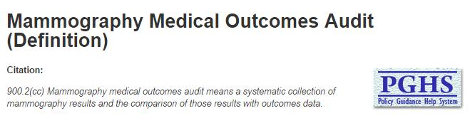
8 PGHS
9 PGHS Subtopics


10 FDA Guidance for MMOA
11 KansasRadiationPhysics.com

12 1994 DHS Publication 12 Lawrence W. Bassett, M.D(Co-chair) E. Edward Hendrick, PhD(Co-chair) Tamsen L. Bassford, MD Priscilla F. Butler,MS Darryl Carter,MD Marydale DeBor, JD Carl J. D Orsi, MD Carol J. Garlinghouse, MSN, RNC Richard F. Jones III, MD Amy S. Langer, MBA J. Leonard Lichtenfeld, MD Janet R. Osuch, MD Lynda N. Reynolds, BS,RT Ellen Shaw de Paredes, MD Richard E. Williams, MD D. Eldridge, M.S. US Dept of Health and Human Services Public Health Service Agency for Health Care Policy and Research Rockville, Maryland AHCPR Publication No October 1994

13 1994 Clinical Determinants

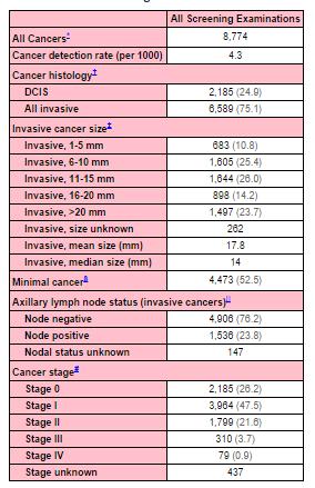
14 MMOA Results at selected sites

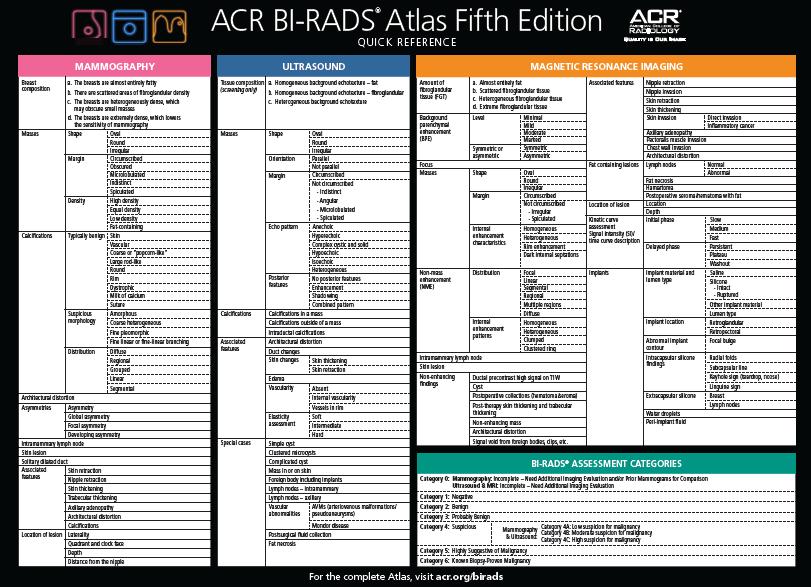
15 2013 BIRADS 5 TH Edition

16 Bi-Rads 5 th Edition, 2013
17 BIRADS
18 Mammography Medical Outcomes Audit Lead Interpreting Physician:, M.D. has the general responsibility of ensuring that the quality assurance program meets all the FDA requirements. No individual shall be assigned or shall retain responsibility for quality assurance tasks unless the lead interpreting physician has determined that the individual s qualifications for, and performance of, the assignment are adequate. Audit Interpreting Physician:, M.D. will review the medical outcomes audit data at least once every 12 months, recording the dates of the audit period(s) and shall be responsible for analyzing results based on this audit. This individual shall also be responsible for documenting the results, notifying other interpreting physicians of their results and the facility aggregate results. If follow-up actions are taken, the reviewing interpreting physician shall also be responsible for documenting the nature of the follow-up. Interpreting Physicians:, M.D., M.D., M.D., M.D., M.D., M.D. All interpreting physicians must follow the facility procedures for corrective action when the images they are asked to interpret are of poor quality. They must all participate in the facility's medical outcomes audit program. QC Technologist:, R.T.(M) is responsible for all individual tasks within the quality assurance program not assigned to the lead interpreting physician or the medical physicist. The tasks are to be performed by the quality control technologist or by other personnel qualified to perform the tasks. When other personnel are utilized for these tasks, the quality control technologist shall ensure that the tasks are completed in such a way as to meet the FDA requirements. MMOA Outcomes/Recall:, R.T.(M) will assist the Lead Interpreting Physician with tasks pertaining to the keeping of totals for all positive readings on patients and for patients who were initially read as negative but were later found out to have pathology(false Negatives). Patients will be tracked until all follow-up is completed and documented as required by the MQSA. Medical Physicist:, M.S., will perform the mammography equipment survey and oversee the equipment-related quality assurance practices of the facility. At a minimum, the medical physicist shall be responsible for performing the surveys and mammography equipment evaluations and providing the facility with the reports as required within 1 month after the survey.
19 Category 0 Categories 1 & 2 "Negative" or "Benign" A CANCER within 1 Year A+... Transferred to Another Facility for Work-up & Audit Purposes B "Additional Imaging Required" Utilize Orig. Flow Sheet, Adding "O" Before Each Class e.g. OA, OS+, etc. Follow-up Unavailable C Screening Algorithm Follow-up Result: Category 1 or 2 D Follow-up Result: Continue to Follow as Cat. 3 E Category 3 "Probably Benign" Additional Follow-up Recommended Follow-up Result: Category 4 or 5 Biopsy Recommended Transfer to Another Facility for Work-up & Audit Purposes F Follow-up Unavailable G Category 6, Categories 4A, 4B, & 4C: CANCER within 1 Year D+... Biopsy Recommended, but not Done H Core Biopsy Follow-up Unavailable I Surgical Biopsy The 2003 ACR BI-RADS reporting system has a new Category 6 for known biopsy-proven malignancy, with no therapy undergone by the patient. This was added so that screening patients already known to have cancer would not skew detection rates. The ACR has also added subcategories for Category 4: 4A, 4B, and 4C to encourage pathologists to initiate further evaluation of benign results and should allow clinicians to better understand follow-up recommendations after biopsy for findings placed in Category 4 subsets. Some other nomenclature changes have been made. Biopsy Recommended, but not Done P CANCER within 1 Year P+... Follow-up in 3-6 Months SF CANCER within 1 Year H+... Benign S Return Follow-up in 3-6 months JF Core Biopsy CANCER within 1 Year S+... Benign J CANCER T... CANCER within 1 Year J+... CANCER K... Categories 4 & 5 "Suspicious or Highly Suggestive of Malignancy" Biopsy Recommended Benign U CANCER within 1 Year U+... Surgical Biopsy Benign L CANCER within 1 Year L+... CANCER V... CANCER M... Follow-up Unavailable R D. Eldridge, M.S. 19
20 Mammography Medical Outcomes Audit Must follow all patients who end up being classified as Category 4 or 5.
21 Manually following biopsies
22 Patient Name: I.D.#: (Last) (First) (Middle) Address: P hone#: Primary Physician: Office #: Date of positive mammogram: Dates of prior mammograms: Done by: R.T.(M) Patient information, history, and other information attached. Read by: M.D. on (date) Mammography Reports attached? Initial Reading Category: 0, 1, 2, 3, 4,or 5 Additional interpretive comments: Details of Follow-up (U=Ultrasound, S=Surgical consult, B=Biopsy, C(months)=Clinical Follow-up Exam) Patient Name: I.D.#: (Last) (First) (Middle) C. Date: Type Follow-up: Done by: M.D. Results: Future Follow-up: Date: Recorded by: Details of Follow-up (U=Ultrasound, S=Surgical consult, B=Radiological Biopsy, C(months)=Clinical Exam, X=eXtra views) A. Date: Type Follow-up: Done by: M.D. Results: Future Follow-up: Date: Recorded by: B. Date: Type Follow-up: Done by: M.D. D. Eldridge, M.S. 22 Results: D. Date: Type Follow-up: Done by: M.D. Results: Future Follow-up: Date: Recorded by: E. Date: Type Follow-up: Done by: M.D. Results: Future Follow-up: Date: Recorded by:
23 Mammography Medical Outcomes Audit # Screenings/year: # Biopsies Recommended: # Biopsies Done: # Cancers Found: Cancer Stage when found:
24 Mammography Medical Outcomes Audit # Screenings: 6408 # Biopsies Recommended: 190 # Biopsies Done: 164 # Cancers Found: 25
25 Mammography Medical Outcomes Audit Biopsy Completion Rate (85%): 86.3% Biopsies Rec./1000 patients(32): 29.7 Cancers found/1000 patients(6): 3.9
26 Mammography Medical Outcomes Audit Disease + Disease - Test + True Positive False Positive Test - False Negative True Negative
27 Mammography Medical Outcomes Audit Disease + Disease - Categories 4, 5, 0+,3+ True Positive False Positive Categories 1,2,3, & 0 False Negative True Negative
28 True Positives (TP). Cancer diagnosed within 1 year after biopsy recommendation based on abnormal mammogram. *True Negative (TN). No known cancer diagnosed within 1 year of normal mammogram. *False Negative (FN). Cancer diagnosed within 1 year of a normal mammogram. Although other definitions of false negative exist, this definition is the most widely applied. *False Positive (FP). Benign disease found at biopsy within 1 year after an abnormal mammogram and recommendation for biopsy or surgical consultation. D. Eldridge, M.S. 28
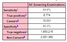
29 *Sensitivity. The probability of detecting a cancer when a cancer exists, or the percentage of all patients found to have breast cancer within 1 year of screening who were correctly diagnosed at the screening session. Sensitivity = TP/(TP+FN) 85-90% *Specificity. The probability of a normal mammogram report when no cancer exists, or the percentage of all patients not found to have breast cancer within 1 year of screening who were correctly identified as normal at the time of screening. Specificity = TN/(FP+TN). D. Eldridge, M.S. 29
30 Disease + Disease - Categories 4, 5, 0+,3+ True Positive False Positive Categories 1,2,3, & 0 False Negative True Negative Sensitivity=TP/(TP+FN) Specificity=TN/(FP+TN) D. Eldridge, M.S. 30
31 Magview

32 Mammography Medical Outcomes Audit
33 Mammography Medical Outcomes Audit Analyzing Biopsy Data
34 Mammography Medical Outcomes Audit Positive predictive value (PPV). Three definitions may be applied, depending on the practice conditions.
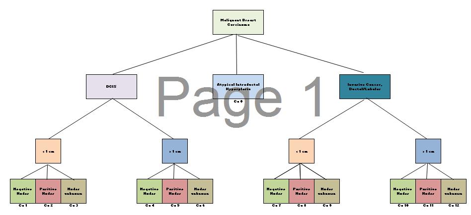
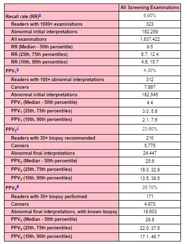
35 *PPV 1 (Abnormal Reading). The percentage of all screening mammography cases that result in a diagnosis of cancer based on abnormal screening examination. PPV 1 = TP/(number of abnormal screening exams), or PPV 1 = TP/(TP+FP 1 ). *PPV 2 (Biopsy recommended). The percentage of all screening mammography cases that result in a diagnosis of cancer based on a recommendation of consideration for biopsy. PPV 2 = TP/(# of cases recommended for biopsy after abnormal screening exams), or PPV 2 = TP/(TP+FP 2 ). *PPV 3 (Biopsy done). The percentage of all screening mammography cases that result in a diagnosis of cancer based on biopsies performed. This is also known as the biopsy yield of malignancy, or the positive biopsy rate. PPV 3 = TP/(number of biopsies), or PPV 3 = TP/(TP+FP 3 ). D. Eldridge, M.S. 35
36 Staging Cancers AJCC Staging of Breast Cancers... T... N... M... is difficult w/o a mandatory regional tumor registry.
37 American Joint Committee On Cancer (AJCC) Staging of Breast Cancer revised: 1/1/03 D. Eldridge, M.S. 37 Stage 0 Tis N0 M0 Stage 1 T1 N0 M0 Stage IIA T0 N1 M0 T1 N1 M0 T2 N0 M0 Stage IIB T2 N1 M0 T3 N0 M0 Stage IIIAT0 N2 M0 T1 N2 M0 T2 N2 M0 T3 N1 M0 T3 N2 M0 Stage IIIBT4 N0 M0 T4 N1 M0 T4 N2 M0 Stage IIICAny T N3 M0 Stage IV Any T Any N M1
38 Tumor T0: No evidence of primary tumor Tis: Carcinoma in situ T1: Tumor > 2 cm T2: 2 cm < Tumor < 5 cm T3: Tumor > 5 cm T4: Tumor (any size) extending to Chest wall or Skin Also: TX, Tis(CDIS), Tis(LCIS), Tis(Paget s), T1mic, T1a, T1b, T1c, T4a, T4, T4c, T4d Metastasis MX: Distant metastasis cannot be assessed M0: No distant metastasis M1: Distant metastasis Lymph Nodes N0: No regional lymph node metastases N1: Mets to ipsilateral axillary lymph nodes fixed or matted, or in clinically apparent ipsilateral internal mammography nodes in the absence of clinically evident axillary lymph node metastasis N3: Mets in ipsilateral infraclaviculary lymph node(s) with or without axillary lymph node involvement, or in clinically apparent ipsilateral internal mammary lymph node(s) and in the presence of clinically evident axillary lymph node mets; or mets in ipsilateral SCLNs with or without axillary or internal mammary lymph node involvement; Also: NX, N2a, N2b, N3a, N3b, N3cpNX, pn0, PN0(I-), PN0(I+), PN0(mol-), PN0(mol+), PN1, PN1mi, PN1a, PN1b, PN1c, pn2, pn2a, pn2b,pn3, pn3a, pn3b, pn3c D. Eldridge, M.S. 38
39 In Situ Carcinomas NOS (not otherwise specified) Intraductal Paget s disease and intraductal Invasive Carcinomas NOS (not otherwise specified) Ductal Inflammatory Medullary, NOS Medullary with lymhoid stroma Mucinous Papillary Tubular Lobular Paget s disease and infiltrating Undifferentiated Squamous cell Adenoid cystic Secretory Cribriform D. Eldridge, M.S. 39
40 EZ Staging
41 EZ Staging
42 Breast Cancer MamtreeTM Malignant Code DCIS Invasive Ca Ductal/Lobular < 1 cm. > 1 cm. < 1 cm. > 1 cm. - Nodes Ca1 + Nodes Ca2 Nodes not sampled Ca3 - Nodes Ca4 + Nodes Ca5 Nodes not sampled Ca6 - Nodes Ca7 + Nodes Ca8 Nodes not sampled Ca9 - Nodes Ca10 + Nodes Ca11 Nodes not sampled Ca12 Patient # Initial Category Malignancy Code Comments 1 0,1,2,3,4,5 2 0,1,2,3,4,5 3 0,1,2,3,4,5 4 0,1,2,3,4,5 5 0,1,2,3,4,5 6 0,1,2,3,4,5 7 0,1,2,3,4,5 8 0,1,2,3,4,5 9 0,1,2,3,4,5 10 0,1,2,3,4,5 11 0,1,2,3,4,5 12 0,1,2,3,4,5 D. Eldridge, M.S. 42

43 Mammography View Abbreviations
44 Assessment Category Meanings

45 chmarks/screening/
46

47 Cancer Detection Rate/Age
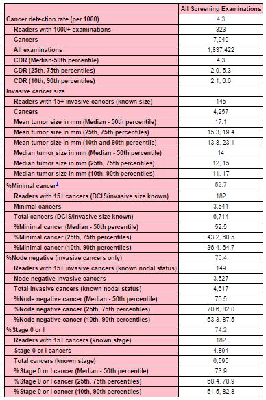
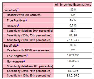
48 2009 BCSC Benchmarks Cancer Detection Rate: 4.7/1000 Median size of invasive Ca: 14.0 mm % Node-negative of invasive Ca: 77.3% % Minimal Ca: 52.6% Abnormal Recall Rate: 10.6% PPV1 (abnormal interpretation): 4.4% PPV2 (rec. for tissue diagnosis): 25.4% PPV3 (biopsy performed): 31.0% Sensitivity: 79.0% Specificity: 89.8%
49 Large Facility
50 What does the Lead Radiologist Want? However, before a complete audit system can be implemented, patient confidentiality and protection of medical audit information from discovery must be insured. Current State peer review statutes protecting peer review activities, such as collection of medical audit data, apply to inpatient facilities. In many States, there is virtually no protection from discovery of quality assurance activities in ambulatory and outpatient settings, where most mammography facilities function.
51 Mammography Medical Outcomes Audit MAMMOGRAPHY We are performing a complex imaging procedure, Mammograms are difficult to interpret, but Patient follow-up might be the toughest part.
52 Mammography Medical Outcomes Audit FOLLOW-UP The mammography medical audit is a systematic collection and analysis of mammography results, which are compared with outcomes data. The result should help ensure quality mammography is done.
53 Mammography Medical Outcomes Audit The MMOA relates technical and interpretive aspects of mammography performance to outcomes.
54 Mammography Medical Outcomes Audit If done correctly, the audit will help.
55 Most Importantly Don t lose a patient who has been recommended for biopsy. Do the best you can.
56 The End Thank you! Duke Eldridge, M.S. KansasRadiationPhysics.com
ACRIN 6666 Therapeutic Surgery Form
 S1 ACRIN 6666 Therapeutic Surgery Form 6666 Instructions: Complete a separate S1 form for each separate area of each breast excised with the intent to treat a cancer (e.g. each lumpectomy or mastectomy).
Completing the Puzzle AJCC TNM Staging Breast. Nicole Catlett, CTR 2017 Kentucky Cancer Registry Fall Conference, September 21 & 22, 2017
 Completing the Puzzle AJCC TNM Staging Breast Nicole Catlett, CTR 2017 Kentucky Cancer Registry Fall Conference, September 21 & 22, 2017 OBJECTIVES Understanding of Breast TNM staging Identify clinical
Chapter 2 Staging of Breast Cancer
 Chapter 2 Staging of Breast Cancer Zeynep Ozsaran and Senem Demirci Alanyalı 2.1 Introduction Five decades ago, Denoix et al. proposed classification system (tumor node metastasis [TNM]) based on the dissemination
STAGE CATEGORY DEFINITIONS
 CLINICAL Extent of disease before any treatment y clinical staging completed after neoadjuvant therapy but before subsequent surgery TX Tis Tis (DCIS) Tis (LCIS) Tis (Paget s) T1 T1mi T1a T1b T1c a b c
LUNG STAGING FORM LATERALITY: LEFT RIGHT BILATERAL
 LUNG STAGING FORM LATERALITY: LEFT RIGHT BILATERAL ( ) Tx Primary tumor cannot be assessed, or tumor proven by the presence of malignant cells in sputum or bronchial washings but not visualized by imaging
Descriptor Definition Author s notes TNM descriptors Required only if applicable; select all that apply multiple foci of invasive carcinoma
 S5.01 The tumour stage and stage grouping must be recorded to the extent possible, based on the AJCC Cancer Staging Manual (7 th Edition). 11 (See Tables S5.01a and S5.01b below.) Table S5.01a AJCC breast
ACR PRACTICE GUIDELINE FOR THE PERFORMANCE OF DIAGNOSTIC MAMMOGRAPHY
 The American College of Radiology, with more than 30,000 members, is the principal organization of radiologists, radiation oncologists, and clinical medical physicists in the United States. The College
The Radiology Aspects
 REQUIREMENTS FOR INTERNATIONAL ACCREDITATION OF BREAST CENTERS/UNITS The Radiology Aspects Miri Sklair-Levy, Israel RADIOLOGY GUIDELINES FOR QUALITY ASSURANCE IN BREAST CANCER SCREENING AND DIAGNOSIS Radiologists
Seventh Edition Staging 2017 Breast
 Seventh Edition Staging 2017 Breast Donna M. Gress, RHIT, CTR Validating science. Improving patient care. No materials in this presentation may be repurposed in print or online without the express written
Breast health and screening
 Breast health and screening Dr Kathy Wong MBBS (HK), FRCR (UK), FHKCR FHKAM (Radiology) Clinical Radiologist Department of Diagnostic and Interventional Radiology Kwong Wah Hospital Questions regarding
BI-RADS Categorization As a Predictor of Malignancy 1
 Susan G. Orel, MD Nicole Kay, BA Carol Reynolds, MD Daniel C. Sullivan, MD BI-RADS Categorization As a Predictor of Malignancy 1 Index terms: Breast, biopsy, 00.1261 Breast neoplasms, localization, 00.125,
BARC/2013/E/019 BARC/2013/E/019. AUDIT OF MAMMOGRAPHY PERFORMED IN OUR HOSPITAL by Surita Kantharia Medical Division
 BARC/2013/E/019 BARC/2013/E/019 AUDIT OF MAMMOGRAPHY PERFORMED IN OUR HOSPITAL by Surita Kantharia Medical Division BARC/2013/E/019 GOVERNMENT OF INDIA ATOMIC ENERGY COMMISSION BARC/2013/E/019 AUDIT OF
MAESTRO TRIAL FINAL RESULTS. Gisela L.G. Menezes, MD, PhD
 MAESTRO TRIAL FINAL RESULTS Gisela L.G. Menezes, MD, PhD MAESTRO Angiogenesis Metastasis How does OA work? Malignant Benign How does OA work? D O How does OA work? Optoacoustic Imaging Fibroadenoma IDC
DESCRIPTION: Percentage of final reports for screening mammograms that are classified as probably benign
 Quality ID #146 (NQF 0508): Radiology: Inappropriate Use of Probably Benign Assessment Category in Screening Mammograms National Quality Strategy Domain: Efficiency and Cost Reduction 2018 OPTIONS F INDIVIDUAL
Health Authority Abu Dhabi
 Health Authority Abu Dhabi Document Title HAAD Standard for Breast Cancer Screening & Diagnosis Document Ref. Number HAAD/BCSD/SD/1.0 Version 1.0 Approval Date 01 July 2012 Effective Date: July 2012 Last
Sentinel nodes. Location: Location: S1.04 Principal clinician. G1.01 Record other relevant information. S2.01 Number of specimens submitted
 Invasive Breast Cancer Histopathology Reporting Proforma Mandatory questions (i.e. protocol standards) are in bold (e.g. S1.01). S1.01 Identification Family name Given name(s) Date of birth DD MM YYYY
Diagnostic Dilemmas of Breast Imaging
 Diagnostic Dilemmas of Breast Imaging Common Causes of Error in Breast Cancer Detection By: Jason Cord, M.D. Mammography: Initial Imaging The standard for detection of breast cancer Screening mammography
now a part of Electronic Mammography Exchange: Improving Patient Callback Rates
 now a part of Electronic Mammography Exchange: Improving Patient Callback Rates Overview This case study explores the impact of a mammography-specific electronic exchange network on patient callback rates
Medical Audit of Diagnostic Mammography Examinations: Comparison with Screening Outcomes Obtained Concurrently
 Katherine E. Dee 1,2 Edward A. Sickles 1 Received July 3, 2000; accepted after revision September 12, 2000. Presented in part at the annual meeting of the American Roentgen Ray Society, Washington, DC,
Mammography. What is Mammography? What are some common uses of the procedure?
 Mammography What is Mammography? Mammography is a specific type of imaging that uses a low-dose x-ray system to examine breasts. A mammography exam, called a mammogram, is used to aid in the early detection
Major Rule Changes. Donna M. Gress, RHIT, CTR Technical Editor, AJCC Cancer Staging Manual First Author, Chapter 1: Principles of Cancer Staging
 AJCC 8 th Edition Staging Major Rule Changes Donna M. Gress, RHIT, CTR Technical Editor, AJCC Cancer Staging Manual First Author, Chapter 1: Principles of Cancer Staging Validating science. Improving patient
Q: How do you clinically code the N if the nodes are stated to be positive on mammogram/us or other imaging? No biopsy of nodes was done.
 Q&A Breast Webinar Q: One of my investigators is interested in knowing when Oncotype DX data collection was implemented. That data is collected in SSFs 22 and 23. I remember that the SSFs for breast were
DESCRIPTION: Percentage of final reports for screening mammograms that are classified as probably benign
 Measure #146 (NQF 0508): Radiology: Inappropriate Use of Probably Benign Assessment Category in Screening Mammograms National Quality Strategy Domain: Efficiency and Cost Reduction 2016 PQRS OPTIONS F
AJCC TNM 6 th Edition Staging Input Data Dictionary
 Catalogue no. 82-225-XIE No. 001 ISSN: 1715-2100 O ISBN: 0-662-41801-8 Manuals AJCC TNM 6 th Edition Staging Input Data Dictionary by Michel Cormier Health Statistics Division Client Custom Services Room
HISTORY OF MQSA AND ACR
 HISTORY OF MQSA AND ACR DEBORAH THAMES R.T. (R)(M)(QM) WHY MQSA? In the United States, there was a lack of standards in mammography imaging. Reporting Imaging Type of imaging screening/diagnostic Equipment
Hong Kong College of Radiologists Mammography Statement
 Hong Kong College of Radiologists Mammography Statement The Hong Kong College of Radiologists would like to give the following comments concerning mammography. Mammography screening: Breast cancer is the
Registrar s Guide to Chapter 1, AJCC Seventh Edition. Overview. Learning Objectives. Describe intent and purpose of AJCC staging
 Registrar s Guide to Donna M. Gress, RHIT, CTR Validating science. Improving patient care. This presentation was supported by the Cooperative Agreement Number DP13-1310 from The Centers for Disease Control
Breast Imaging! Ravi Adhikary, MD!
 Breast Imaging! Ravi Adhikary, MD! ACS Estimated Cancers Statistics 2014! Breast! New Cases in Women! 232,670 (+67,570 in situ)! Deaths in Women! 40,000! Colon! 48,380! 24,040! Cervical! 12,360! 4,020!
Mammographic imaging of nonpalpable breast lesions. Malai Muttarak, MD Department of Radiology Chiang Mai University Chiang Mai, Thailand
 Mammographic imaging of nonpalpable breast lesions Malai Muttarak, MD Department of Radiology Chiang Mai University Chiang Mai, Thailand Introduction Contents Mammographic signs of nonpalpable breast cancer
Overview of the 2005 IOM Report Improving Breast Imaging Quality Standards
 Overview of the 2005 IOM Report Improving Breast Imaging Quality Standards Goals of 2005 Report Study commissioned by US Congress in preparation for reauthorization of MQSA in 2007. to determine whether
AMSER Case of the Month: September 2018
 AMSER Case of the Month: September 2018 60-year-old woman with a left breast mass noted on screening mammography. Catherine McNulty, MS4 Tulane University School of Medicine Dr. Robin Sobolewski Breast
Session 2: The Role of Specialist Radiology Technologists
 Session 2: The Role of Specialist Radiology Technologists Louise M. Henderson, MSPH PhD Assistant Professor, Department of Radiology The University of North Carolina, Chapel Hill Overview Role of the technologist
Jose A Torres, MD 1/12/2017
 Jose A Torres, MD 1/12/2017 Background Globally leading cause of cancer related death in women ~249,000 Americans diagnosed with invasive breast cancer ~40,890 will die of their disease Breast cancer risk
AJCC CANCER STAGE. Site-Specific Instructions - BREAST. Reference: AJCC Cancer Staging Manual, 7 th Edition
 Slide 1 AJCC CANCER STAGE Site-Specific Instructions - BREAST Reference: AJCC Cancer Staging Manual, 7 th Edition In this presentation, we are going to review the AJCC Cancer Staging criteria for the breast
National Accreditation Program For Breast Centers Standards Manual 2017 EDITION. makes a difference ACCREDITATION
 National Accreditation Program For Breast Centers Standards Manual 2017 EDITION makes a difference ACCREDITATION 1 STANDARD 1.1 Level of Responsibility and Accountability The organizational structure of
Case Scenario 1: This case has been slightly modified from the case presented during the live session to add clarity.
 Case Scenario 1: This case has been slightly modified from the case presented during the live session to add clarity. Background: 46 year old married premenopausal female with dense breasts has noticed
Disclosures. Breast Cancer. Breast Imaging Modalities. Breast Cancer Screening. Breast Cancer 6/4/2014
 : Information for the Primary Care Physician Disclosures No financial relationships with commercial entities producing health care products/services. Roxsann Roberts, MD Section Chief, MRI Erlanger/EmCare
Mammography. What is Mammography?
 Scan for mobile link. Mammography Mammography is a specific type of breast imaging that uses low-dose x-rays to detect cancer early before women experience symptoms when it is most treatable. Tell your
Interactive Staging Bee
 Interactive Staging Bee ROBIN BILLET, MA, CTR GA/SC REGIONAL CONFERENCE NOVEMBER 6, 2018? Clinical Staging includes any information obtained about the extent of cancer obtained before initiation of treatment
Understanding Your Pathology Report
 Understanding Your Pathology Report Because every person s breast cancer is unique, it s important to understand the underlying biology of your tumor to personalize your treatment plan. Your physicians
Rare Breast Tumours. 1. Breast Tumours. 1.1 General Results. 1.2 Incidence
 Rare Breast Tumours 1. Breast Tumours 1.1 General Results Table 1. Epithelial Tumours of Breast: Incidence, Trends, Survival Flemish Region 2001-2010 Incidence Trend Survival Females EAPC Relative survival
Collaborative Stage Site-Specific Instructions - BREAST
 Slide 1 Collaborative Stage Site-Specific Instructions - BREAST In this presentation, we are going to review the CS Data Items and coding instructions for the breast primary site. Slide 2 Reading Assignments
Carcinoma mammario: le istologie non frequenti. Valentina Guarneri Università di Padova IOV-IRCCS
 Carcinoma mammario: le istologie non frequenti Valentina Guarneri Università di Padova IOV-IRCCS Histological diversity of breast adenocarcinomas Different histological types are defined according to specific
Challenges to Delivery of High Quality Mammography
 Challenges to Delivery of High Quality Mammography Overview of Current Challenges Barbara Monsees, Washington University Geographic Access, Equity and Impact on Quality Tracy Onega, Dartmouth Medical School
Breast Cancer. Most common cancer among women in the US. 2nd leading cause of death in women. Mortality rates though have declined
 Breast Cancer Most common cancer among women in the US 2nd leading cause of death in women Mortality rates though have declined 1 in 8 women will develop breast cancer Breast Cancer Breast cancer increases
BCSC Glossary of Terms (Last updated 09/16/2009) DEFINITIONS
 DEFINITIONS") Screening mammography scrmam_c BCSC Glossary of Terms (Last updated 09/16/2009) DEFINITIONS The radiologist s indication for exam is the primary determinant of whether a mammogram is screening or diagnostic.
Screening mammography scrmam_c BCSC Glossary of Terms (Last updated 09/16/2009) DEFINITIONS The radiologist s indication for exam is the primary determinant of whether a mammogram is screening or diagnostic.
3/23/2017. Disclaimer. Common Changes in TNM Staging. Overview. Overview. Understanding Terminology. Overview
 Common Changes in TNM Staging Understanding the General Rules of Cancer Staging Thomas P. Baker, MD FCAP March 5, 2017 Disclaimer The identification of specific products or scientific instrumentation is
Screening Mammograms: Questions and Answers
 CANCER FACTS N a t i o n a l C a n c e r I n s t i t u t e N a t i o n a l I n s t i t u t e s o f H e a l t h D e p a r t m e n t o f H e a l t h a n d H u m a n S e r v i c e s Screening Mammograms:
Breast Cancer. Saima Saeed MD
 Breast Cancer Saima Saeed MD Breast Cancer Most common cancer among women in the US 2nd leading cause of death in women 1 in 8 women will develop breast cancer Incidence/mortality rates have declined Breast
Breast Cancer Services in Germany
 Breast Cancer Services in Germany COUNTRY DELEGATES WORKSHOP Ispra (VA) - 13 and 14 March 2013 Karen Budewig, Federal Ministry of Health, Germany Vanessa Kääb-Sanyal, Mammography-Screening-Program, Germany
Chapter 13 Cancer of the Female Breast
 Lynn A. Gloeckler Ries and Milton P. Eisner INTRODUCTION This study presents survival analyses for female breast cancer based on 302,763 adult cases from the Surveillance, Epidemiology, and End Results
BREAST PATHOLOGY. Fibrocystic Changes
 BREAST PATHOLOGY Lesions of the breast are very common, and they present as palpable, sometimes painful, nodules or masses. Most of these lesions are benign. Breast cancer is the 2 nd most common cause
Current Status of Supplementary Screening With Breast Ultrasound
 Current Status of Supplementary Screening With Breast Ultrasound Stephen A. Feig, M.D., FACR Fong and Jean Tsai Professor of Women s Imaging Department of Radiologic Sciences University of California,
ROLE OF MRI IN SCREENING, DIAGNOSIS AND MANAGEMENT OF BREAST CANCER. B.Zandi Professor of Radiology
 ROLE OF MRI IN SCREENING, DIAGNOSIS AND MANAGEMENT OF BREAST CANCER B.Zandi Professor of Radiology Introduction In the USA, Breast Cancer is : The Most Common Non-Skin Cancer The Second Leading cause of
UPMC 1 Delineation of Privileges Request Criteria Summary Sheet
 UPMC 1 Facility: Specialty: UPMC Altoona Radiology KNOWLEDGE TRAINING Successful Completion of an ACGME/AOA, accredited program. The successful completion of an approved (ACGME/AOA) post graduate residency
Session 4: Test instruments to assess interpretive performance challenges and opportunities Overview of Test Set Design and Use
 Session 4: Test instruments to assess interpretive performance challenges and opportunities Overview of Test Set Design and Use Robert A. Smith, PhD American Cancer Society Test Sets vs. Audits Benefits
Interval Cancers in BreastScreen Aotearoa
 Interval Cancers in BreastScreen Aotearoa 2008 2009 Released 2018 nsu.govt.nz Citation: National Screening Unit. 2018. Interval Cancers in BreastScreen Aotearoa 2008 2009. Wellington: Ministry of Health.
B REAST STAGING FORM. PATHOLOGIC Extent of disease through completion of definitive surgery. CLINICAL Extent of disease before any treatment
 B REAST STAGING FORM CLINICAL Extent of disease before any treatment y clinical staging completed after neoadjuvant therapy but before subsequent surgery (DCIS) (LCIS) (Paget s) mi c a b c d TUMOR SIZE:
Effective Health Care Program
 Comparative Effectiveness Review Number 19 Effective Health Care Program Comparative Effectiveness of Core-Needle and Open Surgical Biopsy for the Diagnosis of Breast Lesions Executive Summary Background
Improving Reading Time of Digital Breast Tomosynthesis with Concurrent Computer Aided Detection
 White Paper Improving Reading Time of Digital Breast Tomosynthesis with Concurrent Computer Aided Detection WHITE PAPER 2 3 Abstract PowerLook Tomo Detection, a concurrent computer-aided detection (CAD)
Breast Tomosynthesis. What is breast tomosynthesis?
 Scan for mobile link. Breast Tomosynthesis Breast tomosynthesis is an advanced form of mammography, a specific type of breast imaging that uses low-dose x-rays to detect cancer early when it is most treatable.
Assessing an Emerging Nationwide Population-based Mammography Screening Program in Taiwan
 J Radiol Sci 2011; 36: 1-7 Assessing an Emerging Nationwide Population-based Mammography Screening Program in Taiwan Huay-Ben Pan 1,2,3 Giu-Cheng Hsu 4 Huei-Lung Liang 1,2 Chen-Pin Chou 1,2 Yen-Chi Wang
Breast Pathology. Breast Development
 Breast Pathology Lecturer: Hanina Hibshoosh, M.D. Reading: Kumar, Cotran, Robbins, Basic Pathology, 6th Edition, pages 623-635 Breast Development 5th week - thickening of the epidermis - milk line 5th
Imaging in breast cancer. Mammography and Ultrasound Donya Farrokh.MD Radiologist Mashhad University of Medical Since
 Imaging in breast cancer Mammography and Ultrasound Donya Farrokh.MD Radiologist Mashhad University of Medical Since A mammogram report is a key component of the breast cancer diagnostic process. A mammogram
Mammographic features and correlation with biopsy findings using 11-gauge stereotactic vacuum-assisted breast biopsy (SVABB)
") Original article Annals of Oncology 14: 450 454, 2003 DOI: 10.1093/annonc/mdh088 Mammographic features and correlation with biopsy findings using 11-gauge stereotactic vacuum-assisted breast biopsy (SVABB)
Original article Annals of Oncology 14: 450 454, 2003 DOI: 10.1093/annonc/mdh088 Mammographic features and correlation with biopsy findings using 11-gauge stereotactic vacuum-assisted breast biopsy (SVABB)
BreastScreen Victoria Annual Statistical Report
 BreastScreen Victoria Annual Statistical Report 29 BREASTSCREEN VICTORIA: ANNUAL STATISTICAL REPORT, 29 Produced by: BreastScreen Victoria Coordination Unit Level, 3 Pelham Street, Carlton South Victoria
B REAST STAGING FORM. PATHOLOGIC Extent of disease through completion of definitive surgery. CLINICAL Extent of disease before any treatment
 B REAST STAGING FORM Extent of disease before any treatment y clinical staging completed after neoadjuvant therapy but before subsequent surgery (DCIS) (LCIS) (Paget s) mi a b c a b c d TUMOR SIZE: S TAGE
Quality ID #263: Preoperative Diagnosis of Breast Cancer National Quality Strategy Domain: Effective Clinical Care
 Quality ID #263: Preoperative Diagnosis of Breast Cancer National Quality Strategy Domain: Effective Clinical Care 2018 OPTIONS FOR INDIVIDUAL MEASURES: REGISTRY ONLY MEASURE TYPE: Process DESCRIPTION:
Maram Abdaljaleel, MD Dermatopathologist and Neuropathologist University of Jordan, School of Medicine
 Maram Abdaljaleel, MD Dermatopathologist and Neuropathologist University of Jordan, School of Medicine The most common non-skin malignancy of women 2 nd most common cause of cancer deaths in women, following
Contents 1 The Windows of Susceptibility to Breast Cancer 2 The So Called Pre-Neoplastic Lesions and Carcinoma In Situ
 Contents 1 The Windows of Susceptibility to Breast Cancer... 1 1.1 Introduction... 1 1.2 Risk Factor and Etiological Agents... 2 1.3 The Concept of the Windows of Susceptibility to Carcinogenesis... 5
Collecting Cancer Data: Breast. Prizes! Collecting Cancer Data: Breast 8/4/ NAACCR Webinar Series 1. NAACCR Webinar Series
 Collecting Cancer Data: Breast NAACCR 2008 2009 Webinar Series Prizes! Question of the Month! The participant that submits the best question of the session will receive a fbl fabulous Pi Prize! Shannon
Breast Imaging Donald L. Renfrew, MD
 This free educational material is provided by 333 N. Commercial Street, Suite 100, Neenah, WI 54956 Donald L. Renfrew, MD Breast cancer is the most frequent non-skin cancer diagnosis in women, with an
Survey of Mammography Facilities
 Survey of Mammography Facilities Summary: Survey completed? Yes Date completed: / / No Comments: Instructions: This survey is part of a study being conducted by [insert surveillance institution] breast
Kish chakrabarti, Ph.D. Senior Physicist CDRH/FDA
 Facility Certification Extension Requirements, Quality Assurance and Medical Physicists role for Hologic Selenia Dimensions Digital Breast Tomosynthesis (DBT) System Kish chakrabarti, Ph.D. Senior Physicist
«àπ π â Õ μ «å «π Áß μâ π π ßæ π ª
 «æ å μ Ù-ı ªï Ë Ûapple Ë Ù μ.. -.. ÚııÙ Reg 4-5 Med J Vol. 30 No. 4 Oct - Dec 2011 π æπ åμâπ Original Article «μ»ÿ º æ.., Cheerawan Tansupaphon M.D., «.«. ß «π Thai Board of Diagnostic Radiology ÿà ß π
December 2002 Monthly Memo
 FLORIDA CANCER DATA SYSTEM December 2002 Monthly Memo CODING COMPLEX MORPHOLOGIC DIAGNOSES CODING COMPLEX BREAST HISTOLOGIES Apply these guidelines in priority order. Use the first guideline that applies
4/10/2018. SEER EOD and Summary Stage. Overview KCR 2018 SPRING TRAINING. What is SEER EOD? Ambiguous Terminology General Guidelines
 SEER EOD and Summary Stage KCR 2018 SPRING TRAINING Overview What is SEER EOD Ambiguous Terminology General Guidelines EOD Primary Tumor EOD Regional Nodes EOD Mets SEER Summary Stage 2018 Site Specific
Basement membrane in lobule.
 Bahram Memar, MD Basement membrane in lobule. Normal lobule-luteal phase Normal lobule-follicular phase Lactating breast Greater than 95% are adenocarcinomas in situ carcinomas and invasive carcinomas.
Breast Cancer. For breast cancer, the mortality risk varies with the stage of the cancer.
 CREATED EXCLUSIVELY FOR FINANCIAL PROFESSIONALS Rx FOR SUCCESS Breast Cancer Breast cancer is the most common cancer in women in the United States and second only to lung cancer as a cause of cancer deaths.
Q&A. Fabulous Prizes. Collecting Cancer Data: Breast 4/4/13. NAACCR Webinar Series Collecting Cancer Data Breast
 Collecting Cancer Data Breast NAACCR 2012 2013 Webinar Series Q&A Please submit all questions concerning webinar content through the Q&A panel. Reminder: If you have participants watching this webinar
Clinical Trials of Proton Therapy for Breast Cancer. Andrew L. Chang, MD 張維安 Study Chair
 Clinical Trials of Proton Therapy for Breast Cancer Andrew L. Chang, MD 張維安 Study Chair AndrewLChangMD@gmail.com Disclosure Proton Center Development Corporation Scripps San Diego Proton Therapy Center
The American College of Radiology Breast Ultrasound Accreditation Program: Frequently Asked Questions (Revised: July 31, 2017)
") The American College of Radiology Breast Ultrasound Accreditation Program: Frequently Asked Questions (Revised: July 31, 2017) Table of Contents APPLICATION - GENERAL... 1 MOVED FACILITIES AND UNITS...
The American College of Radiology Breast Ultrasound Accreditation Program: Frequently Asked Questions (Revised: July 31, 2017) Table of Contents APPLICATION - GENERAL... 1 MOVED FACILITIES AND UNITS...
Breast pathology. 2nd Department of Pathology Semmelweis University
 Breast pathology 2nd Department of Pathology Semmelweis University Breast pathology - Summary - Benign lesions - Acute mastitis - Plasma cell mastitis / duct ectasia - Fat necrosis - Fibrocystic change/
Breast Cancer Update 2018 The Latest in Diagnosis and Treatment SARATH K, PALAKODETI, DO, FAACS GENERAL, BREAST, AND COSMETIC SURGEON TOLEDO CLINIC
 Breast Cancer Update 2018 The Latest in Diagnosis and Treatment SARATH K, PALAKODETI, DO, FAACS GENERAL, BREAST, AND COSMETIC SURGEON TOLEDO CLINIC Objectives Identify breast lesions and masses, and know
Mammography Education, Inc.
 Hands-on Breast Screening and Diagnosis Course * Screening of 315 full field digital mammography cases. * Reading a mixture of normals and proven abnormals at high resolution viewing stations. * Immediate
Breast Cancer. Dr Rodney Itaki Anatomical Pathology Discipline Division of Pathology
 Breast Cancer Dr Rodney Itaki Anatomical Pathology Discipline Division of Pathology Muscles Muscles underneath the breasts separating them from the ribs Breast has no muscle tissue 2 Female Breast Anatomy
Learning Objectives. ! At the end of the presentation,students will be able to:
 Learning Objectives! At the end of the presentation,students will be able to: Define Breast Cancer Identify the controllable and uncontrollable risk factors of breast cancer Recognize the 4 stages of breast
American College of Radiology/Society of Breast Imaging Curriculum for Resident and Fellow Education in Breast Imaging
 American College of Radiology/Society of Breast Imaging Curriculum for Resident and Fellow Education in Breast Imaging Edward A. Sickles, MD a, Liane E. Philpotts, MD b, Brett T. Parkinson, MD c, Debra
George Cernile Artificial Intelligence in Medicine Toronto, ON. Carol L. Kosary National Cancer Institute Rockville, MD
 George Cernile Artificial Intelligence in Medicine Toronto, ON Carol L. Kosary National Cancer Institute Rockville, MD Using RCA A system to convert free text pathology reports into a database of discrete
University of Washington Radiology Review Course: Strange and Specific Diagnoses. Case #1
 University of Washington Radiology Review Course: Strange and Specific Diagnoses Katherine E. Dee, MD Seattle Breast Center Via Radiology 2014 Case #1 37 year old presents with bilateral palpable lumps.
Exercise 15: CSv2 Data Item Coding Instructions ANSWERS
 Exercise 15: CSv2 Data Item Coding Instructions ANSWERS CS Tumor Size Tumor size is the diameter of the tumor, not the depth or thickness of the tumor. Chest x-ray shows 3.5 cm mass; the pathology report
FDA Executive Summary
 Meeting of the Radiological Devices Advisory Panel On October 24, 22, the panel will discuss, make recommendations, and vote on a premarket approval application supplement (P83/S) to expand the indications
Amammography report is a key component of the breast
 Review Article Writing a Mammography Report Amammography report is a key component of the breast cancer diagnostic process. Although mammographic findings were not clearly differentiated between benign
Recall and Cancer Detection Rates for Screening Mammography: Finding the Sweet Spot
 Women s Imaging Original Research Grabler et al. Optimal Recall and Cancer Detection Rates for Screening Mammography Women s Imaging Original Research Paula Grabler 1 Dominique Sighoko 2 Lilian Wang 3
TMIST: Frequently Asked Questions
 TMIST: Frequently Asked Questions Key Topics for Site Investigators and Staff This document answers frequently asked questions about the Tomosynthesis Mammographic Imaging Screening Trial (TMIST/EA1151);
Minimizing Errors in Diagnostic Pathology
 Shahla Masood, M.D. Professor and Chair Department of Pathology and Laboratory Medicine University of Florida College of Medicine-Jacksonville Medical Director, Shands Jacksonville Breast Health Center
BREAST MRI. Elizabeth A. Rafferty, M.D. Avon Comprehensive Breast Center Massachusetts General Hospital Harvard Medical School
 BREAST MRI Elizabeth A. Rafferty, M.D. Avon Comprehensive Breast Center Massachusetts General Hospital Harvard Medical School BREAST MRI Any assessment of the breast parenchyma requires the administration
2 Malignant Breast Disease: Diagnosis and Assessment
 2 Malignant Breast Disease: Diagnosis and Assessment Nimmi Arora and Rache M. Simmons Pearls and Pitfalls Screening mammography has allowed for earlier detection of breast cancer. Image-guided biopsy assists
DATA STANDARDS AND QUALITY CONTROL MEMORANDUM DSQC #
 DATA STANDARDS AND QUALITY CONTROL MEMORANDUM DSQC #2006-01 CATEGORY: CLARIFICATION SUBJECT: RESCINDMENT - DSQC MEMORANDUM 2002-08 Coding Complex Morphologic Diagnoses (revised 8/02) EFFECTIVE: For Cases
LYMPHATIC DRAINAGE AXILLARY (MOSTLY) INTERNAL MAMMARY SUPRACLAVICULAR
 INTERNAL MAMMARY SUPRACLAVICULAR") BREAST LYMPHATIC DRAINAGE AXILLARY (MOSTLY) INTERNAL MAMMARY SUPRACLAVICULAR HISTOLOGY LOBE: (10 in whole breast) LOBULE: (many per lobe) ACINUS/I, aka ALVEOLUS/I: (many per lobule) DUCT(S): INTRA- or
BREAST LYMPHATIC DRAINAGE AXILLARY (MOSTLY) INTERNAL MAMMARY SUPRACLAVICULAR HISTOLOGY LOBE: (10 in whole breast) LOBULE: (many per lobe) ACINUS/I, aka ALVEOLUS/I: (many per lobule) DUCT(S): INTRA- or