Plain Abdominal Radiography & GI series 腹部與腸胃道之放射線學
|
|
|
- Easter Leonard
- 6 years ago
- Views:
Transcription

1 Plain Abdominal Radiography & GI series 腹部與腸胃道之放射線學 陳潤秋台北市立聯合醫院
2 Jen-Ai H.

3 Standing abdomen Jen-Ai H.

4 Supine KUB Jen-Ai H. medicalcenter.osu.edu

5 Jen-Ai H.
6 Checklist: bones and stones, gas and mass Jen-Ai H. Free air Bowel gas pattern Bowel wall Pathologic extra or intraperitoneal gas Gas bubbles in organ parenchyma Intraluminal gas collections in tubular structures or hollow viscera Intraperitoneal fluid collections

7 Checklist: bones and stones, gas and mass Jen-Ai H. Presence or nonvisualization of physiologic soft-tissue structures (psoas muscle, flank stripe, hepatic, splenic and renal outlines, bladder, perivesical fat, rectal air) Soft-tissue mass Pathologic calcification or foreign bodies Pathologic skeletal changes Lung bases
8 Gas: GI tract Jen-Ai H. Esophagogram, Stomach, Small bowel: duodenum, jejunum, and ileum. Large bowel: cecum, ascending, transverse, and descending colon, rectum.


9 Gastric volvulus ebsco.smartimagebase.com Jen-Ai H.


10 Jen-Ai H. 1 week newborn, vomiting after birth

11 Jen-Ai H. GI series: 1. a.esophagogram b.ugi series c.small bowel series Enteroclysis e.lgi series 2. a.single contrast b.double contrast c.water-soluble contrast


12 Jen-Ai H. UGI series


13 UGI: Esophagus A muscular tube about 20-24cm. Three phase of peristalsis: Primary: major stripping wave initiated by swallowing. Secondary: similar stripping waves but by irritation or distention along esophageal wall. Tertiary: segmental contraction, non-peristaltic.


14 Stomach: Cardiac Fundus Body Antrum Pyloric canal Duodenum: Duodenal bulb 2nd to 4th portion of duodenum (retroperitoneum, fixed). Jen-Ai H.


15 Small bowel series Jen-Ai Single H. contrast: jejunum,ileum enteroclysis

16 Jen-Ai H. LGI series


17 Jen-Ai H. LGI series: cecum, ascending,transversedescending, sigmoid, rectum, terminal ileum


18 GI lesions Jen-Ai H. 1. Mucosa 2. Submucosa, muscle 3. Serosa 4. Extraluminal


19 Jen-Ai H. Mucosa


20 Jen-Ai H. submucosa

21 Jen-Ai H. Serosa


22 Jen-Ai H. Extraluminal


23 Anatomy position: Hiatal Hernia Sliding hernia: GE junction is above the diaphragm Paraesophageal hernia: Part of fundus herniated Jen-Ai H. above the diaphragm

24 Anatomy abnormal: Achalasia The gastroesophgeal sphincter fails to relax. Primary and secondary peristalsis must absent Low esophgeal sphincter must fail to relax completely on swallowing.. Jen-Ai H. Bird s sign


25 Disease: Esophageal cancer Squamous cell carcinoma Annular Ulcerative Jen-Ai H.


26 Benign gastric ulcer 1. Projection of ulcer crater beyond wall. 2. Radiating fold to edge of ulcer. 3. Ulcer in the center of swelling lesion. 4. Collar sign, Hampton s line. 5. Incisura opposite. Jen-Ai H.


27 Simeon Abramson, M.D. Thomas E. Herbener, M.D. Jen-Ai H. Benign gastric ulcer


28 Malignant ulcer 1. Intraluminal ulcer crater. 2. Abrupt between ulcer and regional fold. 3. Thick rigid fold surrounding lesion. 4. Ulcer crater wide and eccentric 5. Carmen s meniscus sign Jen-Ai H.


29 Jen-Ai H.

30 Jen-Ai H. Gastric cancer
31 Distention limited to the small bowel Jen-Ai H.


32 Jen-Ai H. Ileus: Gall stone ileus


33 Ileus: Torsion of a Meckel diverticulum and enterolith Jen-Ai H.


 R. J.")
34 Duodenal ulcer: Ulcer crater, linear ulcer Healed ulcer--cloverleaf or butterfly appearance All benign, if multiple or postbulbar Zollinger-Ellison syndrome (excessive gastrin secretion--gastrinoma) R. J. Alfidi, M.D. Jen-Ai H.

35 Jen-Ai H. Submucosal tumor in duodenum smooth surface, less acute angle


36 Duodenum external compression widening the C sweep-- pancreatic head lesion Jen-Ai H.
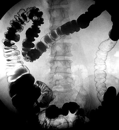
37 Crohn s disease Recurrent inflammatory condition of bowel of unknown etiology Aphthoid ulcer deep ulcer with fistula and sinuses Skip lesions (ulcer scattered in GI) cobblestone mucosal pattern String sign, wall thickening, stenosis Jen-Ai H.


38 Jen-Ai H.
39 Distention limited to the colon Most frequently cause, especially in elderly patients, is colorectal carcinoma (60%) Jen-Ai H.


40 Jen-Ai H. Sigmoid colon cancer with obstruction


41 Jen-Ai H. Colon cutoff sign in acute pancreatitis


42 Jen-Ai H. Colon cutoff sign in acute pancreatitis


43 Jen-Ai H. Volvulus of colon

44 coffee bean sign in sigmoid volvulus Jen-Ai H.


45 Pathologic thickening of bowel wall Pseudomembranous colitis Jen-Ai H.
46 Jen-Ai H. Colon polyp Size: 1-2cm: 10% malignant, >2cm: 50% malignant, Stalk: Single or Multiple: Familial polyposis syndromes. Adenomaous polyp (most commonly): tubular, tubulovillous,villouw Location: rectosignmoid 70%


47 Jen-Ai H. staclk


48 Jen-Ai H. Colon cancer Ulcerative Annular (apple core) Polypoid Plaque like Scirrhous carcinoma Kim LH


49 Jen-Ai H. Protruding lesion Colon cancer Polypoid or plaque like
50 Ulcerative colitis Shallow ulcer,mainly from left side. Loss of haustration, circumferential bowel involvement. Pseudopolyps ( rode like, Y-shape,linear ) Toxic megacolon. Backwash ileitis (mild) Cancer coexisting, rare. Jen-Ai H.


51 Jen-Ai H.


52 Diverticular Disease Herniated mucosa through bowel wall Diverticulum,95% in sigmoid colon Diverticulosis Diverticulitis: Extravasation of barium Intramural abscess, fistula Jen-Ai H.

53 Lymphoma Submucosal pattern produces thumbprinting sign Jen-Ai H.

54 Fixed intraperitoneal gas on a supine film Jen-Ai H.
55 Pneumoperitoneum (free air) on supine film Jen-Ai H. Football sign Visible falciform ligament (the laces of the football) Rigler s sign Gas in the hepatorenal recess (Morison s pouch) Para- and prehepatic gas Inverted V sign

56 falciform ligament outlined by free air Jen-Ai H.


57 Pneumoperitoneum:Rigler s sign Jen-Ai H.


58 Jen-Ai H. NEC with pneumoperitoneum


59 Jen-Ai H. Liver abscess


60 Emphysematous pyelonephritis and cystitis Jen-Ai H.


61 Jen-Ai H. Calcification and calculi


62 Jen-Ai H. Gall bladder stone


63 Jen-Ai H. staghorn stone


64 Jen-Ai H. Porcelain gall bladder


65 Intraperitoneal fluid collection Jen-Ai H.


66 Jen-Ai H. Pseudocyst of pancreas


67 Jen-Ai H. Intraabdominal mass


68 Ingested watch battery in fundus of stomach and then in transverse colon Jen-Ai H.


69 Jen-Ai H. Skeletal change

70 bones and stones, gas and mass Jen-Ai H.
71 Summary Jen-Ai H. Bones, stones, gas and mass Esophagus Anatomy abnormal: Achalasia Benign gastric ulcer Malignant ulcer Colon polyp Ulcerative colitis Pneumoperitoneum (free air) on supine film
Abdominal radiology 腹部放射線學
 Abdominal radiology 腹部放射線學 台北醫學大學 - 市立萬芳醫院 留偉順 laowilson@hotmail.com The Normal Abdominal Series Chest Supine abdomen Erect abdomen Left lateral decubitus abdomen Learning objectives Understanding normal
GIT RADIOLOGY. Water-soluble contrast media (e.g. gastrograffin) are the other available agents.which doesn t cause inflammatory peritonitis..
 are the other available agents.which doesn t cause inflammatory peritonitis..") GIT RADIOLOGY Imaging techniques-general principles: Contrast examinations: Barium sulphate is the best contrast for GIT (with good mucosal coating & excellent opacification & being inert); but is contraindicated
GIT RADIOLOGY Imaging techniques-general principles: Contrast examinations: Barium sulphate is the best contrast for GIT (with good mucosal coating & excellent opacification & being inert); but is contraindicated
Gastrointestinal Tract. Anatomy of GI Tract. Anatomy of GI Tract. (Effective February 2007) (1%-5%)
 (1%-5%)") Gastrointestinal Tract (Effective February 2007) (1%-5%) Anatomy of GI Tract Esophagus bulls-eye or target EG junction seen on sagittal scan posterior to left lobe of liver and anterior to aorta Anatomy
Gastrointestinal Tract (Effective February 2007) (1%-5%) Anatomy of GI Tract Esophagus bulls-eye or target EG junction seen on sagittal scan posterior to left lobe of liver and anterior to aorta Anatomy
Radiology of the abdomen Lecture -1-
 Radiology of the abdomen Lecture -1- Objectives To know radiology modalities used in abdomen imaging mainly GI tract. To know advantages and disadvantages of each modality. To know indications and contraindications
Medical application of transabdominal ultrasound in gastrointestinal diseases
 Medical application of transabdominal ultrasound in gastrointestinal diseases Hsiu-Po Wang Department of Emergency Medicine National Taiwan University Hospital Real-time ultrasound has become a standard
Plain abdomen The standard films are supine & erect AP views (alternative to erect, lateral decubitus film is used in ill patients).
.") Plain abdomen The standard films are supine & erect AP views (alternative to erect, lateral decubitus film is used in ill patients). The stomach can be readily identified by its location, gastric rugae
Plain abdomen The standard films are supine & erect AP views (alternative to erect, lateral decubitus film is used in ill patients). The stomach can be readily identified by its location, gastric rugae
UNDERSTANDING X-RAYS: ABDOMINAL IMAGING THE ABDOMEN
 UNDERSTANDING X-RAYS: ABDOMINAL IMAGING THE ABDOMEN Radiology Enterprises radiologyenterprises@gmail.com www.radiologyenterprises.com STOMACH AND SMALL BOWEL STOMACH AND SMALL BOWEL Swallowed air is a
Introduction and Definitions
 Bowel obstruction Introduction and Definitions Accounts for 5% of all acute surgical admissions Patients are often extremely ill requiring prompt assessment, resuscitation and intensive monitoring Obstruction
LOOKING FOR AIR IN ALL THE WRONG PLACES Richard M. Gore, MD North Shore University Health System University of Chicago Evanston, IL
 SIGNIFICANCE OF EXTRALUMINAL ABDOMINAL GAS: LOOKING FOR AIR IN ALL THE WRONG PLACES Richard M. Gore, MD North Shore University Health System University of Chicago Evanston, IL SCBT/MR 2012 October 26,
Gastrointestinal Disorders. Disorders of the Esophagus 3/7/2013. Congenital Abnormalities. Achalasia. Not an easy repair. Types
 Gastrointestinal Disorders Congenital Abnormalities Disorders of the Esophagus Types Stenosis Atresia Fistula Newborn aspirates while feeding. Pneumonia Not an easy repair Achalasia Lack of relaxation
This lab activity is aligned with Visible Body s Human Anatomy Atlas app. Learn more at visiblebody.com/professors
 1 This lab activity is aligned with Visible Body s Human Anatomy Atlas app. Learn more at visiblebody.com/professors 2 A. Digestive System Overview To Start: Go to the Views menu and scroll down to the
The Digestive System
 The Digestive System Identify the Structure and Function. Mesentery of the Large Intestine The mesentery functions to connect the visceral organs to the abdominal wall. Identify the Structure. Nasal Cavity
Abdominal Imaging: Luminal organs. Rowland Illing MA BMBCh DM FLS MRCS(Eng) FRCR
 FRCR") Abdominal Imaging: Luminal organs Rowland Illing MA BMBCh DM FLS MRCS(Eng) FRCR Aims Reference text & resources Management of a patient Imaging what and when to use What to ask and how to describe Segments
Abdominal Imaging: Luminal organs Rowland Illing MA BMBCh DM FLS MRCS(Eng) FRCR Aims Reference text & resources Management of a patient Imaging what and when to use What to ask and how to describe Segments
Digestive System. In one end and out the other.
 Digestive System In one end and out the other. Overview Every cell in the body needs nourishment, yet most cells cannot leave their position in the body and travel to a food source, so the food must be
No Disclosures. Approach to Abdominal Radiographs
 Approach to Abdominal Radiographs Tapas K. Tejura, M.D. Assistant Professor of Clinical Radiology Keck Medical Center of USC tapas.tejura@med.usc.edu No Disclosures 34-year-old male with acute abdominal
Pathology of Intestinal Obstruction. Dr. M. Madhavan, MBBS., MD., MIAC, Professor of Pathology Saveetha Medical College
 Pathology of Intestinal Obstruction Dr. M. Madhavan, MBBS., MD., MIAC, Professor of Pathology Saveetha Medical College Pathology of Intestinal Obstruction Objectives list the causes of intestinal obstruction
The Digestive System. Chapter 25
 The Digestive System Chapter 25 Introduction Structure of the digestive system A tube that extends from mouth to anus Accessory organs are attached Functions include Ingestion Movement Digestion Absorption
CT Evaluation of Bowel Wall Thickening. Dr: Adel El Badrawy; M.D. Lecturer of Radio Diagnosis Faculty of Medicine Mansoura University.
 CT Evaluation of Bowel Wall Thickening By Dr: Adel El Badrawy; M.D. Lecturer of Radio Diagnosis Faculty of Medicine Mansoura University. The CT findings of bowel wall thickening includes 1 Degree of thickening.
GASTROINTESTINAL SYSTEM
 GASTROINTESTINAL SYSTEM Topographic Anatomy of the Abdomen Surface Landmarks Xiphoid process T9/T10 Inferior costal margin L2/L3 Iliac Crest L4 level ASIS L5/S1 level Pubic symphysis level of greater trochanter
Lab 5 Digestion and Hormones of Digestion. 7/16/2015 MDufilho 1
 Lab 5 Digestion and Hormones of Digestion 1 Figure 23.1 Alimentary canal and related accessory digestive organs. Mouth (oral cavity) Tongue* Parotid gland Sublingual gland Submandibular gland Salivary
- Digestion occurs during periods of low activity - Produces more energy than it uses. - Mucosa
 Introduction Digestive System Chapter 29 Provides processes to break down molecules into a state easily used by cells - A disassembly line: Starts at the mouth and ends at the anus Digestive functions
INVESTIGATIONS OF GASTROINTESTINAL DISEAS
 INVESTIGATIONS OF GASTROINTESTINAL DISEAS Lecture 1 and 2 دز اسماعيل داود فرع الطب كلية طب الموصل Radiological tests of structure (imaging) Plain X-ray: May shows soft tissue outlines like liver, spleen,
Nasogastric tube. Stomach. Pylorus. Duodenum 1. Duodenum 2. Duodenum 3. Duodenum 4
 Esophagus Barium Swallow Stomach and Duodenum 4 year old Upper GI Nasogastric tube Stomach and Duodenum 4 year old Upper GI Nasogastric tube Stomach Pylorus Duodenum 1 Duodenum 2 Duodenum 3 Duodenum 4
3/16/2016. Food--mixture of carbohydrates, proteins, and lipids
 Food--mixture of carbohydrates, proteins, and lipids Food being broken down into small molecules Takes place in the alimentary canal Complete digestive system 4 layers of tissue (in book) Lumen 1) MECHANICAL/PHYSICAL--
Radiology of GI system diseases
 GI Cycle - Lecture 12 436 Teams Radiology of GI system diseases Objectives 1. 2. 3. To know common GIT Pathologies presentation. To understand step wise approach in requesting GIT Radiology Investigations.
National Museum of Health and Medicine
 National Museum of Health and Medicine Otis Historical Archives Bower Photograph Collection Date of Records: 1910s-1920s Size: 1 box Finding Aid: by Eric W. Boyle (2012) Biographical Note: Col. Morris
- Digestion occurs during periods of low activity - Produces more energy than it uses. 3 Copyright 2016 by Elsevier Inc. All rights reserved.
 Introduction Digestive System Chapter 29 Provides processes to break down molecules into a state easily used by cells - A disassembly line: Starts at the mouth and ends at the anus Digestive functions
Imaging Evaluation of Polyps. CT Colonography: Sessile Adenoma. Polyps, DALMs & Megacolon Objectives
 Polyps, DALMs & Megacolon: Pathology and Imaging of the Colon and Rectum Angela D. Levy and Leslie H. Sobin Washington, DC Drs. Levy and Sobin have indicated that they have no relationships which, in the
Two main groups Alimentary canal continuous coiled hollow tube Accessory digestive organs
 Digestion Breakdown of ingested food Absorption of nutrients into the blood Metabolism Production of cellular energy (ATP) Constructive and degradative cellular activities Two main groups Alimentary canal
CEA (CARCINOEMBRYONIC ANTIGEN)
") (CARCINOEMBRYONIC ANTIGEN) 428 C15.3 Malignant neoplasm of upper third of esophagus C15.4 Malignant neoplasm of middle third of esophagus C15.5 Malignant neoplasm of lower third of esophagus C15.8 Malignant
(CARCINOEMBRYONIC ANTIGEN) 428 C15.3 Malignant neoplasm of upper third of esophagus C15.4 Malignant neoplasm of middle third of esophagus C15.5 Malignant neoplasm of lower third of esophagus C15.8 Malignant
Patient. Male 76 year old C.C: abdominal pain
 Patient Male 76 year old C.C: abdominal pain Bowel stool retention Suspected pulmonary TB at right upper lung Infiltration in right lower lung Pleural thickening at the Right chest Localized dilated small
BELLWORK DEFINE: PERISTALSIS CHYME RUGAE Remember the structures of the digestive system 1
 BELLWORK DEFINE: PERISTALSIS CHYME RUGAE 2.07 Remember the structures of the digestive system 1 STANDARD 8) Outline basic concepts of normal structure and function of all body systems, and explain how
Polyps Adenomas Lipomas
 30 Chapter 2 CT Colonography Chapter 2 Polyps Adenomas Lipomas Case 8 Case 9 Case 10 Case 11 Case 12 Case 13 Case 14 Case 15 Case 16 Case 17 Case 18 Pseudopolyp Polyp after intravenous administration of
The Digestive System. Chapter
 The Digestive System Chapter 15.1 Functions: mechanical and chemical breakdown of food *absorption of nutrients Consists of alimentary canal and accessory organs Wall of the Alimentary Canal 15.2 Characteristics
FY 2016 MCRCEDP Approved ICD-10 Code List
 Approved List C18.0 Malignant neoplasm of cecum C18.1 Malignant neoplasm of appendix C18.2 Malignant neoplasm of ascending colon C18.3 Malignant neoplasm of hepatic flexure C18.4 Malignant neoplasm of
Gastrointestinal Tract Imaging. Objectives. Reference. VMB 960 April 6, Stomach Small Intestine Colon. Radiography & Ultrasound
 Gastrointestinal Tract Imaging VMB 960 April 6, 2009 Stomach Small Intestine Colon Objectives Radiography & Ultrasound Contrast Examination of the Small Intestine Reference Chapters 45 47 Pages 750 805
The Digestive System and Body Metabolism
 14 PART B The Digestive System and Body Metabolism PowerPoint Lecture Slide Presentation by Jerry L. Cook, Sam Houston University ESSENTIALS OF HUMAN ANATOMY & PHYSIOLOGY EIGHTH EDITION ELAINE N. MARIEB
Lab Monitor Images Dissection of the Abdominal Vasculature + Lower Digestive System
 Lab Monitor Images Dissection of the Abdominal Vasculature + Lower Digestive System Stomach & Duodenum Frontal (AP) View Nasogastric tube 2 1 3 4 Stomach Pylorus Duodenum 1 Duodenum 2 Duodenum 3 Duodenum
Billing Guideline. Subject: Colorectal Cancer Screening Exams (Invasive Procedures) Effective Date: 1/1/14 Last revision effective 4/16
 Effective Date: 1/1/14 Last revision effective 4/16") Billing Guideline Subject: Colorectal Cancer Screening Exams (Invasive Procedures) Effective Date: 1/1/14 Last revision effective 4/16 Florida Hospital Care Advantage plans include full coverage of in-network
Billing Guideline Subject: Colorectal Cancer Screening Exams (Invasive Procedures) Effective Date: 1/1/14 Last revision effective 4/16 Florida Hospital Care Advantage plans include full coverage of in-network
ORGANS OF THE DIGESTIVE SYSTEM
 ORGANS OF THE DIGESTIVE SYSTEM OBJECTIVES: 1. List and describe the major activities of the digestive system. 2. Identify and give the functions of the organs in and along the digestive tract. MAJOR ACTIVITIES
Includes mouth, pharynx, esophagus, stomach, small intestine, large intestine, rectum, anus. Salivary glands, liver, gallbladder, pancreas
 Chapter 14 The Digestive System and Nutrition Digestive System Brings Nutrients Into the Body The digestive system includes Gastrointestinal (GI) tract (hollow tube) Lumen: space within this tube Includes
A. Incorrect! The esophagus connects the pharynx and the stomach.
 Human Physiology - Problem Drill 19: Digestive Physiology and Nutrition Question No. 1 of 10 Instructions: (1) Read the problem and answer choices carefully, (2) Work the problems on paper as 1. This organ
GASTROINTESTINAL SYSTEM
 GASTROINTESTINAL SYSTEM I. Topographic Anatomy of the Abdomen A. Surface landmarks 1. Xiphoid process 2. Costal margin 3. Iliac crest 4. ASIS 5. Pubic symphysis 6. Inguinal groove B. Anterior abdominal
GASTROINTESTINAL IMAGING STUDY GUIDE
 GASTROINTESTINAL IMAGING STUDY GUIDE Pharynx Diverticula Foreign bodies Trauma o Motility Disorders Esophagus Diverticula Trauma Esophagitis Barrett esophagus Rings, webs, and strictures Varices Benign
The Digestive System Laboratory
 The Digestive System Laboratory 1 The Digestive Tract The alimentary canal is a continuous tube stretching from the mouth to the anus. Liver Gallbladder Small intestine Anus Parotid, sublingual, and submaxillary
THE DIGESTIVE SYSTEM THE FOUR STEPS OF DIGESTION
 THE DIGESTIVE SYSTEM WHAT IS THE DIGESTIVE SYSTEM? the digestive system is a long, muscular tube The entire system is approximately 9m in length and is usually a one way tract WHY DO WE NEED A DIGESTIVE
Tools of the Gastroenterologist: Introduction to GI Endoscopy
 Tools of the Gastroenterologist: Introduction to GI Endoscopy Objectives Endoscopy Upper endoscopy Colonoscopy Endoscopic retrograde cholangiopancreatography (ERCP) Endoscopic ultrasound (EUS) Endoscopic
Development of pancreas and Small Intestine. ANATOMY DEPARTMENT DR.SANAA AL-AlSHAARAWY DR.ESSAM Eldin Salama
 Development of pancreas and Small Intestine ANATOMY DEPARTMENT DR.SANAA AL-AlSHAARAWY DR.ESSAM Eldin Salama OBJECTIVES At the end of the lecture, the students should be able to : Describe the development
Digestive System 7/15/2015. Outline Digestive System. Digestive System
 Digestive System Biology 105 Lecture 18 Chapter 15 Outline Digestive System I. Functions II. Layers of the GI tract III. Major parts: mouth, pharynx, esophagus, stomach, small intestine, large intestine,
Biology Human Anatomy Abdominal and Pelvic Cavities
 Biology 351 - Human Anatomy Abdominal and Pelvic Cavities Please place your name and I.D. number on the back of the last page of this exam. You must answer all questions on this exam. Because statistics
USMLE Step 1 Problem Drill 17: Gastrointestinal System
 USMLE Step 1 Problem Drill 17: Gastrointestinal System Question No. 1 of 10 1. A surgeon is planning to remove a patient s gallbladder endoscopically. During the procedure, the endoscope will traverse
Incidental Esophageal Findings on Chest CT. Amira Hussien, MD, Elliot Fishman, MD, Bouchra Younes, MD, Ahmed Hatw. Johns Hopkins Medical Institution
 Incidental Esophageal Findings on Chest CT Amira Hussien, MD, Elliot Fishman, MD, ouchra Younes, MD, Ahmed Hatw. Johns Hopkins Medical Institution I have nothing to disclose. DISCLOSURE INTRODUCTION Although
Duodenum retroperitoneal
 Duodenum retroperitoneal C shaped Initial region out of stomach into small intestine RETROperitoneal viscus Superior 1 st part duodenal cap ; moves upwards and backwards to lie on the R crura medial to
consists of: Muscular, hollow tube (= digestive tract ) + Various accessory organs
 + Various accessory organs") DIGESTIVE SYSTEM consists of: Muscular, hollow tube (= digestive tract ) + Various accessory organs FUNCTION Individual parts function in: ingestion mechanical digestion chemical and enzymatic digestion
DIGESTIVE SYSTEM consists of: Muscular, hollow tube (= digestive tract ) + Various accessory organs FUNCTION Individual parts function in: ingestion mechanical digestion chemical and enzymatic digestion
X-ray Corner. Imaging of the Small Bowel. Pantongrag-Brown L. Case 1. A 63-year-old man presented with abdominal pain, nausea and vomiting.
 THAI J 42 Imaging of the Small Bowel GASTROENTEROL 2015 X-ray Corner Imaging of the Small Bowel Pantongrag-Brown L Small bowel is the longest tubular organ in the body, about 18-22 feet. It is anchored
DIGESTIVE SYSTEM CLASS NOTES. tube along with several
 DIGESTIVE SYSTEM CLASS NOTES Digestion Breakdown of food and the of nutrients in the bloodstream. Metabolism Production of for and cellular activities. The digestive system is composed of the canal which
The Role of Ultrasound in the Assessment of Inflammatory Bowel Disease
 The Role of Ultrasound in the Assessment of Inflammatory Bowel Disease Dr. Richard A. Beable Consultant Gastrointestinal Radiologist Queen Alexandra Hospital Portsmouth Hospitals NHS Trust Topics for Discussion
ABDOMEN - GI. Duodenum
 TALA SALEH ABDOMEN - GI Duodenum - Notice the shape of the duodenum, it looks like capital G shape tube which extends from the pyloroduodenal junction to the duodenojejunal junction. - It is 10 inches
Ingestion Digestion- Absorption- Elimination
 DIGESTIVE SYSTEM 1 FUNCTIONS Organization GI tract==mouth anus Accessory organs Salivary glands, liver, pancreas, gallbladder Major Functions: Ingestion-mouth, teeth, tongue Digestion- chemical and mechanical
Nordic Forum - Trauma & Emergency Radiology. Bowel Obstruction: Imaging Update
 Nordic Forum - Trauma & Emergency Radiology Bowel Obstruction: Imaging Update Borut Marincek Institute of Diagnostic Radiology University Hospital Zurich, Switzerland Acute Abdomen Bowel Obstruction Bowel
(A) Diarrhea. (B) Stomach cramps. (C) Dehydration due to excess fluid loss. (D) A, B, and C are correct. (E) Only answer B is correct.
 Diarrhea. (B) Stomach cramps. (C) Dehydration due to excess fluid loss. (D) A, B, and C are correct. (E) Only answer B is correct.") Human Anatomy - Problem Drill 21: The Digestive System Question No. 1 of 10 1. A 26-year-old male is treated in the emergency department for severe gastrointestinal disturbance. Which of the following
Human Anatomy - Problem Drill 21: The Digestive System Question No. 1 of 10 1. A 26-year-old male is treated in the emergency department for severe gastrointestinal disturbance. Which of the following
References. GI Biopsies. What Should Pathologists Assistants Know About Gastrointestinal Histopathology? James M Crawford, MD, PhD
 What Should Pathologists Assistants Know About Gastrointestinal Histopathology? James M Crawford, MD, PhD jcrawford1@nshs.edu Executive Director and Senior Vice President for Laboratory Services North
The stomach is formed of three parts: -
 The stomach is formed of three parts: - (a) CARDIAC STOMACH: - It receives the oesophagus through Cardiac aperture guarded by a cardiac sphincter which prevents regurgitation of food. (b) FUNDIC PART:
GI Tract Lynn Ta Jennifer Zhang July 6, 2006 GI TRACT. 1) Other Names: Gastrointestinal tract Digestive tract Alimentary tract
 Other Names: Gastrointestinal tract Digestive tract Alimentary tract") GI Tract Lynn Ta Jennifer Zhang July 6, 2006 GI TRACT 1) Other Names: Gastrointestinal tract Digestive tract Alimentary tract 2) Definition/Location: Digestion and absorption are the primary functions
GI Tract Lynn Ta Jennifer Zhang July 6, 2006 GI TRACT 1) Other Names: Gastrointestinal tract Digestive tract Alimentary tract 2) Definition/Location: Digestion and absorption are the primary functions
Safe Answers For The American Board of Surgery Certifying Exam & Recertifying Exam
 Safe Answers For The American Board of Surgery Certifying Exam & Recertifying Exam By Sarmad Aji, MD., FACS. A comprehensive review of the most commonly asked questions on the American Board of Surgery
KRISHNA TEJA PHARMACY COLLEGE HUMAN ANATOMY AND PHYSIOLOGY. DIGESTIVE SYSTEM Dr.B.Jyothi
 KRISHNA TEJA PHARMACY COLLEGE HUMAN ANATOMY AND PHYSIOLOGY DIGESTIVE SYSTEM Dr.B.Jyothi Prof, Dept. Of Pharmacology KTPC The Digestive System Food undergoes six major processes: 1. Ingestion : process
Small Bowel and Colon Surgery
 Small Bowel and Colon Surgery Why Do I Need a Small Bowel Resection? A variety of conditions can damage your small bowel. In severe cases, your doctor may recommend removing part of your small bowel. Conditions
Oesophageal Disorders
 Oesophageal Disorders Anatomy Upper sphincter Oesophageal body Diaphragm Lower sphincter Gastric Cardia Symptoms Of Oesophageal Disorders Dysphagia Odynophagia Heartburn Atypical Chest Pain Regurgitation
The Digestive System 1
 The Digestive System 1 Digestion Processing of food Types Mechanical (physical) Chew Tear Grind Mash Mix Chemical Catabolic reactions Enzymatic hydrolysis Carbohydrate Protein Lipid 2 Digestion Phases
... Inflammatory disorder of the colon that occurs as a complication of antibiotic treatment.
 Definition Inflammatory disorder of the colon that occurs as a complication of antibiotic treatment. " Epidemiology Humans represent the main reservoir of Clostridium difficile, which is not part of the
An overview of the digestive system. mouth pharynx esophagus stomach small intestine large intestine rectum anus
 An overview of the digestive system mouth pharynx esophagus stomach small intestine large intestine rectum anus Why GIT? What are the main steps in the digestive process? Ingestion intake of food via the
Gastroenterology. Certification Examination Blueprint. Purpose of the exam
 Gastroenterology Certification Examination Blueprint Purpose of the exam The exam is designed to evaluate the knowledge, diagnostic reasoning, and clinical judgment skills expected of the certified gastroenterologist
Contrast Radiography
 Contrast Radiography Type of contrast materials [A] Barium sulphate: for evaluation of the gastrointestinal tract [B] Water soluble contrast materials Oral use : Gastrographin IV injection : Urographin,Telebrix
HASPI Medical Anatomy & Physiology 15a Lab Activity
 HASPI Medical Anatomy & Physiology 15a Lab Activity Name(s): Period: Date: The Digestive System Digestion is an important process that involves breaking down food and drink into small molecules that can
U Lecture Objectives. U Nordic Forum Trauma & Emergency Radiology. Bowel obstruction. U Bowel Obstruction: Etiologies
 Nordic Forum Trauma & Emergency Radiology Lecture Objectives Bowel Obstruction To illustrate the spectrum of acute obstruction of the small and the large bowel To explain how these bowel obstructions may
Esophagus: Spectrum of pathologies on Barium Swallow
 Esophagus: Spectrum of pathologies on Barium Swallow Poster No.: C-1426 Congress: ECR 2013 Type: Authors: Keywords: DOI: Educational Exhibit E. Dhamija 1, D. Chandan 1, D. Srivastava 2 ; 1 New Delhi/IN,
Soft palate elevates, closing off the nasopharynx. Hard palate Tongue Bolus Epiglottis. Glottis Larynx moves up and forward.
 The Cephalic Phase Chemical and mechanical digestion begins in the mouth Saliva is an exocrine secretion Salivary secretion is under autonomic control Softens and lubricates food Chemical digestion: salivary
ANATOMY & PHYSIOLOGY ONLINE COURSE - SESSION 13 THE DIGESTIVE SYSTEM
 ANATOMY & PHYSIOLOGY ONLINE COURSE - SESSION 13 THE DIGESTIVE SYSTEM The digestive system also known as the alimentary canal or gastrointestinal tract consists of a series of hollow organs joined in a
Lab activity manual - Histology of the digestive system. Lab activity 1: esophagus stomach - small intestines
 Lab activity manual - Histology of the digestive system Jeanne Adiwinata Pawitan Prerequisite: Histology of the 4 basic tissues In this module we learn about the histology of the digestive system, from
* Produces various chemicals to break. down the food. * Filters out harmful substances * Gets rid of solid wastes
 * * Produces various chemicals to break down the food * Filters out harmful substances * Gets rid of solid wastes * *Mouth *Pharynx *Oesophagus *Stomach *Small and large intestines * *Changes the physical
Back to Basics: What Imaging Test should I order? Jeanne G. Hill, M.D. Pediatric Radiology Medical University of South Carolina
 Back to Basics: What Imaging Test should I order? Jeanne G. Hill, M.D. Pediatric Radiology Medical University of South Carolina Disclosure Neither I nor any member of my immediate family has a relevant
Clinical Management of Obscure- Overt Gastrointestinal Bleeding. Presented by Dr. 張瀚文
 Clinical Management of Obscure- Overt Gastrointestinal Bleeding Presented by Dr. 張瀚文 Definition Obscure: : hard to understand; not clear. Overt: : public; not secret. Occult: : hidden from the knowledge
Gastrointestinal Pathology. August 2007
 Gastrointestinal Pathology August 2007 Case 1 Dysphagia and halitosis Case 1 Dilatation of the oesophagus with a smooth narrowing of its lower end. The large volume of contained fluid indicates delayed
Role of radiology and imaging in the daignosis of acute abdominal conditions
 Role of radiology and imaging in the daignosis of acute abdominal conditions Miah MAY Introduction In our day to day practice we have to face many of the acute abdominal conditions. As we know acute abdomen
Bio 104 Digestive System
 13 Lecture Outline: Digestive System Hole s HAP [Chapters 17 & 18] General Characteristics of the Alimentary Canal A. Functions 1. Ingestion 2. Mechanical digestion 3. Chemical digestion 4. Propulsion
LECTURE 11 & 12: ABDOMINAL VISCERA ABDOMINAL CONTENTS DIVISION. The location of abdominal viscera is divided into 4 quadrants:
 LECTURE 11 & 12: ABDOMINAL VISCERA ABDOMINAL CONTENTS DIVISION The location of abdominal viscera is divided into 4 quadrants: - horizontal line across the umbilicus divides the upper quadrants from the
McHenry Western Lake County EMS System Paramedic, EMT-B and PHRN Optional Continuing Education 2018 #10 Acute GI Bleeds
 McHenry Western Lake County EMS System Paramedic, EMT-B and PHRN Optional Continuing Education 2018 #10 Acute GI Bleeds Gastrointestinal bleeding is a very common problem in emergency medicine. Between
The focus of this week s lab will be pathology of the gastrointestinal and hepatobiliary systems.
 GASTROINTESTINAL AND HEPATOBILIARY SYSTEMS The focus of this week s lab will be pathology of the gastrointestinal and hepatobiliary systems. GASTROINTESTINAL SYSTEM AND HEPATOBILIARY SYSTEM We will examine
The Alimentary System
 The Alimentary System Contrast Medium: 1. Barium Examinations of different parts of the GI tract require different densities of Barium suspension. The escape of Barium into the peritoneal cavity is extremely
Topics for discussion. Pediatric General Surgery. Physiology. Surgical Newborns. Neonatal Intestinal Obstruction
 Topics for discussion Pediatric General Surgery Professor General & Thoracic Surgery What makes Pediatric Surgery unique? Neonatal intestinal obstruction Abdominal wall defects Inguinal hernias Appendicitis
What's Obesity all about?
 Disclaimer This movie is an educational resource only and should not be used to make a decision on Obesity management. All decisions about obesity management should be made in conjunction with your doctor
DR JAIKISHOR JOTHIRAJ MD POST GRADUATE DEPT OF RADIODIAGNOSIS
 DR JAIKISHOR JOTHIRAJ MD POST GRADUATE DEPT OF RADIODIAGNOSIS YASHODAMMAL 70 YRS OD LADY had C/o diffuse lower abdominal pain 20 days h/o blood in stools 4 days h/o vomiting 2 days h/o burning micturation
X-ray Corner. Imaging of The Colon. Pantongrag-Brown L
 110 Imaging of The Colon X-ray Corner Imaging of The Colon Pantongrag-Brown L Imaging modalities used in colon include plain radiographs, barium enema, US, CT, PET CT and MRI. Barium enema (BE) is declining
Supplementary Online Content
 Supplementary Online Content Tran AH, Ngor EWM, Wu BU. Surveillance colonoscopy in elderly patients: a retrospective cohort study. JAMA Intern Med. Published online August 11, 2014. doi:10.1001/jamainternmed.2014.3746
GI tract pathology MCQs
 GI tract pathology MCQs 1):The most common cause of intestinal obstruction is A. volvulus B. neoplasm C. intussusception D. hernia E. adhesions 2):A filling defect on a barium examination of the gastrointestinal
Bowel obstruction and tumors
 Bowel obstruction and tumors Intestinal Obstruction Obstruction of the GI tract may occur at any level, but the small intestine is most often involved because of its relatively narrow lumen. Causes: Hernias
Energy, Chemical Reactions and Enzymes
 Phosphorylation Hydrolysis Energy, Chemical Reactions and Enzymes Chapter 2 (selections) What is Energy? Energy is the capacity to do work Potential Energy Kinetic Energy Chemical Bond Energy Like a rechargeable
Nutrition and the Digestive System
 C H A P T E R 16 Nutrition and the Digestive System OBJECTIVES After studying this chapter, you should be able to: 1. List and describe the five basic activities of the digestive process. 2. List the four
The Digestive System
 The Digestive System Combining Forms Append/o appendix Hepat/o liver Appendic/o appendix Lingu/o tongue Col/o colon Myc/o fungus Dent/o teeth Odont/o teeth Enter/o intestine Or/o mouth Esophag/o esophagus
Digestive System. Digestive System. Digestion is the process of reducing food to small molecules that can be absorbed into the body.
 Digestive System Digestion is the process of reducing food to small molecules that can be absorbed into the body. 2 Types of Digestion Mechanical digestion physical breakdown of food into small particles