GYNAECOLOGICAL CANCER CLINICAL GUIDELINES
|
|
|
- Homer Jones
- 6 years ago
- Views:
Transcription


1 GYNAECOLOGICAL CANCER CLINICAL GUIDELINES Gynae-Oncology Expert Advisory Group Document Information Title: Cancer Alliance Gynae Cancer Clinical Guidelines Author: Gynae EAG Members Circulation List: See page 2 Contact Details: Mr A Sproston Gynae EAG Chair Telephone: Version History: Date: Version: v2.6 Review Date: May 2020 Document Control Version Date Summary Review Date V V Amendments to EAG and NCA Lead Clinician Updated Updated Population tables Amendments to pages8,10-13,15,16,18-20,29,30,32 and chemotherapy appendix May 2020 May 2017 V NSSG Review amendments marked in yellow May 2017 V TYA follow up on completion of first line treatment added May 2017 V Page44 Lead Clinician updated May 2016 V reviewed May
2 Guidelines agreed by: Gynae EAG members agreed the Guidelines on: Date Agreed: ed to group xxxx for endorsement at the next meeting. Review Date: May 20 2
3 CONTENTS INTRODUCTION... 6 CARCINOMA OF THE VULVA... 7 FIGO STAGING REFERRAL CRITERIA... 7 INVESTIGATIONS... 8 SITE OF TREATMENT... 8 TREATMENT... 8 RADIOTHERAPY / CHEMORADIOTHERAPY... 9 FOLLOW UP NOTES TRIALS GROINS VLL... ERROR! BOOKMARK NOT DEFINED. CARCINOMA OF THE VAGINA FIGO STAGING REFERRAL CRITERIA INVESTIGATIONS SITE OF TREATMENT TREATMENT RADIOTHERAPY / CHEMORADIOTHERAPY FOLLOW UP NOTES TRIALS CARCINOMA OF THE CERVIX FIGO STAGING REFERRAL CRITERIA INVESTIGATIONS SITE OF TREATMENT TREATMENT INDICATIONS FOR ADJUVANT TREATMENT RADIOTHERAPY / CHEMORADIOTHERAPY RECURRENT DISEASE FOLLOW UP TRIALS CARCINOMA OF THE UTERINE CORPUS FIGO STAGING
4 REFERRAL CRITERIA INVESTIGATIONS SITE OF TREATMENT TREATMENT RADIOTHERAPY / CHEMOTHERAPY FOLLOW UP NOTES TRIALS NIL UTERINE SARCOMAS FIGO STAGING REFERAL CRITERIA INVESTIGATIONS SITE OF TREATMENT TREATMENT RADIOTHERAPY / CHEMOTHERAPY FOLLOW UP NOTES TRIALS CARCINOMA OF THE OVARY REFERAL CRITERIA EMERGENCY ADMISSIONS INVESTIGATIONS SITE OF TREATMENT TREATMENT RADIOTHERAPY / CHEMOTHERAPY BORDERLINE TUMOURS NON EPITHELIAL TUMOURS RECURRENT OVARIAN CANCER FOLLOW UP GENETIC RISK ASSESSMENT NOTES TRIALS ICON 8B GESTATIONAL TROPHOBLASTIC DISEASE EAG GUIDELINES FOR TEENAGE AND YOUNG ADULTS APPENDIX 1 TEENAGE AND YOUNG ADULT PATHWAY FOR INITIAL MANAGEMENT36 4
5 APPENDIX 2- TYA FOLLOW UP ON COMPLETION OF FIRST LINE TREATMENT APPENDIX 3 CONTACT DETAILS APPENDIX 4 NHS SPECIALISED SERVICES PATHWAY APPENDIX 1-2WW REFERRAL FORM APPENDIX 2 NCA CHEMOTHERAPY ALGORITHM
6 GUIDELINES FOR THE MANAGEMENT OF PATIENTS WITH GYNAECOLOGICAL MALIGNANCIES INTRODUCTION In the last decade there has been a change in the management of women diagnosed with gynaecological malignancy. These alterations were precipitated by the Calman Hine report followed by publication of the RCOG/BGCS Joint Working Group document and culminating in the NHS Improving Outcomes Guidance (IOG) document. Implicit to all these developments has been the realisation that the best interests of patients are served by a multi-disciplinary approach to treatment that draws upon a team of medical, nursing and allied professionals. This document will be a consensus drawn up and ratified by members of the North of England Network Gynaecological Site Specific Group to guide all clinicians and other healthcare professionals in the investigation and management of patients with gynaecological malignancy. Whilst it is usually feasible to treat the majority of patients according to the patterns of care described, this document is a guideline. Although evidence based this document is not intended to be prescriptive or serve as a textbook of gynaecological malignancy. Thus it provides a starting point outlining the best consensus agreed care pathways accessible at the time of writing. A minority of patients will still require different treatment to that described herein and these decisions should be reached during discussion at Centre MDT s. Original document produced by Mr Jeremy Twigg and Mr Richard Edmondson on behalf of the Network Gynaecological Oncology Site Specific Group. 6
7 CARCINOMA OF THE VULVA FIGO STAGING 2009 Stage 0 Carcinoma in situ - VIN III Stage I Tumour confined to the vulva IA Lesions 2 cm in size, confined to the vulva or perineum and with stromal invasion 1.0 mm*, no nodal metastasis IB Lesions >2 cm in size or with stromal invasion >1.0 mm, confined to the vulva or perineum, with negative nodes Stage II Tumour of any size with extension to adjacent perineal structures (1/3 lower urethra, 1/3 lower vagina, anus) with negative nodes Stage III Tumour of any size with or without extension to adjacent perineal structures (1/3 lower urethra, 1/3 lower vagina, anus) with positive inguino-femoral lymph nodes IIIA (i) With 1 lymph node metastasis ( 5 mm), or (ii) 1 2 lymph node metastasis(es) (<5 mm) IIIB (i) With 2 or more lymph node metastases ( 5 mm), or (ii) 3 or more lymph node metastases (<5 mm) IIIC With positive nodes with extracapsular spread Stage IV Tumour invades other regional (2/3 upper urethra, 2/3 upper vagina), or distant structures IVA Tumour invades any of the following: (i) upper urethral and/or vaginal mucosa, bladder mucosa, rectal mucosa, or fixed to pelvic bone, or (ii) fixed or ulcerated inguino-femoral lymph nodes IVB Any distant metastasis including pelvic lymph nodes *The depth of invasion is defined as the measurement of the Tumour from the epithelial-stromal junction of the adjacent most superficial dermal papilla to the deepest point of invasion. Pathology should be reported to the standards laid out by the Royal College of Pathologists (rcpath.org-vulva) REFERRAL CRITERIA A woman who presents with an unexplained vulval lump suspicious of malignancy should be referred to a rapid access gynaecology clinic under the 2-week rule. In a woman who presents with vulval bleeding, pruritus, pain and ulceration, it is reasonable to use a period of treat, watch and wait as a method of management unless clinical examination suggests a possibility of a cancer. This should include the offer of active follow up until these symptoms resolve or a diagnosis is confirmed. On referral to a rapid access clinic investigations such as colposcopy and non-excisional biopsy should be considered. All women who have a histologically confirmed diagnosis of vulval cancer should be referred to the Gynaecological Oncology Centre MDT. The Unit Lead Gynaecologist is responsible for this and should ensure all relevant diagnostic / staging information is made available for MDT discussion. 7
8 INVESTIGATIONS Pathology Diagnostic biopsy, preferentially an non-excisional biopsy Excision biopsy should be avoided if possible so that laterality of the lesion can be assessed if cancer is confirmed Radiology CT chest abdo/pelvis if pelvic or para-aortic node involvement suspected Haematology/biochemistry FBC, U&E, LFT Radical treatment should not be undertaken without prior biopsy confirmation of malignancy Review of pathology at MDT Check that patient is up to date with smears if appropriate If patients are reluctant to undergo node dissections, ultrasound or MRI may be used to monitor groin nodes with FNA where appropriate following discussion at central MDT SITE OF TREATMENT Patients with vulval cancer should be discussed at the central MDT and referred for treatment at the Centre. TREATMENT EARLY CENTRAL DISEASE Radical WLE / Radical vulvectomy and bilateral groin node dissection (GND) Groin node dissection should be omitted in stage 1a squamous, verrucous, basal cell carcinoma and melanoma GND may be ipsilateral. Where facilities and expertise exist patients may be considered for sentinel node biopsy in lieu of formal groin node dissection. Jeremy please can you add info based upon GROINS2 here EARLY LATERAL DISEASE Defined as disease > 1cm from the midline (see notes below) Radical WLE / radical vulvectomy & GND GND may be ipsilateral. Where facilities and expertise exist patients may be considered for sentinel node biopsy in lieu of formal groin node dissection. ADVANCED LOCAL DISEASE INVOLVING THE ANAL SPHINCTER 8
9 Either: Radical anovulvectomy and BGND & formation of end colostomy Or Concomitant chemoradiotherapy followed by consideration of radical surgery Neoadjuvant chemotherapy followed by radical surgery INDICATIONS FOR ADJUVANT TREATMENT 2 involved inguino-femoral nodes Single nodal metastatic deposit >5mm or with extra capsular spread Macroscopic metastatic disease Extranodal metastatic disease ADVANCED / RECURRENT DISEASE If inguinal nodes are histologically positive but unresectable and the primary tumour is resectable, give pelvic radiotherapy after radical vulvectomy. Vulva included in field if excision margins <1cm. Unresectable primary tumour is treated with concomitant chemo-radiotherapy. If nodes are clinically negative, consider nodal resection first. No inguinal or pelvic RT is necessary if nodes are histologically uninvolved. Large mobile malignant nodes can be debulked by simple resection before RT. Individualisation (according to age, obesity etc) is mandatory under these circumstances. Nodal areas, where necessary, are included in the radiation fields If both vulva and nodes are unresectable, treat with concomitant chemo-radiotherapy or downstaging chemotherapy first. Patients with advanced disease and poor performance status may be offered palliative radiotherapy RADIOTHERAPY / CHEMORADIOTHERAPY RADIOTHERAPY Radiotherapy is only rarely appropriate as a primary treatment as the vulva has a poor tolerance to radiation. It may be considered in patients unfit for surgery or in attempting to preserve anal sphincter function. Standard post-operative radiotherapy for inguinal node positivity should consist of Gy in fractions +/- weekly cisplatin 40mg/m 2 chemotherapy ideally using an IMRT/VMAT radiotherapy solution to the ipsilateral inguinal and external iliac node areas on the involved side(s). Radiotherapy can be considered as an adjuvant or salvage treatment option to the vulva if there is an involved surgical margin where further surgical excision would compromise anal sphincter function. An individualised IMRT/VMAT photon treatment plan or direct electron field should be considered. 9
10 Radiotherapy should be considered for the palliation of metastatic disease. Doses used include 8Gy single fraction, 20Gy in 5 fractions or 30Gy in 10 fractions using parallel opposed fields to help with symptoms including bleeding and pain. Local recurrences not suitable for excision may be treated with radio-active implants. CHEMORADIOTHERAPY Concurrent chemotherapy and radiotherapy may be considered as an option for locally advanced disease given either with curative intent or as a pre-operative treatment to enhance the prospect of successful surgical clearance. The external beam radiotherapy comprises 45 Gy in 25 fractions as described above and the concurrent chemotherapy may be either weekly Cisplatin (as per carcinoma of cervix), 2 cycles of Mitomycin-C and 5FU or 2 cycles of Cisplatin and 5FU. A phase two external beam treatment such as 12.6Gy in 7 fractions may be applied to residual disease after chemoradiation if surgery is not to be undertaken. Regarding management of the groins in patients with inoperable vulvar cancer; to help prevent unnecessary delays in proceeding with chemoradiotherapy, if there are no pathologically enlarged nodes, consider irradiating the groin and distal pelvic lymph nodes upto the common iliac bifurcation to 45Gy in 25 fractions using IMRT/VMAT. Surgically unresectable nodes could also be irradiated but consider boosting to 57.6Gy in 32 fractions. Lymphadenectomy could then be considered during clinical follow up if required on evidence of relapse. Down staging chemotherapy In patients who have large, bulky locally advanced tumours and/or bulky inoperable inguinal lymph nodes with performance status 0-1 and with adequate renal function (EDTA-GFR of 60ml/min) consider using 2-3 cycles of 3-weekly Cisplatin 70mg/m 2 and Paclitaxel 175mg/m 2 chemotherapy prior to radical surgical excision or chemoradiotherapy. FOLLOW UP Patients should be followed up according to local protocol but this should aim to be under the care of the Lead Unit Gynaecologist who initially referred the patient. Duration of follow up and intervals between follow up visits should be according to local protocol but generally should be for a period of five years disease free with intervals of 3 months for the first year, 4 months the second year, six months for the third year and then annually until the fifth anniversary of treatment. All patients should have the contact details of their key worker so that they can have early local review for unexpected symptoms. All patients with recurrent cancer should be referred back to the specialist teams at the Queen Elizabeth Hospital / James Cook University Hospital. All patients should have the contact details of their key worker so that they can arrange early interval review for unexpected symptoms. 10
11 * Please note a Network wide review of follow-up guidelines is currently underway with new guidelines due to be finalised in January 2017 after which the guidelines will be updated NOTES If excision margin is inadequate (<8 mm), re-operation is an option. Frozen section of inguinal nodes, if suspicious, may be useful to decide whether immediate contralateral node dissection should be performed in lateralised tumours. Contralateral node dissection should be performed if unilateral dissection confirms node positivity. Adenocarcinoma of vulva, look for underlying primary. Melanoma treat as for melanoma elsewhere. Sentinel node sampling vs total groin node dissection is an option but guidance should be sought from the institutional melanoma MDT. Basal cell carcinoma local excision. Superficial invasion - <1mm depth of invasion and 2cm in diameter. Centralised disease - Involves a midline structure or is too close to the midline to be cleared by a surgical margin of 1cm. Lateralised disease (see above) - Excision with a surgical margin of 1cm does not encroach on midline structures and is neither anterior to the clitoris or posterior to the posterior fourchette Unfavourable vulval primary - excision margins <1cm free of tumour. Wide local excision (WLE) - excision of lesion with 1cm normal skin margin. Radical wide local excision - excision extends down to deep perineal fascia and clears the tumour by a margin of at least 1cm in all directions. TRIALS 11
12 CARCINOMA OF THE VAGINA FIGO STAGING Stage I Confined to vaginal wall. Stage II Confined to subvaginal tissue. Stage III Extension to pelvic sidewall. Stage IVa Bladder or rectum involved. Stage IVb Distant metastases. REFERRAL CRITERIA Any woman with a suspicious vaginal lesion should undergo histological biopsy under the care of the unit lead gynaecologist to exclude malignancy. If vaginal malignancy is confirmed referral should then be made to the Gynaecological Oncology Centre MDT for further management. The Unit Lead Gynaecologist is responsible for this and should ensure all relevant diagnostic / staging information is made available for MDT discussion. INVESTIGATIONS As for carcinoma of the cervix SITE OF TREATMENT Diagnosis can be made at Unit level. Treatment must be at the Centre TREATMENT Treatment of vaginal cancers must be at centres STAGE I Surgery - if 1mm invasion - no further treatment. - If >1mm invasion further excision or vaginal brachytherapy. STAGE II + III: External beam radiotherapy + vaginal brachytherapy RADIOTHERAPY / CHEMORADIOTHERAPY RADIOTHERAPY Radiotherapy is the most commonly used treatment for vaginal cancer and is effective with good cosmetic results in many patients. Treatment will normally consist of external beam radiotherapy followed by brachytherapy. In patients with early stage I disease brachytherapy alone may be used. For stages I, IIA and early IIB tumours, standard radical radiotherapy will consist of Gy in daily fractions to the pelvis using 4 fields. This will be followed by fractionated high dose rate vaginal brachytherapy to a dose of Gy in 2-3 fractions. For stages IIB, III and IVA tumours standard radical radiotherapy will consist of Gy in 25 fractions including treatment to the groins for advanced disease involving the lower third of 12
13 the vagina. Phase two radiotherapy will be individualised and may require a second phase of external beam radiotherapy in addition to vaginal brachytherapy as above. CHEMOTHERAPY Chemotherapy is not part of standard treatment for carcinoma of vagina but is increasingly being considered as concurrent therapy with radical radiotherapy using a weekly Cisplatin regimen as per carcinoma of the cervix. Similarly, palliative chemotherapy as per carcinoma of cervix may be considered for recurrent or metastatic disease FOLLOW UP Patients should be followed up according to local protocol but this should aim to be under the care of the Lead Unit Gynaecologist who initially referred the patient. Duration of follow up and intervals between follow up visits should be according to local protocol but generally should be for a period of five years disease free with intervals of 3 months for the first year, 4 months the second year, six months for the third year and then annually until the fifth anniversary of treatment. All patients should have the contact details of their key worker so that they can have early local review for unexpected symptoms. All patients with recurrent cancer should be referred to the specialist teams at the Queen Elizabeth Hospital / James Cook University Hospital. * Please see note on page 10 NOTES Melanoma treat as for melanoma in other locations. Management should be undertaken in conjunction with Melanoma MDT TRIALS Nil 13
14 CARCINOMA OF THE CERVIX FIGO STAGING Stage I The carcinoma is strictly confined to the cervix (extension to the corpus would be disregarded) IA Invasive carcinoma which can be diagnosed only by microscopy, with deepest invasion 5 mm and largest extension 7 mm IA1 Measured stromal invasion of 3.0 mm in depth and extension of 7.0 mm IA2 Measured stromal invasion of >3.0 mm and not > 5.0 mm with an extension of not >7.0 mm IB Clinically visible lesions limited to the cervix uteri or pre-clinical cancers greater than stage IA* IB1 Clinically visible lesion 4.0 cm in greatest dimension IB2 Clinically visible lesion >4.0 cm in greatest dimension Stage II IIA IIB Stage III IIIA IIIB Stage IV IVA IVB IIA1 IIA2 Cervical carcinoma invades beyond the uterus, but not to the pelvic wall or to the lower third of the vagina Without parametrial invasion Clinically visible lesion 4.0 cm in greatest dimension Clinically visible lesion >4 cm in greatest dimension With obvious parametrial invasion The tumour extends to the pelvic wall and/or involves lower third of the vagina and/or causes hydronephrosis or non-functioning kidney** Tumour involves lower third of the vagina, with no extension to the pelvic wall Extension to the pelvic wall and/or hydronephrosis or non-functioning kidney The carcinoma has extended beyond the true pelvis or has involved (biopsy proven) the mucosa of the bladder or rectum. A bullous edema, as such, does not permit a case to be allotted to Stage IV Spread of the growth to adjacent organs Spread to distant organs *All macroscopically visible lesions even with superficial invasion are allotted to stage IB carcinomas. Invasion is limited to a measured stromal invasion with a maximal depth of 5.0 mm and a horizontal extension of not >7.0 mm. Depth of invasion should not be >5.0 mm taken from the base of the epithelium of the original tissue superficial or glandular. The depth of invasion should always be reported in mm, even in those cases with early (minimal) stromal invasion (~1 mm). The involvement of vascular/lymphatic spaces should not change the stage allotment. **On rectal examination, there is no cancer-free space between the tumour and the pelvic wall. hydronephrosis or non-functioning kidney are included, unless they are known to be due to another cause. All cases with Pathology should be reported to the standards laid out by the Royal College of Pathologists (rcpath.org-cervix) REFERRAL CRITERIA A woman who presents with a smear suggesting invasion should be seen in an appropriate clinic within 2 weeks. Women with high grade smear cytology should be seen under the timelines mandated by NICE guidelines. 14
15 All newly diagnosed patients (and those with recurrent cancer) should be referred to the Gynaecological Oncology Centre MDT. The Unit Lead Gynaecologist is responsible for this and should ensure all relevant diagnostic / staging information is made available for MDT discussion. INVESTIGATIONS Further investigations should be carried out at the Unit unless otherwise directed by the Gynaecological Oncology Centre MDT Pathology All pathology showing suspected / actual carcinoma of the cervix should be reviewed at MDT LLETZ biopsy, attempt excise in 1 piece if small lesion so that accurate histological determination of tumour size can be made. Perform small loop if large lesion Haematology / Biochemistry FBC U+E LFT Radiology (for stage 1A2 disease and beyond) CT Chest abdo and Pelvis MRI Surgical EUA ± cystoscopy jointly by gynaecological oncologist with clinical oncologist if advanced disease anticipated. Cystoscopy may be omitted if lesions are small and are clinically & radiologically confined to the cervix. Perform EUA immediately prior to RHND if tumour considered to be confined to cervix. SITE OF TREATMENT Stage 1A1 disease can be managed locally in the Unit following discussion at the central MDT. Management of disease beyond this stage must be at the Centre. TREATMENT STAGE IA1 Conization or Loop biopsy only, provided margins are negative of pre invasive and invasive disease Total hysterectomy (abdominal/ laparoscopic) Intracavitary brachytherapy if not a surgical candidate. STAGE IA2 Laparoscopic pelvic lymphadenectomy if cone/loop margin clear Total hysterectomy (abdominal laproscopic or vaginal ) pelvic lymphadenectomy if the cone margin is clear Repeat a cone if the margins of the original loop were involved or uncertain or close or if original loop not considered adequate and re-stage according to results 15
16 Radiotherapy STAGE IB1 Radical hysterectomy + pelvic lymphadenectomy (± BSO) individualised for age, obesity and co-existing medical conditions. For small volume disease adequate conisation with node dissection may be considered adequate Continue surgery if pelvic nodes enlarged but resectable. Consider discontinuing hysterectomy if extra-cervical extension could compromise resection lines but remove enlarged resectable nodes & consider removal of common iliac and lower para-aortic nodes Radical trachelectomy + node dissection in selected women wishing to preserve fertility and agreed by specialist MDT. PET/CT should be considered on a named patient basis for patients considering trachelectomy. Chemoradiotherapy if medically unfit for surgery STAGE IB2 Either Radical hysterectomy + pelvic lymphadenectomy (± BSO) individualised for age, obesity and co-existing medical conditions. If patient prefers surgery, inform them of an 80% risk of requiring post-operative radiotherapy. Or Chemoradiotherapy STAGE IIA Either Radical hysterectomy + pelvic lymphadenectomy (± BSO) individualised for age, obesity and co-existing medical conditions and minimal involvement of upper vagina. Or Chemoradiotherapy STAGE IIB Chemoradiotherapy STAGE III Chemoradiotherapy STAGE IVA Chemoradiotherapy Surgical diversion where indicated STAGE IVB 16
17 Palliative radiotherapy or chemotherapy to symptomatic metastatic sites INDICATIONS FOR ADJUVANT TREATMENT Either 1 major risk factor - >1 positive node - 1 node with extracapsular disease or >5mm metastatic deposit - involved surgical margin(s) - simple hysterectomy performed in the presence of unrecognised invasive carcinoma showing parametrial infiltration Or consider if 2 or more minor factors - excision margins within 5 mm - lymphovascular space invasion - primary tumour >4cm RADIOTHERAPY / CHEMORADIOTHERAPY PRIMARY CHEMORADIATION Stage IIA, IIB, III and IV (i.e. where the tumour is too extensive for complete surgical excision) is normally treated with a combination of external beam radiotherapy with weekly Cisplatin, followed by brachytherapy For patients with stage IA2 and IB1 tumours unsuitable for surgery or prolonged attendance for external beam treatment, therapy may be with intracavitary brachytherapy alone Radical radiotherapy will normally comprise external beam radiotherapy to the pelvis to a dose of Gy in fractions followed by a dose of Gy in 2-3 fractions of high dose rate brachytherapy prescribed to Point A. All patients undergoing primary radical radiotherapy should be considered for concurrent chemotherapy using single agent Cisplatin at a dose of 40 mg/m2 weekly during each of the 5 weeks of external beam radiotherapy. ADJUVANT CHEMORADIATION Postoperative external beam radiotherapy to the pelvis should be recommended for those who fulfil the criteria for adjuvant treatment above. Standard post-operative radiotherapy consists of external beam radiotherapy with a 4-field technique to a dose of Gy in fractions and if no contraindications this should be given with concurrent weekly Cisplatin as above. Weekly Cisplatin 40 mg/m2 should normally be capped at a maximum dose of mg and should only be given if the GFR is >40 ml/min as measured by EDTA clearance or the Cockroft and Gault formula. 17
18 CHEMOTHERAPY Neoadjuvant chemotherapy remains the subject of investigation and consideration of using such treatment should only be used in the context of a clinical trial. Chemotherapy for metastases may be of value in palliation where there is pain from tumour pressure or infiltration following failed radiotherapy. Recommended regimens include single agent Cisplatin (or Carboplatin) or combination treatments including Cisplatin plus Topotecan and Cisplatin (or Carboplatin) plus Taxol. Combination treatments may give a higher response rate but at the expense of significantly greater toxicity. Only Cisplatin plus Topotecan has shown a survival advantage over Cisplatin alone RECURRENT DISEASE All patients with recurrent disease should be discussed at the MDT prior to planning further management. Investigations Radiology MRI (pelvis) & CT (chest/abdomen) in the first instance PET CT when recurrent/persistent disease demonstrated on MRI/CT/histology and exenteration is being considered EUA if suggestive of central pelvic recurrence Biochemistry Tumour markers (SCC Ag, CEA) Management Chemotherapy: as per metastatic disease above Chemoradiotherapy if para-aortic node recurrence Surgery: Exenteration. Patients eligible for consideration of exenteration must be discussed at the Central MDT. Treatment must be individualised to the patient s age, obesity and co-existing medical conditions. Specialty input from colorectal, urology and plastic surgery colleagues should be considered. Consider Laterally Extended Endopelvic Resection (LEER) for pelvic recurrence involving the side wall and treatment may be individualised as discussed as MDT. FOLLOW UP Patients should be followed up according to local protocol but this should aim to be under the care of the Lead Unit Gynaecologist who initially referred the patient. Duration of follow up and intervals between follow up visits should be according to local protocol but generally should be for a period of five years disease free with intervals of 3 months for the first year, 4 months the second year, six months for the third year and then annually until the fifth anniversary of treatment. All patients should have the contact details of their key worker so that they can have early local review for unexpected symptoms. 18
19 All patients suitable for cytoreductive treatment for recurring cancer should be referred to the specialist teams at the Queen Elizabeth Hospital / James Cook University Hospital. * Please see note on page 10 TRIALS SHAPE 19
20 CARCINOMA OF THE UTERINE CORPUS FIGO STAGING 2009 Stage I* Tumour confined to the corpus uteri IA* No or less than half myometrial invasion IB* Invasion equal to or more than half of the myometrium Stage II* Tumour invades cervical stroma, but does not extend beyond the uterus** Stage III* Local and/or regional spread of the Tumour IIIA* Tumour invades the serosa of the corpus uteri and/or adnexae # IIIB* Vaginal and/or parametrial involvement # IIIC* Metastases to pelvic and/or para-aortic lymph nodes # IIIC1* Positive pelvic nodes IIIC2* Positive para-aortic lymph nodes with or without positive pelvic lymph nodes Stage IV* Tumour invades bladder and/or bowel mucosa, and/or distant metastases IVA* Tumour invasion of bladder and/or bowel mucosa IVB* Distant metastases, including intra-abdominal metastases and/or inguinal lymph nodes **Endocervical glandular involvement only should be considered as Stage I and no longer as Stage II #Positive cytology has to be reported separately without changing the stage Pathology should be reported to the standards laid out by the Royal College of Pathologists (rcpath.org) REFERRAL CRITERIA All women felt to be at risk of endometrial cancer should be referred to the local unit lead to be seen under the two week rule in a rapid access clinic. All diagnosed cancers should be referred to the MDT for discussion and registration, even though treatment may occur solely in the Unit. Cases managed at unit level should be managed by the unit lead or nominated deputy. The Unit Lead Gynaecologist is responsible for ensuring all relevant diagnostic / staging information is made available for MDT discussion. INVESTIGATIONS Histology Endometrial biopsy All pathology should be reported by EQA participating Gynae pathologist. Radiology Chest X-ray MRI it the preferred technique to estimate myometrial invasion for low risk histology (G1 G2 endometrioid type) to determine treatment at Unit or Centre 20
21 Consider CT chest abdomen pelvis if histological confirmed high grade cell type (to exclude extra uterine / nodal metastasis) Haematology FBC Biochemistry U&E LFT Consider CA125,CEA SITE OF TREATMENT All suspected low risk cancers (G1 G2 Stage IA Endometrioid type) may be treated at the Unit. Surgery should be performed by the designated surgeon who must be a core member of the central MDT. All high risk types (i.e. grade 3 endometrioid, clear cell and serous type) should be referred for treatment at the Centre. TREATMENT Surgery should be undertaken for all types of endometrial cancers regardless of type if there a likelihood of resecting the primary tumour. Primary surgical treatment offers a higher cure rate than radical radiotherapy specifically for the primary treatment of endometrioid endometrial cancer. Laparoscopic / Open assessment - Consider POD washings for cytological examination. - Careful assessment of abdominal cavity. - Type 1 Total Abdominal Hysterectomy, bilateral salpingo-oophorectomy (TAH, BSO) & assessment of pelvic & para-aortic nodes with removal of suspicious nodes - Consider Laparoscopic hysterectomy / BSO if no extrauterine disease - Routine pelvic lymphadenectomy is not associated with a survival benefit - Lymphadenectomy may be considered in the presence of bulky nodes or high grade cell types - Omental biopsy / omentectomy for high grade tumours such as clear cell, serous type Patients Deemed Medically Unfit For Surgery - External beam radiotherapy and brachytherapy but exact schedule will need to be individualised according to the extent of patient s disease and their performance status. RADIOTHERAPY / CHEMOTHERAPY EXTERNAL BEAM RADIOTHERAPY Adjuvant external beam radiotherapy is recommended for endometrioid tumours if histologically they are: Stage IA (FIGO 2009) G3 with LVSI 21
22 Stage IB with endocervical glandular involvement Stage II (G123) (additional vaginal brachytherapy also given) Stage IIIABC (G123) Stage IVA (G123) Adjuvant external beam radiotherapy is recommended for unfavourable tumours (e.g. clear cell, serous carcinosarcoma) if histologically they are: Stage IB Stage II Stage III ( chemotherapy) Stage IVA ( chemotherapy) External beam radiotherapy is given by a 4-field technique to a dose of Gy in fractions +/- brachytherapy ADJUVANT VAGINAL BRACHYTHERAPY ALONE Adjuvant vaginal brachytherapy should be offered to patients with endometrioid tumours if histologically they are: Stage IA G3 (without LVSI) Stage IB G1,2 A suitable treatment regimen would be 21 Gy in 3 fractions using the high dose-rate micro Selectron machine. Dose is prescribed at 0.5 cm from applicator surface, aiming to irradiate the upper one third of the vagina. RADIOTHERAPY AS PRIMARY TREATMENT This should be considered for patients who are deemed to be inoperable / of poor performance status after careful surgical/anaesthetic assessment. Ideally these patients should be treated with radical intent using a combination of external beam radiotherapy and brachytherapy but the exact schedule will need to be individualised according to the extent of disease and the performance status of the patient. RADIOTHERAPY FOR TREATMENT OF VAGINAL VAULT/ PELVIC SIDEWALL RECURRENCE Where clinical examination and imaging indicates a localised vaginal vault histologically proven recurrence in a previously un-irradiated patient, radical radiotherapy should be offered in the hope of eradicating the recurrence without resorting to exenterative surgery. A careful individualised treatment plan with external beam radiotherapy and brachytherapy will usually be required. PALLIATIVE RADIOTHERAPY Patients with recurrent or metastatic disease and localised symptoms should be considered for palliative radiotherapy. 22
23 HORMONE THERAPY At present there is little role for hormone therapy in the primary or adjuvant treatment of endometrial cancer. For patients unfit for surgery or radiotherapy at diagnosis or with recurrent or metastatic cancer, treatment with progestogens should be considered. Megestrol 160mg daily or medroxyprogesterone acetate (MPA) 200mg or 400mg daily are appropriate Hormone therapy can lead to long term remission in endometrial cancer. GnRH analogues or aromatase inhibitors may be an effective second line hormonal therapy after progestogens have failed. CHEMOTHERAPY There is no routine role for adjuvant chemotherapy in the radical treatment of endometrioid endometrial cancer. A number of trials are currently being developed, some using chemotherapy in combination with radiotherapy (egg PORTEC-3). Carboplatin/Cisplatin, Doxorubicin and Paclitaxel alone or in combination have previously been shown to produce response in patients diagnosed with endometrial cancer. For high grade high stage tumours (IIIA and beyond; clear cell, uterine serous carcinoma and carcinosarcoma) the use of post-operative adjuvant Carboplatin and Taxol should be considered. Where disease is confined to the pelvis or pelvis and para-aortic nodes adjuvant radiotherapy should also be given. Patients with recurrent or metastatic disease not palliated by radiotherapy or hormone treatment may be considered for single agent or combination cytotoxic chemotherapy Surgery for recurrent disease Patients eligible for consideration of exenteration must be discussed at the Central MDT. Treatment must be individualised to the patient s age, obesity and co-existing medical conditions. Specialty input from colorectal, urology and plastic surgery colleagues should be considered FOLLOW UP Patients should be followed up according to local protocol but this should aim to be under the care of the Lead Unit Gynaecologist who initially referred the patient. Duration of follow up and intervals between follow up visits should be according to local protocol but generally should be for a period of five years disease free with intervals of 3 months for the first year, 4 months the second year, six months for the third year and then annually until the fifth anniversary of treatment. All patients should have the contact details of their key worker so that they can have early local review for unexpected symptoms. All patients with recurrent cancer should be referred to the specialist teams at the Queen Elizabeth Hospital / James Cook University Hospital. * Please see note on page 10 23
24 NOTES The diagnosis of endometrial hyperpalasia is difficult, and is not totally robust or reproducible. In some 25-30% of diagnoses of atypical hyperplasia there may be an underlying endometrial adenocarcinoma. It will be difficult for the centre pathologists to review every diagnosis of endometrial hyperplasia but a pragmatic approach would be to review cases of atypical hyperplasia or ones of malignancy to ensure that any possible high grade categories are suitably selected for procedures at the cancer centre. FIGO staging has been updated in 2009 partly in response to changes in the understanding of the histology of uterine tumours. Carcinosarcoma is now staged as for epithelial tumours of the endometrium. The term MMT for carcinosarcoma has been dropped in these guidelines. Carcinosarcomas can have an element of any type of carcinoma (usually serous or endometrioid) and any type of sarcoma, usually homologous (either sarcoma NOS, leiomyosarcoma or stromal sarcoma but sometimes heterologous (such as rhabdomyosarcoma, chondrosarcoma, etc). Staging should be undertaken as laid out in FIGO 2009 recommendations. UPSC / USPA is a misnomer and should be called uterine serous carcinoma since this histological type does not need to be papillary. This type of tumour has a well documented tendency for peritoneal disseminations and needs to be accurately staged. This means that at least omental sampling (if not an omentectomy) should be performed where this tumour is suspected pre-operatively even if the omentum is macroscopically normal so that microscopic involvement can be excluded. TRIALS Nil 24
25 UTERINE SARCOMAS FIGO STAGING 2009 (1) Leiomyosarcomas and Endometrial Stromal Sarcomas (ESS)* Stage I II III IA IB IIA IIB IIIA IIIB IIIC Definition Tumour limited to uterus <5 cm >5 cm Tumour extends to the pelvis Adnexal involvement Tumour extends to extrauterine pelvic tissue Tumour invades abdominal tissues (not just protruding into the abdomen). one site > one site Metastasis to pelvic and/or para-aortic lymph nodes IV IVA Tumour invades bladder and/or rectum IVB Distant metastasis (2) Adenosarcomas* Stage Definition I II III IA IB IC IIA IIB IIIA IIIB IIIC Tumour limited to uterus Tumour limited to endometrium/endocervix with no myometrial invasion Less than or equal to half myometrial invasion More than half myometrial invasion Tumour extends to the pelvis Adnexal involvement Tumour extends to extrauterine pelvic tissue Tumour invades abdominal tissues (not just protruding into the abdomen). one site > one site Metastasis to pelvic and/or para-aortic lymph nodes IV IVA Tumour invades bladder and/or rectum IVB Distant metastasis (3) Carcinosarcomas Carcinosarcomas should be staged as carcinomas of the endometrium. *Note: Simultaneous Tumours of the uterine corpus and ovary/pelvis in association with ovarian/pelvic endometriosis should be classified as independent primary Tumours. 25
26 REFERAL CRITERIA All newly diagnosed patients (and those with recurrent cancer) should be referred to the Gynaecological Oncology Centre MDT. The Unit Lead Gynaecologist is responsible for this and should ensure all relevant diagnostic / staging information is made available for MDT discussion. Uterine sarcomas are a heterogeneous group of tumours with no standardized treatment protocols and few controlled studies evaluating different therapeutic options. Treatment must therefore include reference to the Sarcoma MDT as detailed below: Mr Craig Gerrand MDT Co-ordinator Freeman Hospital Tel: High Heaton Fax: Newcastle upon Tyne NE7 7DN Craig.Gerrand@nuth.nhs.uk INVESTIGATIONS Histology Endometrial biopsy Histology may come from atypical sources e.g. uterine polyps, cervical fibroids All pathology should be reviewed by the Gynaecological Oncology Centre MDT. Radiology Chest X-ray CT chest abdomen pelvis should be undertaken if histologically confirmed high grade cell type (to exclude extra uterine / nodal metastasis) MRI may be needed to determine extent of local tumour PET CT may have a role in the pre-operative workup of sarcoma patients. Use of this modality should be agreed at MDT Haematology FBC Biochemistry U&E LFT CA125/CEA SITE OF TREATMENT Patients with uterine sarcomas should be treated at the Centre. TREATMENT The only treatment of any proven curative value for uterine sarcomas is surgery. POD washings for cytological examination. Careful assessment of abdominal cavity. Type 1 Total Abdominal Hysterectomy, bilateral salpingo-oophorectomy (TAH, BSO) Pelvic / para-aortic lymphadenectomy should be considered if indicated by imaging or in the presence of clinically suspicious disease ± removal of other surgically resectable bulky disease BSO may be omitted for younger patients if tumour has arisen in a fibroid 26
27 Spread for leiomyosarcomas tends to be haematogenous so full surgical staging may not be required RADIOTHERAPY / CHEMOTHERAPY Adjuvant radiotherapy after complete resection of uterine sarcomasis associated with a decreased risk of local recurrence but no benefit in overall survival has been demonstrated. For recurrent / metastatic disease, palliative radiotherapy or chemotherapy, should be considered. Approved chemotherapy regimens include either single agent Doxorubicin or Doxorubicin and Ifosfamide. For endometrial stromal sarcomas hormonal therapy with either a progestogen or an aromatase inhibitor should also be considered. FOLLOW UP Patients should be followed up according to local protocol but this should aim to be under the care of the Lead Unit Gynaecologist who initially referred the patient. Duration of follow up and intervals between follow up visits should be according to local protocol but generally should be for a period of five years disease free with intervals of 3 months for the first year, 4 months the second year, six months for the third year and then annually until the fifth anniversary of treatment. All patients should have the contact details of their key worker so that they can have early local review for unexpected symptoms. All patients with recurrent cancer should be referred to the specialist teams at the Queen Elizabeth Hospital / James Cook University Hospital. * Please see note on page 10 NOTES Carcinosarcomas are probably metaplastic carcinomas but the official term is still carcinosarcoma. They are by definition all high grade. Endometrial stromal sarcomas are no longer assigned a grade. Undifferentiated uterine sarcomas are high grade and come under a separate category. Leiomyosarcomas are subdivided into low and high grade. The low grade sarcomas are: Endometrial stromal sarcoma Low grade leiomyosarcoma Adenosarcoma The high grade sarcomas are: High grade leiomyosarcoma Adenosarcoma with sarcomatous overgrowth Undifferentiated uterine sarcoma TRIALS Nil 27
28 CARCINOMA OF THE OVARY Pathology should be reported to the standards laid out by the Royal College of Pathologists (rcpath.org-ovary) 28
29 REFERAL CRITERIA All women felt to be at risk of ovarian cancer should be referred to the local unit lead to be seen under the two week rule in a rapid access clinic. All patients who are calculated to have a high risk of malignancy index (RMI >200) or otherwise suspected to have ovarian or primary peritoneal cancer by virtue of symptoms / radiological / biochemical / cytological or histological evidence suggestive of ovarian malignancy/primary peritoneal cancer, or significant clinical suspicion, should be discussed at the Centre MDT. The Unit Lead Gynaecologist is responsible for this and should ensure all relevant diagnostic / staging information is made available for MDT discussion. All patients should ideally be discussed at the Centre MDT although it is acknowledged that because of the nature of the disease this may not always be possible pre-operatively. EMERGENCY ADMISSIONS Emergency admissions of patients with confirmed or suspected ovarian cancers should be notified to the unit lead clinician as soon as practically possible to facilitate further discussion in the appropriate central MDT. INVESTIGATIONS Investigations should be carried out at the Unit unless otherwise directed by the Gynaecological Oncology Centre MDT Pathology If ascites/ fluid is drained it should always be sent for cytological analysis All histology / cytology showing suspected / actual carcinoma of the ovary should be reviewed at MDT Radiology Transvaginal and abdominal ultrasound scans should be performed as a first line examination CT Chest Adbo Pelvis is considered first line imaging. Further imaging (CT/MRI) will be at the discretion of the unit lead and/or central MDT Following discussion at the centre MDT Image guided biopsy may be required to establish a histological organ of origin Haematology/biochemistry FBC, U&E, LFT, CA125, CEA are mandatory. CA19-9, CA153, FP, HCG, LDH, oestradiol, Inhibin levels are at the discretion of the referring clinician / MDT discussion dependent on patient age and suspected alternative primary sources Other Lower and upper endoscopy may be indicated if there is possibility of gastrointestinal malignancy. Laparoscopic guided biopsy may be required to establish the diagnosis 29
30 SITE OF TREATMENT Any patient with an RMI > 200 should be referred for treatment at the Centre. Generally, patients with an RMI < 200 may be treated at the Unit in accordance with RCOG guideline 34, although there may be instances where, following discussion at the Centre MDT, treatment centrally may be offered. Calculating the RMI (risk of malignancy index) U 1 point for each of the following Multilocular lesion Solid areas Bilateral lesions ascites Intra abdominal mets U=1 If ultrasound score is 1 U=3 If ultrasound score is 2-5 M M=1 premenopausal M=3 Postmenopausal (more than 1 year of amenorrhea or women over 50 who have had a previous hysterectomy RMI = U x M x CA 125 TREATMENT Complete/Optimal surgical cytoreduction offers the patient the best chance of cure / prolonged remission. Vertical incision Peritoneal washing, careful inspection and palpation of all peritoneal surfaces and pelvic and para-aortic nodes, biopsy of any suspected metastasis TAH, BSO and omentectomy. Bulk reduction of any other macroscopic tumour aiming for complete resection (bowel surgery/ splenectomy / pancreatic tail resection, partial gastrectomy, paraaortic and coeliac node dissection, peritoneal / diaphragmatic stripping / resection if appropriate), careful documentation of size and site of residual disease or stage 1 consider fertility preserving treatment in conjuction with specialist MDT discussion. Relative contraindications for surgery may include: Patient unfit for surgery. Stage IV disease or PPC where optimal or complete cytoreduction is unlikely to be achieved in primary surgery. 30
31 Primary peritoneal carcinoma with no obvious pelvic mass or debulkable disease. Where primary surgery is not appropriate, neoadjuvant chemotherapy followed by delayed debulking surgery (DDS) may be considered All cases who were treated with incomplete surgery resulting in sub-optimal cytoreduction particularly where this was carried out by a non gynaecological oncologist should be reassessed after 3 cycles and the role of interval debulking surgery considered. RADIOTHERAPY / CHEMOTHERAPY Radiotherapy There is no role for radiotherapy in the primary management of ovarian cancer although this modality may be beneficial for isolated metastases or symptomatic pelvic disease Chemotherapy All appropriate patients should be offered platin based chemotherapy +/- paclitaxel. Post operative baseline CT imaging may be undertaken as determined by local protocol. All patients receiving neoadjuvant chemotherapy will be rediscussed in the MDT after three cycles with assessment of tumour response (clinical/biochemical & radiological) to consider delayed / interval debulking surgery. Chemotherapy should commence as soon after primary surgery as possible. For patients whose tumour progresses during first line therapy or within six months following its completion should be considered for second line chemotherapeutic management. Options include topotecan, pegylated liposomal doxorubicin hydrochloride (Caelyx), oral etoposide or tamoxifen. Such patients care should continue to be reviewed by the multidisciplinary team. All chemotherapy for ovarian cancer should be given under the direct supervision of a medical or clinical oncologist member of the multidisciplinary team (MDT) BORDERLINE TUMOURS Borderline ovarian tumours (BOTs) are a heterogeneous group of tumours ranging from tumours with a benign natural history to premalignant lesions capable of malignant transformation. Where a BOT is diagnosed intra operatively (i.e. with frozen section) full staging should be carried out as for a malignant tumour to include appendicectomy for mucinous tumours. If the diagnosis of BOT is made as an incidental finding following surgery then the case (but not the patient) should be referred to the centre MDT for histological review and discussion. Referral should include the operation note in addition to the histology. MDT review and discussion should include a discussion of the role of further surgery dependent upon the histology, fertility desires and completeness of the primary surgery. If the patient is to be considered for further surgery then this should be carried out at the centre. 31
32 Patients may be followed up for 5 years. This should include the use of ultrasound where the contralateral ovary remains in situ, and consideration of tumour markers where these were raised at primary diagnosis. The diagnosis of recurrent disease should always include histological confirmation. In cases of tumours with pseudomyxoma peritonei/ intra abdominal mucinous tumour referral to the Manchester or Basingstoke pseudomyxoma centre should be considered. NON EPITHELIAL TUMOURS Sex-cord Stromal Tumours Laparotomy and surgery as for epithelial tumours Consider chemotherapy (Platinum/Adriamycin/Cyclophosphamide) for any residual or recurrent disease. Consider inhibin as a tumour marker Germ Cell Tumours Tumour markers are CA125, FP, HCG, LDH Paediatric cases should be referred to Paediatric Oncology at the Royal Victoria Infirmary for radiological guided biopsy +In selected cases with bilateral ovarian involvement consider unilateral salpingooophorectomy (fertility sparing surgery) Post operative chemotherapy: Platinum, Etoposide, Bleomycin for 4 cycles or until tumour marker negative 32
33 RECURRENT OVARIAN CANCER If patient is < 6 months from stopping, combination first line treatment second line options include topotecan, liposomal doxorubicin tamoxifen, etoposide. If patient is >6 months from stopping chemo, re expose to carboplatin +/- paclitaxel. If platinum fails above second line options should be used Rising CA125 without clinical or radiographic evidence of recurrence - assume recurrence, but only start chemotherapy when there are clinical signs and/or symptoms of disease Radiotherapy may be used for isolated, symptomatic, chemoresistant recurrences Surgery may be considered for patients who have isolated surgically resectable disease Tamoxifen may be considered for patients considered unsuitable for cytotoxic therapy FOLLOW UP Patients should be followed up according to local protocol but this should aim to be under the care of the Lead Unit Gynaecologist who initially referred the patient. Duration of follow up and intervals between follow up visits should be according to local protocol but generally should be for a period of five years disease free with intervals of 3 months for the first year, 4 months the second year, six months for the third year and then annually until the fifth anniversary of treatment. All patients should have the contact details of their key worker so that they can have early local review for unexpected symptoms. All patients with recurrent cancer suitable for cytoreductive treatment for recurring cancer should be referred to the specialist teams at the Queen Elizabeth Hospital / James Cook University Hospital * Please see note on page 10 GENETIC RISK ASSESSMENT Familial ovarian cancer accounts for 5%-10% of all cases. Patients with a family history of breast or ovarian cancer should be referred for a risk assessment to the local genetics dept, Dr Fiona Douglas in Newcastle, Dr Paul Brennan in Teesside and Dr Alex Henderson in Cumbria. Genetic testing Mutation analysis of relevant known cancer predisposition genes can be undertaken in consenting affected individuals from families assessed to be at high risk (>20% lifetime risk of carcinoma of the ovary). Predictive genetic testing can be offered to at-risk first degree relatives of known mutation carriers following the identification of a pathogenic mutation in a cancer predisposition gene. Surveillance/prophylaxis For women assessed to be at high genetic risk of ovarian cancer there are three options: No intervention Ovarian surveillance in the form of annual transvaginal ultrasound scan combined with an annual CA125 blood test from the age of 35. Ovarian surveillance is unproven and 33
34 any surveillance should only take place within the context of the UK Familial Ovarian Cancer Screening Study (UKFOCSS). Prophylactic bilateral salpingo-oophorectomy and peritoneal washings. It is essential that this procedure includes removal of the Fallopian tubes since the relative risk of Fallopian tube carcinoma is high in BRCA1/2 associated familial breast/ovarian cancer. There remains a small residual risk (<1%) of intra-peritoneal cancer that is histologically similar to carcinoma of the ovary following prophylactic bilateral salpingooophorectomy NOTES DDS Delayed debulking surgery: this happens when the patient undergoes a course of neoadjuvant (upfront) chemotherapy before having primary surgery IDS Interval debulking surgery: this occurs where a patient who has previously had an attempt a primary debulking surgery undergoes a second attempt at debulking surgery during their primary chemotherapy course Many cases have the possibility of a gynaecological malignancy raised in a cytology specimen (often of a possible ovarian origin on an ascitic fluid). Caution, however, must be used when ascribing a malignant/possible malignant process to an ovarian origin on grounds of morphology alone, except possibly in a serous type malignancy. The use of immunohostochemistry can be of help in such cases, if sufficient atypical cells are present to allow for separation from background cells and interpretation of patterns of staining. Frozen section can be of use in the diagnosis of malignancy preoperatively. Whilst this can be of help to the surgeon, there must be sufficient throughput to ensure that the reporting pathologist has sufficient experience and exposure to cases to ensure a robust service and any such service must be subject to regular audit. A robust protocol for its use should also be agreed with clinical users before commencing. TRIALS ICON 8B GESTATIONAL TROPHOBLASTIC DISEASE Gestational trophoblastic disease should be managed according to protocols issued by the National Trophoblastic centres. All cases should be registered with the national centres and in most cases this will not involve the regional centres. 34
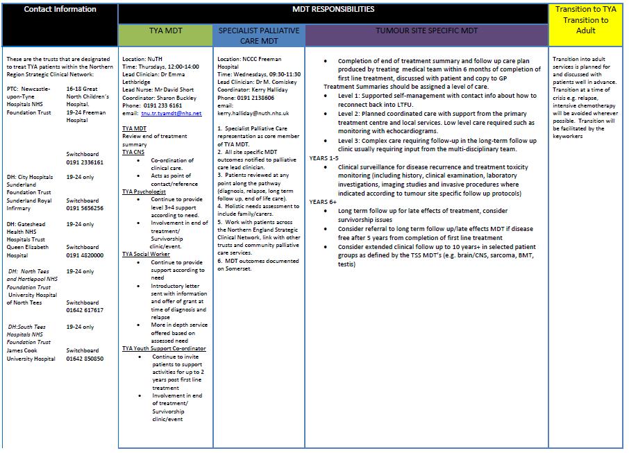
35 EAG GUIDELINES FOR TEENAGE AND YOUNG ADULTS Teenage and Young Adults Peer Review Measures Topic 11-1C (Functions of the Network Site Specific Groups for TYA) 1. Teenage and Young Adult Pathway for initial Management The EAG has received the document named NCA Teenage and Young Adult Cancer Pathway Guidance Paper and agrees to follow the generic TYA Pathway with any site specific variations to be documented. Please see Appendix 1 for pathway. 2. Teenage and Young Adult Pathway for Follow up on completion of first line treatment The EAG has received the document named NCA Teenage and Young Adult Cancer Pathway for follow up on completion of first line treatment and aged to follow the generic TYA pathway. Please see Appendix 2 for pathway. 3. Pathways for cases involving Specialised NHS services (Only Gynae and Sarcoma) The Gynae EAG and SAG reviewed and agreed the Specialised NHS Service pathway for patient s age years. This is attached in Appendix 3 35

36 Appendix 1 Teenage and Young Adult Pathway for initial Management 36

37 Appendix 2- TYA follow up on completion of first line treatment 37
38 38
39 Appendix 3 Contact Details List of designated MDTs at Principal Treatment Centre and TYA Designated Hospitals (19-24 years) Name of NHS Trust and designated hospital site Name of MDT TYA Lead Clinician TYA Lead Nurse Contact Number All MDTs: Principal Treatment Centre Breast Dr Emma Lethbridge David Short Colorectal Gynaeoncology (diagnostic) Haematology Head & Neck Lung Neurooncology (Brain/Spinal, Pituitary, Skull Base) Sarcoma Specialist Skin Specialist pancreatic Supra T-cell Lymphoma Teenage and Young Adult MDT Testicular Thyroid Specialist Upper GI Specialist Urology david.short@nuth.nhs.uk (Dect48858) Gateshead Health NHS Foundation Trust - at Queen Elizabeth Hospital Specialist Gynaeoncology Ms Christine Ang rachel.mugnai@ghnt.nhs.uk City Hospitals Sunderland NHS Foundation Trust - at Sunderland Royal Hospital North Tees and Hartlepool NHS Foundation Trust - at University Hospital of North Tees South Tees Hospital NHS Foundation Trust - at James Cook University Hospital Haematology Specialist Urology (testicular only ) Dr Scott Marshall Faye Laverick faye.armstrong@chsft.nhs.uk All MDTs: Dr Padmaja Kat Dawson Haematology Lokireddy Katherine.Dawson@nth.nhs.uk Local Urology Thyroid Breast Colorectal Lung Local Upper GI All MDTs: Dr Dianne Plews Jill Linton Specialist Gynaeoncology jill.linton@stees.nhs.uk Breast Colorectal Haematology Head & Neck Lung Neurooncology Specialist Skin Thyroid Specialist Upper GI Specialist Urology ext
40 Appendix 4 NHS Specialised Services Pathway 40
41 41

42 APPENDIX 1-2ww REFERRAL FORM 42
Staging and Treatment Update for Gynecologic Malignancies
 Staging and Treatment Update for Gynecologic Malignancies Bunja Rungruang, MD Medical College of Georgia No disclosures 4 th most common new cases of cancer in women 5 th and 6 th leading cancer deaths
GYNAECOLOGICAL CANCER CLINICAL GUIDELINES
 Northern England Strategic Clinical Networks GYNAECOLOGICAL CANCER CLINICAL GUIDELINES Gynae-Oncology NSSG on behalf of NECN Document Information Title: NECN Gynae Cancer Clinical Guidelines Author: Gynae
Guideline for the Management of Vulval Cancer
 Version History Guideline for the Management of Vulval Cancer Version Date Brief Summary of Change Issued 2.0 20.02.08 Endorsed by the Governance Committee 2.1 19.11.10 Circulated at NSSG meeting 2.2 13.04.11
North of Scotland Cancer Network Clinical Management Guideline for Carcinoma of the Uterine Cervix
 THIS DOCUMENT North of Scotland Cancer Network Carcinoma of the Uterine Cervix UNCONTROLLED WHEN PRINTED DOCUMENT CONTROL Prepared by A Kennedy/AG Macdonald/Others Approved by NOT APPROVED Issue date April
PRINCESS MARGARET CANCER CENTRE CLINICAL PRACTICE GUIDELINES GYNECOLOGIC CANCER CERVIX
 PRINCESS MARGARET CANCER CENTRE CLINICAL PRACTICE GUIDELINES GYNECOLOGIC CANCER CERVIX Site Group: Gynecology Cervix Author: Dr. Stephane Laframboise 1. INTRODUCTION 3 2. PREVENTION 3 3. SCREENING AND
North of Scotland Cancer Network Clinical Management Guideline for Endometrial Cancer
 THIS DOCUMENT North of Scotland Cancer Network Clinical Management Guideline for Endometrial Cancer Based on WOSCAN CMG with further extensive consultation within NOSCAN UNCONTROLLED WHEN PRINTED DOCUMENT
Proposed All Wales Vulval Cancer Guidelines. Dr Amanda Tristram
 Proposed All Wales Vulval Cancer Guidelines Dr Amanda Tristram Previous FIGO staging FIGO Stage Features TNM Ia Lesion confined to vulva with
Type I. Type II. Excess estrogen Lynch Endometrioid adenocarcinoma PTEN. High grade More aggressive Serous, Clear Cell p53
 Type I Excess estrogen Lynch Endometrioid adenocarcinoma PTEN Type II High grade More aggressive Serous, Clear Cell p53 Stage I IA IB Stage II Stage III IIIA IIIB IIIC IIIC1 IIIC2 Stage IV IVA IVB nodes
Cervical Cancer: 2018 FIGO Staging
 Cervical Cancer: 2018 FIGO Staging Jonathan S. Berek, MD, MMS Laurie Kraus Lacob Professor Stanford University School of Medicine Director, Stanford Women s Cancer Center Senior Scientific Advisor, Stanford
Coversheet for Network Site Specific Group Agreed Documentation
 Coversheet for Network Site Specific Group Agreed Documentation This sheet is to accompany all documentation agreed by Pan Birmingham Cancer Network Site Specific Groups. This will assist the Network Governance
The International Federation of Gynecology and Obstetrics (FIGO) updated the staging
 updated the staging") Continuing Education Column Revised FIGO Staging System Hee Sug Ryu, MD Department of Obstetrics and Gynecology, Ajou University School of Medicine E - mail : hsryu@ajou.ac.kr J Korean Med Assoc 2010;
Continuing Education Column Revised FIGO Staging System Hee Sug Ryu, MD Department of Obstetrics and Gynecology, Ajou University School of Medicine E - mail : hsryu@ajou.ac.kr J Korean Med Assoc 2010;
3/25/2019. Rare uterine cancers ~3% Leiomyosarcoma Carcinosarcoma (MMMT) Endometrial Stromal Sarcomas Aggressive tumors High Mortality Rates
 Endometrial Stromal Sarcomas Aggressive tumors High Mortality Rates") J. Anthony Rakowski D.O., F.A.C.O.O.G. MSU SCS Board Review Coarse Rare uterine cancers ~3% Leiomyosarcoma Carcinosarcoma (MMMT) Endometrial Stromal Sarcomas Aggressive tumors High Mortality Rates Signs
J. Anthony Rakowski D.O., F.A.C.O.O.G. MSU SCS Board Review Coarse Rare uterine cancers ~3% Leiomyosarcoma Carcinosarcoma (MMMT) Endometrial Stromal Sarcomas Aggressive tumors High Mortality Rates Signs
C ORPUS UTERI C ARCINOMA STAGING FORM (Carcinosarcomas should be staged as carcinomas)
") CLINICAL C ORPUS UTERI C ARCINOMA STAGING FORM PATHOLOGIC Extent of disease before S TAGE C ATEGORY D EFINITIONS Extent of disease through any treatment completion of definitive surgery y clinical staging
CLINICAL C ORPUS UTERI C ARCINOMA STAGING FORM PATHOLOGIC Extent of disease before S TAGE C ATEGORY D EFINITIONS Extent of disease through any treatment completion of definitive surgery y clinical staging
New Cancer Cases By Site Breast 28% Lung 14% Colo-Rectal 10% Uterus 6% Thyroid 5% Lymphoma 4% Ovary 3%
 Uterine Malignancy New Cancer Cases By Site 2010 Breast 28% Lung 14% Colo-Rectal 10% Uterus 6% Thyroid 5% Lymphoma 4% Ovary 3% Cancer Deaths By Site 2010 Lung 26% Breast 15% Colo-Rectal 9% Pancreas 7%
Adjuvant Therapies in Endometrial Cancer. Emma Hudson
 Adjuvant Therapies in Endometrial Cancer Emma Hudson Endometrial Cancer Most common gynaecological cancer Incidence increasing in Western world 1-2% cancer deaths 75% patients postmenopausal 97% epithelial
Chapter 2: Initial treatment for endometrial cancer (including histologic variant type)
") Chapter 2: Initial treatment for endometrial cancer (including histologic variant type) CQ01 Which surgical techniques for hysterectomy are recommended for patients considered to be stage I preoperatively?
Chapter 2: Initial treatment for endometrial cancer (including histologic variant type) CQ01 Which surgical techniques for hysterectomy are recommended for patients considered to be stage I preoperatively?
MPH Quiz. 1. How many primaries are present based on this pathology report? 2. What rule is this based on?
 MPH Quiz Case 1 Surgical Pathology from hysterectomy performed July 11, 2007 Final Diagnosis: Uterus, resection: Endometrioid adenocarcinoma, Grade 1 involving most of endometrium, myometrial invasion
C ORPUS UTERI C ARCINOMA STAGING FORM (Carcinosarcomas should be staged as carcinomas)
") C ORPUS UTERI C ARCINOMA STAGING FORM CLINICAL Extent of disease before any treatment y clinical staging completed after neoadjuvant therapy but before subsequent surgery Tis * T1 I T1a IA NX N0 N1 N2
C ORPUS UTERI C ARCINOMA STAGING FORM CLINICAL Extent of disease before any treatment y clinical staging completed after neoadjuvant therapy but before subsequent surgery Tis * T1 I T1a IA NX N0 N1 N2
SCAN Gynaecological Group. Clinical Management Protocols vulval cancer
 SE Scotland Cancer Network SCAN Gynaecological Group Clinical Management Protocols vulval cancer 2009 www.scan.scot.nhs.uk August 2001 updated annually, most recently INTRODUCTION The South East Scotland
PRINCESS MARGARET CANCER CENTRE CLINICAL PRACTICE GUIDELINES GYNECOLOGIC CANCER VULVAR
 PRINCESS MARGARET CANCER CENTRE CLINICAL PRACTICE GUIDELINES GYNECOLOGIC CANCER VULVAR Last Revision Date July 2015 1 Site Group: Gynecologic Cancer Vulvar Author: Dr. Stephane Laframboise 1. INTRODUCTION
Endometrial Cancer. Incidence. Types 3/25/2019
 Endometrial Cancer J. Anthony Rakowski DO, FACOOG MSU SCS Board Review Coarse Incidence 53,630 new cases yearly 8,590 deaths yearly 4 th most common malignancy in women worldwide Most common GYN malignancy
North of Scotland Cancer Network Clinical Management Guideline for Cancer of the Ovary
 North of Scotland Cancer Network Cancer of the Ovary Based on WOSCAN CMG with further extensive consultation within NOSCAN UNCONTROLLED WHEN PRINTED DOCUMENT CONTROL Prepared by NOSCAN Gynaecology Cancer
Referral and Management Guidelines for Gynaecological Cancers within North Trent
 North Trent Cancer Network Referral and Management Guidelines for Gynaecological Cancers within North Trent Final Version 3.0 August 2011 Review date : June 2013 Produced by the North Trent Cancer Network
Gynaecology NSSG (Lancs & South Cumbria) Uterine Cancer Guidelines (V4.0)
 Uterine Cancer Guidelines (V4.0)") Gynaecology NSSG (Lancs & South Cumbria) Uterine Cancer Guidelines (V4.0) ** VALID ON DATE OF PRINTING ONLY all guidelines available on the Strategic Clinical Network website : GMLSC SCN Date first published
Gynaecology NSSG (Lancs & South Cumbria) Uterine Cancer Guidelines (V4.0) ** VALID ON DATE OF PRINTING ONLY all guidelines available on the Strategic Clinical Network website : GMLSC SCN Date first published
Case Scenario 1. History
 History Case Scenario 1 A 53 year old white female presented to her primary care physician with post-menopausal vaginal bleeding. The patient is not a smoker and does not use alcohol. She has no family
Cervical Cancer 3/25/2019. Abnormal vaginal bleeding
 Cervical Cancer Abnormal vaginal bleeding Postcoital, intermenstrual or postmenopausal Vaginal discharge Pelvic pain or pressure Asymptomatic In most patients who are not sexually active due to symptoms
PORTEC-4. Patient seqnr. Age at inclusion (years) Hospital:
 Hospital:") May 2016 Randomisation Checklist Form 1, page 1 of 2 Patient seqnr. Age at inclusion (years) Hospital: Eligible patients should be registered and randomised via the Internet at : https://prod.tenalea.net/fs4/dm/delogin.aspx?refererpath=dehome.aspx
May 2016 Randomisation Checklist Form 1, page 1 of 2 Patient seqnr. Age at inclusion (years) Hospital: Eligible patients should be registered and randomised via the Internet at : https://prod.tenalea.net/fs4/dm/delogin.aspx?refererpath=dehome.aspx
Cervical cancer presentation
 Carcinoma of the cervix: Carcinoma of the cervix is the second commonest cancer among women worldwide, with only breast cancer occurring more commonly. Worldwide, cervical cancer accounts for about 500,000
Invasive Cervical Cancer: Squamous Cell, Adenocarcinoma, Adenosquamous
 Note: If available, clinical trials should be considered as preferred treatment options for eligible patients (www.mdanderson.org/gynonctrials). Other co-morbidities are taken into consideration prior
Algorithms for management of Cervical cancer
 Algithms f management of Cervical cancer Algithms f management of cervical cancer are based on existing protocols and guidelines within the ESGO comunity and prepared by ESGO Educational Committe as a
Essex & East Suffolk Gynae Cancer Supra-Network
 Essex & East Suffolk Gynae Cancer Supra-Network Gynaecological Cancers Referral, Diagnosis and Management Guidelines Constitution Version Number 9.0 Author Members of the NSSG Date Written August 2010
Janjira Petsuksiri, M.D
 GYN malignancies Janjira Petsuksiri, M.D Outlines Cervical cancer Endometrial cancer Ovarian cancer Vaginal cancer Vulva cancer 2 CA Cervix Epidemiology - Second most common female cancer Risk factors
UTERINE SARCOMA EXAMPLE OF A UTERINE SARCOMA USING PROPOSED TEMPLATE
 UTERINE SARCOMA EXAMPLE OF A UTERINE SARCOMA USING PROPOSED TEMPLATE Case: Adenosarcoma with heterologous elements and stromal overgrowth o TAH, BSO, omentectomy, staging biopsies of cul-de-sac, bladder
Staging. Carcinoma confined to the corpus. Carcinoma confined to the endometrium. Less than ½ myometrial invasion. Greater than ½ myometrial invasion
 5 th of June 2009 Background Most common gynaecological carcinoma in developed countries Most cases are post-menopausal Increasing incidence in certain age groups Increasing death rates in the USA 5-year
MRI in Cervix and Endometrial Cancer
 28th Congress of the Hungarian Society of Radiologists RCR Session Budapest June 2016 MRI in Cervix and Endometrial Cancer DrSarah Swift St James s University Hospital Leeds, UK Objectives Cervix and endometrial
Study Title The SACS trial - Phase II Study of Adjuvant Therapy in CarcinoSarcoma of the Uterus
 Study Title The SACS trial - Phase II Study of Adjuvant Therapy in CarcinoSarcoma of the Uterus Investigators Dr Bronwyn King, Peter MacCallum Cancer Centre Dr Linda Mileshkin, Peter MacCallum Cancer Centre
Endometrial Cancer. Saudi Gynecology Oncology Group (SGOG) Gynecological Cancer Treatment Guidelines
 Gynecological Cancer Treatment Guidelines") Saudi Gynecology Oncology Group (SGOG) Gynecological Cancer Treatment Guidelines Endometrial Cancer Emad R. Sagr, MBBS, FRCSC Consultant Gynecology Oncology Security forces Hospital, Riyadh Epidemiology
Saudi Gynecology Oncology Group (SGOG) Gynecological Cancer Treatment Guidelines Endometrial Cancer Emad R. Sagr, MBBS, FRCSC Consultant Gynecology Oncology Security forces Hospital, Riyadh Epidemiology
Vaginal intraepithelial neoplasia
 Vaginal intraepithelial neoplasia The terminology and pathology of VAIN are analogous to those of CIN (VAIN I-III). The main difference is that vaginal epithelium does not normally have crypts, so the
NAACCR Webinar Series /7/17
 COLLECTING CANCER DATA: UTERUS 2017 2018 NAACCR WEBINAR SERIES Q&A Please submit all questions concerning webinar content through the Q&A panel. Reminder: If you have participants watching this webinar
Gynecologic Cancer InterGroup Cervix Cancer Research Network. Management of Cervical Cancer in Resource Limited Settings.
 Management of Cervical Cancer in Resource Limited Settings Linus Chuang MD Conflict of Interests None Cervical cancer is the fourth most common malignancy in women worldwide 530,000 new cases per year
Gynecologic Oncologist. Surgery Chemotherapy Radiation Therapy Hormonal Therapy Immunotherapy. Cervical cancer
 Gynecologic Oncology Pre invasive vulvar, vaginal, & cervical disease Vulvar Cervical Endometrial Uterine Sarcoma Fallopian Tube Ovarian GTD Gynecologic Oncologist Surgery Chemotherapy Radiation Therapy
ARROCase: Locally Advanced Endometrial Cancer
 ARROCase: Locally Advanced Endometrial Cancer Charles Vu, MD (PGY-3) Faculty Advisor: Peter Y. Chen, MD, FACR Beaumont Health (Royal Oak, MI) November 2016 Case 62yo female with a 3yr history of vaginal
Index. B Bilateral salpingo-oophorectomy (BSO), 69
, 69") A Advanced stage endometrial cancer diagnosis, 92 lymph node metastasis, 92 multivariate analysis, 92 myometrial invasion, 92 prognostic factors FIGO stage, 94 histological grade, 94, 95 histologic cell
A Advanced stage endometrial cancer diagnosis, 92 lymph node metastasis, 92 multivariate analysis, 92 myometrial invasion, 92 prognostic factors FIGO stage, 94 histological grade, 94, 95 histologic cell
Jacqui Morgan March 6, 2019
 Jacqui Morgan March 6, 2019 Case 1 25yo, G2P1 Here for WWE, no problems, healthy, needs refill on OCPs. Pap- Abnormal Glandular Cells-NOS Now What?? Case 1 Colposcopy What findings? Case 1 ECC Cervical
Please complete prior to the webinar. HOSPITAL REGISTRY WEBINAR FEMALE REPRODUCTIVE SYSTEM EXERCISES CASE 1: FEMALE REPRODUCTIVE
 Please complete prior to the webinar. HOSPITAL REGISTRY WEBINAR FEMALE REPRODUCTIVE SYSTEM EXERCISES PHYSICAL EXAMINATION CASE 1: FEMALE REPRODUCTIVE 3/5 Patient presents through the emergency room with
Uterine Cervix. Protocol applies to all invasive carcinomas of the cervix.
 Uterine Cervix Protocol applies to all invasive carcinomas of the cervix. Protocol revision date: January 2005 Based on AJCC/UICC TNM, 6 th edition and FIGO 2001 Annual Report Procedures Cytology (No Accompanying
Uterus Malignancies /5/15
 Collecting Cancer Data: Uterus 2014-2015 NAACCR Webinar Series February 5, 2015 Q&A Please submit all questions concerning webinar content through the Q&A panel. Reminder: If you have participants watching
Uterine Malignancies. Collecting Cancer Data: Uterine Malignancies 10/7/2010. NAACCR Webinar Series 1. Questions. Fabulous Prizes!!!
 Uterine October 7, 2010 NAACCR 2010-2011 Webinar Series Session 1 1 Questions Please use the Q&A panel to submit your questions Send questions to All Panelist 2 Fabulous Prizes!!! 3 NAACCR 2010-2011 Webinar
Vulvar Carcinoma. Definition: Cases should be classified as carsinoma of the vulva when the primary site growth is in the vulva Malignant melanoma sho
 Carcinoma Vulva & Vagina Subdivisi Onkologi Ginekologi Bagian Obgin FK USU Vulvar Carcinoma. Definition: Cases should be classified as carsinoma of the vulva when the primary site growth is in the vulva
Clinical guideline Published: 27 April 2011 nice.org.uk/guidance/cg122
 Ovarian cancer: recognition and initial management Clinical guideline Published: 27 April 2011 nice.org.uk/guidance/cg122 NICE 2018. All rights reserved. Subject to Notice of rights (https://www.nice.org.uk/terms-and-conditions#notice-ofrights).
3/25/2019. J. Anthony Rakowski D.O., F.A.C.O.O.G. MSU SCS Board Review Coarse
 J. Anthony Rakowski D.O., F.A.C.O.O.G. MSU SCS Board Review Coarse 1 4 th most common GYN cancer 5% of malignancies of GYN type. 4850 new cases annually 1030 deaths Cigarette smoking Vulvar dystrophy (Lichen
MUSCLE - INVASIVE AND METASTATIC BLADDER CANCER
 10 MUSCLE - INVASIVE AND METASTATIC BLADDER CANCER Recommendations from the EAU Working Party on Muscle Invasive and Metastatic Bladder Cancer G. Jakse (chairman), F. Algaba, S. Fossa, A. Stenzl, C. Sternberg
Enterprise Interest None
 Enterprise Interest None Cervical Cancer -Management of late stages ESP meeting Bilbao Spain 2018 Dr Mary McCormack PhD FRCR Consultant Clinical Oncologist University College Hospital London On behalf
Cervical Cancer Guidelines L and SC Network July Introduction:
 Cervical Cancer Guidelines L and SC Network July 2018 Introduction: There was a total number of 442 cases of cervix cancer diagnosed in Lancashire and South Cumbria Cancer Network in the period 2005 2009
One of the commonest gynecological cancers,especially in white Americans.
 Gynaecology Dr. Rozhan Lecture 6 CARCINOMA OF THE ENDOMETRIUM One of the commonest gynecological cancers,especially in white Americans. It is a disease of postmenopausal women with a peak incidence in
2009 USCAP Gyn Pathology Evening Session Case #3. Richard J. Zaino, MD Hershey Medical Center Penn State University Hershey, PA
 2009 USCAP Gyn Pathology Evening Session Case #3 Richard J. Zaino, MD Hershey Medical Center Penn State University Hershey, PA rzaino@psu.edu Clinical history Middle aged woman with an exophytic mass of
Case Scenario 1. Pathology report Specimen from mediastinoscopy Final Diagnosis : Metastatic small cell carcinoma with residual lymphatic tissue
 Case Scenario 1 Oncology Consult: Patient is a 51-year-old male with history of T4N3 squamous cell carcinoma of tonsil status post concurrent chemoradiation finished in October two years ago. He was hospitalized
 TOC NCCN Categories of Evidence and Consensus Category 1: Based upon high-level evidence, there is uniform NCCN consensus that the intervention is appropriate. Category 2A: Based upon lower-level evidence,
ENDOMETRIAL CANCER Updated Apr 2017 by: Dr. Jenny Ko (Medical Oncologist, Abbotsford Cancer Centre)
") ENDOMETRIAL CANCER Updated Apr 2017 by: Dr. Jenny Ko (Medical Oncologist, Abbotsford Cancer Centre) Source: UpToDate 2017, ASCO/CCO/Alberta provincial guidelines, NCCN Reviewed by: Dr. Sarah Glaze (Gynecologic
ENDOMETRIAL CANCER Updated Apr 2017 by: Dr. Jenny Ko (Medical Oncologist, Abbotsford Cancer Centre) Source: UpToDate 2017, ASCO/CCO/Alberta provincial guidelines, NCCN Reviewed by: Dr. Sarah Glaze (Gynecologic
Pathway Gynaecology Cancer & Diagnostic Protocol for Inter Trust transfer
 NICaN Pathway Gynaecology Cancer & Diagnostic Protocol for Inter Trust transfer Timed schedules to enable the proactive management of the patient from point of receipt of referral to first definitive treatment
What is endometrial cancer?
 Uterine cancer What is endometrial cancer? Endometrial cancer is the growth of abnormal cells in the lining of the uterus. The lining is called the endometrium. Endometrial cancer usually occurs in women
Gynecological Cancers in Primary Care
 Gynecological Cancers in Primary Care Nora M. Lersch MSN CRNP AOCNP Division of Gynecological Oncology Objectives Identify the incidence of ovarian, cervical, vulvar and endometrial cancer Identify common
Guideline for the Follow-up of Patients with Gynaecological Malignancies
 Guideline for the Follow-up of Patients with Gynaecological Malignancies Version History Version Date Summary of Change/Process 2.0 20.02.08 Endorsed by the Governance Committee 2.1 18.11.10 Circulated
17 th ESO-ESMO Masterclass in clinical Oncology
 17 th ESO-ESMO Masterclass in clinical Oncology Cervical and endometrial Cancer Cristiana Sessa IOSI Bellinzona, Switzerland Berlin, March 28 th, 2018 Presenter Disclosures None Cervical Cancer Estimated
Epithelial Ovarian Cancer
 Epithelial Ovarian Cancer GYNE/ONC Practice Guideline Dr. Alex Hammond Dr. Ian Kerr Dr. Akira Sugimoto Dr. Stephen Welch Kay Faroni Christine Gawlik Kerri Thornton Approval Date: This guideline is a statement
Chapter 8 Adenocarcinoma
 Page 80 Chapter 8 Adenocarcinoma Overview In Japan, the proportion of squamous cell carcinoma among all cervical cancers has been declining every year. In a recent survey, non-squamous cell carcinoma accounted
Clinical Audit Data: 01 October 2015 to 30 September West of Scotland Cancer Network. Gynaecological Cancer Managed Clinical Network
 Gynaecological Cancer Managed Clinical Network Audit Report Ovarian Cancer Quality Performance Indicators Cervical Cancer Quality Performance Indicators Endometrial Cancer Quality Performance Indicators
CPC on Cervical Pathology
 CPC on Cervical Pathology Dr. W.K. Ng Senior Medical Officer Department of Clinical Pathology Pamela Youde Nethersole Eastern Hospital Cervical Smear: High Grade SIL (CIN III) Cervical Smear: High Grade
Vagina. 1. Introduction. 1.1 General Information and Aetiology
 Vagina 1. Introduction 1.1 General Information and Aetiology The vagina is part of internal female reproductive system. It is an elastic, muscular tube that connects the outside of the body to the cervix.
ICRT รศ.พญ.เยาวล กษณ ชาญศ ลป
 ICRT รศ.พญ.เยาวล กษณ ชาญศ ลป Brachytherapy การร กษาด วยร งส ระยะใกล Insertion การสอดใส แร Implantation การฝ งแร Surface application การวางแร physical benefit of brachytherapy - very high dose of radiation
Bladder Cancer Guidelines
 Bladder Cancer Guidelines Agreed by Urology CSG: October 2011 Review Date: September 2013 Bladder Cancer 1. Referral Guidelines The following patients should be considered as potentially having bladder
Role and Techniques of Surgery in Carcinoma Cervix. Dr Vanita Jain Additional Professor OBGYN PGIMER, Chandigarh
 Role and Techniques of Surgery in Carcinoma Cervix Dr Vanita Jain Additional Professor OBGYN PGIMER, Chandigarh Points for Discussion Pattern of spread Therapeutic options Types of surgical procedures
STUMPed for a Diagnosis Contemporary Management of Uterine Sarcomas
 UCSF Helen Diller Family Comprehensive Cancer Center Disclosures I have no financial disclosures STUMPed for a Diagnosis Contemporary Management of Uterine Sarcomas Lee-may Chen, MD Department of Obstetrics,
Sarah Burton. Lead Gynae Oncology Nurse Specialist Cancer Care Cymru
 Sarah Burton Lead Gynae Oncology Nurse Specialist Cancer Care Cymru Gynaecological Cancers Cervical Cancers Risk factors Presentation Early sexual activity Multiple sexual partners Smoking Human Papiloma
University of Kentucky. Markey Cancer Center
 University of Kentucky Markey Cancer Center Invasive Cancer of the Vagina and Urethra Fred Ueland, MD No matter what you accomplish in your life, the size of your funeral will still be determined by the
The Role of Radiation in the Management of Gynecologic Cancers. Scott Glaser, MD
 The Role of Radiation in the Management of Gynecologic Cancers Scott Glaser, MD Nothing to disclose DISCLOSURE Outline The role of radiation in: Endometrial Cancer Adjuvant Medically inoperable Cervical
UPDATE IN THE MANAGEMENT OF INVASIVE CERVICAL CANCER
 UPDATE IN THE MANAGEMENT OF INVASIVE CERVICAL CANCER Susan Davidson, MD Professor Department of Obstetrics and Gynecology Division of Gynecologic Oncology University of Colorado- Denver Anatomy Review
1. Written information to patient /GP: fax ASAP to GP & offer copy of consultation letter.
 Skin Cancer follow up guidelines If NEW serious diagnosis given: 1. Written information to patient /GP: fax ASAP to GP & offer copy of consultation letter. 2. Free prescription information details. 3.
Case 1. Gynaecology Case Presentation. Objectives. Disclosures 22/10/ year old female Clinical history: Assess right ovarian cyst
 Gynaecology Case Presentation Organ Imaging 2016 University of Toronto Sarah Johnson 39 year old female Clinical history: Assess right ovarian cyst Clinically diagnosed endometriosis Started fertility
Shared Care Pathway for Soft Tissue Sarcomas Presenting to Site Specialised MDTs. Gynaecological sarcomas Version 1
 Shared Care Pathway for Soft Tissue Sarcomas Presenting to Site Specialised MDTs Gynaecological sarcomas Version 1 Background This guidance is to provide direction for the management of patients with sarcomas
ARRO Case: Early-stage Endometrial Cancer
 ARRO Case: Early-stage Endometrial Cancer Ankit Modh, MD (PGY-4) Faculty Advisor: Mohamed A Elshaikh, MD Department of Radiation Oncology Henry Ford Cancer Institute Case Presentation 70 y/o African American
ENDOMETRIAL CANCER: A GUIDE FOR PATIENTS
 ENDOMETRIAL CANCER: A GUIDE FOR PATIENTS PATIENT INFORMATION BASED ON ESMO CLINICAL PRACTICE GUIDELINES This guide for patients has been prepared by Reliable Cancer Therapies (RCT) as a service to patients,
ENDOMETRIAL CANCER. Endometrial cancer is a great concern in UPDATE. For personal use only. Copyright Dowden Health Media
 For mass reproduction, content licensing and permissions contact Dowden Health Media. UPDATE ENDOMETRIAL CANCER Are lymphadenectomy and external-beam radiotherapy valuable in women who have an endometrial
MUSCLE-INVASIVE AND METASTATIC BLADDER CANCER
 MUSCLE-INVASIVE AND METASTATIC BLADDER CANCER (Text update March 2008) A. Stenzl (chairman), N.C. Cowan, M. De Santis, G. Jakse, M. Kuczyk, A.S. Merseburger, M.J. Ribal, A. Sherif, J.A. Witjes Introduction
MANAGEMENT OF CERVICAL CANCER
 MANAGEMENT OF CERVICAL CANCER Dr. Ujeen Shrestha Malla* and Prof. Dr. Zhang Shui Rong Department of Obstetrics and Gynaecology, Clinical Medical College of Yangtze University, Jingzhou Central Hospital,
Upper GI Malignancies Imaging Guidelines for the Management of Gastric, Oesophageal & Pancreatic Cancers 2012
 Upper GI Malignancies Imaging Guidelines for the Management of Gastric, Oesophageal & Pancreatic Cancers 2012 Version Control This is a controlled document please destroy all previous versions on receipt
North of Scotland Cancer Network Clinical Management Guideline for Metastatic Malignancy of Undefined Primary Origin (MUO)
") North of Scotland Cancer Network Clinical Management Guideline for Metastatic Malignancy of Undefined Primary Origin (MUO) UNCONTROLLED WHEN PRINTED DOCUMENT CONTROL Original Prepared by NMcL April 2016
North of Scotland Cancer Network Clinical Management Guideline for Metastatic Malignancy of Undefined Primary Origin (MUO) UNCONTROLLED WHEN PRINTED DOCUMENT CONTROL Original Prepared by NMcL April 2016
Gynecologic Malignancies. Kristen D Starbuck 4/20/18
 Gynecologic Malignancies Kristen D Starbuck 4/20/18 Outline Female Cancer Statistics Uterine Cancer Adnexal Cancer Cervical Cancer Vulvar Cancer Uterine Cancer Endometrial Cancer Uterine Sarcoma Endometrial
receive adjuvant chemotherapy
 Women with high h risk early stage endometrial cancer should receive adjuvant chemotherapy Michael Friedlander The Prince of Wales Cancer Centre and Royal Hospital for Women The Prince of Wales Cancer
CARCINOMA CERVIX. Dr. PREETHI REDDY. B. M S OBG II yr POST GRADUATE.
 CARCINOMA CERVIX Dr. PREETHI REDDY. B M S OBG II yr POST GRADUATE. Introduction Cervical cancer is the second most common female malignancy worldwide. It is responsible for 4,66,000 deaths annually worldwide
29 Cancer of the Uterine Corpus
 29 Cancer of the Uterine Corpus Robbert Soeters INTRODUCTION Malignancies affecting the uterine corpus are endometrial adenocarcinoma and uterine sarcomas. ENDOMETRIAL ADENOCARCINOMA Endometrial adenocarcinoma
North of Scotland Cancer Network Clinical Management Guideline for Oropharyngeal Cancer
 Nth of Scotland Cancer Netwk Clinical Management Guideline f Oropharyngeal Cancer UNCONTROLLED WHEN PRINTED Based on NHST CMG with further extensive consultation within NOSCAN DOCUMENT CONTROL Original
UTERINE SARCOMAS CURRENT THERAPEUTIC OPTIONS
 Review Journal of Translational Medicine and Research, volume 19, no. 1-2, 2014 UTERINE SARCOMAS CURRENT THERAPEUTIC OPTIONS N. Bacalbaæa 1, A. Traistaru 2, I. Bãlescu 3 1 Carol Davila University of Medicine
Cancer of Unknown Primary (CUP) Protocol
 Protocol") 1 Department of Oncology. Cancer of Unknown Primary (CUP) Protocol Version: Document type: Document sponsor Designation Document author [ s] Designation[s] Approving committee / Group Ratified by: Date
1 Department of Oncology. Cancer of Unknown Primary (CUP) Protocol Version: Document type: Document sponsor Designation Document author [ s] Designation[s] Approving committee / Group Ratified by: Date
Guidelines for the Management of Bladder Cancer West Midlands Expert Advisory Group for Urological Cancer
 Guidelines for the Management of Bladder Cancer West Midlands Expert Advisory Group for Urological Cancer West Midlands Clinical Networks and Clinical Senate Coversheet for Network Expert Advisory Group
Gynaecological Oncology Cases
 Gynaecological Oncology Cases 1. Tamoxifen and the endometrium 2. Cancer and the older woman Dr Julie M Lamont Consultant Gynaecological Oncologist Epworth Freemasons Hospital 21 st April 2015 Mrs FS 66
Cancer of the corpus uteri
 DOI: 10.1002/ijgo.12612 FIGO CANCER REPORT 2018 Cancer of the corpus uteri Frédéric Amant 1,2,3, * Mansoor Raza Mirza 4 Martin Koskas 5 Carien L. Creutzberg 6 1 Division of Gynecologic Oncology, University
LCA Gynaecological Cancer Clinical Guidelines
 LCA Gynaecological Cancer Clinical Guidelines July 2014 LCA GYNAECOLOGICAL CANCER CLINICAL GUIDELINES Contents Introduction... 5 Executive summary... 6 1 The Structure of Gynaecology Oncology Services...