PROSTATE CANCER (CORE/NEEDLE BIOPSY) STRUCTURED REPORTING PROTOCOL (2nd Edition 2018)
|
|
|
- Adrian Lang
- 5 years ago
- Views:
Transcription
1 PROSTATE CANCER (CORE/NEEDLE BIOPSY) STRUCTURED REPORTING PROTOCOL (2nd Edition 2018) Incorporating the: International Collaboration on Cancer Reporting (ICCR) Prostate Cancer Core Needle Biopsy Specimen Dataset 1
2 Core Document versions: World Health Organization (WHO). Classification of tumours. Pathology and genetics of the urinary system and male genital organs. 4 th edition. 2
3 ISBN: Publications number (SHPN): (CI) Online copyright RCPA 2018 This work (Protocol) is copyright. You may download, display, print and reproduce the Protocol for your personal, non-commercial use or use within your organisation subject to the following terms and conditions: 1. The Protocol may not be copied, reproduced, communicated or displayed, in whole or in part, for profit or commercial gain. 2. Any copy, reproduction or communication must include this RCPA copyright notice in full. 3. With the exception of Chapter 6 - the checklist, no changes may be made to the wording of the Protocol including any Standards, Guidelines, commentary, tables or diagrams. Excerpts from the Protocol may be used in support of the checklist. References and acknowledgments must be maintained in any reproduction or copy in full or part of the Protocol. 4. In regard to Chapter 6 of the Protocol - the checklist: o o o o o o The wording of the Standards may not be altered in any way and must be included as part of the checklist. Guidelines are optional and those which are deemed not applicable may be removed. Numbering of Standards and Guidelines must be retained in the checklist, but can be reduced in size, moved to the end of the checklist item or greyed out or other means to minimise the visual impact. Additional items for local use may be added but must not be numbered as a Standard or Guideline, in order to avoid confusion with the RCPA checklist items. Formatting changes in regard to font, spacing, tabulation and sequencing may be made. Commentary from the Protocol may be added or hyperlinked to the relevant checklist item. Apart from any use as permitted under the Copyright Act 1968 or as set out above, all other rights are reserved. Requests and inquiries concerning reproduction and rights should be addressed to RCPA, 207 Albion St, Surry Hills, NSW 2010, Australia. First published: July 2018, 2nd Edition (version 2.0) 3
4 Disclaimer The Royal College of Pathologists of Australasia ("College") has developed these protocols as an educational tool to assist pathologists in reporting of relevant information for specific cancers. While each protocol includes standards and guidelines which are indicators of minimum requirements and recommendations, the protocols are a first edition and have not been through a full cycle of use, review and refinement. Therefore, in this edition, the inclusion of standards and guidelines in each document are provided as an indication of the opinion of the relevant expert authoring group, but should not be regarded as definitive or as widely accepted peer professional opinion. The use of these standards and guidelines is subject to the clinician s judgement in each individual case. The College makes all reasonable efforts to ensure the quality and accuracy of the protocols and to update the protocols regularly. However subject to any warranties, terms or conditions which may be implied by law and which cannot be excluded, the protocols are provided on an "as is" basis. The College does not warrant or represent that the protocols are complete, accurate, error-free, or up to date. The protocols do not constitute medical or professional advice. Users should obtain appropriate medical or professional advice, or where appropriately qualified, exercise their own professional judgement relevant to their own particular circumstances. Users are responsible for evaluating the suitability, accuracy, currency, completeness and fitness for purpose of the protocols. Except as set out in this paragraph, the College excludes: (i) all warranties, terms and conditions relating in any way to; and (ii) all liability (including for negligence) in respect of any loss or damage (including direct, special, indirect or consequential loss or damage, loss of revenue, loss of expectation, unavailability of systems, loss of data, personal injury or property damage) arising in any way from or in connection with; the protocols or any use thereof. Where any statute implies any term, condition or warranty in connection with the provision or use of the protocols, and that statute prohibits the exclusion of that term, condition or warranty, then such term, condition or warranty is not excluded. To the extent permitted by law, the College's liability under or for breach of any such term, condition or warranty is limited to the resupply or replacement of services or goods. 4
5 Contents Scope...6 Abbreviations...7 Definitions...8 Introduction Authority and development Clinical information and surgical handling Specimen handling and macroscopic findings Microscopic findings Ancillary studies findings Synthesis and overview Structured checklist Formatting of pathology reports Appendix 1 Pathology request form for prostate cancer Appendix 2 Guidelines for formatting of a pathology report Appendix 3 Example of a pathology report References
6 Scope This protocol contains standards and guidelines for the structured reporting of prostate core needle biopsies. The dataset applies to invasive carcinomas of the prostate gland. Rare urothelial carcinomas arising within the prostate are also included. There are separate protocols for radical prostatectomy and transurethral resection (TUR) specimens. Structured reporting aims to improve the completeness and usability of pathology reports for clinicians, and improve decision support for cancer treatment. This protocol can be used to define and report the minimum data set but the structure is scalable and can equally accommodate a maximum data set or fully comprehensive report. 6
7 Abbreviations AJCC CG CS EPE FISH ICCR ISUP LIS LVI PBS PIN PSA RCPA SVI TNM UICC WHO American Joint Committee on Cancer Commentary for a guideline Commentary for a standard extraprostatic extension Fluorescent in-situ hybridization International Collaboration on Cancer Reporting International Society of Urological Pathology laboratory information system lymphovascular invasion Pharmaceutical Benefits Scheme prostatic intraepithelial neoplasia prostate specific antigen Royal College of Pathologists of Australasia seminal vesicle involvement tumour-node-metastasis International Union Against Cancer World Health Organization 7
8 Definitions The table below provides definitions for general or technical terms used in this protocol. Readers should take particular note of the definitions for standard, guideline and commentary, because these form the basis of the protocol. Ancillary study Clinical information Commentary An ancillary study is any pathology investigation that may form part of a cancer pathology report but is not part of routine histological assessment. Patient information required to inform pathological assessment, usually provided with the specimen request form, also referred to as pre-test information. Commentary is text, diagrams or photographs that clarify the standards (see below) and guidelines (see below), provide examples and help with interpretation, where necessary (not every standard or guideline has commentary). Commentary is used to: define the way an item should be reported, to foster reproducibility explain why an item is included (e.g. how does the item assist with clinical management or prognosis of the specific cancer). cite published evidence in support of the standard or guideline state any exceptions to a standard or guideline. In this document, commentary is prefixed with CS (for commentary on a standard) or CG (for commentary on a guideline), numbered to be consistent with the relevant standard or guideline, and with sequential alphabetic lettering within each set of commentaries (eg CS1.01a, CG2.05b). General commentary General commentary is text that is not associated with a specific standard or guideline. It is used: to provide a brief introduction to a chapter, if necessary for items that are not standards or guidelines but are included in the protocol as items of potential importance, for which there is currently insufficient evidence to recommend their inclusion. (Note: in future reviews of protocols, such items may be reclassified as either standards or guidelines, in line with diagnostic and prognostic advances, following evidentiary review). 8
9 Guideline Guidelines are recommendations; they are not mandatory, as indicated by the use of the word should. Guidelines cover items that are unanimously agreed should be included in the dataset but are not supported by NHMRC level III-2 evidence. 1 These elements may be clinically important and recommended as good practice but are not yet validated or regularly used in patient management. Guidelines include key information other than that which is essential for clinical management, staging or prognosis of the cancer such as macroscopic observations and interpretation, which are fundamental to the histological diagnosis and conclusion eg macroscopic tumour details, block identification key, may be included as either required or recommended elements by consensus of the expert committee. Such findings are essential from a clinical governance perspective, because they provide a clear, evidentiary decision-making trail. Guidelines are not used for research items. In this document, guidelines are prefixed with G and numbered consecutively within each chapter (eg G1.10). Macroscopic findings Microscopic findings Predictive factor Prognostic factor Standard Measurements, or assessment of a biopsy specimen, made by the unaided eye. In this document, the term microscopic findings refers to histomorphological assessment. A predictive factor is a measurement that is associated with response or lack of response to a particular therapy. A prognostic factor is a measurement that is associated with clinical outcome in the absence of therapy or with the application of a standard therapy. It can be thought of as a measure of the natural history of the disease. Standards are mandatory, as indicated by the use of the term must. Standards are essential for the clinical management, staging or prognosis of the cancer. These elements will either have evidentiary support at Level III-2 or above (based on prognostic factors in the NHMRC levels of evidence 1 document). In rare circumstances, where level III-2 evidence is not available an element may be made a Standard where there is unanimous agreement in the expert committee. An appropriate staging system eg Pathological TNM staging would normally be included as a required element.these elements must be recorded and at the discretion of the pathologist included in the pathology report according to the needs of the recipient of the report. The summation of all standards represents the minimum dataset for the cancer. In this document, standards are prefixed with S and numbered consecutively within each chapter (eg S1.02). 9
10 Structured report Synoptic report Synthesis A report format which utilises standard headings, definitions and nomenclature with required information. A structured report in condensed form (as a synopsis or precis). Synthesis is the process in which two or more pre-existing elements are combined, resulting in the formation of something new. The Oxford dictionary defines synthesis as the combination of components or elements to form a connected whole. In the context of structured pathology reporting, synthesis represents the integration and interpretation of information from two or more modalities to derive new information. 10
11 Introduction Prostate cancer Prostate cancer is the most common non-cutaneous related cancer with 19,233 new cases reported in Australia and 3129 in New Zealand in It is also the third most common cause of cancer death, in both Australia and New Zealand in ,3 Both the number of new cases and the number of deaths from prostate cancer are increasing, partly driven by the ageing of the population. 4 There is a wide variation in the biological behaviour of prostate cancer. Most tumours are relatively slow-growing; however, a significant minority have the propensity for aggressive behaviour, including metastasis, and such tumours can be fatal. 5 Benefits of structured reporting The pathology report lays the foundation for a patient s cancer journey and conveys information which: Provides the definitive diagnosis Includes critical information for Tumour-Node-Metastasis (TNM) staging Evaluates the adequacy of the surgical excision Provides morphological and biological prognostic markers which determine personalised cancer therapy However, the rapid growth in ancillary testing such as immunohistochemistry, flow cytometry, cytogenetics, and molecular studies, have made the task of keeping abreast of advances on specific cancer investigations extremely difficult for pathologists. The use of structured reporting checklists by pathologists ensures that all key elements are included in the report specifically those which have clinical management, staging or prognostic implications. Consequently minimum or comprehensive datasets for the reporting of cancer have been developed 6,7 around the world. Both the United Kingdom, 8 and United States 9 have produced standardised cancer reporting protocols or datasets for national use for many years. The use of cancer reporting checklists improves completeness and quality of cancer reporting and thereby ensures an improved outcome for cancer patients. This has long term cost implications for public health by ensuring the most effective and timely treatment based on accurate and complete information. The use of a structured reporting format also facilitates easy extraction of the necessary information by secondary users of the information ie cancer registries. Importance of histopathological reporting Information from pathology reports on core biopsy and transurethral resection (TUR) specimens, particularly Gleason grade and pathological stage, has a key role in the rational planning of patient management and is a major component of the most common nomograms used to guide clinical decision making. 10 International Collaboration on Cancer Reporting The International Collaboration on Cancer Reporting (ICCR), founded in 2011 by the Australasian (RCPA), US (CAP) and UK (RCPath) Colleges of Pathology and 11
12 the Canadian Association of Pathology (CAP-ACP) in association with the Canadian Partnership Against Cancer (CPAC), was established to explore the possibilities of a collaborative approach to the development of common, internationally standardised and evidence-based cancer reporting protocols for surgical pathology specimens. The ICCR, recognising that standardised cancer datasets have been shown to provide significant benefits for patients and efficiencies for organisations through the ease and completeness of data capture undertook to use the best international approaches and the knowledge and experience of expert pathologists, and produce cancer datasets which would ensure that cancer reports across the world will be of the same high quality ensuring completeness, consistency, clarity, conciseness and above all, clinical utility. Representatives from the four countries participating in the initial collaboration undertook a pilot project in 2011 to develop four cancer datasets - Lung, Melanoma, Prostate (Radical Prostatectomy), and Endometrium. Following on from the success of this pilot project, the ICCR was joined by the European Society of Pathology (ESP) in 2013 and in 2014 incorporated a not-for-profit organisation focussed on the development of internationally agreed evidencebased datasets developed by world leading experts. The ICCR Datasets are made freely available from its website Design of this protocol This structured reporting protocol has been developed using the ICCR dataset on prostate cancer (core needle biopsy specimens) as the foundation. This protocol includes all of the ICCR cancer dataset elements as well as additional information, elements and commentary as agreed by the RCPA expert committee. It provides a comprehensive framework for the assessment and documentation of pathological features of prostate cancer in core needle biopsy specimens ICCR dataset elements for prostate cancer in core needle biopsy specimens are included verbatim. ICCR Required elements are mandatory and therefore represented as standards in this document. ICCR Recommended elements, that is, those which are not mandatory but are recommended, may be included as guidelines or upgraded to a standard based on the consensus opinion of the local expert committee. The ICCR elements are identified in each chapter with the ICCR logo placed before the Standard or Guideline number or bullet and the ICCR element description and commentary is boarded by a grey box as shown below: G2.01 A block identification key listing the nature and origin of all tissue blocks should be recorded. Additional commentary by the RCPA expert committee may be added to an ICCR element but is not included in the grey bordered area nor indicated with an ICCR logo eg 12
13 G2.03 If present, the laterality of the lymph nodes submitted may be recorded as left, right or bilateral. CS2.03a If present, record site and number. All lymph node tissue should be submitted for histological examination. Further information on the ICCR is available at Checklist Consistency and speed of reporting is improved by the use of discrete data elements recorded from the checklist. Items suited to tick boxes are distinguished from more complex elements requiring free text or narrative. A structured or discrete approach to responses is favoured, however the pathologist is encouraged to include free text or narrative where necessary to document any other relevant issues, to give reasons for coming to a particular opinion and to explain any points of uncertainty. Report format The structure provided by the following chapters, headings and subheadings describes the elements of information and their groupings, but does not necessarily represent the format of either a pathology report (Chapter 7) or checklist (Chapter 6). These, and the structured pathology request form (Appendix 1) are templates that represent information from this protocol, organised and formatted differently to suit different purposes. Key documentation Guidelines for Authors of Structured Cancer Pathology Reporting Protocols, Royal College of Pathologists of Australasia, World Health Organization (WHO). Classification of tumours. Pathology and genetics of the urinary system and male genital organs. Humphrey PA, Moch H, Reuter VE, Ulbright TM editors. 4 th edition. Lyon, France: IARC Press; Changes since last edition Inclusion of all ICCR agreed REQUIRED and RECOMMENDED elements. 13
14 Authority and development This section provides information about the process undertaken to develop this protocol. This 2 nd edition of the protocol is an amalgam of two separate processes: 1. This protocol is based on the ICCR dataset prostate cancer core needle biopsy specimens 1 st edition. All ICCR elements from this dataset, both required (mandatory) and recommended (optional), are included in this protocol, verbatim. (It should be noted that RCPA feedback from all Anatomical Pathology fellows and specifically the local expert committee was sought during the development process of the ICCR dataset.) Details of the ICCR development process and the international expert authoring committee responsible for the ICCR dataset are available on the ICCR website: iccr-cancer.org. 2. Additional elements, values and commentary have been included as deemed necessary by the local expert committee. In addition, the standard inclusions of RCPA protocols eg example reports, request information etc, have also been added. Authorship 2 nd edition Clinical Professor James Kench (Chair and lead author), Pathologist Dr David Clouston, Pathologist Professor Brett Delahunt, Pathologist Adjunct Professor Warick Delprado, Pathologist Dr Thomas Eade, Radiation Oncologist Dr Lisa Horvath, Medical Oncologist Dr Krishan Rasiah, Urologist Professor Hema Samaratunga, Pathologist Associate Professor Jurgen Stahl, Pathologist Authorship 1 st edition (2014) Professor James Kench (Chair), Pathologist Dr David Clouston, Pathologist Professor Brett Delahunt, Pathologist Professor Warick Delprado, Pathologist Dr Thomas Eade, Radiation Oncologist Associate Professor David Ellis, Pathologist Dr Lisa Horvath, Medical Oncologist Dr Krishan Rasiah, Urologist Dr Hema Samaratunga, Pathologist Associate Professor Jurgen Stahl, Pathologist 14
15 Editorial manager Meagan Judge, Royal College of Pathologists of Australasia Acknowledgements The genitourinary expert committee wish to thank all the pathologists and clinicians who contributed to the discussion around this document. 15
16 Stakeholders ACT Health ACT Cancer Registry Australian Cancer Network Australian Commission on Safety and Quality in Health Care Australian Digital Health Agency Australian Institute of Health and Welfare Cancer Australia Cancer Council ACT Cancer Council Queensland Cancer Council Victoria Cancer Council Western Australia Cancer Institute NSW Cancer Services Advisory Committee (CanSAC) Cancer Voices NSW Clinical Oncology Society of Australia (COSA) Department of Health, Australia Department of Health, New Zealand Faculty of Radiation Oncology Genito-Urinary Group (FROGG) Health Informatics Society of Australia (HISA) Independent Review Group of Pathologists Medical Software Industry Association (MSIA) National Pathology Accreditation Advisory Council (NPAAC) New Zealand Cancer Registry Northern Territory Cancer Registry Pathology Australia Public Pathology Australia Queensland Cooperative Oncology Group (QCOG) RCPA Anatomical Pathology Advisory Committee (APAC) Representatives from laboratories specialising in anatomical pathology across Australia Royal Australasian College of Physicians (RACP) South Australia Cancer Registry Standards Australia Tasmanian Cancer Registry The Australian and New Zealand Urogenital and Prostate Cancer Trials Group (ANZUP) The Medical Oncology Group of Australia The Prostate Cancer Foundation of Australia (PCFA) 16
17 The Prostate Cancer Foundation of New Zealand (PCFNZ) The Royal Australasian College of Surgeons (RACS) The Royal Australian and New Zealand College of Radiologists (RANZCR) The Royal Australian College of General Practitioners (RACGP) The Royal College of Pathologists of Australasia (RCPA) The Urological Society Of Australia And New Zealand (USANZ) Western Australia Clinical Oncology Group (WACOG) Development process This protocol has been developed following the ten-step process set out in Guidelines for Authors of Structured Cancer Pathology Reporting Protocols. 17 Where no reference is provided, the authority is the consensus of the local expert group for local inclusions and the ICCR Dataset Authoring Committee for ICCR components denoted with the ICCR logo. 17
18 1 Pre-analytical This chapter relates to information that should be recorded on receipt of the specimen in the laboratory. The pathologist is reliant on the quality of information received from the clinicians or requestor. Some of this information may be received in generic pathology request forms; however, the additional information required by the pathologist specifically for the reporting of Prostate Core Needle Biopsies is outlined in Appendix 1. Appendix 1 also includes a standardised request information sheet that may be useful in obtaining all relevant information from the requestor. Surgical handling procedures affect the quality of the specimen and recommendations for appropriate surgical handling are included in Appendix 1. S1.01 All demographic information provided on the request form and with the specimen must be recorded. CS1.01a CS1.01b CS1.01c The Royal College of Pathologists of Australasia (RCPA) The Pathology Request-Test-Report Cycle Guidelines for Requesters and Pathology Providers must be adhered to. 18 This document specifies the minimum information to be provided by the requesting clinician for any pathology test. Whether or not the patient identifies as Aboriginal and/ or Torres Strait Islander. This is in support of a government initiative to monitor the health of indigenous Australians particularly in relation to cancer. The patient s health identifiers may include the patient s Medical Record Number as well as a national health number such as a patient s Individual Healthcare Identifier (IHI) (Australia) or the National Healthcare Identifier (New Zealand). S1.02 All clinical information as documented on the request form must be recorded verbatim. CS1.02a CS1.02b The request information may be recorded as a single text (narrative) field or it may be recorded in a structured format. The copy doctors requested on the request form must be recorded. S1.03 The pathology accession number of the specimen must be recorded. S1.04 The principal clinician involved in the patient s care and responsible for investigating the patient must be recorded. CS1.04a The principle clinician should provide key information regarding the clinical presentation of the patient. Follow up may be required with the principle clinician for a 18
19 number of reasons: The clinical assessment and staging may be incomplete at the time of biopsy. The pathology request is often authored by the clinician performing the biopsy rather than the clinician who is investigating and managing the patient. The identity of this clinician is often not indicated on the pathology request form In practice therefore, it is important in such cases that the reporting pathologist should be able to communicate with the managing clinician for clarification. CS1.04b The Australian Healthcare identifiers i.e. Healthcare Provider Identifier - Individual (HPI-I) and Healthcare Provider Identifier - Organisation (HPI-O) should be included, where possible, to identify the principal clinician involved in the patient's care. G1.01 Any clinical information received in other communications from the requestor or other clinician should be recorded together with the source of that information. 19
20 2 Specimen handling and macroscopic findings This chapter relates to the procedures required after the information has been handed over from the requesting clinician, and the specimen has been received in the laboratory. Specimen handling Detailed fixation and specimen handling instructions are available from the RCPA online Cut-up Manual: The tissue from each specimen container must be submitted and processed as a separate specimen. 19 Preferably the urologist should submit each needle core in a separate container, so that each specimen jar will contain only one core. This is in accordance with the current consensus recommendations from the College of American Pathologists, International Society of Urological Pathology and Association of Directors of Anatomic and Surgical Pathology. 20 When there are 2 or more cores per container, fragmentation, if present, precludes accurate assessment of i) the number of cores received; ii) the number of positive cores if carcinoma is present in more than one fragment and; iii) the extent of tumour in each core. These are important prognostic parameters, particularly with respect to the optimal selection of patients for active surveillance protocols. 20,21 There is a greater tendency to core fragmentation when >1 core is submitted in a container. 22 Furthermore, if >2 cores are submitted in one tissue cassette/block it is difficult to align them all within one plane during embedding. Since foci of prostate carcinoma in needle core biopsies are often small, this may lead to the carcinoma not being represented on the slide and a false negative diagnosis being rendered. Deeper sections may not necessarily avoid this problem if the area of interest has been lost during block trimming in an attempt to cut a full face section. Macroscopic findings S2.01 Details of each submitted specimen should be recorded as follows: a) the location of each specimen as stated on the specimen containers b) the total number of cores in the specimen container 20
21 c) the length of core(s) must be measured (in millimetres). CS2.01a Information on specimens submitted for histopathological examination, including location, number of needle cores and length of cores, is regarded as an integral and essential part of a pathology report. 23 The length of the cores should be measured in the wet specimen before tissue processing and paraffin embedding. Preferably there should be only 1 needle core in each specimen jar. However, if 2 or more needle cores are submitted in one container and there is some fragmentation, it may not be possible to reliably determine the number of involved cores. In this situation the urologist should state on the pathology request/requisition form how many cores were submitted in each jar to avoid counting fragments of the one core as separate cores (particularly with cores <6 mm long) and providing misleading information on tumour extent. 20 Where more than 5 cores are submitted in a specimen jar, e.g. with saturation/template biopsies, a range may be submitted for length of the cores rather than measuring each one individually. S2.02 A block identification key listing the nature and origin of all tissue blocks must be recorded. CS2.02a The origin/designation of all tissue blocks should be recorded and it is preferable to document this information in the final pathology report. If this information is not included in the final pathology report, it should be available on the laboratory computer system and relayed to the reviewing pathologist. 23 Specifically for needle core biopsy cases, this information may be helpful in interpreting specimens where the urologist has submitted more than one needle core per specimen container. Recording the origin/designation of tissue blocks also facilitates retrieval of blocks, for example for further immunohistochemical or molecular analysis, research studies or clinical trials. G2.01 A descriptive or narrative field should be provided to record any macroscopic information that is not recorded in the above standards and guidelines, and that would normally form part of the macroscopic description. CG2.01a The traditional macroscopic narrative recorded at the time of specimen dissection is often reported separately from the cancer dataset. Although this remains an option, it is recommended that macroscopic information be recorded within the overall 21
22 structure of this protocol. CG2.01b Much of the information recorded in a traditional macroscopic narrative is covered in the standards and guidelines above and in many cases, no further description is required. 22
23 3 Microscopic findings This section relates to purely histological or morphological assessment. Information derived from multiple investigational modalities, or from two or more chapters, is described in Chapter 5. S3.01 The histological findings must be recorded either: i) individually; or ii) at case level (ie. global or composite reporting) CS3.01a i) Individual specimen reporting has some advantages over case level reporting since it: - Delineates the region with the highest score and allows urologists to decide if nerve sparing operations can be done on one side only. - Facilitates surgical decision making, including wide dissection at apex and bladder neck as well as nerve sparing - Partin tables, etc use the highest core s score, not the average or composite. ii) Alternatively, depending on local practice, case level reporting may be used instead. However, case level or composite reporting may not be appropriate for targeted biopsies. S3.02 The histological tumour type must be recorded. CS3.02a Choose from the following values: Adenocarcinoma (Acinar) Other (specify) Refer to Appendix 4. CS3.02b The vast majority (>95%) of prostate cancers are acinar adenocarcinomas. 24 Other types of carcinoma are rarer but must be recorded if present, since some variants, such as ductal adenocarcinoma, small cell carcinoma, sarcomatoid carcinoma and urothelialtype adenocarcinoma, have a significantly poorer prognosis The tumour type should be assigned in line with the 2016 World Health Organisation (WHO) classification and mixtures of different types should 23
24 be indicated. 24 Subtypes of prostate carcinoma are often identified in combination with acinar type carcinoma, and in such cases the tumour type should be classified according to the subtype. Urothelial carcinomas arising in the urinary bladder or urethra are dealt with in separate datasets; however, those rare urothelial carcinomas arising within the prostate are included in this dataset. Information on histological tumour type may be recorded at a specimen level or at a case level depending on local practice. The response type No evidence of primary tumour should only be used if specimen level reporting is utilised. S3.03 The Histological grade must be recorded 16,30,31 using the a. Gleason grading system (ISUP 2014) b. The International Society of Urological Pathology (ISUP) Grade (also known as Grade Group) (See Tables 1 and 2 and Figure 1) CS3.03a The Gleason grading system is the foundation of grading for prostatic adenocarcinoma. The Gleason score is traditionally obtained by adding the two predominant Gleason patterns or doubling the pattern in cases with uniform grade. This was modified in the International Society of Urological Pathology (ISUP) 2005 revision by always including the highest grade in the Gleason score of needle biopsies, regardless of its amount. 30 At a subsequent ISUP consensus conference in 2014, the Gleason system was further modified and many of the decisions taken at this meeting have been included in the 4th edition of the WHO classification. It was decided that Gleason pattern 4 should include fused or poorly formed glands, glomerulations and all cribriform patterns of acinar adenocarcinoma. A grouping of the Gleason scores into 5 grade categories was proposed and this was endorsed by the ISUP Council (March 2015). Over the past decades Gleason scores below 6 have become less commonly used, especially on needle biopsies. There is also an understanding that Gleason score 7 tumours have a worse prognosis if there is a predominant pattern 4 (4+3) than if pattern 3 dominates (3+4). Both the Gleason score and the ISUP grade (Grade group) should always be reported for the sake of clarity. The ISUP consensus conference also recommended that the percentage of Gleason pattern 4 be reported in cases with ISUP grades 2 or 3. The rationale for this is to indicate if the tumour is bordering on the lower 24
25 or higher ends of Gleason score 7. In some jurisdictions, Gleason score 7 tumours with 10% pattern 4 are considered for active surveillance. 32 The percentage of Gleason pattern 4 and 5 is reported by some pathologists 33 but this was not endorsed by the WHO classification working group. This element is thus optional. Depending on local practice, the different elements of grade data may be reported on either core or specimen level or as a composite (global) grade based on all cancer present in the biopsy cores or a combination of both. The grade groups and associated definitions are outlined in Table 1. Both the Gleason score and the ISUP grade (Grade group) should always be reported for the sake of clarity. Table 1: ISUP grading system, core/needle biopsies and transurethral resection of the prostate (TURP) specimens ISUP grade (Grade group) Gleason score Definition Grade Only individual discrete well-formed glands Grade 2 3+4=7 Predominantly well-formed glands with lesser component (*) of poorly- formed/fused/cribriform glands Grade 3 4+3=7 Predominantly poorly-formed/fused/cribriform glands with lesser component (**) of well-formed glands 4+4=8 Only poorly-formed/fused/cribriform glands Grade 4 3+5=8 5+3=8 Predominantly well-formed glands and lesser component (*) lacking glands (or with necrosis) Predominantly lacking glands (or with necrosis) and lesser component (**) of well-formed glands Grade Lack gland formation (or with necrosis) with or without poorly formed/fused/cribriform glands * Any component of the high-grade pattern (i.e. even if less than 5%) is included in the grade. ** The low-grade pattern is included in the grade only if it is at least 5%. 25

26 Figure 1 Gleason grade 5 carcinoma Table 2 Gleason scoring of unusual patterns Pattern Morphology Comment Vacuoles Cytoplasmic change seen in all grades Grade as if vacuoles were absent, on the underlying architecture Mucin extravasation Grade based on glandular architecture Mucinous fibroplasia Collagenous micronodules Grade based on glandular architecture Glomeruloid structures Grade as 4 Foamy gland change Grade based on glandular architecture Small cell neuroendocrine Do not assign a grade G3.01 The percentage of Gleason pattern 4/5, where the Gleason scores 7, should be recorded for each specimen. S3.04 Tumour extent in each separately received specimen must be recorded. CS3.04a An estimate of the extent of carcinoma in each specimen is important. The following methods are recommended: i) The number of positive cores over the total number of cores. and ii) the length of tissue involved by carcinoma (in mm) 26
27 or iii) the linear extent of prostatic tissue involved by carcinoma as a percentage. CS3.04b Number of biopsy cores positive for cancer and linear extent of cancer in the cores correlate with tumour volume, postoperative stage and outcome. 10,34-37 Number of positive cores should be reported but may be difficult to determine because of fragmentation when multiple cores have been submitted together. The number of positive cores should not be greater than the number of cores taken (as specified in Clinical Information ). Site specific labelling and single core submission facilitates the assessment of cancer extent. 19 Linear extent should be reported and may be recorded either as millimetres cancer length or % cancer in each core or as a composite measure of cancer involvement in all cores. 38 CS3.04c The methods for reporting of discontinuous cancer remain controversial. Whether intervening benign tissue is included or subtracted from the extent measurement may determine eligibility for active surveillance. The tumour extent of a discontinuous cancer can be reported by either: a) including the intervening benign tissue, e.g. a 1 mm focus of carcinoma separated from a second 1mm focus by 10 mm of intervening benign tissue would be reporting as spanning a distance of 12 mm (see Fig 2a); or b) subtracting the intervening benign tissue, e.g. a 1 mm focus of carcinoma separated from a second 1mm focus by 10 mm of intervening benign tissue would be reporting as having a tumour extent of 2 mm (see Fig 2b). 39 In either case, the presence of discontinuous cancer should be noted and the method used in calculating extent clearly stated. 27
28 Figure 2 a) Measurement of discontinuous carcinoma in a core/needle biopsy b) S3.05 The presence or absence of perineural invasion must be recorded. CS3.05a The significance of perineural invasion in prostate core biopsy specimens is uncertain. Some studies show a correlation with extraprostatic extension (EPE) in the corresponding radical prostatectomy specimens or an association with adverse outcome in patients treated with radical prostatectomy or external beam radiation ,43-45 Other investigators have questioned prognostic value of biopsy perineural invasion in univariate or multivariate analyses A systematic review of the literature concluded that the weight of evidence suggested that in clinically localised disease perineural invasion was a significant prognostic factor for EPE and subsequent local recurrence. 50 In advanced disease perineural invasion is common and probably not of prognostic significance. It should also be noted that nerves are not necessarily observed in biopsy material, therefore it is not always possible to assess the possibility of perineural invasion. G3.02 The presence or absence of seminal vesicle invasion should be recorded. CG3.02a Seminal vesicle invasion (SVI) is rarely identified in needle biopsies cores, hence its absence does not need to be explicitly stated. However, if seminal vesicle/ejaculatory duct invasion is present it should be recorded and the following comments apply. 28
29 SVI is defined as involvement of the muscular wall of the extraprostatic portion of the seminal vesicle. 51 If possible seminal vesicle tissue is present (either unintentionally or intentionally, as in a targeted biopsy) and involved by carcinoma, this may be significant since it indicates that the tumour could be pt3b in the American Joint Committee on Cancer (AJCC)/Union for International Cancer Control (UICC) Staging system. 52,53 However, assessment of SVI is problematic in needle biopsy specimens since it is impossible to reliably distinguish between extraprostatic seminal vesicle and intraprostatic seminal vesicle tissue, therefore it is important not to over interpret invasion of the latter two structures as SVI since their involvement by tumour does not constitute pt3b disease. Unless one is dealing with a targeted seminal vesicle biopsy, it is recommended to report tumour involvement of such structures in a needle core biopsy as seminal vesicle/ejaculatory duct invasion rather than as SVI. S3.06 The presence or absence of lymphovascular invasion must be recorded. CS3.06a Lymphovascular invasion (LVI) is rarely identified in needle biopsies cores, hence its absence does not need to be explicitly stated. However, if LVI is present it should be recorded and the following comments apply. Invasion of lymphatic or blood vessels (i.e. thin-walled endothelial-lined spaces) is uncommonly identified in needle core biopsy specimens and there is little published data on the significance of LVI specifically relating to prostate core biopsies. However, there is good evidence that LVI is a significant independent prognostic indicator of increased risk of recurrence post radical prostatectomy; therefore, if LVI is identified in a needle core it may well be significant and its presence should be recorded. The presence of LVI does not affect assignment of the AJCC/UICC T category. 29
 became accepted terminology at a 1996 consensus conference, and replaces earlier ambiguous terms such capsular penetration, perforation, or invasion.")
30 Figure 3 Lymphovascular invasion in a small vessel S3.07 The presence of extraprostatic extension (EPE) must be recorded. CS3.07a Extraprostatic extension (EPE) became accepted terminology at a 1996 consensus conference, and replaces earlier ambiguous terms such capsular penetration, perforation, or invasion. 58 In radical prostatectomy specimens EPE is an independent prognostic indicator of increased risk of recurrence post radical prostatectomy and is important in assignment of the AJCC/UICC T category. 59,60 There is limited data specifically on the significance of EPE in needle core biopsies given that it is relatively uncommonly identified; however, it may be occasionally be seen and should be reported when present since it indicates that the tumour is at least pt3a in the TNM system. 52 In needle cores it is defined as tumour admixed with adipocytes, usually at the end of a biopsy core. It is recommended that the site of any EPE present is recorded since this information is useful for correlation with magnetic resonance imaging (MRI) results and may assist the urologist or radiation oncologist with the technical aspects of treatment planning. 30
 A B A: EPE in a core/needle biopsy, H&E stain at 100x scale B: EPE in a core/needle biopsy, H&E stain at 400x scale S3.")
 is an uncommon finding in needle biopsies cores, hence its absence does not need to be explicitly stated.")
31 G3.03 The location of extraprostatic extension (EPE) may be recorded. Figure 4 Extraprostatic extension (EPE) A B A: EPE in a core/needle biopsy, H&E stain at 100x scale B: EPE in a core/needle biopsy, H&E stain at 400x scale S3.08 The presence or absence of intraductal carcinoma of the prostate (IDC-P) must be recorded. CS3.08a Intraductal carcinoma of the prostate (IDC-P) is an uncommon finding in needle biopsies cores, hence its absence does not need to be explicitly stated. However, if IDC-P is present it should be recorded and the following comments apply. IDC-P is usually associated with invasive prostate cancer, however, occasionally isolated IDC-P is found without invasive carcinoma; this latter situation is rare and beyond the scope of this dataset. IDC-P has been well characterised at the histological and molecular levels over the past decade and its clinical significance is now also better understood. 61 The diagnosis of IDC-P is based on morphology and the key criteria include: 1) large calibre glands that are more than twice the diameter of normal nonneoplastic peripheral glands; 2) preserved (at least focally) basal cells identified on H&E staining (or with basal cell markers, such as p63, keratin 34βE12 and keratin 5/6, however, the use of immunohistochemistry to identify basal cells is optional, rather than mandatory, for the diagnosis of IDC-P); 3) significant nuclear atypia including enlargement and anisonucleosis; and 4) comedonecrosis, which is often but not always present. 62,63 It is important to distinguish IDC-P from 31


32 high grade prostatic intraepithelial neoplasia (HGPIN): compared to IDC-P, HGPIN has less architectural and cytological atypia, and cribriform HGPIN is rare. IDC-P is strongly associated with high volume, high grade invasive prostate carcinoma and metastatic disease, hence the presence of IDC-P in a biopsy, even if invasive carcinoma cannot be identified, mandates immediate repeat biopsy or definitive therapy (depending on the clinical situation) In a cohort treated with radiation +/- androgen deprivation therapy, the presence of IDC-P in the needle biopsy was an independent predictor of early biochemical recurrence and metastasis. 68 There was a strong consensus (82%) at the recent ISUP consensus meeting (Chicago 2014) that IDC-P should not be assigned an ISUP or Gleason grade. 31 Figure 5 Intraductal carcinoma of the prostate IDC-P A B A: IDC-P in a core/needle biopsy, 100x scale B: IDC-P in a core/needle biopsy, 200x scale G3.04 The presence or absence of co-existent pathology should be recorded. CG3.04a In some cases clinical management decisions may be aided by knowledge of coexisting pathology, such as HGPIN, glandular atypia suspicious for malignancy (atypical small acinar proliferation), granulomatous prostatitis etc. If there is carcinoma present, the presence of HGPIN is generally not significant, except perhaps occasionally in the situation where the carcinoma is of very limited extent. Even if no cancer is identified in the specimen, the significance of finding HGPIN in needle core biopsies has been controversial with some studies finding an 32
33 increased risk for detection of prostatic adenocarcinoma in subsequent biopsies, while others did not. 69,70 Recent studies, including one analysing data from a large Canadian cohort, found that this risk was related to the extent of HGPIN, i.e. the number of involved sites; only patients with multifocal HGPIN had a significantly increased risk of prostate cancer Low grade prostatic intraepithelial neoplasia (PIN) should not be reported. Likewise, if there is carcinoma present in a specimen, the presence of glandular atypia suspicious for malignancy (atypical small acinar proliferation) is generally not significant, except perhaps occasionally in the situation where the carcinoma is of very limited extent. In specimens where there is no cancer identified but glandular atypia is present, the risk of carcinoma being present in subsequent biopsies is approximately 50% Active prostatitis and granulomatous prostatitis may cause a rise in serum prostate-specific antigen (PSA), although inflammatory lesions may coexist with carcinoma and it is important not to assume that their presence always accounts for an unexplained increase in a patient s PSA. G3.05 Comments should be included, if appropriate. CG3.05a Free text entry to allow any additional, unusual or unexpected findings, such as seminal vesicle invasion, to be reported. 33
34 4 Ancillary studies findings No ancillary tests are currently used on a routine diagnostic basis for prostate cancer. However, several have been proposed and there may be a sufficient evidence base in the future for including ancillary prognostic or predictive biomarkers or tests in later editions of this protocol. 34
35 5 Synthesis and overview Information that is synthesised from multiple modalities and therefore cannot reside solely in any one of the preceding chapters is described here. For example, tumour stage is synthesised from multiple classes of information clinical, macroscopic and microscopic. By definition, synthetic elements are inferential rather than observational, often representing high-level information that is likely to form part of the Summary or Diagnosis section in the final formatted report. Overarching case comment is synthesis in narrative format. Although it may not necessarily be required in any given report, the provision of the facility for overarching commentary in a cancer report is essential. G5.01 The Diagnostic summary section of the final formatted report should include relevant information for clinical decision making. CG5.01a CG5.01b CG5.01c CG5.01d It is appropriate to draw all the specimens together at the end of the report as a summary, particularly as specimen numbers can sometimes pass fourteen to twenty per patient. The summary should include relevant positive and negative findings. A summary Gleason Score should be included. It is controversial as to whether just the highest score or both the highest and a composite/global score (reflecting the overall grading of all the specimens received) should be included. 30 Clinicians need a single score for nomograms but will vary in which they prefer. Hence, to fit local practice a composite/global score may be given in addition to the highest score. If this option is utilised, then the type of score should be clearly identified in the report. The summary can be formatted as follows: Tumour Type Location of positive cores Adenocarcinoma (or variant) State location of positive cores Gleason Score Highest (eg: 4+4=8) Composite* (eg: 4+3=7) Perineural invasion Lymphovascular invasion Extraprostatic extension Present / Not identified Present / Not identified Present / No extraprostatic tissue present / Not identified 35
36 Intraductal ca. of prostate Comment Present/ Not identified free text for any unusual aspects *Optional element. May not be appropriate when targeted biopsies have been taken. S5.01 The reporting system must provide a field for free text or narrative in which the reporting pathologist can give overarching case comment, if required. CS5.01a This field may be used, for example, to: explain the decision-making pathway, or any elements of clinicopathological ambiguity, or factors affecting diagnostic certainty, thereby allowing communication of diagnostic subtlety or nuance that is beyond synoptic capture give recommendations for further action or investigation document further consultation or results still pending. CS5.01b Use of this field is at the discretion of the reporting pathologist. G5.02 The edition/version number of the RCPA protocol on which the report is based should be included on the final report. CG5.02a For example, the pathology report may include the following wording at the end of the report: the data fields within this formatted report are aligned with the criteria as set out in the RCPA document XXXXXXXXXX XXXX Edition dated XXXXXXX. 36
37 6 Structured checklist The following checklist contains all the standards and guidelines for this protocol in the simplest possible form. The summation of all standards is equivalent to the minimum data set for prostate cancer. For emphasis, standards (mandatory elements) are formatted in bold font. S6.01 The structured checklist provided below may be modified as required but with the following restrictions: a. All standards and their respective naming conventions, definitions and value lists must be adhered to. b. Guidelines are not mandatory but are best practice and where used, must follow the naming conventions, definitions and value lists given in the protocol. G6.01 The order of information and design of the checklist may be varied according to the laboratory information system (LIS) capabilities and as described in Functional Requirements for Structured Pathology Reporting of Cancer Protocols. 78 CG6.01a CG6.01b Where the LIS allows dissociation between data entry and report format, the structured checklist is usually best formatted to follow pathologist workflow. In this situation, the elements of synthesis or conclusions are necessarily at the end. The report format is then optimised independently by the LIS. Where the LIS does not allow dissociation between data entry and report format, (for example where only a single text field is provided for the report), pathologists may elect to create a checklist in the format of the final report. In this situation, communication with the clinician takes precedence and the checklist design is according to principles given in Chapter 7. G6.02 Where the checklist is used as a report template (see G6.01), the principles in Chapter 7 and Appendix 2 apply. CG6.02a All extraneous information, tick boxes and unused values should be deleted. G6.03 Additional comment may be added to an individual response where necessary to describe any uncertainty or nuance in the selection of a prescribed response in the checklist. Additional comment is not required where the prescribed response is adequate. 37
38 Values in italics are conditional on previous responses. Values in all caps are headings with sub values. S/G Item description Response type Conditional Pre-analytical S1.01 Demographic information provided S1.02 Clinical information provided on request form Not provided OR Text OR Structured entry as below: CLINICAL INFORMATION Previous history of prostate cancer (including the Gleason grade and score of previous specimens if known) Previous biopsy (specify date and where performed) Text Text Previous therapy Text Other Text Specimens provided Text 38
39 S/G Item description Response type Conditional Pre-biopsy serum PSA Clinical stage Numeric: ng/ml Text S1.03 Pathology accession number Alpha-numeric S1.04 Principal clinician caring for the patient Text G1.01 Comments Text Macroscopic findings S2.01 SPECIMEN(S) SUBMITTED Location of specimens Numeric: sequential indicator for each specimen (container) received AND Text: location Notes: Note that a sequential indicator, the location for each specimen must be recorded for each specimen received. 39
40 S/G Item description Response type Conditional Total number of cores Numeric: Notes: Length of core(s) Numeric: mm Note that the total number of cores must be recorded for each specimen received. AND/OR Fragments Notes: S2.02 Block identification key Text G2.01 Other macroscopic comment Text For each core received in S2.01 a length of core is recorded. Microscopic findings S3.02 Histological tumour type Multi selection value list (select all that apply): Adenocarcinoma (Acinar) Other (specify) 40
41 S/G Item description Response type Conditional S3.03 HISTOLOGICAL GRADE Conditional on presence of positive specimens in S3.02 GLEASON SCORE Indeterminate (specify reason) OR complete the following: Primary pattern/grade Numeric: (1-5) Secondary pattern/grade Numeric: (1-5) International Society of Urological Pathology (ISUP) Grade (Grade Group/Prognostic Group) G3.01 Percentage Gleason pattern 4/5 (applicable for Gleason scores 7) Single selection value list: ISUP Grade (Grade Group) 1 (Gleason score 6) ISUP Grade (Grade Group) 2 (Gleason score 3+4=7) ISUP Grade (Grade Group) 3 (Gleason score 4+3=7) ISUP Grade (Grade Group) 4 (Gleason score 8) ISUP Grade (Group Group) 5 (Gleason score 9-10) Indeterminate (specify reason) Numeric: % OR 41
42 S/G Item description Response type Conditional Not identified S3.04 TUMOUR EXTENT Notes: Note that tumour extent (whatever options) must be recorded for each positive specimen recorded in S3.02. Conditional on presence of positive specimens in S3.02 Number of positive cores ( 5mm) and the total number of cores Numeric: / AND Length of tissue involved by carcinoma Numeric: mm OR Linear extent of prostatic tissue involved by carcinoma Numeric: % 42
43 S/G Item description Response type Conditional S3.05 Perineural invasion Single selection value list: Not identified Present Conditional on presence of positive specimens in S3.02 Notes: Perineural invasion should be recorded for each positive specimen recorded in S3.02. Negative findings can be recorded per specimen or alternatively they can be recorded in the Summary section. G3.02 Seminal vesicle invasion Single selection value list: Not identified Present Conditional on presence of positive specimens in S3.02 Notes: Seminal vesicle/ejaculatory duct invasion should be recorded for each positive specimen recorded in S3.02. Negative findings can be recorded per specimen or alternatively they can be recorded in the Summary section. S3.06 Lymphovascular invasion Single selection value list: Not identified Present Conditional on presence of positive specimens in S3.02 Notes: Lymphovascular invasion should be recorded for each 43
44 S/G Item description Response type Conditional positive specimen recorded in S3.02. Negative findings can be recorded per specimen or alternatively they can be recorded in the Summary section. S3.07 Extraprostatic extension (EPE) Single selection value list: Notes: Not identified Present The presence of EPE is assessed for each positive specimen recorded in S3.02. Negative findings can be recorded per specimen or alternatively they can be recorded in the Summary section. Conditional on presence of positive specimens in S3.02 If present, consider reporting G3.03. G3.03 Location(s) of EPE Multi selection value list (select all that apply): Right base Right mid Right apex Left base Left mid Left apex Other (specify) Conditional on presence of EPE S3.08 Intraductal carcinoma of prostate (IDC-P) Single selection value list: Not identified Present 44
45 S/G Item description Response type Conditional Notes: The presence of IDC-P should be recorded for each specimen received in S2.02. Negative findings can be recorded per specimen or alternatively they can be recorded in the Summary section. G3.04 Coexistent pathology Single selection value list: G3.05 Additional microscopic comment Text None identified Present (specify) Synthesis and overview G5.01 Diagnostic summary Text/Table: Tumour Type Location Adenocarcinoma (or variant) State location of positive cores Gleason Score : Highest (eg: 4+4=8) Composite* (eg 4+3=7) Perineural invasion Lymphovascular present / not identified present / not identified 45
46 S/G Item description Response type Conditional invasion Extraprostatic extension Intraductal ca. of prostate Comment present / no extraprostatic tissue present / not identified present/not identified free text for any unusual aspects *Optional S5.01 Overarching comment Text G5.02 Edition/version number of the RCPA protocol on which the report is based Text 46
47 7 Formatting of pathology reports Good formatting of the pathology report is essential for optimising communication with the clinician, and will be an important contributor to the success of cancer reporting protocols. The report should be formatted to provide information clearly and unambiguously to the treating doctors, and should be organised with their use of the report in mind. In this sense, the report differs from the structured checklist, which is organised with the pathologists workflow as a priority. Uniformity in the format as well as in the data items of cancer reports between laboratories makes it easier for treating doctors to understand the reports; it is therefore seen as an important element of the systematic reporting of cancer. For guidance on formatting pathology reports, please refer to Appendix 2. An example of a pathology report is shown in Appendix 3. 47
48 Appendix 1 Pathology request information and surgical handling procedures This appendix describes the information that should be collected before the pathology test. Some of this information can be provided on generic pathology request forms; any additional information required specifically for the reporting of bladder cancer may be provided by the clinician on a separate request information sheet. An example request information sheet is included below. Elements which are in bold text are those which pathologists consider to be required information. Those in non-bold text are recommended. Also included in this appendix are the procedures that are recommended before handover of specimens to the laboratory. Clinical information relating to presenting symptoms and spread of disease including pretreatment prostate specific antigen (PSA) are necessary for staging of the tumour. Details of previous therapy are required because this often impacts upon the grading of the tumour and this needs to be taken into account by the examining pathologist. Similar information is required regardless of whether the specimen is a core biopsy, transurethral resection (TUR) or radical prostatectomy. Diagnosis of prostate cancer in thin core biopsy, TUR of prostate and prostatectomy specimens cannot be made on clinical grounds alone; rather, the diagnosis relies on histological examination of the specimen (see Chapter 3). The histopathology report forms part of a patient s permanent medical record and includes information that informs appropriate management. As such, the report provides a method for the recording relevant clinical information that will be permanently available even in the absence of the patient s detailed clinical notes. Patient information Adequate demographic and request information should be provided with the specimen. Items relevant to cancer reporting protocols include: i ii iii sex patient name date of birth iv identification and contact details of requesting doctor v date of request Whether or not the patient identifies as Aboriginal and/ or Torres Strait Islander. This is in support of a government initiative to monitor the health of indigenous Australians particularly in relation to cancer. The patient s health identifiers should be provided. 48
49 The patient s health identifiers may include the patient s Medical Record Number as well as a national health number such as a patient s Individual Healthcare Identifier (IHI) (Australia) or the National Healthcare Identifier (New Zealand). The Australian Healthcare identifiers i.e. Healthcare Provider Identifier - Individual (HPI-I) and Healthcare Provider Identifier - Organisation (HPI- O) should be use, where possible, to identify the requesting doctor. Clinical Information Clinical information should be provided including the following. It is the responsibility of the clinician requesting the pathological examination of a specimen to provide information that will have an impact on the diagnostic process or affect its interpretation. The use of a standard pathology requisition/request form including a checklist of important clinical information is strongly encouraged to help ensure that relevant clinical data is provided by the clinicians with the specimen. Information about prior biopsies or treatment aids interpretation of the microscopic findings and accurate pathological diagnosis, while knowledge of the number of needle cores taken from each site aids pathological assessment of the number of involved cores. Radiation and/or endocrine therapy for prostate cancer have a profound effect on the morphology of both the cancer and the benign prostatic tissue. For this reason, information about any previous therapy is important for the accurate assessment of needle core biopsies. Following irradiation, benign acinar epithelium shows nuclear enlargement and nucleolar prominence, 79 while basal cells may show cytological atypia, nuclear enlargement and nuclear smudging. 80 There may also be increased stromal fibrosis, which may resemble tumour-induced desmoplasia. These changes may persist for a considerable period, having been reported up to 72 months after treatment, and are more pronounced in patients who have undergone brachytherapy compared to those who have received external beam radiation therapy. 80,81 It is important to document any previous radiotherapy to help the pathologist to interpret changes accurately. Radiation may be associated with apparent upgrading of prostate cancer in prostatectomy specimens. 82 Likewise, neoadjuvant androgen deprivation therapy (ADT) may induce morphological changes in both prostate cancer and benign tissue. Androgen blockade induces basal cell hyperplasia and cytoplasmic vacuolation in benign prostatic tissue, although this is unlikely to be confused with malignancy. 83 More significantly from a diagnostic point of view, neoadjuvant ADT may increase the risk of overlooking acinar adenocarcinoma on low power microscopic examination due to collapse of glandular lumina, cytoplasmic pallor and shrinking of nuclei The effect of androgen blockage on prostate cancer is variable and an apparent upgrading of the cancer has been reported in a number of studies. 82,83 Hence, it has been suggested that in biopsies undertaken following either 49
50 radiotherapy or androgen deprivation therapy, tumours that show significant treatment effect should not be graded. 87 The Gleason grade and score of prostate cancer in any previously submitted specimen should also be provided by the clinician as this allows assessment of any progression of the tumour towards a higher grade/more undifferentiated state, which itself may be of prognostic significance. If the patient is on active surveillance this information should also be included. Previous history of prostate cancer (including the Gleason grade and score of previous specimens if known) In many cases the clinical history will influence the ultimate diagnosis and may provide information which will assist in providing prognostic information. Specifically, the Gleason grade and score of prostate cancer in any previously submitted specimen should be provided. This permits assessment of any progression of the tumour towards a more undifferentiated state, which itself is of prognostic significance. Previous biopsy (specify date and where performed) Previous therapy should be described. Radiation and /or endocrine therapy for prostate cancer has a profound effect on the morphology of both the cancer and the benign prostatic tissue. For this reason, information about any previous therapy is important for the accurate assessment of biopsies. Following irradiation, benign, acinar epithelium shows nuclear enlargement and nucleolar prominence 79 while basal cells may show cytologic atypia, nuclear enlargement and nuclear smudging. 80 There may also be increased stromal fibrosis, which may resemble tumour-induced desmoplasia. These changes may persist for a considerable period, have been reported up to 72 months after treatment and are more pronounced in patients who have undergone brachytherapy compared to those who have received external beam radiation therapy. 80,81 It is important to document any previous radiotherapy to help the pathologist to interpret changes accurately. Failure to do so could lead to an incorrect diagnosis of malignancy. Radiotherapy has a variable effect on prostate carcinoma. There is debate as to whether or not Gleason grading is appropriate 79,88 because radiation may be associated with apparent upgrading in prostatectomy specimens. 82 It has been suggested that in biopsies undertaken following radiotherapy, tumours that do not show any radiation effect should be graded, while tumours that show a treatment effect should not. 87 Neoadjuvant androgen blockade induces basal cell hyperplasia and cytoplasmic vacuolation in benign prostatic tissue, and this is unlikely to be confused with malignancy. 83 The effect of androgen blockage on prostate cancer is variable and an apparent 50
51 upgrading of the cancer has been reported in a number of studies. 82,83 The current consensus is that these tumours should not be graded. 89 The nature of the specimens provided must be clearly stated. Thin core biopsy is only undertaken for diagnostic purposes in men in which cancer is suspected on clinical grounds or following testing in asymptomatic patients. Core biopsies of prostate from different sites must be submitted in separate specimen pots. Preferably, each individual core/needle biopsy should be submitted in its own specimen jar. If the urologist includes >1, they should specify the number present in that specimen jar. This will facilitate accurate mapping of any cancer present and thereby inform surgical clearance and decisions regarding the appropriateness of undertaking nerve sparing procedures. Accurate mapping of cancers can also assist in planning of radiotherapy. The prebiopsy serum PSA value should be recorded. The clinician requesting the pathological examination should provide information on the pre-biopsy serum prostate-specific antigen (PSA) level. The use of a standard pathology requisition/request form including a checklist of important clinical information is strongly encouraged to help ensure that important clinical data is provided by the clinicians with the specimen. Despite criticisms about the utility of PSA-based prostate cancer screening, most prostate cancers are detected in asymptomatic men on the basis of PSA testing. Although PSA levels provide some indication of the likelihood of discovering cancer within a biopsy of the prostate, a diagnosis of malignancy should be based on histological findings and should not be influenced by PSA levels. In addition, serum PSA is a key parameter in some nomograms widely used to pre-operatively predict the American Joint Committee on Cancer (AJCC)/Union of International Cancer Control (UICC) pathological T category of prostate cancer or the risk of recurrence following radical prostatectomy and to guide clinical decision making with respect to disease management. 90 If the patient is on 5-alpha-reductase inhibitor medications, such as finasteride or dutasteride, this should be recorded as it may lower serum PSA levels and affect interpretation of serum PSA values for detecting prostate cancer Serum PSA is essential for stage grouping in the 7 th Edition of the AJCC/UICC TNM staging system. 94 In addition, serum PSA is a key parameter in some nomograms widely used to estimate the risk of recurrence post-operatively and guide clinical decision making on adjuvant therapy Despite criticisms about the utility of PSA-based prostate cancer screening, most prostate cancers are detected in asymptomatic 51
52 men on the basis of PSA testing. Although PSA levels provide some indication of the likelihood of discovering cancer within a biopsy of the prostate, a diagnosis of malignancy should be based on histological findings and should not be influenced by PSA levels. Elevated PSA levels may be due to acute or granulomatous prostatitis and, for this reason, any clinical evidence of prostatitis should be reported to the pathologist as part of the clinical history. 98 In isolation, PSA levels have a moderately high sensitivity but a low specificity for prostate cancer. Beyond the age of 50 years, PSA levels increase due to an increasing incidence of benign prostatic hyperplasia. There is no cut-off point for PSA levels that is diagnostic for cancer, although age related medians and reference intervals improve the predictive power of this test (see the RCPA Position Paper). 99,100 Elevated levels of serum PSA also provide prognostic information; in particular, pretreatment PSA levels have been correlated with tumour grade and stage. 101 The clinical stage should be recorded. The clinician requesting the pathological examination should provide information on the clinical stage. The use of a standard pathology requisition/request form including a checklist of important clinical information is strongly encouraged to help ensure that important clinical data is provided by the clinicians with the specimen. Along with pre-biopsy serum PSA, clinical stage is a vital parameter in some nomograms widely used to preoperatively predict the pathological T category of prostate cancer and to guide clinical decision making with respect to disease management. 90 Comments should be included, if appropriate. Space for free text should be included to encourage reporting of ambiguity, or for the addition of other comments. 52

53 Example request information sheet 53
54 The above Request Information Sheet is published to the RCPA website. 54
55 Appendix 2 Guidelines for formatting of a pathology report Layout Headings and spaces should be used to indicate subsections of the report, and heading hierarchies should be used where the LIS allows it. Heading hierarchies may be defined by a combination of case, font size, style and, if necessary, indentation. Grouping like data elements under headings and using white space assists in rapid transfer of information. 102 Descriptive titles and headings should be consistent across the protocol, checklist and report. When reporting on different tumour types, similar layout of headings and blocks of data should be used, and this layout should be maintained over time. Consistent positioning speeds data transfer and, over time, may reduce the need for field descriptions or headings, thus reducing unnecessary information or clutter. Within any given subsection, information density should be optimised to assist in data assimilation and recall. The following strategies should be used: Configure reports in such a way that data elements are chunked into a single unit to help improve recall for the clinician. 102 Reduce clutter to a minimum. 102 Thus, information that is not part of the protocol (eg billing information or Snomed codes) should not appear on the reports or should be minimised. Reduce the use of formatting elements (eg bold, underlining or use of footnotes) because these increase clutter and may distract the reader from the key information. Where a structured report checklist is used as a template for the actual report, any values provided in the checklist but not applying to the case in question must be deleted from the formatted report. Reports should be formatted with an understanding of the potential for the information to mutate or be degraded as the report is transferred from the LIS to other health information systems. As a report is transferred between systems: text characteristics such as font type, size, bold, italics and colour are often lost tables are likely to be corrupted as vertical alignment of text is lost when fixed font widths of the LIS are rendered as proportional fonts on screen or in print spaces, tabs and blank lines may be stripped from the report, disrupting the formatting supplementary reports may merge into the initial report. 55
56 Appendix 3 Examples of pathology reports These are example reports only, formats will vary with Laboratory System functionality and local design. The table in example 2 is regarded as an optimal method of rendition, however it is recognised that it may not be achievable in many existing Laboratory Systems. In general the laboratory should assess the capabilities of all end users to ensure faithful reproduction with the electronic transmission of any pathology report. Current Australian Standards stipulate that the report be sent both as a text version (atomic or otherwise) and that it be accompanied by a version rendered specifically as intended by the pathologist / laboratory. One way to achieve this is using a pdf format as below. 56

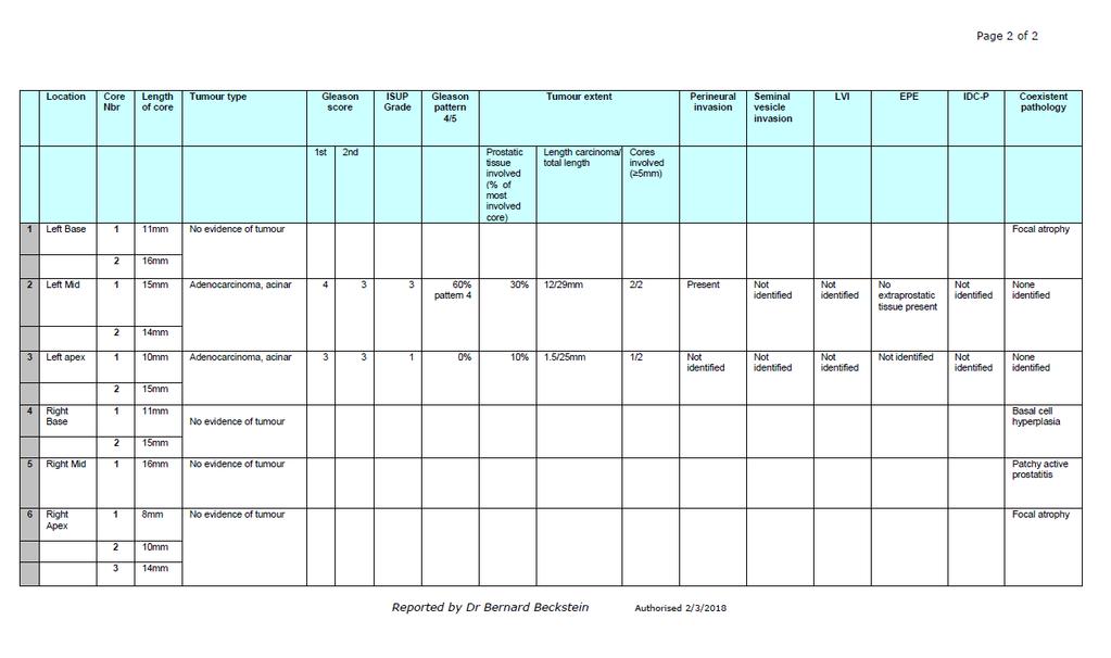
57 Example report 1 57
58 58
59 59

60 Example report 2 60
61 61
PROSTATE CANCER (TRANSURETHRAL RESECTION AND ENUCLEATION) STRUCTURED REPORTING PROTOCOL (1 st Edition 2018)
 STRUCTURED REPORTING PROTOCOL (1 st Edition 2018)") PROSTATE CANCER (TRANSURETHRAL RESECTION AND ENUCLEATION) STRUCTURED REPORTING PROTOCOL (1 st Edition 2018) Incorporating the: International Collaboration on Cancer Reporting (ICCR) Prostate Cancer - Transurethral
PROSTATE CANCER (TRANSURETHRAL RESECTION AND ENUCLEATION) STRUCTURED REPORTING PROTOCOL (1 st Edition 2018) Incorporating the: International Collaboration on Cancer Reporting (ICCR) Prostate Cancer - Transurethral
PROSTATE CANCER (RADICAL PROSTATECTOMY) STRUCTURED REPORTING PROTOCOL (3rd Edition 2018)
 STRUCTURED REPORTING PROTOCOL (3rd Edition 2018)") PROSTATE CANCER (RADICAL PROSTATECTOMY) STRUCTURED REPORTING PROTOCOL (3rd Edition 2018) Incorporating the: International Collaboration on Cancer Reporting (ICCR) Prostate Cancer - Radical Prostatectomy
PROSTATE CANCER (RADICAL PROSTATECTOMY) STRUCTURED REPORTING PROTOCOL (3rd Edition 2018) Incorporating the: International Collaboration on Cancer Reporting (ICCR) Prostate Cancer - Radical Prostatectomy
<INSERT CANCER NAME> STRUCTURED REPORTING PROTOCOL (<Insert whether 1 st, 2 nd etc> Edition> <Insert year published here e.g.
 STRUCTURED REPORTING PROTOCOL ( Edition> )
CARCINOMA OF THE URINARY BLADDER STRUCTURED REPORTING PROTOCOL
 CARCINOMA OF THE URINARY BLADDER STRUCTURED REPORTING PROTOCOL (RADICAL CYSTECTOMY, PARTIAL CYSTECTOMY, DIVERTICULECTOMY, CYSTOPROSTATECTOMY) (2nd Edition 2018) Incorporating the: International Collaboration
CARCINOMA OF THE URETHRA STRUCTURED REPORTING PROTOCOL. (1st Edition 2018)
") CARCINOMA OF THE URETHRA STRUCTURED REPORTING PROTOCOL (URETHRECTOMY) (1st Edition 2018) Incorporating the: International Collaboration on Cancer Reporting (ICCR) Dataset for the reporting of carcinoma
CARCINOMA OF THE URETHRA STRUCTURED REPORTING PROTOCOL (URETHRECTOMY) (1st Edition 2018) Incorporating the: International Collaboration on Cancer Reporting (ICCR) Dataset for the reporting of carcinoma
URINARY TRACT CARCINOMA - TRANSURETHRAL RESECTION AND BIOPSY STRUCTURED REPORTING PROTOCOL (1st Edition 2018)
") URINARY TRACT CARCINOMA - TRANSURETHRAL RESECTION AND BIOPSY STRUCTURED REPORTING PROTOCOL (1st Edition 2018) Incorporating the: International Collaboration on Cancer Reporting (ICCR) Dataset for the reporting
URINARY TRACT CARCINOMA - TRANSURETHRAL RESECTION AND BIOPSY STRUCTURED REPORTING PROTOCOL (1st Edition 2018) Incorporating the: International Collaboration on Cancer Reporting (ICCR) Dataset for the reporting
S1.04 PRINCIPAL CLINICIAN G1.01 COMMENTS S2.01 SPECIMEN LABELLED AS G2.01 *SPECIMEN DIMENSIONS (PROSTATE) S2.03 *SEMINAL VESICLES
 S2.03 *SEMINAL VESICLES") Prostate Cancer Histopathology Reporting Proforma (Radical Prostatectomy) Includes the International Collaboration on Cancer reporting dataset denoted by * Family name Given name(s) Date of birth Indigenous
Prostate Cancer Histopathology Reporting Proforma (Radical Prostatectomy) Includes the International Collaboration on Cancer reporting dataset denoted by * Family name Given name(s) Date of birth Indigenous
S1.04 Principal clinician. G1.01 Comments. G2.01 *Specimen dimensions (prostate) S2.02 *Seminal vesicles
 S2.02 *Seminal vesicles") Prostate Cancer Histopathology Reporting Proforma (Radical Prostatectomy) Includes the International Collaboration on Cancer reporting dataset denoted by * Family name Given name(s) Date of birth Sex Male
Prostate Cancer Histopathology Reporting Proforma (Radical Prostatectomy) Includes the International Collaboration on Cancer reporting dataset denoted by * Family name Given name(s) Date of birth Sex Male
CARCINOMA OF THE RENAL PELVIS AND URETER STRUCTURED REPORTING PROTOCOL. URETERECTOMY) (1st Edition 2018)
 (1st Edition 2018)") CARCINOMA OF THE RENAL PELVIS AND URETER STRUCTURED REPORTING PROTOCOL (NEPHROURETERECTOMY AND URETERECTOMY) (1st Edition 2018) Incorporating the: International Collaboration on Cancer Reporting (ICCR)
CARCINOMA OF THE RENAL PELVIS AND URETER STRUCTURED REPORTING PROTOCOL (NEPHROURETERECTOMY AND URETERECTOMY) (1st Edition 2018) Incorporating the: International Collaboration on Cancer Reporting (ICCR)
CARCINOMA OF RENAL TUBULAR ORIGIN (excision specimens) STRUCTURED REPORTING PROTOCOL. (2nd Edition 2018)
 STRUCTURED REPORTING PROTOCOL. (2nd Edition 2018)") CARCINOMA OF RENAL TUBULAR ORIGIN (excision specimens) STRUCTURED REPORTING PROTOCOL (2nd Edition 2018) Incorporating the International Collaboration on Cancer Reporting (ICCR) Dataset for the reporting
CARCINOMA OF RENAL TUBULAR ORIGIN (excision specimens) STRUCTURED REPORTING PROTOCOL (2nd Edition 2018) Incorporating the International Collaboration on Cancer Reporting (ICCR) Dataset for the reporting
Neoplasia of the Testis - Retroperitoneal Lymphadenectomy. STRUCTURED REPORTING PROTOCOL (1st Edition 2018)
") Neoplasia of the Testis - Retroperitoneal Lymphadenectomy STRUCTURED REPORTING PROTOCOL (1st Edition 2018) Incorporating the: International Collaboration on Cancer Reporting (ICCR) Neoplasia of the Testis
Neoplasia of the Testis - Retroperitoneal Lymphadenectomy STRUCTURED REPORTING PROTOCOL (1st Edition 2018) Incorporating the: International Collaboration on Cancer Reporting (ICCR) Neoplasia of the Testis
Update on Reporting Prostate Cancer Pathology
 Update on Reporting Prostate Cancer Pathology Dr. Andrew J. Evans MD, PhD, FACP, FRCPC Consultant in Genitourinary Pathology University Health Network, Toronto, ON None Disclosures Learning Objectives
Prostate cancer ~ diagnosis and impact of pathology on prognosis ESMO 2017
 Prostate cancer ~ diagnosis and impact of pathology on prognosis ESMO 2017 Dr Puay Hoon Tan Division of Pathology Singapore General Hospital Prostate cancer (acinar adenocarcinoma) Invasive carcinoma composed
CARCINOMA OF THE PENIS STRUCTURED REPORTING PROTOCOL. (1st Edition 2018)
") CARCINOMA OF THE PENIS STRUCTURED REPORTING PROTOCOL (1st Edition 2018) Incorporating the: International Collaboration on Cancer Reporting (ICCR) Dataset for the reporting of Carcinoma of the Penis and
CARCINOMA OF THE PENIS STRUCTURED REPORTING PROTOCOL (1st Edition 2018) Incorporating the: International Collaboration on Cancer Reporting (ICCR) Dataset for the reporting of Carcinoma of the Penis and
Protocol for the Examination of Specimens From Patients With Carcinoma of the Prostate Gland
 Protocol for the Examination of Specimens From Patients With Carcinoma of the Prostate Gland Version: Protocol Posting Date: June 2017 Includes ptnm requirements from the 8 th Edition, AJCC Staging Manual
TUMOURS OF THE HEART, PERICARDIUM AND GREAT VESSELS STRUCTURED REPORTING PROTOCOL (1 st Edition 2016)
") TUMOURS OF THE HEART, PERICARDIUM AND GREAT VESSELS STRUCTURED REPORTING PROTOCOL (1 st Edition 2016) Based on the: International Collaboration on Cancer Reporting (ICCR) Neoplasms of the Heart, Pericardium
TUMOURS OF THE HEART, PERICARDIUM AND GREAT VESSELS STRUCTURED REPORTING PROTOCOL (1 st Edition 2016) Based on the: International Collaboration on Cancer Reporting (ICCR) Neoplasms of the Heart, Pericardium
1. Background. Position Statement
 Position Statement Subject: Structured Pathology Reporting of Cancer Approval Date: March 2015 Review Date: March 2019 Reviewed By: Board of Directors Number: 2/2015 The purpose of structured reporting
Procedures Needle Biopsy Transurethral Prostatic Resection Suprapubic or Retropubic Enucleation (Subtotal Prostatectomy) Radical Prostatectomy
 Radical Prostatectomy") Prostate Gland Protocol applies to invasive carcinomas of the prostate gland. Protocol web posting date: July 2006 Protocol effective date: April 2007 Based on AJCC/UICC TNM, 6 th edition Procedures Needle
Prostate Gland Protocol applies to invasive carcinomas of the prostate gland. Protocol web posting date: July 2006 Protocol effective date: April 2007 Based on AJCC/UICC TNM, 6 th edition Procedures Needle
Prostate Pathology: Prostate Carcinoma, variants and Gleason Grading (Part 1)
") Prostate Pathology: Prostate Carcinoma, variants and Gleason Grading (Part 1) Jae Y. Ro, MD, PhD June 7, 2012 Ten Leading Cancer Types for the Estimated New Cancer Cases and Deaths By Sex, United States,
Prostate Pathology: Prostate Carcinoma, variants and Gleason Grading (Part 1) Jae Y. Ro, MD, PhD June 7, 2012 Ten Leading Cancer Types for the Estimated New Cancer Cases and Deaths By Sex, United States,
MESOTHELIOMA IN THE PLEURA AND PERITONEUM STRUCTURED REPORTING PROTOCOL (1 st Edition 2016)
") MESOTHELIOMA IN THE PLEURA AND PERITONEUM STRUCTURED REPORTING PROTOCOL (1 st Edition 2016) Based on the: International Collaboration on Cancer Reporting (ICCR) Mesothelioma of the Pleura and Peritoneum
MESOTHELIOMA IN THE PLEURA AND PERITONEUM STRUCTURED REPORTING PROTOCOL (1 st Edition 2016) Based on the: International Collaboration on Cancer Reporting (ICCR) Mesothelioma of the Pleura and Peritoneum
3/23/2017. Significant Changes in Prostate Cancer Classification, Grading, Staging and Reporting. Disclosure of Relevant Financial Relationships
 Disclosure of Relevant Financial Relationships Staging and Reporting of Prostate Cancer: Major Changes in 8 th Edition AJCC Staging and CAP Cancer Checklists USCAP requires that all planners (Education
INTRADUCTAL LESIONS OF THE PROSTATE. Jonathan I. Epstein
 INTRADUCTAL LESIONS OF THE PROSTATE Jonathan I. Epstein Topics Prostatic intraepithelial neoplasia (PIN) Intraductal adenocarcinoma (IDC-P) Intraductal urothelial carcinoma Ductal adenocarcinoma High Prostatic
STRUCTURED REPORTING PROTOCOL FOR EXCISIONS AND COLPOSCOPIC BIOPSIES PERFORMED FOR THE DIAGNOSIS AND TREATMENT OF PRE-INVASIVE CERVICAL NEOPLASIA (1
 STRUCTURED REPORTING PROTOCOL FOR EXCISIONS AND COLPOSCOPIC BIOPSIES PERFORMED FOR THE DIAGNOSIS AND TREATMENT OF PRE-INVASIVE CERVICAL NEOPLASIA (1 st edition 2017) i ISBN: 978-1-76000-576-4 (online)
PSA. HMCK, p63, Racemase. HMCK, p63, Racemase
 Case 1 67 year old male presented with gross hematuria H/o acute prostatitis & BPH Urethroscopy: small, polypoid growth with a broad base emanating from the left side of the verumontanum Serum PSA :7 ng/ml
Diagnosis, pathology and prognosis including variant pathology
 PROSTATE CANCER Diagnosis, pathology and prognosis including variant pathology No Conflict of Interest Universitat Autónoma de Barcelona F.Algaba Section of Pathology PROSTATE CANCER Diagnosis, pathology
Structured Pathology Reporting of Cancer Newsletter
 Structured Pathology Reporting of Cancer Newsletter Welcome to the September 2010 edition of the Structured Pathology Reporting of Cancer newsletter. This newsletter is intended to provide information
Quality ID #250 (NQF 1853): Radical Prostatectomy Pathology Reporting National Quality Strategy Domain: Effective Clinical Care
: Radical Prostatectomy Pathology Reporting National Quality Strategy Domain: Effective Clinical Care") Quality ID #250 (NQF 1853): Radical Prostatectomy Pathology Reporting National Quality Strategy Domain: Effective Clinical Care 2018 OPTIONS FOR INDIVIDUAL MEASURES: REGISTRY ONLY MEASURE TYPE: Process
Quality ID #250 (NQF 1853): Radical Prostatectomy Pathology Reporting National Quality Strategy Domain: Effective Clinical Care 2018 OPTIONS FOR INDIVIDUAL MEASURES: REGISTRY ONLY MEASURE TYPE: Process
Prostate Cancer Grading, Staging and Reporting: An Update Cristina Magi-Galluzzi, MD, PhD
 Prostate Cancer Grading, Staging and Reporting: An Update Cristina Magi-Galluzzi, MD, PhD Director, Genitourinary Pathology R.J. Tomsich Pathology & Laboratory Medicine Institute Professor of Pathology,
Definition of Synoptic Reporting
 Definition of Synoptic Reporting The CAP has developed this list of specific features that define synoptic reporting formatting: 1. All required cancer data from an applicable cancer protocol that are
Measure #250 (NQF 1853): Radical Prostatectomy Pathology Reporting National Quality Strategy Domain: Effective Clincial Care
: Radical Prostatectomy Pathology Reporting National Quality Strategy Domain: Effective Clincial Care") Measure #250 (NQF 1853): Radical Prostatectomy Pathology Reporting National Quality Strategy Domain: Effective Clincial Care 2016 PQRS OPTIONS FOR INDIVIDUAL MEASURES: CLAIMS, REGISTRY DESCRIPTION: Percentage
Measure #250 (NQF 1853): Radical Prostatectomy Pathology Reporting National Quality Strategy Domain: Effective Clincial Care 2016 PQRS OPTIONS FOR INDIVIDUAL MEASURES: CLAIMS, REGISTRY DESCRIPTION: Percentage
THYMIC EPITHELIAL TUMOURS. STRUCTURED REPORTING PROTOCOL (1 st Edition 2016)
") THYMIC EPITHELIAL TUMOURS STRUCTURED REPORTING PROTOCOL (1 st Edition 2016) Based on the: International Collaboration on Cancer Reporting (ICCR) Thymic Epithelial Tumours Dataset www.iccr-cancer.org i
THYMIC EPITHELIAL TUMOURS STRUCTURED REPORTING PROTOCOL (1 st Edition 2016) Based on the: International Collaboration on Cancer Reporting (ICCR) Thymic Epithelial Tumours Dataset www.iccr-cancer.org i
Structured Pathology Reporting of Cancer Newsletter
 Structured Pathology Reporting of Cancer Newsletter Welcome to the second quarterly edition of the Structured Pathology Reporting of Cancer newsletter. This newsletter is intended to provide information
Prostate cancer staging and datasets: The Nitty-Gritty. What determines our pathological reports? 06/07/2018. Dan Berney Maastricht 2018
 Prostate cancer staging and datasets: The Nitty-Gritty What determines our pathological reports? Dan Berney Maastricht 2018 Biopsy reporting. How not to do it. The TNM 8 th edition. Changes good and bad
ACCME/Disclosures. Cribriform Lesions of the Prostate. Case
 Cribriform Lesions of the Prostate Ming Zhou, MD, PhD Departments of Pathology and Urology New York University Langone Medical Center New York, NY Ming.Zhou@NYUMC.ORG ACCME/Disclosures The USCAP requires
Structured Pathology Reporting of Cancer Newsletter
 Structured Pathology Reporting of Cancer Newsletter December 2012. Issue 12. Index : (click on a title below to go directly to that story) Welcome to the last edition of the Structured Pathology Reporting
Patient identifiers Date of request Accession/Laboratory number. CLINICAL STAGE (Note 3)
") Prostate Core Needle Biopsy Histopathology Reporting Guide Part 1 - Clinical Information/Specimen Receipt Family/Last name Given name(s) Date of birth DD MM YYYY Patient identifiers Date of request Accession/Laboratory
Prostate Core Needle Biopsy Histopathology Reporting Guide Part 1 - Clinical Information/Specimen Receipt Family/Last name Given name(s) Date of birth DD MM YYYY Patient identifiers Date of request Accession/Laboratory
3/28/2017. Disclosure of Relevant Financial Relationships. GU Evening Subspecialty Case Conference. Differential Diagnosis:
 GU Evening Subspecialty Case Conference Rajal B. Shah, M.D. VP, Medical Director, Urologic Pathology Miraca Life Sciences, Irving, Texas Clinical Associate Professor of Pathology Baylor College of Medicine,
Recommendations for the Reporting of Prostate Carcinoma
 Recommendations for the Reporting of Prostate Carcinoma Association of Directors of Anatomic and Surgical Pathology * ADASP Reporting Guidelines It has been evident for decades that pathology reports are
MALIGNANT SALIVARY GLAND NEOPLASMS STRUCTURED REPORTING PROTOCOL (1 st Edition 2013)
") MALIGNANT SALIVARY GLAND NEOPLASMS STRUCTURED REPORTING PROTOCOL (1 st Edition 2013) Core Document versions: AJCC Cancer Staging Manual 7 th edition (including errata corrected with 5th reprint 10 th Aug
MALIGNANT SALIVARY GLAND NEOPLASMS STRUCTURED REPORTING PROTOCOL (1 st Edition 2013) Core Document versions: AJCC Cancer Staging Manual 7 th edition (including errata corrected with 5th reprint 10 th Aug
A re-audit of Prostate biopsies from January to December 2010 and 2013.
 A re-audit of Prostate biopsies from January to December 2010 and 2013. Dr. M S Siddiqui Consultant Histopathologist University Hospital of North Tees Stockton on Tees. Objectives To assess and compare
AJCC Cancer Staging 8 th Edition. Prostate Chapter 58. Executive Committee, AJCC. Professor and Director, Duke Prostate Center
 AJCC Cancer Staging 8 th Edition Prostate Chapter 58 Judd W Moul, MD, FACS Executive Committee, AJCC Professor and Director, Duke Prostate Center Duke University Durham, North Carolina Validating science.
Carcinoma of the Urinary Bladder Histopathology
 Carcinoma of the Urinary Bladder Histopathology Reporting Proforma (Radical & Partial Cystectomy, Cystoprostatectomy) Includes the International Collaboration on Cancer reporting dataset denoted by * Family
Outline (1) Outline (2) Concepts in Prostate Pathology. Peculiarities of Prostate Cancer. Peculiarities of Prostate Cancer
 Outline (2) Concepts in Prostate Pathology. Peculiarities of Prostate Cancer. Peculiarities of Prostate Cancer") Concepts in Prostate Pathology Murali Varma Cardiff, UK wptmv@cf.ac.uk Sarajevo Nov 2013 Outline (1) Peculiarities of prostate cancer Peculiarities of prostate needle biopsy Needle bx vs. TURP Prostate
Concepts in Prostate Pathology Murali Varma Cardiff, UK wptmv@cf.ac.uk Sarajevo Nov 2013 Outline (1) Peculiarities of prostate cancer Peculiarities of prostate needle biopsy Needle bx vs. TURP Prostate
Ritu Nayar, MD Professor and Vice Chair of Pathology Northwestern University, Feinberg School of Medicine Chicago, IL
 Ritu Nayar, MD Professor and Vice Chair of Pathology Northwestern University, Feinberg School of Medicine Chicago, IL email: r-nayar@northwestern.edu Nothing to disclose College of American Pathologists
MEDICAL POLICY Genetic and Protein Biomarkers for Diagnosis and Risk Assessment of
 POLICY: PG0367 ORIGINAL EFFECTIVE: 08/26/16 LAST REVIEW: 09/27/18 MEDICAL POLICY Genetic and Protein Biomarkers for Diagnosis and Risk Assessment of Prostate Cancer GUIDELINES This policy does not certify
Protocol for the Examination of Biopsy Specimens From Patients With Carcinoma of the Prostate Gland
 Protocol for the Examination of Biopsy Specimens From Patients With Carcinoma of the Prostate Gland Version: ProstateBiopsy 4.0.3.1 Protocol Posting Date: February 2019 Accreditation Requirements The use
Grading Prostate Cancer: Recent Changes and Refinements
 USPSTF: 2012 Report on serum PSA Screening Recommendation rating of D Reduced screening, Reduced biopsies, reduced incidence Refinements currently occurring in 2017. WHY? Grading Prostate Cancer: Recent
Gleason Scoring System 2017 JASREMAN DHILLON, MD ASSOCIATE PROFESSOR, DEPARTMENT OF ANATOMIC PATHOLOGY, MOFFITT CANCER CENTER, TAMPA, FLORIDA
 Gleason Scoring System 2017 JASREMAN DHILLON, MD ASSOCIATE PROFESSOR, DEPARTMENT OF ANATOMIC PATHOLOGY, MOFFITT CANCER CENTER, TAMPA, FLORIDA Learners Objectives u Latest changes per ISUP 2014 that impact
Case #1: 75 y/o Male (treated and followed by prostate cancer oncology specialist ).
.") SOLID TUMORS WORKSHOP Cases for review Prostate Cancer Case #1: 75 y/o Male (treated and followed by prostate cancer oncology specialist ). January 2009 PSA 4.4, 20% free; August 2009 PSA 5.2; Sept 2009
SOLID TUMORS WORKSHOP Cases for review Prostate Cancer Case #1: 75 y/o Male (treated and followed by prostate cancer oncology specialist ). January 2009 PSA 4.4, 20% free; August 2009 PSA 5.2; Sept 2009
5/21/2018. Difficulty in Underdiagnosing Prostate Cancer. Diagnosis of Prostate Cancer. Evaluation of Prostate Cancer and Atypical on Needle Biopsy
 Evaluation of Prostate Cancer and Atypical on Needle Biopsy Jonathan I. Epstein Difficulty in Underdiagnosing Prostate Cancer Limited tissue on needle biopsy (1 cm. x
Intraductal carcinoma of the prostate on needle biopsy: histologic features and clinical significance
 & 2006 USCAP, Inc All rights reserved 0893-3952/06 $30.00 www.modernpathology.org Intraductal carcinoma of the prostate on needle biopsy: histologic features and clinical significance Charles C Guo 1 and
NUMERATOR: Reports that include the pt category, the pn category and the histologic grade
 Quality ID #100 (NQF 0392): Colorectal Cancer Resection Pathology Reporting: pt Category (Primary Tumor) and pn Category (Regional Lymph Nodes) with Histologic Grade National Quality Strategy Domain: Effective
TOPICS FOR DISCUSSION
 INTERNATIONAL SOCIETY OF UROLOGIC PATHOLOGY PATHOLOGIC STAGING OF SELECT UROLOGIC MALIGNANCIES Mahul B. Amin, MD Professor and Chairman Pathology and Laboratory Medicine Cedars-Sinai Medical Center Los
NPQR Quality Payment Program (QPP) Measures 21_18247_LS.
 Measures 21_18247_LS.") NPQR Quality Payment Program (QPP) Measures 21_18247_LS MEASURE ID: QPP 99 MEASURE TITLE: Breast Cancer Resection Pathology Reporting pt Category (Primary Tumor) and pn Category (Regional Lymph Nodes)
NPQR Quality Payment Program (QPP) Measures 21_18247_LS MEASURE ID: QPP 99 MEASURE TITLE: Breast Cancer Resection Pathology Reporting pt Category (Primary Tumor) and pn Category (Regional Lymph Nodes)
Prostate Case Scenario 1
 Prostate Case Scenario 1 H&P 5/12/16: A 57-year-old Hispanic male presents with frequency of micturition, urinary urgency, and hesitancy associated with a weak stream. Over the past several weeks, he has
Audit. Public Health Monitoring Report on 2006 Data. National Breast & Ovarian Cancer Centre and Royal Australasian College of Surgeons.
 National Breast & Ovarian Cancer Centre and Royal Australasian College of Surgeons Audit Public Health Monitoring Report on 2006 Data November 2009 Prepared by: Australian Safety & Efficacy Register of
Prostate cancer grading: a decade after the 2005 modified system
 2018 USCAP, Inc All rights reserved 0893-3952/18 $32.00 S47 Prostate cancer grading: a decade after the 2005 modified system Jonathan I Epstein 1,2,3 1 Department of Pathology, The Johns Hopkins Medical
Q&A. Overview. Collecting Cancer Data: Prostate. Collecting Cancer Data: Prostate 5/5/2011. NAACCR Webinar Series 1
 Collecting Cancer Data: Prostate NAACCR 2010-2011 Webinar Series May 5, 2011 Q&A Please submit all questions concerning webinar content through the Q&A panel Overview NAACCR 2010-2011 Webinar Series 1
2016 WHO CLASSIFICATION OF TUMOURS OF THE PROSTATE. Peter A. Humphrey, MD, PhD Yale University School of Medicine New Haven, CT
 2016 WHO CLASSIFICATION OF TUMOURS OF THE PROSTATE Peter A. Humphrey, MD, PhD Yale University School of Medicine New Haven, CT 2016 WHO CLASSIFICATION OF TUMOURS OF THE PROSTATE AUTHORS : PROSTATE CHAPTER
Uropathology January Jon Oxley
 Uropathology January 2012 Jon Oxley Background to seminar These slides were available to view via the web from scanned slides The junior pathologists answered questions on them via the web The answers
A Framework for Optimal Cancer Care Pathways in Practice
 A to Guide Care Cancer Care A for Care in Practice SUPPORTING CONTINUOUS IMPROVEMENT IN CANCER CARE Developed by the National Cancer Expert Reference Group to support the early adoption of the A to Guide
Introduction & Descriptors
 AJCC 8 th Edition Staging Introduction & Descriptors Donna M. Gress, RHIT, CTR Technical Editor, AJCC Cancer Staging Manual First Author, Chapter 1: Principles of Cancer Staging Validating science. Improving
College of American Pathologists. Pathology Performance Measures included in CMS 2012 PQRS
 College of American Pathologists Pathology Performance Measures included in CMS 2012 PQRS Breast Cancer Resection Pathology Reporting Measure #99 pt category (primary tumor) and pn category (regional lymph
ANATOMICAL PATHOLOGY TARIFF
 ANATOMICAL PATHOLOGY TARIFF A GUIDE TO UTILISATION. The following guidelines have been agreed by consensus of Anatomical Pathologists who are members of the Anatomical Pathologist s Group, or the National
AJCC 8 th Edition Staging. Introduction & Descriptors. Learning Objectives. This webinar is sponsored by
 AJCC 8 th Edition Staging Introduction & Descriptors Donna M. Gress, RHIT, CTR Technical Editor, AJCC Cancer Staging Manual First Author, Chapter 1: Principles of Cancer Staging Validating science. Improving
Carcinoma of the Renal Pelvis and Ureter Histopathology
 Carcinoma of the Renal Pelvis and Ureter Histopathology Reporting Proforma (NEPHROURETERECTOMY AND URETERECTOMY) Includes the International Collaboration on Cancer reporting dataset denoted by * Family
Urinary Bladder, Ureter, and Renal Pelvis
 Urinary Bladder, Ureter, and Renal Pelvis Protocol applies to all carcinomas of the urinary bladder, ureter, and renal pelvis. Protocol revision date: January 2005 Based on AJCC/UICC TNM, 6th edition Procedures
Introduction. Key Words: high-grade prostatic intraepithelial neoplasia, HGPIN, radical prostatectomy, prostate biopsy, insignificant prostate cancer
 Prostate cancer after initial high-grade prostatic intraepithelial neoplasia and benign prostate biopsy Premal Patel, MD, 1 Jasmir G. Nayak, MD, 1,2 Zlatica Biljetina, MD, 4 Bryan Donnelly, MD 3, Kiril
Pathology of the Prostate. PathoBasic Tatjana Vlajnic
 Pathology of the Prostate PathoBasic 24.01.17 Tatjana Vlajnic Overview Adenocarcinoma of the prostate Grading Special variants Mimickers of prostate adenocarcinoma Atrophy Inflammatory conditions Granulomatous
Human Papillomavirus Testing in Head and Neck Carcinomas
 Human Papillomavirus Testing in Head and Neck Carcinomas Guideline from the College of American Pathologists Early Online Release Publication: Archives of Pathology & Laboratory Medicine 12/18/2017 Overview
Table of contents. Page 2 of 40
 Page 1 of 40 Table of contents Introduction... 4 1. Background Information... 6 1a: Referral source for the New Zealand episodes... 6 1b. Invasive and DCIS episodes by referral source... 7 1d. Age of the
*OPERATIVE PROCEDURE. Serum tumour markers within normal limits S1.04 PRINCIPAL CLINICIAN
 Neoplasia of the Testis - Orchidectomy Histopathology Reporting Proforma Includes the International Collaboration on Cancer reporting dataset denoted by * Family name Given name(s) Date of birth Indigenous
Although current American Cancer Society guidelines
 ORIGINAL ARTICLE Diffuse Adenosis of the Peripheral Zone in Prostate Needle Biopsy and Prostatectomy Specimens Tamara L. Lotan, MD* and Jonathan I. Epstein, MD*w z Abstract: We have observed a group of
Prostate Immunohistochemistry. Literature Interpretation: Caveats. Must be aware of staining pattern of antibody in the relevant tissue
 IHC Interpretation: General Principles (1) Prostate Immunohistochemistry Murali Varma Cardiff, UK wptmv@cf.ac.uk Sarajevo Nov 2013 Must be aware of staining pattern of antibody in the relevant tissue Nuclear/cytoplasmic/membranous
Prostate Overview Quiz
 Prostate Overview Quiz 1. The path report reads: Gleason 3 + 4 = 7. The Gleason s score is a. 3 b. 4 c. 7 d. None of the above 2. The path report reads: Moderately differentiated adenocarcinoma of the
MUSCLE-INVASIVE AND METASTATIC BLADDER CANCER
 MUSCLE-INVASIVE AND METASTATIC BLADDER CANCER (Text update March 2008) A. Stenzl (chairman), N.C. Cowan, M. De Santis, G. Jakse, M. Kuczyk, A.S. Merseburger, M.J. Ribal, A. Sherif, J.A. Witjes Introduction
The Role of the Pathologist Active Surveillance for Prostate Cancer
 The Role of the Pathologist Active Surveillance for Prostate Cancer Thomas M. Wheeler, M.D. W. L. Moody, Jr., Professor and Chair Department of Pathology & Immunology Baylor College of Medicine Houston,
Skin lesions suspicious for melanoma: New Zealand excision margin guidelines in practice
 Skin lesions suspicious for melanoma: excision margin guidelines in practice Tess Brian MBBS; 1 Michael B. Jameson MBChB, FRACP, FRCP, PhD 2,3 1 Department of Plastic and Reconstructive Surgery, Waikato
Disclosures. Outline. What IS tumor budding?? Tumor Budding in Colorectal Carcinoma: What, Why, and How. I have nothing to disclose
 Tumor Budding in Colorectal Carcinoma: What, Why, and How Disclosures I have nothing to disclose Soo-Jin Cho, MD, PhD Assistant Professor UCSF Dept of Pathology Current Issues in Anatomic Pathology 2017
Guideline. Associated Documents ASCO CAP 2018 GUIDELINES and SUPPLEMENTS -
 Guideline Subject: ASCO CAP 2018 HER2 Testing for Breast Cancer Guidelines - Recommendations for Practice in Australasia Approval Date: December 2018 Review Date: December 2022 Review By: HER2 testing
They Do Look Alike : Mimics of Prostate Cancer in Biopsy Samples
 They Do Look Alike : in Biopsy Samples Gladell P. Paner, MD Departments of Pathology and Surgery (Urology) University of Chicago, IL USA Gladell.paner@uchospitals.edu Benign in Needle Biopsy 1. Benign
A schematic of the rectal probe in contact with the prostate is show in this diagram.
 Hello. My name is William Osai. I am a nurse practitioner in the GU Medical Oncology Department at The University of Texas MD Anderson Cancer Center in Houston. Today s presentation is Part 2 of the Overview
OPEN ACCESS ORIGINAL ARTICLE
 OPEN ACCESS ORIGINAL ARTICLE Interobserver Variability of Gleason Score and Completeness of Histopathology Report in Prostatic Adenocarcinoma in Prostate Needle Biopsy Specimens among General Pathologists
Greater Manchester & Cheshire Guidelines for Pathology Reporting for Oesophageal and Gastric Malignancy
 Greater Manchester & Cheshire Guidelines for Pathology Reporting for Oesophageal and Gastric Malignancy Authors: Dr Gordon Armstrong, Dr Sue Pritchard 1. General Comments 1.1 Cancer reporting: Biopsies
AJCC 8 th Edition Staging. Introduction & Descriptors. Learning Objectives. This webinar is sponsored by
 AJCC 8 th Edition Staging Introduction & Descriptors Donna M. Gress, RHIT, CTR Technical Editor, AJCC Cancer Staging Manual First Author, Chapter 1: Principles of Cancer Staging Validating science. Improving
Prostatic ductal adenocarcinoma: An aggressive variant that is underdiagnosed and undersampled on transrectal ultrasound (TRUS)-guided needle biopsy
-guided needle biopsy") Original research Prostatic ductal adenocarcinoma: An aggressive variant that is underdiagnosed and undersampled on transrectal ultrasound (TRUS)-guided needle biopsy Previn Gulavita, MD; * Shaheed W.
Original research Prostatic ductal adenocarcinoma: An aggressive variant that is underdiagnosed and undersampled on transrectal ultrasound (TRUS)-guided needle biopsy Previn Gulavita, MD; * Shaheed W.
GUIDELINES ON NON-MUSCLE- INVASIVE BLADDER CANCER
 GUIDELINES ON NON-MUSCLE- INVASIVE BLADDER CANCER (Limited text update December 21) M. Babjuk, W. Oosterlinck, R. Sylvester, E. Kaasinen, A. Böhle, J. Palou, M. Rouprêt Eur Urol 211 Apr;59(4):584-94 Introduction
BreastScreen Victoria Annual Statistical Report
 BreastScreen Victoria Annual Statistical Report 29 BREASTSCREEN VICTORIA: ANNUAL STATISTICAL REPORT, 29 Produced by: BreastScreen Victoria Coordination Unit Level, 3 Pelham Street, Carlton South Victoria
PLEASE TURN OFF YOUR CELL PHONES
 CAP Companion Meeting at 2017 USCAP Mission Control The Value of Cancer Protocols, Staging Manuals, and Key Revisions to Select Tumor Sites Sub-topic Title> PLEASE TURN OFF YOUR CELL PHONES Moderators:
Coordinate Expression of Cytokeratins 7 and 20 in Prostate Adenocarcinoma and Bladder Urothelial Carcinoma
 Anatomic Pathology / CYTOKERATINS 7 AND 20 IN PROSTATE AND BLADDER CARCINOMAS Coordinate Expression of Cytokeratins 7 and 20 in Prostate Adenocarcinoma and Bladder Urothelial Carcinoma Nader H. Bassily,
Patient identifiers Date of request Accession/Laboratory number FOR EACH SPECIMEN - COMPLETE THE FOLLOWING:
 Prostate Core Needle Biopsy Histopathology Reporting Guide Part 2 - Specimen Level Reporting Family/Last name Given name(s) Date of birth Patient identifiers Date of request Accession/Laboratory number
Prostate Biopsy Interpretation: An Illustrated Guide
 Prostate Biopsy Interpretation: An Illustrated Guide Authors Prostate Biopsy Interpretation: An Illustrated Guide Rajal B. Shah, M.D. Director, Urologic Pathology Caris Life Sciences 6655 North MacArthur
GUIDELINES ON PROSTATE CANCER
 10 G. Aus (chairman), C. Abbou, M. Bolla, A. Heidenreich, H-P. Schmid, H. van Poppel, J. Wolff, F. Zattoni Eur Urol 2001;40:97-101 Introduction Cancer of the prostate is now recognized as one of the principal
Chapter 2. Understanding My Diagnosis
 Chapter 2. Understanding My Diagnosis With contributions from Nancy L. Brown, Ph.D.,Palo Alto Medical Foundation Research Institute; and Patrick Swift, M.D., Alta Bates Comprehensive Cancer Program o Facts
DATA STANDARDS AND QUALITY CONTROL MEMORANDUM DSQC #
 DATA STANDARDS AND QUALITY CONTROL MEMORANDUM DSQC #2006-01 CATEGORY: CLARIFICATION SUBJECT: RESCINDMENT - DSQC MEMORANDUM 2002-08 Coding Complex Morphologic Diagnoses (revised 8/02) EFFECTIVE: For Cases
CANCER REPORTING IN CALIFORNIA: ABSTRACTING AND CODING PROCEDURES California Cancer Reporting System Standards, Volume I
 CANCER REPORTING IN CALIFORNIA: ABSTRACTING AND CODING PROCEDURES California Cancer Reporting System Standards, Volume I Changes and Clarifications 16 th Edition April 15, 2016 Quick Look- Updates to Volume
NUMERATOR: Reports that include the pt category, the pn category and the histologic grade
 Measure #99 (NQF 0391): Breast Cancer Resection Pathology Reporting: pt Category (Primary Tumor) and pn Category (Regional Lymph Nodes) with Histologic Grade National Quality Strategy Domain: Effective
Australian Organisation Launches Large International Clinical Trials in Prostate Cancer
 Australian Organisation Launches Large International Clinical Trials in Prostate Cancer Media Release Sydney 8 April, 2014: The Australian and New Zealand Urogenital and Prostate (ANZUP) Cancer Trials
ARTHUR PURDY STOUT SOCIETY COMPANION MEETING: DIFFICULT NEW DIFFERENTIAL DIAGNOSES IN PROSTATE PATHOLOGY. Jonathan I. Epstein.
 1 ARTHUR PURDY STOUT SOCIETY COMPANION MEETING: DIFFICULT NEW DIFFERENTIAL DIAGNOSES IN PROSTATE PATHOLOGY Jonathan I. Epstein Professor Pathology, Urology, Oncology The Reinhard Professor of Urological