Image processing mammography applications
|
|
|
- Dorcas Simpson
- 5 years ago
- Views:
Transcription
1 Image processing mammography applications Isabelle Bloch LTCI, Télécom ParisTech Mammography p.1/27
2 Image processing for mammography 1. Answering needs for systematic screening, diagnosis, interventional applications. 2. Several imaging modalities. 3. Here: focus on X-ray mammography and tomosynthesis. Mammography p.2/27

3 A few words on imaging modalities Mammography: capability to image microcalcifications Mammography p.3/27

4 A few words on imaging modalities Echography: lesion differentiation, needle guidance Mammography p.3/27

5 A few words on imaging modalities MRI (using Gd): local extension assessment, diagnosis after treatment Mammography p.3/27

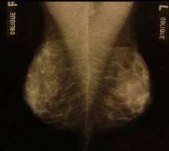
6 Typical views cranio-caudal medio-lateral-oblique Mammography p.4/27
7 Image processing chain From native image... Image correction: gain / offset defect pixels modulation transfer function compensation Post-processing: log transformation thickness equalization constrast enhancement CAD... Display: lighting monitor calibration VOI / LUT... to visualization Next: illustrations from S. Muller, GE Healthcare Mammography p.5/27

8 Image correction Mammography p.6/27

9 Image correction Mammography p.6/27

10 FTM compensation Mammography p.7/27

11 Thickness equalization Mammography p.8/27

12 Thickness equalization Mammography p.8/27

13 Thickness equalization Mammography p.8/27

14 Contrast enhancement Mammography p.9/27

15 Contrast enhancement Mammography p.9/27

16 Contrast enhancement Mammography p.9/27

17 Computer assisted detection: CAD Mammography p.10/27

18 Computer assisted detection: CAD Mammography p.10/27
19 CAD methods Filtering and enhancement: preferably using local methods local statistics, wavelets... compromise under-enhancement (can cause FN) / over-enhancement (FP) Segmentation: thresholding and region growing edge detection and deformable models template matching Markov random fields left/right differences multiscale fuzzy methods Quantitative measures: intensity, shape, texture, clusters Classification: artificial neural networks kernel-based methods (SVM...) decision trees Evaluation: specificity and sensitivity ROC curve: true positives as a function of false positives Mammography p.11/27

20 Tomosynthesis Mammography p.12/27

21 Tomosynthesis Mammography p.12/27

22 Tomosynthesis Mammography p.12/27
23 Tomosynthesis Greater conspicuity of lesions. Borders of lesions more clearly defined. Reduced call-back rate - almost eliminates recall for superimposed structures (summation shadows). Accurate 3-D location. Better differentiates benign from malignant. Mammography p.12/27
24 CAD for tomosynthesis PhD thesis of G. Peters, with GE Healthcare Back-Projection 3D Detection 3D Analysis 3D Decision A 2D Detection 2D Analysis Back-Projection Re-Projection 3D Analysis 3D Decision B 2D Detection 2D Analysis 2D Decision Back-Projection C Choice: Strategy B Mammography p.13/27

25 Algorithm scheme Raw Image Candidate Detection Fuzzy Segmentation Fuzzy Feature Extraction Partial Defuzzification Acquisition Geometry Back-Projection / Re-Projection Attribute Aggregation Classification Mammography p.14/27


26 Dense kernel detector Using wavelets and brackground density estimation: Mammography p.15/27

27 Segmentation result: circumscribed lesion Original image Reference contour Initialization Region-based Contour-based Hybrid Mammography p.16/27

28 Segmentation result: spiculated lesion Original image Reference contour Initialization Region-based Contour-based Hybrid Mammography p.17/27


29 Hypothesis testing for a radiological finding Detection Radiological Finding Hypothesis A: Circumscribed Mass Hypothesis B: Spiculated Mass Active Contour Model Circumscribed Mass Active Contour Model Spiculated Mass Fuzzy Active Contour Fuzzy Active Contour Feature Extraction Feature Extraction Fuzzy Attributes Fuzzy Attributes Fuzzy Decision Tree Fuzzy Decision Tree Confidence Degree in Hypothesis A Confidence Degree in Hypothesis B Decision Mammography p.18/27

30 Features from fuzzy contours Features: area, compacicty, mean gradient along the contour, homogeneity... Mammography p.19/27


31 Algorithm scheme for aggregation on particle level Projected Views Set of 2D Particles Set of 2D Fuzzy Attributes Partial Defuzzification Attribute Aggregation Fuzzy Particle Maps Correspondance Computation Cumulated Fuzzy Attribute Pixel Aggregation Re-Projected Fuzzy Particle Volume Fuzzy Particle Volume Re-Projection Mammography p.20/27


32 Complete processing chain original image wavelet filtering active contour segmentation assuming spiculated mass active contour segmentation assuming circumscribed mass wavelet filter response fuzzy active contour fuzzy active contour partial defuzzification feature extraction feature extraction partial defuzzification fuzzy particle map fuzzy attributes fuzzy attributes fuzzy particle map aggregation aggregation aggregation aggregation fuzzy particle volume cumulated fuzzy attributes cumulated fuzzy attributes fuzzy particle volume fuzzy decision tree fuzzy decision tree memberhip degree memberhip degree aggregation memberhip degree Mammography p.21/27

33 Automated detection of opacities and architectural distorsions in tomosynthesis PhD thesis of G. Palma, with GE Healthcare Mammography p.22/27
34 Global scheme: two channels approach DBT Volume Sub sampled volume Fuzzy 3D map Seeds Sub sampling Fuzzy connected filter (by slice) Thresholding A contrario detection (slice by slice processing) 2D and 3D aggregation Convergence regions Seeds Segmentation Contours Feature extraction Feature extraction Attributes Attributes Classification Classification Classes Masses detection Classes Architectural distortions detection Suspicious regions Mammography p.23/27

35 Makers from connected filters Mammography p.24/27
36 A contrario detection K c,q,r = (αr < cq < r) 1 if (tan(θ) cq αr) 0 otherwise. θ = angle between cq and orientation at point q. Z c,r = q Ω/αr< cq <r K c,q,r Mammography p.25/27
37 A contrario detection Z c,r λ r is ǫ-meaningful if the expectation of its number of occurrences in the image is less than ǫ (Desolneux et al., IJCV, 2000) λ r = min { λ N/P[Z c,r λ] ǫ } M where M it the number of pairs (c,r) to be considered. A contrario detection: computing {λr }, computing orientations, computing Zc,r for each (c,r), detection of ǫ-meaningful events (Zc,r > λ r ). Mammography p.25/27

38 A contrario detection Mammography p.25/27
39 Results Dense kernel detection: Mammography p.26/27

40 Results Convergence detection: Mammography p.26/27
41 Performances Performance of the whole dense kernel detection channel Sensitivity tag1 tag Number of false positives per breast Mammography p.27/27
42 Performances Performance of the a contrario detector for spiculated lesions only, and for architectural distortions and highly spiculated lesions Sensitivity Number of false positives per breast Sensitivity Number of false positives per breast Mammography p.27/27
43 Performances Performance of the suspicious convergence detection channel Sensitivity Number of false positives per breast Mammography p.27/27

44 Performances Performance of the complete detection process, after the aggregation step Sensitivity (%) Specificity (# of false positives per breast) Plane of a DBT volume exhibiting a strongly spiculated lesion, used in the convergence channel evaluation. Although this lesion is not detected by this channel, it is correctly detected by the dense kernel channel, and therefore by the final fusion step. Mammography p.27/27
Mammography is a most effective imaging modality in early breast cancer detection. The radiographs are searched for signs of abnormality by expert
 Abstract Methodologies for early detection of breast cancer still remain an open problem in the Research community. Breast cancer continues to be a significant problem in the contemporary world. Nearly
arxiv: v2 [cs.cv] 8 Mar 2018
![arxiv: v2 [cs.cv] 8 Mar 2018](/thumbs/87/97094636.jpg "arxiv: v2 [cs.cv] 8 Mar 2018") Automated soft tissue lesion detection and segmentation in digital mammography using a u-net deep learning network Timothy de Moor a, Alejandro Rodriguez-Ruiz a, Albert Gubern Mérida a, Ritse Mann a, and
Automated soft tissue lesion detection and segmentation in digital mammography using a u-net deep learning network Timothy de Moor a, Alejandro Rodriguez-Ruiz a, Albert Gubern Mérida a, Ritse Mann a, and
CHAPTER 2 MAMMOGRAMS AND COMPUTER AIDED DETECTION
 9 CHAPTER 2 MAMMOGRAMS AND COMPUTER AIDED DETECTION 2.1 INTRODUCTION This chapter provides an introduction to mammogram and a description of the computer aided detection methods of mammography. This discussion
Image processing for cardiac and vascular applications
 Image processing for cardiac and vascular applications Isabelle Bloch Isabelle.Bloch@telecom-paristech.fr http://perso.telecom-paristech.fr/bloch LTCI, Télécom ParisTech Cardio-vascular imaging p.1/26
Since its introduction in 2000, digital mammography has become
 Review Article Smith A, PhD email : Andrew.smith@hologic.com Since its introduction in 2000, digital mammography has become an accepted standard of care in breast cancer screening and has paved the way
Contrast-Enhanced Breast Tomosynthesis: Combining the Best of Both Worlds for Better Breast-Cancer Diagnosis
 Contrast-Enhanced Breast Tomosynthesis: Combining the Best of Both Worlds for Better Breast-Cancer Diagnosis T Wu (twu2@partners.org), E Rafferty, R Moore, D Kopans, Massachusetts General Hospital, Boston,
Detection of architectural distortion using multilayer back propagation neural network
 Available online www.jocpr.com Journal of Chemical and Pharmaceutical Research, 2015, 7(2):292-297 Research Article ISSN : 0975-7384 CODEN(USA) : JCPRC5 Detection of architectural distortion using multilayer
Classification of Mammograms using Gray-level Co-occurrence Matrix and Support Vector Machine Classifier
 Classification of Mammograms using Gray-level Co-occurrence Matrix and Support Vector Machine Classifier P.Samyuktha,Vasavi College of engineering,cse dept. D.Sriharsha, IDD, Comp. Sc. & Engg., IIT (BHU),
MIT International Journal of Electronics and Communication Engineering Vol. 3, No. 1, Jan. 2013, pp
 MIT International Journal of Electronics and Communication Engineering Vol. 3, No. 1, Jan. 013, pp. 43 47 43 A Novel Technique to Detect Abnormal Masses from Digital Mammogram Saurabh Verma Email: saurav.v84@gmail.com
LUNG CANCER continues to rank as the leading cause
 1138 IEEE TRANSACTIONS ON MEDICAL IMAGING, VOL. 24, NO. 9, SEPTEMBER 2005 Computer-Aided Diagnostic Scheme for Distinction Between Benign and Malignant Nodules in Thoracic Low-Dose CT by Use of Massive
Brain Tumor segmentation and classification using Fcm and support vector machine
 Brain Tumor segmentation and classification using Fcm and support vector machine Gaurav Gupta 1, Vinay singh 2 1 PG student,m.tech Electronics and Communication,Department of Electronics, Galgotia College
Radiation Dosimetry in Digital Breast Tomosynthesis. March, 2015 William J. O Connel, Dr. Ph, Senior Medical Physicist
 Radiation Dosimetry in Digital Breast Tomosynthesis March, 2015 William J. O Connel, Dr. Ph, Senior Medical Physicist Imagination at work. Syllabus 1. Introduction 2. Dosimetry in Mammography 3. Dosimetry
Classification of mammogram masses using selected texture, shape and margin features with multilayer perceptron classifier.
 Biomedical Research 2016; Special Issue: S310-S313 ISSN 0970-938X www.biomedres.info Classification of mammogram masses using selected texture, shape and margin features with multilayer perceptron classifier.
Policy Library Clinical Advantages of Digital Breast Tomosynthesis in Symptomatic Patients
 Policy Library Clinical Advantages of Digital Breast Tomosynthesis in Symptomatic Patients Version: 1 Approved by: Faculty of Clinical Radiology Council Date of approval: Click and type: day month and
Computer aided detection of clusters of microcalcifications on full field digital mammograms
 Computer aided detection of clusters of microcalcifications on full field digital mammograms Jun Ge, a Berkman Sahiner, Lubomir M. Hadjiiski, Heang-Ping Chan, Jun Wei, Mark A. Helvie, and Chuan Zhou Department
NAÏVE BAYES CLASSIFIER AND FUZZY LOGIC SYSTEM FOR COMPUTER AIDED DETECTION AND CLASSIFICATION OF MAMMAMOGRAPHIC ABNORMALITIES
 NAÏVE BAYES CLASSIFIER AND FUZZY LOGIC SYSTEM FOR COMPUTER AIDED DETECTION AND CLASSIFICATION OF MAMMAMOGRAPHIC ABNORMALITIES 1 MARJUN S. SEQUERA, 2 SHERWIN A. GUIRNALDO, 3 ISIDRO D. PERMITES JR. 1 Faculty,
Mammogram Analysis: Tumor Classification
 Mammogram Analysis: Tumor Classification Literature Survey Report Geethapriya Raghavan geeragh@mail.utexas.edu EE 381K - Multidimensional Digital Signal Processing Spring 2005 Abstract Breast cancer is
Improved Intelligent Classification Technique Based On Support Vector Machines
 Improved Intelligent Classification Technique Based On Support Vector Machines V.Vani Asst.Professor,Department of Computer Science,JJ College of Arts and Science,Pudukkottai. Abstract:An abnormal growth
Automated Approach for Qualitative Assessment of Breast Density and Lesion Feature Extraction for Early Detection of Breast Cancer
 Automated Approach for Qualitative Assessment of Breast Density and Lesion Feature Extraction for Early Detection of Breast Cancer 1 Spandana Paramkusham, 2 K. M. M. Rao, 3 B. V. V. S. N. Prabhakar Rao
Update of Digital Breast Tomosynthesis. Susan Orel Roth, MD
 Update of Digital Breast Tomosynthesis Susan Orel Roth, MD NCI estimates that : Why DBT? Approximately 20% of breast cancers are missed at mammography screening Average recall rates approximately 10%
Automatic Classification of Breast Masses for Diagnosis of Breast Cancer in Digital Mammograms using Neural Network
 IJSTE - International Journal of Science Technology & Engineering Volume 1 Issue 11 May 2015 ISSN (online): 2349-784X Automatic Classification of Breast Masses for Diagnosis of Breast Cancer in Digital
Mammographic Cancer Detection and Classification Using Bi Clustering and Supervised Classifier
 Mammographic Cancer Detection and Classification Using Bi Clustering and Supervised Classifier R.Pavitha 1, Ms T.Joyce Selva Hephzibah M.Tech. 2 PG Scholar, Department of ECE, Indus College of Engineering,
Financial Disclosures
 Financial Disclosures 3D Mammography: The Latest Developments in the Breast Imaging Arena I have no financial disclosures Dr. Katharine Lampen-Sachar Breast and Body Radiologist Radiology Associates of
8/31/2016 HIDING IN PLAIN SITE, ARCHITECTURAL DISTORTIONS AND BREAST ASYMMETRIES ARCHITECTURAL DISTORTIONS ARCHITECTURAL DISTORTIONS
 HIDING IN PLAIN SITE, ARCHITECTURAL DISTORTIONS AND BREAST ASYMMETRIES DEBORAH THAMES R.T. (R)(M)(QM) ARCHITECTURAL DISTORTIONS Definition is disruption of the natural flow of breast pattern towards the
Mammographic imaging of nonpalpable breast lesions. Malai Muttarak, MD Department of Radiology Chiang Mai University Chiang Mai, Thailand
 Mammographic imaging of nonpalpable breast lesions Malai Muttarak, MD Department of Radiology Chiang Mai University Chiang Mai, Thailand Introduction Contents Mammographic signs of nonpalpable breast cancer
International Journal of Advance Research in Engineering, Science & Technology
 Impact Factor (SJIF): 3.632 International Journal of Advance Research in Engineering, Science & Technology e-issn: 2393-9877, p-issn: 2394-2444 (Special Issue for ITECE 2016) An Efficient Image Processing
Corporate Medical Policy
 Corporate Medical Policy File Name: Origination: Last CAP Review: Next CAP Review: Last Review: digital_breast_tomosynthesis 3/2011 6/2016 6/2017 11/2016 Description of Procedure or Service Conventional
Characterization of the breast region for computer assisted Tabar masking of paired mammographic images
 Characterization of the breast region for computer assisted Tabar masking of paired mammographic images Paola Casti, Arianna Mencattini, Marcello Salmeri Dept. of Electronic Engineering, University of
Computer Aided Detection of Abnormalities in Mammograms
 Computer Aided Detection of Abnormalities in Mammograms I W Hutt 1, S M Astley 1 & C R M Boggis 2 1: Wolfson Image Analysis Unit, Dept of Medical Biophysics, Manchester University, Oxford. Rd, Manchester,
Opportunities and Innovations in Digital Mammography John M. Sandrik, Ph.D. GE Healthcare Milwaukee, WI
 Opportunities and Innovations in Digital Mammography John M. Sandrik, Ph.D. GE Healthcare Milwaukee, WI john.sandrik@med.ge.com with many thanks to Vince Polkus, Advanced Applications Product Mgr. 1 Content
Digital breast tomosynthesis
 GE Healthcare Digital breast tomosynthesis Daniel B. Kopans, M.D., F.A.C.R. Professor of Radiology Harvard Medical School Senior Radiologist - Breast Imaging Division Massachusetts General Hospital Since
Look differently. Invenia ABUS. Automated Breast Ultrasound
 Look differently. Invenia ABUS Automated Breast Ultrasound InveniaTM ABUS from GE Healthcare offers a view beyond mammography, with breast screening technology that looks differently. 40 % The unseen risk.
Leonard M. Glassman MD
 BI-RADS The New BI-RADS Leonard M. Glassman MD FACR Former Chief of Breast Imaging American Institute for Radiologic Pathology Washington Radiology Associates, PC Breast Imaging Reporting and Data System
AN ALGORITHM FOR EARLY BREAST CANCER DETECTION IN MAMMOGRAMS
 AN ALGORITHM FOR EARLY BREAST CANCER DETECTION IN MAMMOGRAMS Isaac N. Bankman', William A. Christens-Barryl, Irving N. Weinberg2, Dong W. Kim3, Ralph D. Semmell, and William R. Brody2 The Johns Hopkins
Mammogram Analysis: Tumor Classification
 Mammogram Analysis: Tumor Classification Term Project Report Geethapriya Raghavan geeragh@mail.utexas.edu EE 381K - Multidimensional Digital Signal Processing Spring 2005 Abstract Breast cancer is the
Mammography limitations. Clinical performance of digital breast tomosynthesis compared to digital mammography: blinded multi-reader study
 Clinical performance of digital breast tomosynthesis compared to digital mammography: blinded multi-reader study G. Gennaro (1), A. Toledano (2), E. Baldan (1), E. Bezzon (1), C. di Maggio (1), M. La Grassa
Simulation of spiculated breast lesions
 Simulation of spiculated breast lesions Premkumar Elangovan* a, Faisal Alrehily b, R Ferrari Pinto a, Alaleh Rashidnasab a,c, David R Dance b,d, Kenneth C Young b,d, Kevin Wells a a Centre for Vision,
Threshold Based Segmentation Technique for Mass Detection in Mammography
 Threshold Based Segmentation Technique for Mass Detection in Mammography Aziz Makandar *, Bhagirathi Halalli Department of Computer Science, Karnataka State Women s University, Vijayapura, Karnataka, India.
Digital Breast Tomosynthesis from a first idea to clinical routine
 International Master Programm Biomedical Engineering Digital Breast Tomosynthesis from a first idea to clinical routine Historical background 2D imaging of 3D objects has important limitations Jörg Barkhausen
CHAPTER - 2 LITERATURE REVIEW
 CHAPTER - 2 LITERATURE REVIEW Currently, there is an increasing interest for establishing automatic systems that screens a huge number of people for vision threatening diseases like diabetic retinopathy
10.4 Computer-Aided Detection and Diagnosis in Mammography
 10.4 Computer-Aided Detection and Diagnosis in Mammography Mehul P. Sampat, Mia K. Markey, and Alan C. Bovik The University of Texas at Austin 1 Introduction...1195 2 Computer-Aided Detection of Mammographic
Automated detection of masses on whole breast volume ultrasound scanner: false positive reduction using deep convolutional neural network
 Automated detection of masses on whole breast volume ultrasound scanner: false positive reduction using deep convolutional neural network Yuya Hiramatsu a, Chisako Muramatsu* a, Hironobu Kobayashi b, Takeshi
DETECTING DIABETES MELLITUS GRADIENT VECTOR FLOW SNAKE SEGMENTED TECHNIQUE
 DETECTING DIABETES MELLITUS GRADIENT VECTOR FLOW SNAKE SEGMENTED TECHNIQUE Dr. S. K. Jayanthi 1, B.Shanmugapriyanga 2 1 Head and Associate Professor, Dept. of Computer Science, Vellalar College for Women,
Detection of suspicious lesion based on Multiresolution Analysis using windowing and adaptive thresholding method.
 Detection of suspicious lesion based on Multiresolution Analysis using windowing and adaptive thresholding method. Ms. N. S. Pande Assistant Professor, Department of Computer Science and Engineering,MGM
Compressive Re-Sampling for Speckle Reduction in Medical Ultrasound
 Compressive Re-Sampling for Speckle Reduction in Medical Ultrasound Professor Richard Mammone Rutgers University Email Phone Number Christine Podilchuk, Lev Barinov, Ajit Jairaj and William Hulbert ClearView
EARLY STAGE DIAGNOSIS OF LUNG CANCER USING CT-SCAN IMAGES BASED ON CELLULAR LEARNING AUTOMATE
 EARLY STAGE DIAGNOSIS OF LUNG CANCER USING CT-SCAN IMAGES BASED ON CELLULAR LEARNING AUTOMATE SAKTHI NEELA.P.K Department of M.E (Medical electronics) Sengunthar College of engineering Namakkal, Tamilnadu,
The Radiology Aspects
 REQUIREMENTS FOR INTERNATIONAL ACCREDITATION OF BREAST CENTERS/UNITS The Radiology Aspects Miri Sklair-Levy, Israel RADIOLOGY GUIDELINES FOR QUALITY ASSURANCE IN BREAST CANCER SCREENING AND DIAGNOSIS Radiologists
Mammographic Mass Detection Using a Mass Template
 Mammographic Mass Detection Using a Mass Template Serhat Ozekes, MSc 1 Onur Osman, PhD 1 A.Yilmaz Çamurcu, PhD 2 Index terms: Mass detection Computer aided detection Mammography Objective: The purpose
Computer-aided diagnosis of subtle signs of breast cancer: Architectural distortion in prior mammograms
 Computer-aided diagnosis of subtle signs of breast cancer: Architectural distortion in prior mammograms Rangaraj M. Rangayyan Department of Electrical and Computer Engineering University of Calgary, Calgary,
Spatial Frequency Localization in Mammograms Using Wavelets
 Western Michigan University ScholarWorks at WMU Dissertations Graduate College 12-2009 Spatial Frequency Localization in Mammograms Using Wavelets Tomislav Bujanovic Western Michigan University Follow
Computer-Aided Diagnosis for Microcalcifications in Mammograms
 Computer-Aided Diagnosis for Microcalcifications in Mammograms Werapon Chiracharit Department of Electronic and Telecommunication Engineering King Mongkut s University of Technology Thonburi BIE 690, November
Tomosynthesis and breast imaging update. Dr Michael J Michell Consultant Radiologist King's College Hospital NHS Foundation Trust
 Tomosynthesis and breast imaging update Dr Michael J Michell Consultant Radiologist King's College Hospital NHS Foundation Trust Breast imaging new technology BREAST CANCER FLT PET shows different grades
Improving Reading Time of Digital Breast Tomosynthesis with Concurrent Computer Aided Detection
 White Paper Improving Reading Time of Digital Breast Tomosynthesis with Concurrent Computer Aided Detection WHITE PAPER 2 3 Abstract PowerLook Tomo Detection, a concurrent computer-aided detection (CAD)
Investigating the performance of a CAD x scheme for mammography in specific BIRADS categories
 Investigating the performance of a CAD x scheme for mammography in specific BIRADS categories Andreadis I., Nikita K. Department of Electrical and Computer Engineering National Technical University of
Investigation of multiorientation and multiresolution features for microcalcifications classification in mammograms
 Investigation of multiorientation and multiresolution features for microcalcifications classification in mammograms Aqilah Baseri Huddin, Brian W.-H. Ng, Derek Abbott 3 School of Electrical and Electronic
Computer aided diagnosis in digital mammography: Classification of mass and normal tissue
 University of South Florida Scholar Commons Graduate Theses and Dissertations Graduate School 2003 Computer aided diagnosis in digital mammography: Classification of mass and normal tissue Monika Shinde
Research Article Automated Abnormal Mass Detection in the Mammogram Images Using Chebyshev Moments
 Research Journal of Applied Sciences, Engineering and Technology 5(2): 513-518, 2013 DOI:10.19026/rjaset.5.4983 ISSN: 2040-7459; E-ISSN: 2040-7467 2013 Maxwell Scientific Publication Corp. Submitted: May
A Breast Surgeon s Use of Three Dimensional Specimen Tomosynthesis
 A Breast Surgeon s Use of Three Dimensional Specimen Tomosynthesis Cary S. Kaufman MD, FACS Associate Clinical Professor of Surgery A Breast Surgeon s Use of Three Dimensional Specimen Tomosynthesis Cary
Updates in Mammography. Dr. Yang Faridah A. Aziz Department of Biomedical Imaging University Malaya Medical Centre
 Updates in Mammography Dr. Yang Faridah A. Aziz Department of Biomedical Imaging University Malaya Medical Centre Updates in Mammography Breast Imaging Dr. Yang Faridah A. Aziz Department of Biomedical
Computer-aided detection in DBT (digital breast tomosynthesis)
") Computer-aided detection in DBT (digital breast tomosynthesis) 2015 Summer Prof. Yong Man Ro Image and Video Systems (IVY) Lab., Department of Electrical Engineering, KAIST ymro@kaist.ac.kr IVY Lab (Image
Computer-aided detection in DBT (digital breast tomosynthesis) 2015 Summer Prof. Yong Man Ro Image and Video Systems (IVY) Lab., Department of Electrical Engineering, KAIST ymro@kaist.ac.kr IVY Lab (Image
CURRENTLY FDA APPROVED ARE FULL FIELD DIGITAL MAMMOGRAPHY SYSTEMS AND FILM SCREEN STILL BEING USED AT SOME INSTITUTIONS
 ABBY DUROJAYE,M.D CURRENTLY FDA APPROVED ARE FULL FIELD DIGITAL MAMMOGRAPHY SYSTEMS AND FILM SCREEN STILL BEING USED AT SOME INSTITUTIONS BOTH HAVE BEEN SHOWN TO BE EFFECTIVE TOOLS EARLY DETECTION OF BREAST
CLASSIFICATION OF ABNORMALITY IN B -MASS BY ARCHITECTURAL DISTORTION
 CLASSIFICATION OF ABNORMALITY IN B -MASS BY ARCHITECTURAL DISTORTION #1 Venmathi.A.R., * 2 D.C.Jullie Josphine #1.Dept of ECE, Kings Engineering College * 2. Dept of CSE,Kings Engineering college Abstract-The
The latest developments - Automated Breast Volume Scanning. Dr. med. M. Golatta
 The latest developments - Automated Breast Volume Scanning Dr. med. M. Golatta Automated Breast Volume US: Why? o Mammography is limited in dense breasts: high false negative rate o Many of these tumors
Malignant Breast Cancer Detection Method - A Review. Patiala
 Malignant Breast Cancer Detection Method - A Review 1 Jaspreet Singh Cheema, 2 Amrita, 3 Sumandeep kaur 1,2 Student of M.tech Computer Science, Punjabi University, Patiala 3 Assistant professor, Department
A Reliable Method for Brain Tumor Detection Using Cnn Technique
 IOSR Journal of Electrical and Electronics Engineering (IOSR-JEEE) e-issn: 2278-1676,p-ISSN: 2320-3331, PP 64-68 www.iosrjournals.org A Reliable Method for Brain Tumor Detection Using Cnn Technique Neethu
Digital Imaging and Communications in Medicine (DICOM) Supplement 50: Mammography Computer-Aided Detection SR SOP Class
 Supplement 50: Mammography Computer-Aided Detection SR SOP Class") Digital Imaging and Communications in Medicine (DICOM) Supplement 50: Mammography Computer-Aided Detection SR SOP Class Status: Letter Ballot Text February 2, 2001 DICOM Standards Committee 1300 N. 17
Digital Imaging and Communications in Medicine (DICOM) Supplement 50: Mammography Computer-Aided Detection SR SOP Class Status: Letter Ballot Text February 2, 2001 DICOM Standards Committee 1300 N. 17
Breast tomosynthesis reduces radiologist performance variability compared to digital mammography
 Breast tomosynthesis reduces radiologist performance variability compared to digital mammography Andrew Smith 1, Elizabeth Rafferty 2, Loren Niklason 1 1 Hologic, Inc., Bedford MA, USA 2 Massachusetts
BI-RADS classification in breast tomosynthesis. Our experience in breast cancer cases categorized as BI-RADS 0 in digital mammography
 BI-RADS classification in breast tomosynthesis. Our experience in breast cancer cases categorized as BI-RADS 0 in digital mammography Poster No.: C-0562 Congress: ECR 2017 Type: Scientific Exhibit Authors:
The second chapter (Chapter 7) in the second section is on automated segmentation of the lungs on CT images, provided by Drs. Sensakovic and Armato.
 in the second section is on automated segmentation of the lungs on CT images, provided by Drs. Sensakovic and Armato.") xviii Preface Medical imaging is an indispensable tool of patients healthcare in modern medicine. Machine leaning plays an essential role in the medical imaging field, including Computer-Aided Diagnosis
xviii Preface Medical imaging is an indispensable tool of patients healthcare in modern medicine. Machine leaning plays an essential role in the medical imaging field, including Computer-Aided Diagnosis
Review on Mammogram Mass Detection by Machine Learning Techniques
 Review on Mammogram Mass Detection by Machine Learning Techniques Valliappan Raman, Putra Sumari, H.H.Then, and Saleh Ali K. Al-Omari Abstract Breast cancer continues to be a significant public health
Automated detection scheme of architectural distortion. in mammograms using adaptive Gabor filter
 Automated detection scheme of architectural distortion in mammograms using adaptive Gabor filter Ruriha Yoshikawa 1, Atsushi Teramoto 1, Tomoko Matsubara 2,Hiroshi Fujita 3 1. Graduate School of Fujita
Fundamentals of Breast Tomosynthesis
 Fundamentals of Breast Tomosynthesis Improving the Performance of Mammography Andrew Smith, Ph.D. This white paper is one in a series of research overviws on advanced technologies in women s healthcare.
Lung Cancer Diagnosis from CT Images Using Fuzzy Inference System
 Lung Cancer Diagnosis from CT Images Using Fuzzy Inference System T.Manikandan 1, Dr. N. Bharathi 2 1 Associate Professor, Rajalakshmi Engineering College, Chennai-602 105 2 Professor, Velammal Engineering
Correlation between lesion type and the additional value of digital breast tomosynthesis
 Correlation between lesion type and the additional value of digital breast tomosynthesis Poster No.: C-1604 Congress: ECR 2011 Type: Scientific Exhibit Authors: C. Van Ongeval, L. Cockmartin, A. Van Steen,
Contrast Enhanced Spectral Mammography (CESM) Updates
 Updates") Contrast Enhanced Spectral Mammography (CESM) Updates Georgeta Mihai, PhD, DABR Medical Physicist, BIDMC, Boston Assistant Professor, Harvard Medical School, Boston Disclosures None Acknowledgments: Da
Contrast Enhanced Spectral Mammography (CESM) Updates Georgeta Mihai, PhD, DABR Medical Physicist, BIDMC, Boston Assistant Professor, Harvard Medical School, Boston Disclosures None Acknowledgments: Da
Computer Assisted Radiology and Surgery
 Computer Assisted Radiology and Surgery How Can a Massive Training Artificial Neural Network (MTANN) Be Trained With a Small Number of Cases in the Distinction Between Nodules and Vessels in Thoracic CT?
Classification of benign and malignant masses in breast mammograms
 Classification of benign and malignant masses in breast mammograms A. Šerifović-Trbalić*, A. Trbalić**, D. Demirović*, N. Prljača* and P.C. Cattin*** * Faculty of Electrical Engineering, University of
Breast Imaging Lexicon
 9//201 200 BI RADS th Edition 201 BI RADS th Edition Breast Imaging Lexicon Mammographic Pathology and Assessment Categories Deborah Thames, R.T.(R)(M)(QM) The Advanced Health Education Center Nonmember:
MEM BASED BRAIN IMAGE SEGMENTATION AND CLASSIFICATION USING SVM
 MEM BASED BRAIN IMAGE SEGMENTATION AND CLASSIFICATION USING SVM T. Deepa 1, R. Muthalagu 1 and K. Chitra 2 1 Department of Electronics and Communication Engineering, Prathyusha Institute of Technology
Automatic Computer Aided Diagnosis of Breast Cancer in Dynamic Contrast Enhanced Magnetic Resonance Images. Hongbo Wu
 Automatic Computer Aided Diagnosis of Breast Cancer in Dynamic Contrast Enhanced Magnetic Resonance Images by Hongbo Wu A thesis submitted in conformity with the requirements for the degree of Master of
Emerging Techniques in Breast Imaging: Contrast-Enhanced Mammography and Fast MRI
 Emerging Techniques in Breast Imaging: Contrast-Enhanced Mammography and Fast MRI Lilian Wang, M.D. Breast Imaging Section Department of Radiology Northwestern Medicine Overview Rationale for new imaging
COMPUTER AIDED DIAGNOSIS SYSTEM FOR DIGITAL MAMMOGRAPHY. Mohamed Eltahir Makki Elmanna
 COMPUTER AIDED DIAGNOSIS SYSTEM FOR DIGITAL MAMMOGRAPHY By Mohamed Eltahir Makki Elmanna A Thesis Submitted to the Faculty of Engineering at Cairo University in Partial Fulfillment of the Requirements
Diagnostic Dilemmas of Breast Imaging
 Diagnostic Dilemmas of Breast Imaging Common Causes of Error in Breast Cancer Detection By: Jason Cord, M.D. Mammography: Initial Imaging The standard for detection of breast cancer Screening mammography
Brain Tumour Detection of MR Image Using Naïve Beyer classifier and Support Vector Machine
 International Journal of Scientific Research in Computer Science, Engineering and Information Technology 2018 IJSRCSEIT Volume 3 Issue 3 ISSN : 2456-3307 Brain Tumour Detection of MR Image Using Naïve
A Novel Approach to Breast Ultrasound Image Segmentation Based on the Characteristics of Breast Tissue and Particle Swarm Optimization
 A Novel Approach to Breast Ultrasound Image Segmentation Based on the Characteristics of Breast Tissue and Particle Swarm Optimization Yanhui Guo,, H.D. Cheng,, Jiawei Tian 3, Yingtao Zhang School of Computer
FDA Executive Summary
 Meeting of the Radiological Devices Advisory Panel On October 24, 22, the panel will discuss, make recommendations, and vote on a premarket approval application supplement (P83/S) to expand the indications
Improved Detection of Lung Nodule Overlapping with Ribs by using Virtual Dual Energy Radiology
 Improved Detection of Lung Nodule Overlapping with Ribs by using Virtual Dual Energy Radiology G.Maria Dhayana Latha Associate Professor, Department of Electronics and Communication Engineering, Excel
Contrast-Enhanced Digital Mammography
 2015 ARRS Breast Symposium Contrast-Enhanced Digital Mammography John Lewin, M.D. Diversified Radiology of Colorado CEDM - Outline History Technique Literature Review / Cases Clinical Status Inexpensive,
NMF-Density: NMF-Based Breast Density Classifier
 NMF-Density: NMF-Based Breast Density Classifier Lahouari Ghouti and Abdullah H. Owaidh King Fahd University of Petroleum and Minerals - Department of Information and Computer Science. KFUPM Box 1128.
Digital Breast Tomosynthesis Ready for Routine Screening?
 Digital Breast Tomosynthesis Ready for Routine Screening? Sophia Zackrisson MD, PhD, Assoc Prof of Radiology Skåne University Healthcare, Lund University, Sweden 1 Mammography screening 20% reduced breast
Automating Quality Assurance Metrics to Assess Adequate Breast Positioning in Mammography
 Automating Quality Assurance Metrics to Assess Adequate Breast Positioning in Mammography Gerald R. Kolb, JD, The Breast Group, Sunriver, OR; Kaier Wang, PhD, VolparaSolutions, Wellington, NZ; Ariane Chan,
Current Status of Supplementary Screening With Breast Ultrasound
 Current Status of Supplementary Screening With Breast Ultrasound Stephen A. Feig, M.D., FACR Fong and Jean Tsai Professor of Women s Imaging Department of Radiologic Sciences University of California,
Estimation of Breast Density and Feature Extraction of Mammographic Images
 IJIRST International Journal for Innovative Research in Science & Technology Volume 2 Issue 11 April 2016 ISSN (online): 2349-6010 Estimation of Breast Density and Feature Extraction of Mammographic Images
ISSN Vol.03,Issue.06, May-2014, Pages:
 www.semargroup.org, www.ijsetr.com ISSN 2319-8885 Vol.03,Issue.06, May-2014, Pages:0920-0926 Breast Cancer Classification with Statistical Features of Wavelet Coefficient of Mammograms SHITAL LAHAMAGE
Detecting Architectural Distortion in Mammograms Using a Gabor Filtered Probability Map Algorithm
 Detecting Architectural Distortion in Mammograms Using a Gabor Filtered Probability Map Algorithm O tega Ejofodomi, Michael Olawuyi, Don Onyishi, Godswill Ofualagba To cite this version: O tega Ejofodomi,
Classification of Thyroid Nodules in Ultrasound Images using knn and Decision Tree
 Classification of Thyroid Nodules in Ultrasound Images using knn and Decision Tree Gayana H B 1, Nanda S 2 1 IV Sem, M.Tech, Biomedical Signal processing & Instrumentation, SJCE, Mysuru, Karnataka, India
Research Article A Selective Ensemble Classification Method Combining Mammography Images with Ultrasound Images for Breast Cancer Diagnosis
 Hindawi Computational and Mathematical Methods in Medicine Volume 27, Article ID 4896386, 7 pages https://doi.org/5/27/4896386 Research Article A Selective Ensemble Classification Method Combining Mammography
Amammography report is a key component of the breast
 Review Article Writing a Mammography Report Amammography report is a key component of the breast cancer diagnostic process. Although mammographic findings were not clearly differentiated between benign
Name of Policy: Computer-aided Detection (CAD) Mammography
 Mammography") Name of Policy: Computer-aided Detection (CAD) Mammography Policy #: 112 Latest Review Date: October 2010 Category: Radiology Policy Grade: Active Policy but no longer scheduled for regular literature
Name of Policy: Computer-aided Detection (CAD) Mammography Policy #: 112 Latest Review Date: October 2010 Category: Radiology Policy Grade: Active Policy but no longer scheduled for regular literature
LUNG NODULE DETECTION SYSTEM
 LUNG NODULE DETECTION SYSTEM Kalim Bhandare and Rupali Nikhare Department of Computer Engineering Pillai Institute of Technology, New Panvel, Navi Mumbai, India ABSTRACT: The Existing approach consist