Updates in Mammography. Dr. Yang Faridah A. Aziz Department of Biomedical Imaging University Malaya Medical Centre
|
|
|
- Dinah Webb
- 6 years ago
- Views:
Transcription
1 Updates in Mammography Dr. Yang Faridah A. Aziz Department of Biomedical Imaging University Malaya Medical Centre
2 Updates in Mammography Breast Imaging Dr. Yang Faridah A. Aziz Department of Biomedical Imaging University Malaya Medical Centre

3 Updates in Breast Imaging Digital Mammography including CAD Tomosynthesis Contrast-enhanced Mammography MR breast imaging
4 Breast Disease Breast Disease Breast carcinoma is the commonest cancer in Malaysian women in every race 3738 new cases every year A woman in Malaysia has a 1 in 20 chance of developing breast cancer in her lifetime National Cancer Registry Malaysia report 2003

5 Breast Disease Breast Disease Statistics from National Cancer Registry, 2002 & Charts from
6 Mammography through the years Mammography is shown to be effective in reducing breast cancer mortality through early detection 1 Breast imaging started in 1913 by Albert Salomon Mammography started in the 1960s 2 In 1969, the first x-ray units dedicated to breast imaging were available 2 By 1976, mammography as a screening device became standard practice and its value in the diagnosis of breast carcinoma was recognised 1. Tabar L et al. Beyond randomized controlled trials: organized mammography screening substantially reduces breast cancer mortality. Cancer 2001; 91:
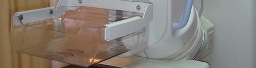
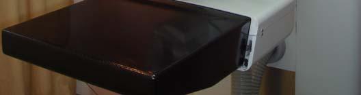
7 Mammography through the years Screen-film mammography (SFM) is the technology of choice Personnel involved are well-trained in this method Low cost and high spatial resolution Ongeval V et al. Current status of digital mammography for screening and diagnosis of breast cancer. Curr opin intern med 2007; 6(1):
8 Mammography through the years In general radiology, transition to digital technology began two decades ago Digital mammography was first introduced in stereotactic biopsy Digital mammography was slow due to difficulty to produce full-field digital detectors In 2005, the first digital mammography was approved Ongeval V et al. Current status of digital mammography for screening and diagnosis of breast cancer. Curr opin intern med 2007; 6(1):
9 Digital vs SFM Digital vs SFM Detection of breast lesion Workflow processes Image quality including image postprocessing Image archival and retrieval
10 Digital vs SFM Digital vs SFM Detection of breast lesion Workflow processes Image quality including image postprocessing Image archival and retrieval
11 Detection of breast lesion Detection of breast lesion Breast is a difficult organ to imaged Breast density ranges from dense (75% or more of breast compose of glandular tissue) to fatty Sensitivity of detection of carcinoma is 62.9% in dense breast compared to 87% in fatty breast 1 1. Carney PA et al. Individual and combined effects of age, breast density, and hormone replacement therapy use on the accuracy of screening mammography. Ann Intern Med 2003;138(3):

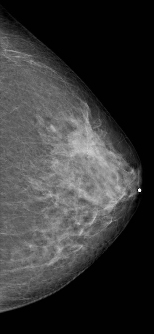
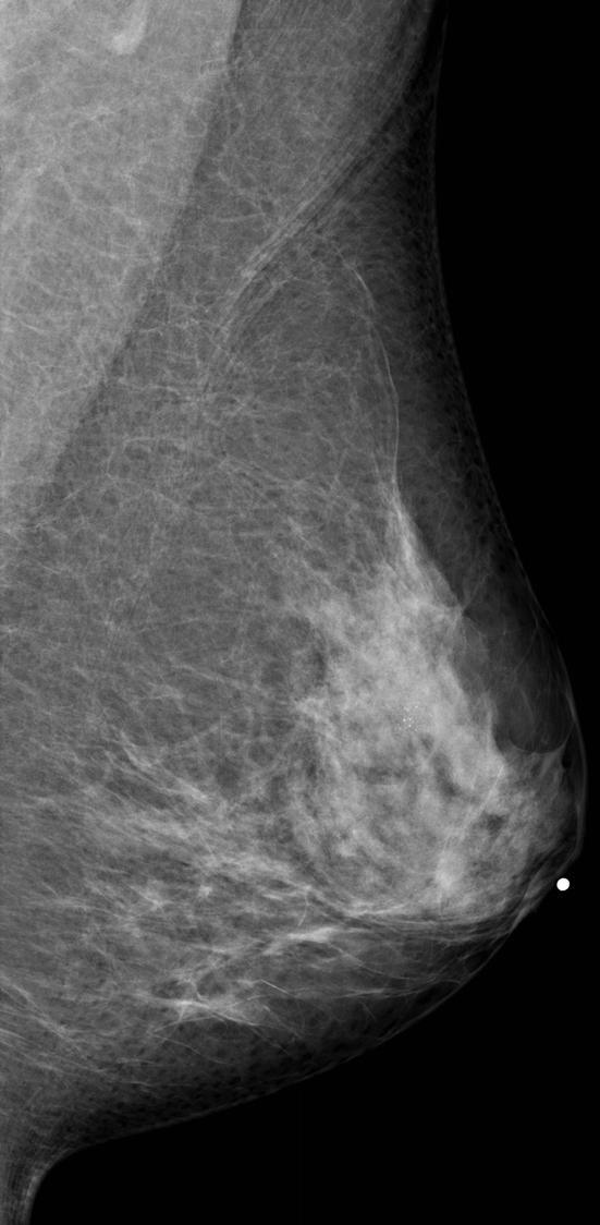
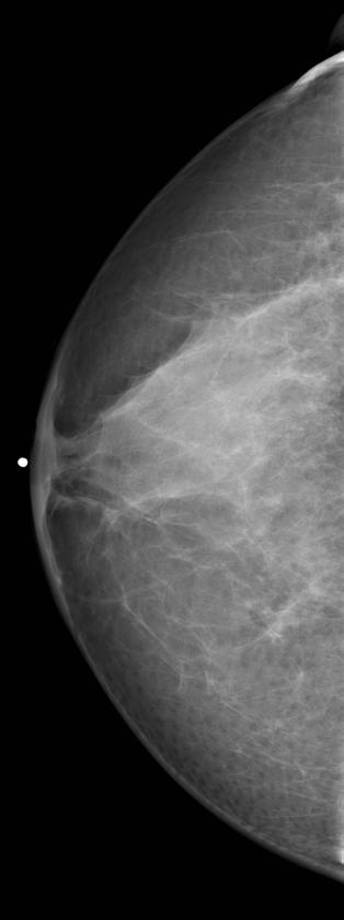
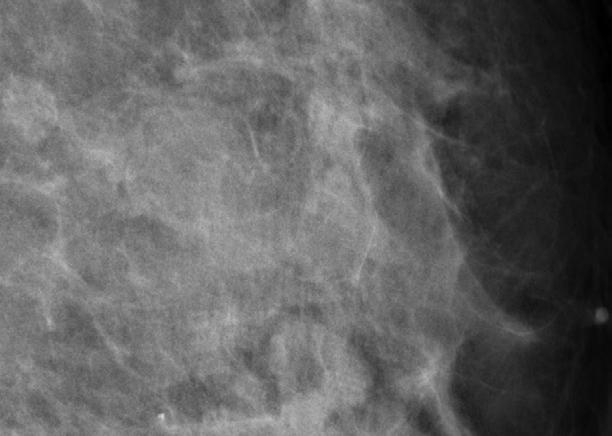
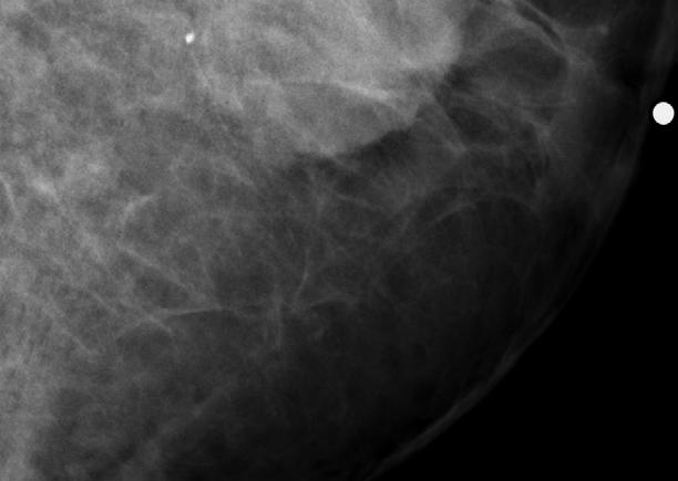
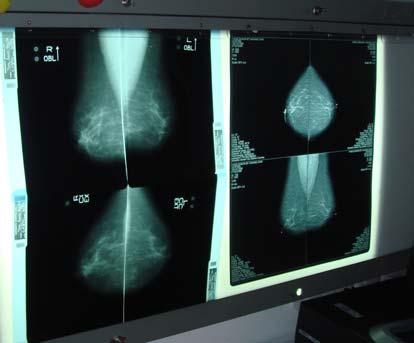
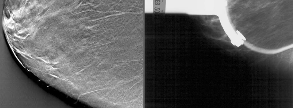
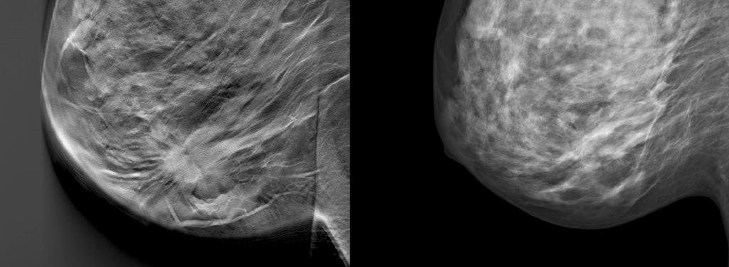
12 Image quality Image quality Fatty backgound

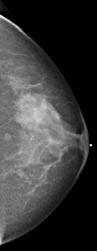
13 Image quality Image quality Moderately dense


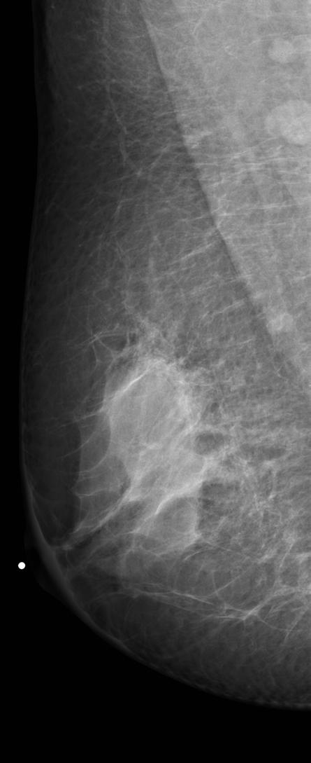
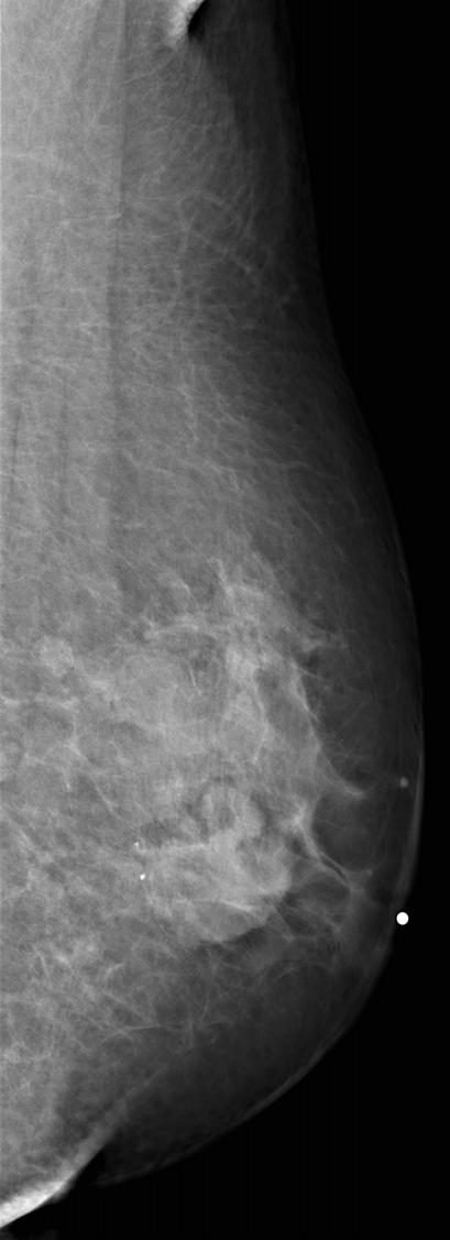
14 Image quality Image quality Dense breast
15 Lesion detection vs breast density Lesion detection vs breast density Women with dense tissue have a four to six times higher risk of breast cancer compared to women with little or no dense tissue Masking of existing lesion by overlying breast tissue Boyd NF et al. Mammographic Density and the Risk and Detection of Breast Cancer. New England Journal of Medicine 2007; 356(3):
16 Digital vs SFM Digital vs SFM Overall diagnostic accuracy of digital and film mammography for breast cancer detection is similar Digital is more accurate in: Women under 50 Women with dense breast Premenopausal or perimenopausal women Due to wide dynamic range of digital, able to display contrasting regions without compromising resolution Pisano ED et al. Diagnostic Performance of Digital versus Film Mammography for Breast-Cancer Screening. New England Journal of Medicine 2005; 353(17):
17 Detection of breast lesion: calcification SFM boasts a high spatial resolution good for detection of microcalcification DM is limited by pixel size However the high contrast resolution of DM shows more calcification compared to SFM increases the ability to characterize calcification better Kim HH, Pisano ED, et al. Comparison of calcification specificity in digital mammography using softcopy display versus screen-film mammography. AJR 2006; 187(1):47-50
18

19 Clustered microcalcification Biopsy: Ductal carcinoma in situ
20 Detection of breast lesion Detection of breast lesion DM also allows for use of software such as computer-aided detection CAD serves as a second reader in a screening mammogram programme

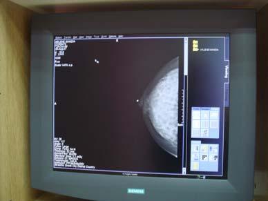
21 Computer aided diagnosis Computer aided diagnosis Diagnosis made by a radiologist taking into account the computer output as a second opinion Causes of missed cancers Poor image quality training of radiographers and QA Misinterpretation training of radiologists Oversight second reader/cad Used mainly in screening mammography Courtesy of Siemens
22 Digital vs SFM Digital vs SFM Detection of breast lesion Workflow processes Image quality including image postprocessing Image archival and retrieval
23 Digital vs SFM Digital vs SFM Reliance on personnel Problems with film processing e.g. artifacts

24 Digital vs SFM Digital vs SFM
25 Digital Imaging Digital Imaging Workflow of different modalities Time (mins) Interpretation and results Processing time Exposure time SFM 1 CRM 2 DM3 Modalities Ranganathan S, Y Faridah, KH Ng. Moving into the digital era: a novel experience with the first full-field digital mammography system in Malaysia. Singapore Med J 2007; 48(9):
26 Workflow processes Workflow processes A 45% reduction in the time taken to perform and process images using DM compared to SFM DM is also useful in stereotactic biopsy and hookwire localisation Hard-copy images of DM is consistent without presence of artifacts Ranganathan S, Y Faridah, KH Ng. Moving into the digital era: a novel experience with the first full-field digital mammography system in Malaysia. Singapore Med J 2007; 48(9):

27 Workflow processes - biopsy Workflow processes - biopsy Use of digital system during biopsy decreases the overall time of procedure as images do not need to be processed for needle placement
28 Digital vs SFM Digital vs SFM Detection of breast lesion Workflow processes Image quality including image post-processing Image archival and retrieval
29 Digital vs SFM Image quality Digital vs SFM Image quality DM is able to manipulate digital information Repeat rate with DM is low DM can change image contrast, zoom and magnify Delineates subcutaneous skin better than SFM However no amount of image manipulation could compensate for a badly taken mammogram!


30 Mammogram mass Mammogram mass benign Sebaceous cyst
31


32 HPE invasive ductal carcinoma
33

34 Biopsy: Mucinous carcinoma

35 Mammogram - mass Mammogram - mass Carcinoma
36 Digital vs SFM Digital vs SFM Detection of breast lesion Workflow processes Image quality including image postprocessing Image archival and retrieval

37 Digital vs SFM Digital vs SFM
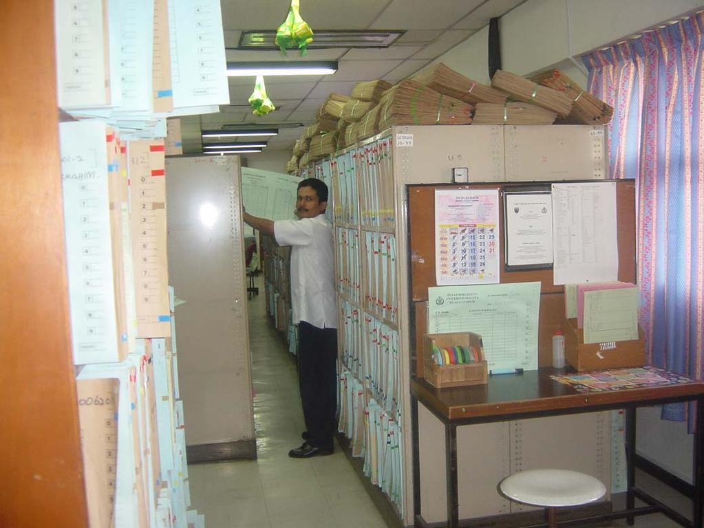
38 Limitation of space

39 MAMMOGRAM FILMS FILM FILM MORE FILM Workflow in a Radiology department with SFM Limited storage Missing films Labour intensive Degradation of films RADIOLOGY DEPARTMENT, WARDS, CLINIC, OTHER HOSPITALS
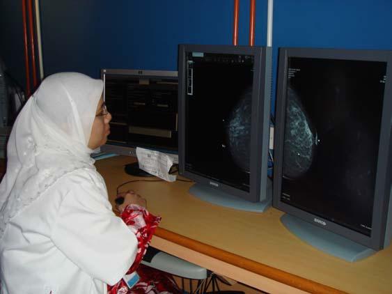
40 MAMMOGRAPHY Workflow in a Radiology department with Digital

41 MAMMOGRAMS P A C S Radiology Dept Clinics Wards Other hospitals
42 Digital Mammography: in summary Digital Mammography: in summary Detection of breast lesion CAD Work-flow process Image quality Image archival Cost Overall sensitivity in screening for breast lesion Accuracy in dense breast Detection of microcalcification Use of software e.g. CAD Processing time Reporting time Ability to manipulate image Consistency of hard-copy image Subcutaneous tissue delineation Image archival and retrieval Cost, cost, cost! DM = SFM DM > SFM DM > SFM DM >> SFM DM >> SFM DM < SFM initially if do soft-copy reporting DM yes, SFM no DM > SFM DM >> SFM DM >> SFM DM >>> SFM


43 Breast Tomosynthesis Breast Tomosynthesis Acquisition of multiple images during Tomo scan Displays the breast in slices Reduces tissue overlap Reduction in compression pressure Dose? Similar to one view mammogram Courtesy of Siemens

44 Breast Tomosynthesis Breast Tomosynthesis Courtesy of Hologic

45 Courtesy of Siemens

46 Courtesy of Siemens
47 Contrast-enhanced Mammography A pre-contrast mammogram is performed An iodine-based contrast media is injected Post-contrasted images are then acquired These images (pre-contrast and contrasted) are subtracted


48 Contrast enhanced Mammography Jong RA et al. Contrast Enhanced Digital Mammography. Radiology 2003; 228:
49 Breast MR Breast MR Has a high sensitivity in detecting breast lesion Has a high negative predictive value No radiation
50 Breast MR: Drawbacks Breast MR: Drawbacks Picks up benign lesion as well Gives rise to higher number of unneccesary biopsies Cost
51 Breast MR Breast MR Not used for general population screening Reserved for young women with high risk of breast carcinoma Performed also in women with suspected multiple breast carcinomas

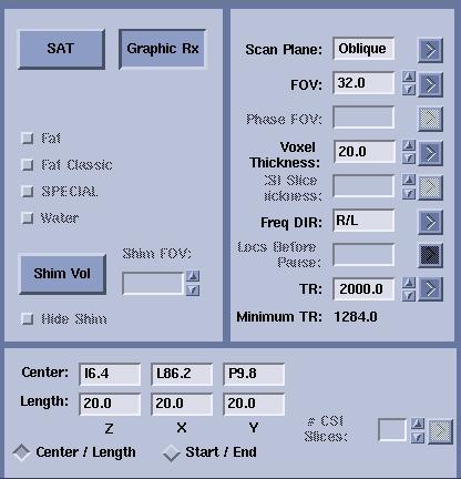
52 Breast MR Breast MR Courtesy of GE
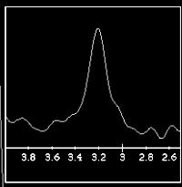
53 Breast MR Breast MR Static T1WI, T2WI, STIR Contrast-enhanced Dynamic contrast-enhanced Spectroscopy
54 Breast MR Breast MR Courtesy of GE

55 Breast MR Breast MR Courtesy of GE

56 Breast MR Breast MR Courtesy of GE

 close to it.")

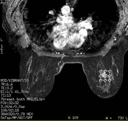
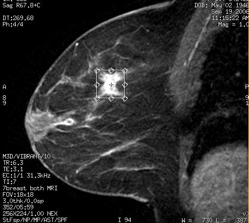
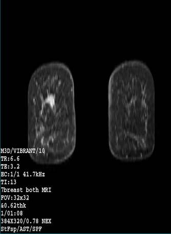
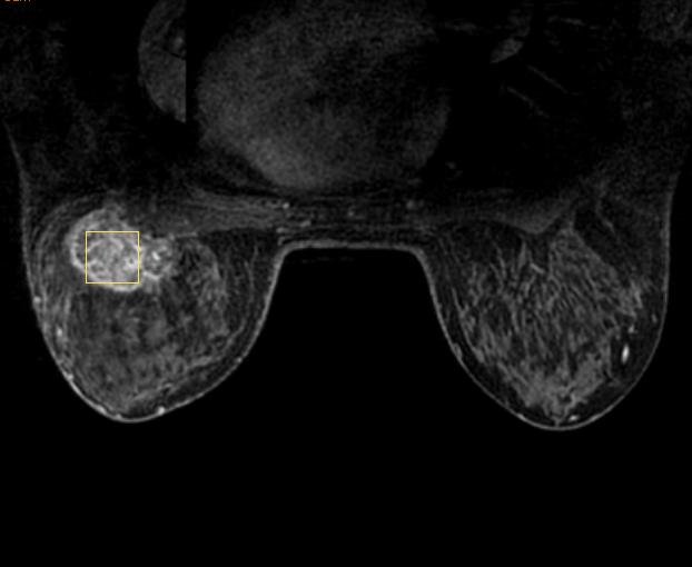
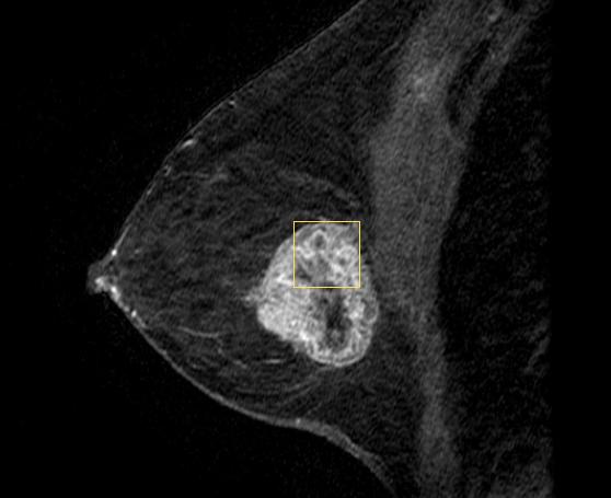
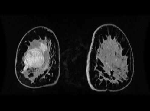
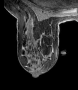
57 Breast MR Breast MR a b c Figure 1 (a) T1 pre contasted (b) Contrast-enhanced T1-WT FLASH 3D showing an ill defined lesion with irregular margin, features typical of a carcinoma (c) Fat suppressed MRI image revealed a smaller satellite lesion (arrow) close to it. (c) The enhancement curve was Type III with an early peak and delayed phase rapid washout. Histology revealed multifocal invasive ductal carcinoma. d

58 Breast MR - Spectroscopy Breast MR - Spectroscopy Courtesy of GE


59 Breast MR - Biopsy Breast MR - Biopsy Courtesy of GE
60 Conclusion Exiting developments in breast imaging mainly due to digital technology Multiple approach to image the breast Do not forget that NOTHING can compensate for an examination that is done badly Perform the best that you can every time!
61 THANK YOU
Digital Breast Tomosynthesis from a first idea to clinical routine
 International Master Programm Biomedical Engineering Digital Breast Tomosynthesis from a first idea to clinical routine Historical background 2D imaging of 3D objects has important limitations Jörg Barkhausen
Financial Disclosures
 Financial Disclosures 3D Mammography: The Latest Developments in the Breast Imaging Arena I have no financial disclosures Dr. Katharine Lampen-Sachar Breast and Body Radiologist Radiology Associates of
Current Status of Supplementary Screening With Breast Ultrasound
 Current Status of Supplementary Screening With Breast Ultrasound Stephen A. Feig, M.D., FACR Fong and Jean Tsai Professor of Women s Imaging Department of Radiologic Sciences University of California,
Here are examples of bilateral analog mammograms from the same patient including CC and MLO projections.
 Good afternoon. It s my pleasure to be discussing Diagnostic Breast Imaging over the next half hour. I m Wei Yang, Professor of Diagnostic Radiology and Chief, the Section of Breast Imaging as well as
Since its introduction in 2000, digital mammography has become
 Review Article Smith A, PhD email : Andrew.smith@hologic.com Since its introduction in 2000, digital mammography has become an accepted standard of care in breast cancer screening and has paved the way
CHAPTER 2 MAMMOGRAMS AND COMPUTER AIDED DETECTION
 9 CHAPTER 2 MAMMOGRAMS AND COMPUTER AIDED DETECTION 2.1 INTRODUCTION This chapter provides an introduction to mammogram and a description of the computer aided detection methods of mammography. This discussion
Emerging Techniques in Breast Imaging: Contrast-Enhanced Mammography and Fast MRI
 Emerging Techniques in Breast Imaging: Contrast-Enhanced Mammography and Fast MRI Lilian Wang, M.D. Breast Imaging Section Department of Radiology Northwestern Medicine Overview Rationale for new imaging
EARLY DETECTION: MAMMOGRAPHY AND SONOGRAPHY
 EARLY DETECTION: MAMMOGRAPHY AND SONOGRAPHY Elizabeth A. Rafferty, M.D. Avon Comprehensive Breast Center Massachusetts General Hospital Harvard Medical School Breast Cancer Screening Early detection of
New Imaging Modalities for better Screening and Diagnosis
 New Imaging Modalities for better Screening and Diagnosis Miri Sklair-Levy, MD Department of Diagnostic Imaging Sheba Medical Center, Sackler School of Medicine, Tel Aviv University Department of Diagnostic
Mammography. What is Mammography?
 Scan for mobile link. Mammography Mammography is a specific type of breast imaging that uses low-dose x-rays to detect cancer early before women experience symptoms when it is most treatable. Tell your
Mammography. What is Mammography? What are some common uses of the procedure?
 Mammography What is Mammography? Mammography is a specific type of imaging that uses a low-dose x-ray system to examine breasts. A mammography exam, called a mammogram, is used to aid in the early detection
The Radiology Aspects
 REQUIREMENTS FOR INTERNATIONAL ACCREDITATION OF BREAST CENTERS/UNITS The Radiology Aspects Miri Sklair-Levy, Israel RADIOLOGY GUIDELINES FOR QUALITY ASSURANCE IN BREAST CANCER SCREENING AND DIAGNOSIS Radiologists
Standard Breast Imaging Modalities. Lilian Wang, M.D. Breast Imaging Section Department of Radiology Northwestern Medicine
 Standard Breast Imaging Modalities Lilian Wang, M.D. Breast Imaging Section Department of Radiology Northwestern Medicine Overview Standard breast imaging modalities Mammography Ultrasound MRI Imaging
Mammographic imaging of nonpalpable breast lesions. Malai Muttarak, MD Department of Radiology Chiang Mai University Chiang Mai, Thailand
 Mammographic imaging of nonpalpable breast lesions Malai Muttarak, MD Department of Radiology Chiang Mai University Chiang Mai, Thailand Introduction Contents Mammographic signs of nonpalpable breast cancer
Breast Tomosynthesis. What is breast tomosynthesis?
 Scan for mobile link. Breast Tomosynthesis Breast tomosynthesis is an advanced form of mammography, a specific type of breast imaging that uses low-dose x-rays to detect cancer early when it is most treatable.
Mammography. Background and Perspective. Mammography Evolution. Background and Perspective. T.R. Nelson, Ph.D. x41433
 - 2015 Background and Perspective 2005 (in US) Women Men Mammography Invasive Breast Cancer Diagnosed 211,240 1,690 Noninvasive Breast Cancer Diagnosed 58,940 Deaths from Breast Cancer 40,410 460 T.R.
EARLY DETECTION: MAMMOGRAPHY AND SONOGRAPHY
 EARLY DETECTION: MAMMOGRAPHY AND SONOGRAPHY Elizabeth A. Rafferty, M.D. Avon Comprehensive Breast Center Massachusetts General Hospital Harvard Medical School Breast Cancer Screening Early detection of
Epworth Healthcare Benign Breast Disease Symposium. Sat Nov 12 th 2016
 Epworth Healthcare Benign Breast Disease Symposium Breast cancer is common Sat Nov 12 th 2016 Benign breast disease is commoner, and anxiety about breast disease commoner still Breast Care Campaign UK
What s New in Breast Imaging. Jennifer A. Harvey, M.D., FACR Professor of Radiology University of Virginia
 What s New in Breast Imaging Jennifer A. Harvey, M.D., FACR Professor of Radiology University of Virginia Disclosure Hologic, Inc. Shareholder and research agreement. Volpara Solutions, Ltd. Shareholder
Correlation between lesion type and the additional value of digital breast tomosynthesis
 Correlation between lesion type and the additional value of digital breast tomosynthesis Poster No.: C-1604 Congress: ECR 2011 Type: Scientific Exhibit Authors: C. Van Ongeval, L. Cockmartin, A. Van Steen,
TOMOSYNTHESIS. Daniela Bernardi. U.O. Senologia Clinica e Screening mammografico APSS Trento, Italy
 TOMOSYNTHESIS Daniela Bernardi U.O. Senologia Clinica e Screening mammografico APSS Trento, Italy BACKGROUND early detection through screening MAMMOGRAPHY is associated with reduced breast cancer morbidity
CURRENT METHODS IN IMAGE GUIDED BREAST BIOPSY
 CURRENT METHODS IN IMAGE GUIDED BREAST BIOPSY Stuart Silver April 24, 2004 OBJECTIVES Review development of current techniques Discuss stereotactic breast biopsy Discuss US guided breast biopsy 1 OBJECTIVES
Contrast Enhanced Spectral Mammography (CESM) Updates
 Updates") Contrast Enhanced Spectral Mammography (CESM) Updates Georgeta Mihai, PhD, DABR Medical Physicist, BIDMC, Boston Assistant Professor, Harvard Medical School, Boston Disclosures None Acknowledgments: Da
Contrast Enhanced Spectral Mammography (CESM) Updates Georgeta Mihai, PhD, DABR Medical Physicist, BIDMC, Boston Assistant Professor, Harvard Medical School, Boston Disclosures None Acknowledgments: Da
Breast Tomosynthesis An additional screening tool in the fight against breast cancer
 What to Expect Breast Tomosynthesis An additional screening tool in the fight against breast cancer Every woman over 40 should be examined for breast cancer once a year. American Cancer Society What to
WHAT TO EXPECT. Breast Tomosynthesis An additional screening tool in the fight against breast cancer HOLOGIC. The Women's Health Company
 WHAT TO EXPECT Breast Tomosynthesis An additional screening tool in the fight against breast cancer HOLOGIC The Women's Health Company ...,. Screening for breast cancer Doctors and scientists agree that
Update of Digital Breast Tomosynthesis. Susan Orel Roth, MD
 Update of Digital Breast Tomosynthesis Susan Orel Roth, MD NCI estimates that : Why DBT? Approximately 20% of breast cancers are missed at mammography screening Average recall rates approximately 10%
Breast Health and Imaging Glossary
 Contact: Lorna Vaughan HerSpace Breast Imaging & Biopsy Associates 300 State Route 35 South W. Long Branch, NJ 07764 732-571-9100, ext. 104 lorna@breast-imaging.com Breast Health and Imaging Glossary Women
Policy Library Clinical Advantages of Digital Breast Tomosynthesis in Symptomatic Patients
 Policy Library Clinical Advantages of Digital Breast Tomosynthesis in Symptomatic Patients Version: 1 Approved by: Faculty of Clinical Radiology Council Date of approval: Click and type: day month and
Breast tomosynthesis reduces radiologist performance variability compared to digital mammography
 Breast tomosynthesis reduces radiologist performance variability compared to digital mammography Andrew Smith 1, Elizabeth Rafferty 2, Loren Niklason 1 1 Hologic, Inc., Bedford MA, USA 2 Massachusetts
Contrast-Enhanced Digital Mammography
 2015 ARRS Breast Symposium Contrast-Enhanced Digital Mammography John Lewin, M.D. Diversified Radiology of Colorado CEDM - Outline History Technique Literature Review / Cases Clinical Status Inexpensive,
Contrast-Enhanced Spectral Mammography
 Contrast-Enhanced Spectral Mammography Illuminating Breast Cancer Detection SenoBright HD TM gehealthcare.com/senobright Mammography is the most reliable imaging technique for breasts, but limitations
Improving Methods for Breast Cancer Detection and Diagnosis. The National Cancer Institute (NCI) is funding numerous research projects to improve
 is funding numerous research projects to improve") CANCER FACTS N a t i o n a l C a n c e r I n s t i t u t e N a t i o n a l I n s t i t u t e s o f H e a l t h D e p a r t m e n t o f H e a l t h a n d H u m a n S e r v i c e s Improving Methods for
CANCER FACTS N a t i o n a l C a n c e r I n s t i t u t e N a t i o n a l I n s t i t u t e s o f H e a l t h D e p a r t m e n t o f H e a l t h a n d H u m a n S e r v i c e s Improving Methods for
Tomosynthesis and breast imaging update. Dr Michael J Michell Consultant Radiologist King's College Hospital NHS Foundation Trust
 Tomosynthesis and breast imaging update Dr Michael J Michell Consultant Radiologist King's College Hospital NHS Foundation Trust Breast imaging new technology BREAST CANCER FLT PET shows different grades
Pitfalls and Limitations of Breast MRI. Susan Orel Roth, MD Professor of Radiology University of Pennsylvania
 Pitfalls and Limitations of Breast MRI Susan Orel Roth, MD Professor of Radiology University of Pennsylvania Objectives Review the etiologies of false negative breast MRI examinations Discuss the limitations
Dense Breasts, Get Educated
 Dense Breasts, Get Educated What are Dense Breasts? The normal appearances to breasts, both visually and on mammography, varies greatly. On mammography, one of the important ways breasts differ is breast
Breast MRI Update. Jeffrey C. Weinreb, MD, FACR Yale University School of Medicine
 Breast MRI Update Jeffrey C. Weinreb, MD, FACR jeffrey.weinreb@yale.edu Yale University School of Medicine I disclose the following financial relationships with relevant commercial interests: Bracco Bayer
Digital Breast Tomosynthesis Ready for Routine Screening?
 Digital Breast Tomosynthesis Ready for Routine Screening? Sophia Zackrisson MD, PhD, Assoc Prof of Radiology Skåne University Healthcare, Lund University, Sweden 1 Mammography screening 20% reduced breast
Tissue Breast Density
 Tissue Breast Density Reporting breast density within the letter to the patient is now mandated by VA law. Therefore, this website has been established by Peninsula Radiological Associates (PRA), the radiologists
9/3/2015. New Mammographic Modality Training. Mammographer. Qualified Radiologic Technologist. Tomosynthesis Training
 FDA Required Breast Tomosynthesis Training New Mammographic Modality Training On any new mammography technology, such as breast tomosynthesis, the Mammography Quality Standards Act (MQSA) (http:fda.gov/radiation-emitting
arxiv: v2 [cs.cv] 8 Mar 2018
![arxiv: v2 [cs.cv] 8 Mar 2018](/thumbs/87/97094636.jpg "arxiv: v2 [cs.cv] 8 Mar 2018") Automated soft tissue lesion detection and segmentation in digital mammography using a u-net deep learning network Timothy de Moor a, Alejandro Rodriguez-Ruiz a, Albert Gubern Mérida a, Ritse Mann a, and
Automated soft tissue lesion detection and segmentation in digital mammography using a u-net deep learning network Timothy de Moor a, Alejandro Rodriguez-Ruiz a, Albert Gubern Mérida a, Ritse Mann a, and
Armed Forces Institute of Pathology.
 Armed Forces Institute of Pathology www.radpath.com Armed Forces Institute of Pathology Breast Disease www.radpath.org Armed Forces Institute of Pathology Interpretation of Breast MRI Leonard M. Glassman
Breast Cancer Screening and Diagnosis
 Breast Cancer Screening and Diagnosis Priya Thomas, MD Assistant Professor Clinical Cancer Prevention and Breast Medical Oncology University of Texas MD Anderson Cancer Center Disclosures Dr. Thomas has
Breast Cancer Imaging
 Breast Cancer Imaging I. Policy University Health Alliance (UHA) will cover breast imaging when such services meet the medical criteria guidelines (subject to limitations and exclusions) indicated below.
Breast Cancer Screening
 Scan for mobile link. Breast Cancer Screening What is breast cancer screening? Screening examinations are tests performed to find disease before symptoms begin. The goal of screening is to detect disease
Look differently. Invenia ABUS. Automated Breast Ultrasound
 Look differently. Invenia ABUS Automated Breast Ultrasound InveniaTM ABUS from GE Healthcare offers a view beyond mammography, with breast screening technology that looks differently. 40 % The unseen risk.
TOMOSYNTHESIS: WORTH ALL THE HYPE?
 X-Ray Associates of New Mexico, P.C. TOMOSYNTHESIS: WORTH ALL THE HYPE? MICHAEL N. LINVER, MD, FACR MAMMOGRAPHY: THE GOOD, THE PRETTY GOOD, & THE NOT SO GOOD MAMMOGRAPHY: THE GOOD, THE PRETTY GOOD, & THE
High Risk Screening: A Multimodality Approach
 High Risk Screening: A Multimodality Approach John Lewin, M.D., FACR, FSBI The Women s Imaging Center Denver, Colorado Disclosures Consultant to Hologic Previously received research funds from Hologic
Contrast enhanced spectral mammography: A literature review
 Contrast enhanced spectral mammography: A literature review Poster No.: R-0172 Congress: RANZCR-AOCR 2012 Type: Authors: Keywords: DOI: Educational Exhibit S. Buzynski, D. Taylor; Perth/AU Breast, Digital
Digital Breast Tomosynthesis in the Diagnostic Environment: A Subjective Side-by-Side Review
 Women s Imaging Original Research Hakim et al. Digital Breast Tomosynthesis Women s Imaging Original Research Christiane M. Hakim 1 Denise M. Chough 1 Marie A. Ganott 1 Jules H. Sumkin 1 Margarita L. Zuley
Opportunities and Innovations in Digital Mammography John M. Sandrik, Ph.D. GE Healthcare Milwaukee, WI
 Opportunities and Innovations in Digital Mammography John M. Sandrik, Ph.D. GE Healthcare Milwaukee, WI john.sandrik@med.ge.com with many thanks to Vince Polkus, Advanced Applications Product Mgr. 1 Content
Compressive Re-Sampling for Speckle Reduction in Medical Ultrasound
 Compressive Re-Sampling for Speckle Reduction in Medical Ultrasound Professor Richard Mammone Rutgers University Email Phone Number Christine Podilchuk, Lev Barinov, Ajit Jairaj and William Hulbert ClearView
Imaging in breast cancer. Mammography and Ultrasound Donya Farrokh.MD Radiologist Mashhad University of Medical Since
 Imaging in breast cancer Mammography and Ultrasound Donya Farrokh.MD Radiologist Mashhad University of Medical Since A mammogram report is a key component of the breast cancer diagnostic process. A mammogram
Outline. Digital Breast Tomosynthesis: Update and Pearls for Implementation. Tomosynthesis Dataset: 2D/3D (Hologic Combo Acquisition)
") Outline Digital Breast Tomosynthesis (DBT) the new standard of care Digital Breast Tomosynthesis: Update and Pearls for Implementation Emily F. Conant, M.D. Professor, Chief of Breast Imaging Department
Outline Digital Breast Tomosynthesis (DBT) the new standard of care Digital Breast Tomosynthesis: Update and Pearls for Implementation Emily F. Conant, M.D. Professor, Chief of Breast Imaging Department
Fundamentals of Breast Tomosynthesis
 Fundamentals of Breast Tomosynthesis Improving the Performance of Mammography Andrew Smith, Ph.D. This white paper is one in a series of research overviws on advanced technologies in women s healthcare.
Radiation Dosimetry in Digital Breast Tomosynthesis. March, 2015 William J. O Connel, Dr. Ph, Senior Medical Physicist
 Radiation Dosimetry in Digital Breast Tomosynthesis March, 2015 William J. O Connel, Dr. Ph, Senior Medical Physicist Imagination at work. Syllabus 1. Introduction 2. Dosimetry in Mammography 3. Dosimetry
Breast health and screening
 Breast health and screening Dr Kathy Wong MBBS (HK), FRCR (UK), FHKCR FHKAM (Radiology) Clinical Radiologist Department of Diagnostic and Interventional Radiology Kwong Wah Hospital Questions regarding
Dense Breasts. A Breast Cancer Risk Factor and Imaging Challenge
 Dense Breasts A Breast Cancer Risk Factor and Imaging Challenge Renee Pinsky, MD University of Michigan Department of Radiology Division of Breast Imaging No Disclosures QUIZ: ARE YOU DENSE? a. Breast
Mammography and Other Screening Tests. for Breast Problems
 301.681.3400 OBGYNCWC.COM Mammography and Other Screening Tests What is a screening test? for Breast Problems A screening test is used to find diseases, such as cancer, in people who do not have signs
Disclosures. Outline. Learning Objectives. Introduction. Introduction. Stereotactic Breast Biopsy vs Mammography: Image Quality and Dose.
 Disclosures Stereotactic Biopsy vs Mammography: and Dose None Vikas Patel, PhD, DABR Upstate Medical Physics 2014 Annual Meeting The American Association of Physicists in Medicine Austin, TX Learning Objectives
A comparison of the accuracy of film-screen mammography, full-field digital mammography, and digital breast tomosynthesis
 Clinical Radiology xxx (2012) 1e6 Contents lists available at SciVerse ScienceDirect Clinical Radiology journal homepage: www.clinicalradiologyonline.net A comparison of the accuracy of film-screen mammography,
Breast Imaging & You
 Breast Imaging & You What s Inside: Breast Imaging... 2 Digital Breast Tomosynthesis (DBT) mammograms... 4 Breast cancer screening... 6 Dense breast tissue... 8 Automated Breast Ultrasound (ABUS)... 9
Assessment of extent of disease: digital breast tomosynthesis (DBT) versus full-field digital mammography (FFDM)
 versus full-field digital mammography (FFDM)") Assessment of extent of disease: digital breast tomosynthesis (DBT) versus full-field digital mammography (FFDM) Poster No.: C-1237 Congress: ECR 2012 Type: Scientific Paper Authors: N. Seo 1, H. H. Kim
Assessment of extent of disease: digital breast tomosynthesis (DBT) versus full-field digital mammography (FFDM) Poster No.: C-1237 Congress: ECR 2012 Type: Scientific Paper Authors: N. Seo 1, H. H. Kim
Henda s Law. Supplemental screening for women with dense breast tissue and increased risk
 . Henda s Law Supplemental screening for women with dense breast tissue and increased risk The 2011 Texas Legislature passed House Bill 2102 which is effective 1st September 2011. The law is informally
Experience with Tomosynthesis (3-D) Mammography in a Mobile Setting and Current Controversies
 Mammography in a Mobile Setting and Current Controversies") Experience with Tomosynthesis (3-D) Mammography in a Mobile Setting and Current Controversies Jennifer Lance, MHSA, MS Jerry McLarty, PhD LSU Feist-Weiller Cancer Center Shreveport LA Sept 15, 2008 A brief
Experience with Tomosynthesis (3-D) Mammography in a Mobile Setting and Current Controversies Jennifer Lance, MHSA, MS Jerry McLarty, PhD LSU Feist-Weiller Cancer Center Shreveport LA Sept 15, 2008 A brief
Bianca den Dekker, MD - PhD student. Prof dr R.M. Pijnappel Prof dr H.M. Verkooijen Dr M. Broeders
 Diagnostic value of Three-dimensional UltRasound in breast cancer screening participants referred with a BI-RADS 0 test result: a comparison of imaging strategies (TURBO) Bianca den Dekker, MD - PhD student
Detection and Classification of Calcifications on Digital Breast Tomosynthesis and 2D Digital Mammography: A Comparison
 Women s Imaging Original Research Spangler et al. Digital Breast Tomosynthesis Versus 2D Digital Mammography Women s Imaging Original Research FOCUS ON: M. Lee Spangler 1 Margarita L. Zuley 2 Jules H.
CURRENTLY FDA APPROVED ARE FULL FIELD DIGITAL MAMMOGRAPHY SYSTEMS AND FILM SCREEN STILL BEING USED AT SOME INSTITUTIONS
 ABBY DUROJAYE,M.D CURRENTLY FDA APPROVED ARE FULL FIELD DIGITAL MAMMOGRAPHY SYSTEMS AND FILM SCREEN STILL BEING USED AT SOME INSTITUTIONS BOTH HAVE BEEN SHOWN TO BE EFFECTIVE TOOLS EARLY DETECTION OF BREAST
Min Jung Kim Department of Medicine The Graduate School, Yonsei University
 Zoomed image of contact mammography versus magnification mammography in the diagnosis of microcalcifications with soft-copy full field digital mammography Min Jung Kim Department of Medicine The Graduate
Screening mammograms in women <50 years of age: Low risk is NOT protective
 Screening mammograms in women
Breast Tomosynthesis
 Breast Tomosynthesis The Use of Breast Tomosynthesis in a Clinical Setting 2 What s Inside Introduction... 1 Initial Hologic Clinical Trial Purpose and Methodology... 1 Clinical Trial Results... 2 Improved
Breast Cancer Screening and High Risk
 Breast Cancer Screening and High Risk Mary Freyvogel, DO Breast Surgeon Clinical Assistant Professor of Surgery University Hospitals Case Medical Center St. John Medical Center / Elyria Medical Center
Volume 14 - Issue 3, Matrix
 Volume 14 - Issue 3, 2014 - Matrix Digital Breast Tomosynthesis for Screening and Diagnosis of Breast Cancer Author ECRI ECRI Institute 29 Broadwater Road Suite 104 Welwyn Garden City AL7 3BQ United Kingdom
Melissa Hartman, DO Women s Health Orlando VA Medical Center
 Melissa Hartman, DO Women s Health Orlando VA Medical Center Most common non-skin cancer and Second deadliest cancer in women Majority are diagnosed by abnormal screening study An approach to breast cancer
Diagnostic Dilemmas of Breast Imaging
 Diagnostic Dilemmas of Breast Imaging Common Causes of Error in Breast Cancer Detection By: Jason Cord, M.D. Mammography: Initial Imaging The standard for detection of breast cancer Screening mammography
FACT: FACT: Breast Cancer Staging. For cancer to occur, something must damage nucleus of the cell. Stage I. Stage II 10/9/2018
 Digital Breast Tomosynthesis (DBT) Pathology Findings: Case Studies and Beyond For cancer to occur, something must damage nucleus of the cell. Advanced Health Education Center www.aheconline.com Copyright
Contrast-Enhanced Breast Tomosynthesis: Combining the Best of Both Worlds for Better Breast-Cancer Diagnosis
 Contrast-Enhanced Breast Tomosynthesis: Combining the Best of Both Worlds for Better Breast-Cancer Diagnosis T Wu (twu2@partners.org), E Rafferty, R Moore, D Kopans, Massachusetts General Hospital, Boston,
Mammography limitations. Clinical performance of digital breast tomosynthesis compared to digital mammography: blinded multi-reader study
 Clinical performance of digital breast tomosynthesis compared to digital mammography: blinded multi-reader study G. Gennaro (1), A. Toledano (2), E. Baldan (1), E. Bezzon (1), C. di Maggio (1), M. La Grassa
Detailed Program of the second BREAST IMAGING AND INTERVENTIONS PROGRAM am am : Clinician s requirements from breast imaging
 Detailed Program of the second BREAST IMAGING AND INTERVENTIONS PROGRAM 2012 Day one, 2 nd November BREAST IMAGING AND INTERVENTIONS PROGRAM 2012 9.00 AM 9.10 am Introduction 9.10 am - 9.30 am : Clinician
Contrast-enhanced Breast MRI RSSA 2013
 Contrast-enhanced Breast MRI RSSA 2013 Prof. dr. Maurice van den Bosch University Medical Center Utrecht, the Netherlands Index 1) Breast cancer 2) Why MRI of the breast 3) Technique 4) Interpretation
Screening Mammograms: Questions and Answers
 CANCER FACTS N a t i o n a l C a n c e r I n s t i t u t e N a t i o n a l I n s t i t u t e s o f H e a l t h D e p a r t m e n t o f H e a l t h a n d H u m a n S e r v i c e s Screening Mammograms:
ORIGINAL ARTICLE EVALUATION OF BREAST LESIONS USING X-RAY MAMMOGRAM WITH HISTOPATHOLOGICAL CORRELATION
 Available online at www.journalijmrr.com INTERNATIONAL JOURNAL OF MODERN RESEARCH AND REVIEWS IJMRR ISSN: 2347-8314 Int. J. Modn. Res. Revs. Volume 3, Issue 10, pp 807-814, October, 2015 ORIGINAL ARTICLE
AN ALGORITHM FOR EARLY BREAST CANCER DETECTION IN MAMMOGRAMS
 AN ALGORITHM FOR EARLY BREAST CANCER DETECTION IN MAMMOGRAMS Isaac N. Bankman', William A. Christens-Barryl, Irving N. Weinberg2, Dong W. Kim3, Ralph D. Semmell, and William R. Brody2 The Johns Hopkins
Breast Tomosynthesis
 Breast Tomosynthesis The Use of Breast Tomosynthesis in a Clinical Setting 2 What s Inside Introduction... 1 Initial Hologic Clinical Trial Purpose and Methodology... 1 Clinical Trial Results... 2 Improved
Proven clinical effectiveness at low radiation dose
 MicroDose Mammography Solutions Proven clinical effectiveness at low radiation dose Several studies provide evidence that Philips MicroDose Mammography* can provide outstanding image quality at 18% to
F r e q u e n t l y A s k e d Q u e s t i o n s. Mammograms
 Mammograms Q: What is a mammogram? A: A mammogram is a safe, low-dose x-ray exam of the breasts to look for changes that are not normal. The results are recorded on x-ray film or directly into a computer
Breast Imaging Essentials
 Breast Imaging Essentials Module 1 Transcript 2016 ASRT. All rights reserved. Breast Imaging Essentials Module 1 - Fundamentals of Breast Imaging 1. ASRT Animation 2. Welcome Welcome to Module 1 of Breast
Hosts. Screening for Dense Breasts. Guest Expert: Regina Hooley, MD Associate Professor of Diagnostic Radiology, Yale School of Medicine
 Hosts Anees MD Susan Higgins MD Associate Professor of Surgical Oncology Professor of Therapeutic Radiology, Obstetrics, Gynecology, and Reproductive Sciences Steven Gore MD Director of Hematologic Malignancies
Mammography is a most effective imaging modality in early breast cancer detection. The radiographs are searched for signs of abnormality by expert
 Abstract Methodologies for early detection of breast cancer still remain an open problem in the Research community. Breast cancer continues to be a significant problem in the contemporary world. Nearly
Breast Density. Information for Health Professionals
 Breast Density Information for Health Professionals BreastScreen NSW provides free screening mammography to asymptomatic women aged 50-74 every two years, with the aim of diagnosing breast cancer at an
Breast Imaging & You
 Breast Imaging & You What s Inside: Breast Imaging... 2 Digital Breast Tomosynthesis (DBT) mammograms... 4 Breast cancer screening... 6 Dense breast tissue... 8 Automated breast ultrasound (ABUS)... 9
MANAGEMENT OF DENSE BREASTS. Nichole K Ingalls, MD, MPH NW Surgical Specialists September 25, 2015
 MANAGEMENT OF DENSE BREASTS Nichole K Ingalls, MD, MPH NW Surgical Specialists September 25, 2015 No financial disclosures National Cancer Institute National Cancer Institute Increased Cancer Risk... DENSITY
Breast Imaging Update: Old Dog New Tricks
 Breast Imaging Update: Old Dog New Tricks Claire McKay, DO M&S Imaging Assoc. San Antonio, TX cmckayhart@juno.com Goals Describe modalities available, old and new Provide understanding of pros and cons
Improving Reading Time of Digital Breast Tomosynthesis with Concurrent Computer Aided Detection
 White Paper Improving Reading Time of Digital Breast Tomosynthesis with Concurrent Computer Aided Detection WHITE PAPER 2 3 Abstract PowerLook Tomo Detection, a concurrent computer-aided detection (CAD)
Successful Breast MRI Program : The ingredients
 Successful Breast MRI Program : The ingredients Dr. Smriti Hari Associate Professor Deptt. Of Radiology All India Institute of Medical Sciences New Delhi How to perform Breast MRI Breast MRI descriptors
Full-Field Digital Versus Screen- Film Mammography: Comparative Accuracy in Concurrent Screening Cohorts
 Accuracy of Mammography Women s Imaging Original Research WOMEN S IMAGING Marco Rosselli Del Turco 1 Paola Mantellini 1 Stefano Ciatto 1 Rita Bonardi 1 Francesca Martinelli 1 Barbara Lazzari 1 Nehmat Houssami
Categorical Classification of Spiculated Mass on Breast MRI
 Categorical Classification of Spiculated Mass on Breast MRI Poster No.: C-1974 Congress: ECR 2013 Type: Authors: Scientific Exhibit Y. Kanda 1, S. Kanao 2, M. Kataoka 2, K. Togashi 2 ; 1 Kyoto City/JP,
Ge elastography cpt codes
 Ge elastography cpt codes Aetna considers digital mammography a medically necessary acceptable alternative to film mammography. Currently, there are no guideline recommendations from leading medical professional
TMIST: Frequently Asked Questions
 TMIST: Frequently Asked Questions Key Topics for Site Investigators and Staff This document answers frequently asked questions about the Tomosynthesis Mammographic Imaging Screening Trial (TMIST/EA1151);
Breast Cancer Screening: Improved Readings With Computers
 Transcript Details This is a transcript of an educational program accessible on the ReachMD network. Details about the program and additional media formats for the program are accessible by visiting: https://reachmd.com/programs/advances-in-medical-imaging/breast-cancer-screening-improvedreadings-with-computers/4023/
Contrast-Enhanced Spectral Mammography helps to improve breast cancer diagnostics
 GE Healthcare Contrast-Enhanced Spectral Mammography helps to improve breast cancer diagnostics SenoBright TM mammography, Midwest U.S. hospital Simple test benefits clinicians and patients A hospital