Lumbar spinal stenosis is narrowing of the spinal canal that results in compression of the cauda
|
|
|
- Gerald Gregory
- 5 years ago
- Views:
Transcription
1 1 CHAPTER 32: CLINICAL RESULTS OF THE IDE TRIAL OF X-STOP INTERSPINOUS SYSTEMS ELIZABETH YU AND JAMES ZUCHERMAN Lumbar spinal stenosis Lumbar spinal stenosis is narrowing of the spinal canal that results in compression of the cauda equina. Stenosis may be present focally at a single level or at multiple levels. Stenosis may be due to congenital/developmental stenosis, acquired/degenerative stenosis or a combination of the two.[1] When stenosis is secondary to degenerative changes, presentation is most common in the population age of greater than fifty. Neurogenic intermittent claudication is the clinical manifestation of lumbar spinal stenosis. Symptoms were first described by Verbiest in 1954 in a case report of seven patient. All had spinal stenosis. Symptoms described included weakness in the bilateral lower extremity and numbness that were present with walking or standing and relieved with rest.[2]. Relief from neurogenic intermittent claudication results from forward flexion and expansion of the spinal canal as well as the neural foramen. Treatment of symptomatic lumbar spinal stenosis ranges from nonoperative treatment to operative treatment. The natural history of spinal stenosis is variable with 45% of patient remaining stable, 15% of patients improving, and 30% of patients declining.[3] Treatment Nonoperative treatment includes medication, bracing, physical therapy, as well as epidural steroid injections. When comparing nonoperative treatment with that of surgical decompression,
2 2 a study by Weinstein, et. al. conducted a multicenter randomized control trial as well as an observational cohort group. The multicenter study included thirteen centers within the US with a follow-up of up to four years. Outcomes were measured at 6 weeks, 3 months, 6 months and yearly with outcome measures of SF-36 and Oswestry Disability index scoring. The study found that patients who have failed 3 months of conservative treatment for symptomatic lumbar spinal stenosis, had significantly improved pain scores and function up through follow-up at 4 years when treated with surgical decompression compared to the continued nonoperative group.[4] Operative intervention The goal of operative intervention is decompression of the thecal sac. Multiple procedures have been described to include, but not limited to, laminectomy, laminotomy, and insertion of an interspinous process distraction system. This chapter will focus on the application of the interspinous process distraction system, specifically the X-stop device. Figure 1 The X-stop is designed to limit terminal extension at stenotic levels of the lumbar spine and relieve symptoms of lumbar spinal stenosis. It is targeted toward patients with intermittent neurogenic claudication. Patients would have failed nonoperative treatment and find relief with flexion. On-label indications are for over 50 years of age, greater than 50 minutes continuous sitting tolerance, and ability to walk more than 50 feet. Multiple studies have reviewed the risks and benefits of the X-Stop. Zucherman, et. al. conducted a multi-center prospective randomized study looking at the first year s results. Nine centers participated in the study with two hundred patients randomized from May of 2000 to July of Patients included had either an MRI or CT which demonstrated one or two levels of stenosis. One hundred patients were randomized into the X-stop group and ninety-one were
3 3 randomized into the nonoperative group. Nine patients withdrew from the study after their randomization. Non-operative treatment included one or more epidural steroid injection, nonsteroidal anti-inflammatory medication, analgesics, as well as physical therapy and/or bracing. Mean age of the X-Stop group was 70 and 69 years of age in the non-operative group. Average duration of symptoms between the two groups was between three to four years. Outcomes were measured using the Medical outcomes study short form-36 (SF-36) and the Zurich claudication questionnaires (ZCQ). Patients were seen at 6 weeks, 6 months, and 1 year after treatment. Preoperative ZCQ scores were not statistically different between the two groups. At each follow-up milestone, patients randomized to the X-stop group had significantly improvement ZCQ scores as well as SF-36 scores compared to the randomized non-operative group. The authors concluded that at 1 year, patients with neurogenic claudication significantly improved with X-stop treatment compared to non-operative treatment with similar reoperation rates.[5] The same group reviewed their results at two and four years.[6, 7] At two years followup, ninety-three of the one-hundred X-stop patients and eight-one of the ninety-one of the nonoperative patients were available for follow-up. Six of the 93 patients in the X-stop group and twenty-four of the 81 patients treated nonoperatively underwent decompression surgery by two years. When reviewing imaging studies, 96% of the X-stop group had maintenance of distraction at 2 years. 48.4% of the X-stop group patients improved in the ZCQ score compared to 4.9% of the nonoperative group at 2 year follow-up.[7] The same group also assessed the quality of life of the patients at 2 years follow-up using the SF-36 scoring. They gathered the bodily pain, general health, mental and physical component, physical functioning, emotion and physical role as well as social function aspect of the outcome form. At two years, they found
4 4 statistically significant improvement in all areas, except general health, emotional role and mental component when compared to the nonoperative group. There was improvement in the majority of the quality of life assessment, to include the physical component as well as physical function and role, improvement in bodily pain, mental health, and social function.[8] At four years, Kondrashov et. al. noted eighteen patients from the original study randomized to the X-Stop were available for follow-up. Average follow-up was 51 months and outcomes were measured based on Oswestry disability index (ODI). Improvement was noted when comparing preoperative ODI of 45 (range 20 to 80) and postoperative ODI of 15 (range 0 to 36). The authors noted that the ODI and ZCQ (also known as the Swiss Spinal Stenosis Questionnaire) outcome scores have been shown to be similar for outcome measures of spinal stenosis patients.[9] They concluded that the X-stop has lasting effects.[6] Five year prospective data from a multicenter study, to include Zucherman et. al., has been gathered and analyzed. Twenty-eight patients with a minimum of five year follow-up had undergone X-stop placement with the same criteria from the original X-stop study discussed previously. Average age was 67.5 years. Zurich claudication questionnaire was used for outcome measures. Preoperative outcomes compared to postoperative outcomes demonstrated an average score improvement of around 1 point for both the symptom severity and physical function, with a change of 0.5 points defined as clinically important. There was one displaced X-stop that was revised within the study time period. The authors concluded that neurogenic claudication treated with the X-stop provides improved patient outcomes out to five years thus far.[10] Biomechanics/Design rationale
5 5 Biomechanical and cadaveric studies have been conducted assessing the central canal and foraminal space changes with flexion and extension of the spine. Schmid et. al. reviewed MRIs of twelve volunteers with a mean age of 28. No patient had an abnormal MRI, to include disc pathology. MRIs were obtain in the seated upright neutral, seated upright flexed, seated upright extended and supine extended position. Two radiologists read the MRIs and measured the anterior-posterior diameter of the central canal, cross sectional area of the canal at the disc and pedicular levels, as well as cross sectional area of the neural foramina. All but two reads were within 10% of each other. In their results, they found there was a mean decrease of cross sectional area of 16.4% when comparing seated upright flexion and extension at the level of the L4-L5 disk; whereas, there was no change in cross sectional area at the pedicular level. When looking at the ligamentum flavum, there was an increase in thickness with seated upright extension compared to flexion of approximately three-fold. When assessing the neural foramen, there was a mean decrease of cross sectional area of 23.2% when comparing seated upright flexion and extension at each disc level of L1 to S1. There was a 19.2% increase in cross sectional area when comparing the seated upright neutral position with the seated upright flexion position. The authors concluded that position of the spine, particularly in seated upright extension, can result in changes in the area of the spinal canal and neural foramen.[11] Fujiwara et. al. studied cadaveric models and assessed the six degrees of motion of the lumbar spine. Average age of the cadavers was 69. All muscle was removed, leaving the ligaments, disc and neural elements. Biomechanical testing included flexion, extension, lateral bending, and rotation. Cross sectional area of the foramen was measured using CT scans. Flexion increased the foraminal area by 11.3% and extension decreased the area by 12.0%. Lateral bending
6 6 decreased the area by 8.4% and opened the contralateral foramen by 8.0%. Rotation showed a decrease in area of 5.7% and opening on the contralateral side of 6.5%.[12] The effect of the X-stop on foraminal and canal area has been studied. Twenty-six patients with lumbar spinal stenosis underwent X-stop placement with review of preoperative and postoperative MRI. The mean age was 71 years old. The MRIs were obtained with the patients in the erect position, sitting neutral, flexion and extension position. The authors noted a statistically significant increased in the spinal canal area in the erect and seated upright neutral position compared to the preoperative area in single level X-stops. At double level X-stops, there was significant increase in area in the erect and seated upright extension position. When looking at the neural foramen, they noted increased area in both the seated upright flexion and extension position.[13] Another study had similar results. A review of 10 patients who underwent the X-stop application and followed for a minimum of nine months demonstrated an increase in the cross sectional area of the dural sac of 22.3% and neural foramen of 36.5% from preoperative to postoperative MRI.[14] Zucherman et. al. s group has also assessed their patients MRI outcomes. Nine patients underwent both preoperative and postoperative MRI evaluation at an average of 6 years postoperatively. MRI was used to assess the average canal and neural foraminal area. Disc height and endplate angles were also measured. There was noted statistically significant increase in canal area from 1.27 cm 2 to 1.42 cm 2 and a foraminal area increase from 0.54 cm 2 to 0.97 cm 2. There was no difference in disc height or endplate angles.[15] Concerns have been raised on the effect of sagittal balance secondary to the device. Siddique s group reviewed the same twenty-six patients noted above and looked at the effects of motion and disc height and angulation on the patients with X-stops. Preoperative and
7 7 postoperative MRIs were obtained in the standing, seated upright neutral, flexion and extension as well as supine position. Disc heights were measured in the seated flexion and extension position as well as supine and erect position. Lumbar motion was measured from superior endplates of L1 and S1 in the upright seated extension and flexion position. The authors results showed that there was no significant difference in lumbar curvature pre and postoperatively. There was noted decreased range of motion in the caudal level of a two-level X-stop placement. There was no change in range of motion for a one-level X-stop placement. There was no significant change in the anterior or posterior disc height pre and postoperatively. The authors concluded that there was no significant change in the sagittal kinematics with X-stop placement on the lumbar spine.[16] Schulte, et. al. conducted a prospective study of twenty patients who underwent an X-stop. All patients had standing flatback radiographic series done and the sagittal balance was assessed preoperatively and postoperatively. The average age of the patients was 68. One level X-stop was done in five patients, two level X-stop in fourteen patients, and three level X-stop in one patient. Sagittal balance was found to have improved in sixteen of the twenty patients and decline in four of the twenty patients. Mean change in sagittal balance was - 2.0cm.[17] Lindsey, et. al., additionally, looked into the results of the range of motion of the lumbar spine as well as the adjacent lumbar levels with X-stop implantation. Seven cadaveric models were employed. L2-L5 were studied with the donors ranging from 17 to 55 years of age. Flexion, extension, lateral bending and axial rotation were assessed. One level X-stop was performed each level independently. It was found that at L3-L4 and L4-L5, the range of motion of extension was significantly reduced at these levels of implantation. The overall range of motion of L2-L5 from flexion and extension was also significantly decreased. There was no
8 8 significant change in axial rotation or lateral bending with and without the implant. The authors therefore concluded that the motion segments adjacent to the X-stop levels are not significantly affected kinematically.[18] Biomechanical effects of the X-stop The X-stop device was devised as a safer less invasive approach for treatment of neurogenic intermittent claudication from lumbar spinal stenosis. Because of its relative safety, rapid recovery and ease of administration under local anesthesia, many elderly, osteoporotic and appropriately fearful patients are especially attracted to the procedures as are their surgeons. The insertional load to failure of the X-stop and the spinous process load to failure was studied by Talwar et. al. Four cadaveric specimens were used to study the insertional load of L3-L4 and L4-L5. The maximum load and mean load of insertion of the device was measured. Seven cadaveric specimens were used measure the load to failure of the spinous process when the device was loaded in the cranial, middle and caudal portion of the spinous process. Bone mineral density was measured on each specimen. The average age of the specimens was 64. The authors found that the mean lateral insertional load of the X-stop was 65.6 Newtons and the mean spinous process load to failure was Newtons. The mean load to failure at the areas of the spinous process was not significantly different. They did find a linear correlation between the mean load to failure of the spinous process and the square of vertebral bone mineral density. The authors concluded that the mean load to failure of the lumbar spinous process is over four times the mean lateral insertional load of the X-stop, concluding that the technique of device placement is relatively reliable when BMD is greater than -2.8.[19]
9 9 Intervertebral disc pressure as well as facet loading and how they are affected have been studied in the cadaveric model. Swanson, et. al. used pressure transducers on eight cadaveric lumbar spines ranging in age of 56 to 80 years of age. Each disc from L2 through L5 was subjected to 700 Newtons of axial pressure with intradiscal pressure measured with the spine in neutral, extended and flexed position. 7.5 Nm was used for the bending moment. An X-stop was then placed between L3-L4 and the test was repeated. The authors reported that there was a significant decrease in the intradiscal pressure at the posterior annulus of L3-4 (level of implantation) as well as a decrease in intradiscal pressure in the nucleus in the neutral and extended positions. There was no significant change in the intradiscal pressures of L2-3 and L4-5. [20] Wiseman, et. al. assessed the pressure on the facet joints in extension with implantation of the X-stop. Seven lumbar cadaveric models were used to assess the L2-3, L3-4, and L4-5 facet joints. All specimens were loaded axially loaded with 700 Newtons and facet joint pressure were measured using pressure films. 15 Nm extension moment was applied and pressure film was remeasured. An X-stop was then placed at L3-4 and the models were retested. Results showed the X-stop significantly reduced the mean pressure, contact area, and force at L3-L4 by 39%, 46%, and 67%, respectively. The adjacent facet joints did not change at L2-L3. The authors concluded that the X-stop decreases the facet load at the implant level and does not affect the load at adjacent facets levels.[21] Economic considerations The cost of the treatment of lumbar spinal stenosis has been reviewed. Skidmore, et. al. compared the cost of the nonoperative treatment versus X-stop and laminectomy versus X-stop
10 10 for lumbar spinal stenosis. A literature review was conducted as well as formulation of a costeffectiveness analysis based on a multicenter, randomized study of 131 patients. Data collection of medical reimbursement, medicare reimbursements, and estimated medical costs were gathered for 2 years time. Quality-adjusted life years were calculated as well. The authors concluded that for moderately affected patients with lumbar spinal stenosis, X-stop placement as an outpatient was more cost effective than a laminectomy and nonoperative treatment.[22] Burnett, et. al., on the other hand, also compared the treatment of nonoperative care, decompression laminectomy, and X-stop placement over 2 years. A literature review was conducted as well as a cost-effectiveness model was created to look at the direct health care costs with the use of mean case numbers and reimbursements of sixteen states as well as the medical reimbursement costs of the three treatment options. Outcomes were measured using qualityadjusted life years. Laminectomies were considered inpatient procedures and X-stops were considered outpatient procedures. The two surgical groups also had the additional cost of the nonoperative group. At two years time, the authors concluded that laminectomy was the most effective treatment, followed by X-stop and lastly nonoperative treatment.[23] Kondrashov et. al. retrospectively reviewed eighteen patients with X-stop placement at one or two levels with age-matched patients who underwent laminectomy decompression. Both groups had similar length follow-up of a little greater than 4 years. The X-stop procedure was performed as an outpatient. Average hospital costs for each procedure were assessed. The authors concluded that when directly comparing the X-stop and laminectomy procedure and their perioperative costs to the hospital, a single level X-stop is approximately one-third the cost of a single level laminectomy and a double level X-stop is approximately half the cost of a two level laminectomy.[24]
11 11 Complications Complications from the use of the X-stop have been reviewed. Bowers, et. al. retrospectively reviewed all patients in their institution who underwent an X-stop placement. Thirteen patients with moderate or severe spinal stenosis had X-stop application at L4-L5 and L3-4. Average age of the patients was 75 years. All but one patient had regular follow-up intervals within two years postoperatively. All patients were contacted at four years postoperatively. All patients had improvement in back and leg pain after surgery; however, ten patients pain returned (77%). The authors noted complications of the X-stop included three patients with spinous process fractures, with resultant laminectomy with fusion. Two patients had new radiculopathies of which one was at the X-stop level and the other was at the adjacent level. Four patients required subsequent laminectomy decompressions. Two patients were counseled on laminectomy. The authors concluded they had an 85% complication rate and that X-stop may not be as beneficial for patients with moderate to severe spinal stenosis and careful patient selection is imperative.[25] Survival analysis was retrospectively assessed by Tuschel et. al. at a single institution forty-six patients who underwent a total of sixty-one X-stops. The mean age was 68.2 years with thirty-one one level X-stop and fifteen patients with two level X-stops. Mean follow-up was 34 months. Five patients did not meet follow-up criteria and fourteen patients underwent revision surgery by latest follow-up. Twenty-nine patients with X-stops remained for review. VAS, ODI, and SF-36 (physical component summary score) significantly improved, but the SF-36 (mental component summary score) did not significantly improve from preoperative to postoperative. When asked to rank satisfaction, 47% were very satisfied, 37% were somewhat satisfied and 16% were not satisfied. Of the fourteen patients that underwent revision, nine
12 12 underwent decompression with or without instrumented fusion, two patients had removal of the X-stop for implant pain, and two patients had removal at an outside institution. The reasoning for the latter two patients was not stated. One patient had a revision of the X-stop due to traumatic dislocation. Kaplan-Meier survivorship curve yielded a 68% survival at 48 months. Eleven patients underwent revision within the first 18 months, of which all had continuing or return of preoperative symptoms. Three patients underwent revision at 3-4 years after the X-stop procedure. These three patients had improvement of symptoms during the above period prior to revision. The authors concluded that the results of the X-stop may not always be favorable and patient selection is critical.[26] Though yet unpublished, Zucherman s group reviewed their complications with the use of the X-stop device. Three-hundred patients were identified from January 2006 to August 2007 with one to three levels X-stop implantation. Average age was 74 years with follow-up ranging from 3 to 4.5 years. Twenty patients (6.7%) encountered complications, of which ten required further surgical intervention. Complications included 6 failures, 5 device migration or spinous process fracture, 3 new radiculopathy, 1 compression fracture, 1 infection, 1 hematoma, and 3 medical complications. All 6 failures went onto revision, 3 were removed and 1 patient underwent laminectomy with posterior fusion.[27] Expanded indications Advancement of the use of the X-stop has been developed. As noted previously, the spinous process load to failure correlates with the patient s bone mineral density. It can be projected that the lower the T-score on a DEXA scan, the higher the risk of spinous process fracture, thus failure of the X-stop device. Idler et. al. created a study to evaluate the use of PMMA injected
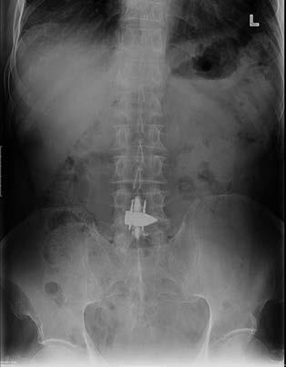
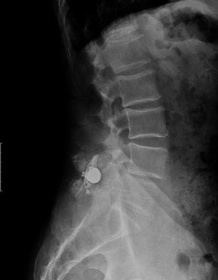
13 13 into the spinous process to enhance the strength of entire X-stop construct. Nine cadavers were used. Motion segments from each cadaver were dissected and divided into the control and PMMA group. Two cubic centimeters of PMMA were injected into the spinous processes with an 11 gauge needle. The mean load failure and stiffness was calculated in each group with axial loading of 1cm/min until failure. Bone mineral densities were obtained from each of the nine specimens, with an average of The mean load to failure of the X-stop construct for the control group was 1250 Newtons. The mean load to failure of the X-stop PMMA construct was 2386 Newtons. They found a linear correlation between the BMD and failure load and stiffness. The authors concluded that augmentation of the spinous process with PMMA increases that failure load as well as stiffness of the X-stop construct and potentially can be incorporated into in vivo use.[28] Zucherman et. al. conducted a prospective study assessing the use of PMMA augmentation in vivo. Two-hundred forty patients with a diagnosis of osteoporosis or osteopenia based on DEXA scan were included. All patients had a minimum one year follow-up. Seventyseven patients were in the PMMA augmentation group with X-stop implantation and onehundred sixty-three patients were in the control group with X-stop implantation only. Oswestry disability index score, walking and standing times, patient satisfaction, as well as visual analog scale were used for outcome measures. Cementation techniques were also assessed separately. Overall, the authors found an average improvement of ODI for the PMMA group was 18.7 and 14.3 for the control group. No statistical difference was noted between the two groups. Standing and walking times as well as satisfaction rates were also similar. When the PMMA group was subclassified based on cementation technique, statistically significant improvement in ODI score was seen in those was grade 3 (full cementation of the spinous process) when compared to the control group.[29] Figure 2 and 3
14 14 Outcomes of the X-stop application and age has been assessed. Romero, et. al. conducted a retrospective review of two hundred fifty-five patients who underwent an X-stop over the course of one year. Minimum follow-up was one year and ODI scores were used to measure outcome. Patients were divided into two groups: greater than 70 years of age and those younger than 70 years of age. The authors found that ODI scores, amount of time standing as well as walking was improved in those patients who were younger than 70 compared to those older than 70. This was statistically significant..[30] In some patients, the degree of spinal stenosis can be quite severe that an X-stop application alone is not enough, particularly severe subarticular and foraminal stenosis. Fuchs, et. al. reviewed the stability of the lumbar spine with graded facetectomies and X-stop implantation. Seven cadaveric specimens were gathered with assessment of L2-L3, L3-L4, and L4-L5. Graded facetectomies included a unilateral medial facetectomy, unilateral total facetectomy and bilateral total facetectomy. Rotation, flexion, extension, and neutral positions were measured using camera images with specimens undergoing facetectomies with and without X-stop application. 700 Newtons of axial loading and 7.5 Nm were used for bending moments. The authors found that bilateral total facetectomies significantly increased flexion and lateral bending range of motion. Unilateral medial facetectomy and unilateral total facetectomy did not result in significant destabilization and may be done in the setting of X-stop application.[31] Facet cysts arise from degenerative facet joints and may be present in lumbar spinal stenosis as space occupying lesions. Abrams, et. al. conducted a retrospective review of two hundred eighty-five patients who underwent X-stop application over the course of five years at a single institution. Fifty-eight (20.4%) of the patients were found to have synovial cysts contributing to stenosis. Mean follow-up was 22 months. Comparison of the ODI and VAS
15 15 were conducted between those patients with and without facet cysts. No statistically significant difference was noted between those with and without facet cysts in patients treated with the X- stop. The authors concluded that the X-stop is successful in the treatment of lumbar stenosis with the presence of facet cysts.[32] The original IDE study by Zucherman, et. al. included patients with up to grade I spondylolisthesis. Anderson, et. al. conducted a multicenter randomized control study looking specifically at the efficacy of X-stop use in patients with grade 1 spondylolisthesis and spinal stenosis. Nine centers randomized one hundred ninety-one patients with symptomatic lumbar spinal stenosis who met the original criteria listed above, as well as spondylolisthesis of 5 to 25%. Patients were seen at six weeks, six, twelve, and twenty four months postoperatively. Zurich claudication questionnaire and SF-36 was used for the outcome measures. No patients in the control group study crossed over to the X-stop group. Five patients in the X-stop group and four patients in the control group underwent surgical decompression with laminectomy. On radiographic review, there was no statistically significant increase in spondylolisthesis or kyphosis at two year radiographic follow-up. At two year follow-up, 63.4% of the X-stop group had improved ZCQ scores compared to 12.9% in the control group. [33] The relationship of scoliosis and the X-stop has been reviewed. Rolfe, et. al. retrospectively reviewed one-hundred seventy-nine patients with a minimum 1 year follow-up between January 2006 and May 2007 who had an X-stop placed, ranging from one to three levels. One hundred-sixteen patients had a less than 10 degree curve, forty-one patients had between an degree curve and twenty-two patients had over a 26 degree curve, with a maximum of 55 degrees. Oswestry disability scores were used to calculate outcome. In their results, the authors found that ODI scores improved in all groups after X-stop placement;
16 16 however, improvement was less profound when the curvature increased beyond 23 to 28 degrees.[34] Summary The X-stop is designed to limit terminal extension at stenotic levels of the lumbar spine and relieve symptoms of lumbar spinal stenosis. This has been confirmed by imaging studies to increase both central canal and foraminal area. It is a procedure that can be performed without general anesthesia and in an outpatient setting for patients with symptomatic lumbar spinal stenosis. It has been shown to be relatively safe in the appropriate patient population with symptomatic relief out to five years. Formatted: Indent: Left: 0", First line: 0" Formatted: Indent: Left: 0", First line: 0"

17 17 Figure 1 Polyetheretherketone X-stop implant. Figure 2 and 3: Upright lumbar radiographs with PMMA augmentation and X-stop.
18 18
19 19 REFERENCES 1. Spivak, J.M., Degenerative lumbar spinal stenosis. J Bone Joint Surg Am, (7): p Verbiest, H., A radicular syndrome from developmental narrowing of the lumbar vertebral canal. J Bone Joint Surg Br, B(2): p Sengupta, D.K. and H.N. Herkowitz, Lumbar spinal stenosis. Treatment strategies and indications for surgery. Orthop Clin North Am, (2): p Weinstein, J.N., et al., Surgical versus nonoperative treatment for lumbar spinal stenosis four-year results of the Spine Patient Outcomes Research Trial. Spine (Phila Pa 1976), (14): p Zucherman, J.F., et al., A prospective randomized multi-center study for the treatment of lumbar spinal stenosis with the X STOP interspinous implant: 1-year results. Eur Spine J, (1): p Kondrashov, D.G., et al., Interspinous process decompression with the X-STOP device for lumbar spinal stenosis: a 4-year follow-up study. J Spinal Disord Tech, (5): p Zucherman, J.F., et al., A multicenter, prospective, randomized trial evaluating the X STOP interspinous process decompression system for the treatment of neurogenic intermittent claudication: two-year follow-up results. Spine (Phila Pa 1976), (12): p Hsu, K.Y., et al., Quality of life of lumbar stenosis-treated patients in whom the X STOP interspinous device was implanted. J Neurosurg Spine, (6): p
20 20 9. Pratt, R.K., J.C. Fairbank, and A. Virr, The reliability of the Shuttle Walking Test, the Swiss Spinal Stenosis Questionnaire, the Oxford Spinal Stenosis Score, and the Oswestry Disability Index in the assessment of patients with lumbar spinal stenosis. Spine (Phila Pa 1976), (1): p Zucherman, J., et. al., Five Year Outcomes in Patients with the Xstop Interspinous Process Device for Neurogenic Intermittant Claudication Due to Lumbar Spinal Stenosis, in North American Spine Society. 2008: Toronto, Canada. 11. Schmid, M.R., et al., Changes in cross-sectional measurements of the spinal canal and intervertebral foramina as a function of body position: in vivo studies on an openconfiguration MR system. AJR Am J Roentgenol, (4): p Fujiwara, A., et al., Morphologic changes in the lumbar intervertebral foramen due to flexion-extension, lateral bending, and axial rotation: an in vitro anatomic and biomechanical study. Spine (Phila Pa 1976), (8): p Siddiqui, M., et al., Influence of X Stop on neural foramina and spinal canal area in spinal stenosis. Spine (Phila Pa 1976), (25): p Lee, J., et al., An interspinous process distractor (X STOP) for lumbar spinal stenosis in elderly patients: preliminary experiences in 10 consecutive cases. J Spinal Disord Tech, (1): p. 72-7; discussion Idler, C., et. al. Long Term Xstop Interspinous Process Decompression Follow-up Comparing Pre and Post Operative MRI Canal and Foramen Area in 8th Annual Global Symposium on Motion Preservation Technology, Spine Arthroplasty Society Miami Beach, Florida.
21 Siddiqui, M., et al., Effects of X-STOP device on sagittal lumbar spine kinematics in spinal stenosis. J Spinal Disord Tech, (5): p Schulte, L.M., et al., Change in sagittal balance with placement of an interspinous spacer. Spine (Phila Pa 1976), (20): p. E Lindsey, D.P., et al., The effects of an interspinous implant on the kinematics of the instrumented and adjacent levels in the lumbar spine. Spine (Phila Pa 1976), (19): p Talwar, V., et al., Insertion loads of the X STOP interspinous process distraction system designed to treat neurogenic intermittent claudication. Eur Spine J, (6): p Swanson, K.E., et al., The effects of an interspinous implant on intervertebral disc pressures. Spine (Phila Pa 1976), (1): p Wiseman, C.M., et al., The effect of an interspinous process implant on facet loading during extension. Spine (Phila Pa 1976), (8): p Skidmore, G., et al., Cost-effectiveness of the X-STOP(R) interspinous spacer for lumbar spinal stenosis. Spine (Phila Pa 1976), (5): p. E Burnett, M.G., S.C. Stein, and R.H. Bartels, Cost-effectiveness of current treatment strategies for lumbar spinal stenosis: nonsurgical care, laminectomy, and X-STOP. J Neurosurg Spine, (1): p Kondrashov DG, H.M., Hsu KY, Zucherman JF (2007) X-STOP versus decompression for neurogenic claudication: economic and clinical analysis. Internet Journal of Minimally Invasive Spinal Technology 1.
22 Bowers, C., et al., Dynamic interspinous process stabilization: review of complications associated with the X-Stop device. Neurosurg Focus, (6): p. E Tuschel, A., et al., Implant survival analysis and failure modes of the X STOP interspinous distraction device. Spine (Phila Pa 1976), Iezza, A., et. al. Analysis of Complications with Xstop. in International Society for the Study of the Lumbar Spine Goteborg, Sweden. 28. Idler, C., et al., A novel technique of intra-spinous process injection of PMMA to augment the strength of an inter-spinous process device such as the X STOP. Spine (Phila Pa 1976), (4): p Zucherman, J., et al., Use PMMA Augmentation in Osteopenic/Osteoporotic Patients Treated with the Xstop Device for Neurogenic Intermittant Claudication, in Proceedings of the International Society For the Study of the Lumbar Spine. May 2009: South Beach, Miami, FL. 30. Romero, N., et al., Outcome versus Age: A Retrospective Patient Chart Review After X- Stop Decompression, in International Society for the Advancement of Spine Surgery. April 2010 Poster: New Orleans, LA. 31. Fuchs, P.D., et al., The use of an interspinous implant in conjunction with a graded facetectomy procedure. Spine (Phila Pa 1976), (11): p ; discussion Abrams, J., et al., Treatment of Facet Cysts Associated with Neurogenic Intermittent Claudication with X-STOP. Submitted for publication, St. Mary's Spine Center: San Francisco, California.
23 Anderson, P.A., C.B. Tribus, and S.H. Kitchel, Treatment of neurogenic claudication by interspinous decompression: application of the X STOP device in patients with lumbar degenerative spondylolisthesis. J Neurosurg Spine, (6): p Rolfe, K.W., et al., Scoliosis and interspinous decompression with the X-STOP: prospective minimum 1-year outcomes in lumbar spinal stenosis. Spine J, (11): p
Chapter 35. Clinical Results of IDE Trial of X- Stop Interspinous Systems Lumbar Spinal Stenosis Treatment 2
 Chapter 35 Clinical Results of IDE Trial of X- Stop Interspinous Systems 35.1 Lumbar Spinal Stenosis 2 35.2 Treatment 2 35.3 Operative Intervention 2 35.4 Biomechanics/Design Rationale 3 35.5 Biomechanical
Evidence Table. Study Type: Randomized controlled trial. Study Aim: To evaluate the safety and efficacy of the X-Stop interspinous implant.
 Evidence Table Clinical Area: Reference: Spinal decompression device for lumbar spinal stenosis Zucherman JF et al. A prospective randomized multi-center study for the treatment of lumbar spinal stenosis
High failure rate of the interspinous distraction device (X-Stop) for the treatment of lumbar spinal stenosis caused by degenerative spondylolisthesis
 for the treatment of lumbar spinal stenosis caused by degenerative spondylolisthesis") Eur Spine J (2008) 17:188 192 DOI 10.1007/s00586-007-0492-x ORIGINAL ARTICLE High failure rate of the interspinous distraction device (X-Stop) for the treatment of lumbar spinal stenosis caused by degenerative
Eur Spine J (2008) 17:188 192 DOI 10.1007/s00586-007-0492-x ORIGINAL ARTICLE High failure rate of the interspinous distraction device (X-Stop) for the treatment of lumbar spinal stenosis caused by degenerative
Lumbar Laminotomy DEFINING APPROPRIATE COVERAGE POSITIONS NASS COVERAGE POLICY RECOMMENDATIONS TASKFORCE
 NASS COVERAGE POLICY RECOMMENDATIONS Lumbar Laminotomy DEFINING APPROPRIATE COVERAGE POSITIONS North American Spine Society 7075 Veterans Blvd. Burr Ridge, IL 60527 TASKFORCE Introduction North American
Current Spine Procedures
 SPINE BOOT CAMP: WHAT YOU DON T KNOW MAY COST YOU! David Abraham, M.D. The Reading Neck and Spine Center Reading, PA Current Spine Procedures Epidural/Transforaminal Injections Lumbar Procedures Laminectomy
5/27/2016. Stand-Alone Lumbar Lateral Interbody Fusion (LLIF) vs. Supplemental Fixation. Disclosures. LLIF Approach
 vs. Supplemental Fixation. Disclosures. LLIF Approach") Stand-Alone Lumbar Lateral Interbody Fusion (LLIF) vs. Supplemental Fixation Joseph M. Zavatsky, M.D. Spine & Scoliosis Specialists Tampa, FL Disclosures Consultant - Zimmer / Biomet, DePuy Synthes Spine,
Stand-Alone Lumbar Lateral Interbody Fusion (LLIF) vs. Supplemental Fixation Joseph M. Zavatsky, M.D. Spine & Scoliosis Specialists Tampa, FL Disclosures Consultant - Zimmer / Biomet, DePuy Synthes Spine,
BSD Journal of Spinal Disorders and Techniques Publish Ahead of Print DOI: /BSD.0b013e31827b671f
 BSD Journal of Spinal Disorders and Techniques Publish Ahead of Print DOI:10.1097/BSD.0b013e31827b671f Two-Year Evaluation of the X-STOP Interspinous Spacer in Different Primary Patient Populations with
5/19/2017. Interspinous Process Fixation with the Minuteman G3. What is the Minuteman G3. How Does it Work?
 Interspinous Process Fixation with the Minuteman G3 LLOYDINE J. JACOBS, MD CASTELLVI SPINE MEETING MAY 13, 2017 What is the Minuteman G3 The world s first spinous process plating system that is: Minimally
Interspinous and Interlaminar Stabilization/Distraction Devices (Spacers)
") Federal Employee Program 1310 G Street, N.W. Washington, D.C. 20005 202.942.1000 Fax 202.942.1125 7.01.107 Page: 1 of 12 Last Review Status/Date: June 2015 Interspinous and Interlaminar Stabilization/Distraction
Federal Employee Program 1310 G Street, N.W. Washington, D.C. 20005 202.942.1000 Fax 202.942.1125 7.01.107 Page: 1 of 12 Last Review Status/Date: June 2015 Interspinous and Interlaminar Stabilization/Distraction
Original Policy Date 12/2013
 MP 7.01.87 Interspinous Distraction Devices (Spacers) Medical Policy Section Surgery I12/2013ssue 12:2012 Original Policy Date 12/2013 Last Review Status/Date Reviewed with literature search/12/2013 Return
Aperius interspinous implant versus open surgical decompression in lumbar spinal stenosis
 The Spine Journal 11 (2011) 933 939 Clinical Study interspinous implant versus open surgical decompression in lumbar spinal stenosis Roberto Postacchini, MD a, Emiliano Ferrari, MD b, Gianluca Cinotti,
PARADIGM SPINE. Interlaminar Technology. Interlaminar Implant
 PARADIGM SPINE Interlaminar Technology Interlaminar Implant SPINAL STENOSIS WITH BACK PAIN THE RATIONALE FOR STABILIZATION For the treatment of spinal stenosis, surgeons have various treatment options.
Cover Page. The handle holds various files of this Leiden University dissertation.
 Cover Page The handle http://hdl.handle.net/1887/29800 holds various files of this Leiden University dissertation. Author: Moojen, Wouter Anton Title: Introducing new implants and imaging techniques for
Interspinous and Interlaminar Stabilization / Distraction Devices (Spacers)
") Interspinous and Interlaminar Stabilization / Distraction Devices (Spacers) Policy Number: 7.01.107 Last Review: 9/2014 Origination: 2/2007 Next Review: 9/2015 Policy Blue Cross and Blue Shield of Kansas
Interspinous and Interlaminar Stabilization / Distraction Devices (Spacers) Policy Number: 7.01.107 Last Review: 9/2014 Origination: 2/2007 Next Review: 9/2015 Policy Blue Cross and Blue Shield of Kansas
Protocol. Interspinous and Interlaminar Stabilization/Distraction Devices (Spacers)
") Interspinous and Interlaminar Stabilization/Distraction Devices (701107) Medical Benefit Effective Date: 10/01/13 Next Review Date: 07/15 Preauthorization No Review Dates: 07/07, 07/08, 09/09, 09/10, 07/11,
Interspinous and Interlaminar Stabilization/Distraction Devices (701107) Medical Benefit Effective Date: 10/01/13 Next Review Date: 07/15 Preauthorization No Review Dates: 07/07, 07/08, 09/09, 09/10, 07/11,
Subject: Interspinous Decompression Devices for Spinal Stenosis (X Stop, Coflex) Guidance Number: MCG-222 Revision Date(s):
 Guidance Number: MCG-222 Revision Date(s):") Subject: Interspinous Decompression Devices for Spinal Stenosis (X Stop, Coflex) Guidance Number: MCG-222 Revision Date(s): Original Effective Date: 3/16/15 DESCRIPTION OF PROCEDURE/SERVICE/PHARMACEUTICAL
Subject: Interspinous Decompression Devices for Spinal Stenosis (X Stop, Coflex) Guidance Number: MCG-222 Revision Date(s): Original Effective Date: 3/16/15 DESCRIPTION OF PROCEDURE/SERVICE/PHARMACEUTICAL
Corporate Medical Policy
 Corporate Medical Policy Image-Guided Minimally Invasive Decompression (IG-MLD) for File Name: Origination: Last CAP Review: Next CAP Review: Last Review: image-guided_minimally_invasive_decompression_for_spinal_stenosis
LUMBAR SPINAL STENOSIS
 LUMBAR SPINAL STENOSIS Always occurs in the mobile segment. Factors play role in Stenosis Pre existing congenital or developmental narrowing of the lumbar spinal canal Translation of one anatomic segment
2/5/2019. Facet Joint Pain. Biomechanics
 Facet Arthropathy as a Pain Source Evaluation and Management Shelby Spine Jan 31 st Feb 2 nd, 2019 Kushagra Verma MD, MS Adult and Pediatric Scoliosis And Spine Deformity Beach Orthopaedics Specialty Institute
PROF. EPIMENIO RAMUNDO ORLANDO
 PROF. EPIMENIO RAMUNDO ORLANDO Lumbar Spinal Stenosis - Definition N.I.C. caused by lumbar stenosis was firstly described by Verbiest (1954)*1 and is characterized by contemporary single or multiple factors:
Scoliosis and interspinous decompression with the X-STOP: prospective minimum 1-year outcomes in lumbar spinal stenosis
 The Spine Journal 10 (2010) 972 978 Clinical Study Scoliosis and interspinous decompression with the X-STOP: prospective minimum 1-year outcomes in lumbar spinal stenosis Kevin W. Rolfe, MD, MPH a, *,
Effects of X-Stop Device on Sagittal Lumbar Spine Kinematics in Spinal Stenosis
 ORIGINAL ARTICLE Effects of X-Stop Device on Sagittal Lumbar Spine Kinematics in Spinal Stenosis Manal Siddiqui, FRCS,* Efthimios Karadimas, MD,* Malcolm Nicol, MRCS,* Francis W. Smith, FRCR,w and Douglas
Innovative Techniques in Minimally Invasive Cervical Spine Surgery. Bruce McCormack, MD San Francisco California
 Innovative Techniques in Minimally Invasive Cervical Spine Surgery Bruce McCormack, MD San Francisco California PCF Posterior Cervical Fusion PCF not currently an ambulatory care procedure Pearl diver
Corporate Medical Policy
 Corporate Medical Policy Interspinous and Interlaminar Stabilization/Distraction Devices File Name: Origination: Last CAP Review: Next CAP Review: Last Review: interspinous_and_interlaminar_stabilization-distraction_devices
Populations Interventions Comparators Outcomes Individuals: With lumbar spinal stenosis
 Image-Guided Minimally Invasive Decompression for Spinal (701126) (Formerly Image-Guided Minimally Invasive Lumbar Decompression for Spinal ) Medical Benefit Effective Date: 10/01/17 Next Review Date:
Am I eligible for the TOPS study? Possibly, if you suffer from one or more of the following conditions:
 Am I eligible for the TOPS study? Possibly, if you suffer from one or more of the following conditions: Radiating leg pain Greater leg / buttock pain than back pain Severe pain sets in when walking as
Japan is currently experiencing an unprecedented era of an. An Interspinous Process Distractor (X STOP) for Lumbar Spinal Stenosis in Elderly Patients
 for Lumbar Spinal Stenosis in Elderly Patients") ORIGINAL ARTICLE An Interspinous Process Distractor (X STOP) for Lumbar Spinal Stenosis in Elderly Patients Preliminary Experiences in 10 Consecutive Cases Jangbo Lee, MD*, Kazutoshi Hida, MD*, Toshitaka
ORIGINAL ARTICLE An Interspinous Process Distractor (X STOP) for Lumbar Spinal Stenosis in Elderly Patients Preliminary Experiences in 10 Consecutive Cases Jangbo Lee, MD*, Kazutoshi Hida, MD*, Toshitaka
A New Approach to Quantify Functional Improvements Following X-Stop Spacer Procedure: A Case Report
 Elmer ress Case Report J Med Cases. 2015;6(5):205-210 A New Approach to Quantify Functional Improvements Following X-Stop Spacer Procedure: A Case Report Marzieh Hajiaghamemar a, d, Morteza Seidi a, Amy
ProDisc-L Total Disc Replacement. IDE Clinical Study
 Total Disc Replacement IDE Clinical Study Study Design TDR vs. circumferential fusion: Multi-center, prospective, randomized trial 17 centers, 292 patients 162 patients 80 fusion patients 50 non-randomized
ProDisc-L Total Disc Replacement. IDE Clinical Study.
 ProDisc-L Total Disc Replacement. IDE Clinical Study. A multi-center, prospective, randomized clinical trial. Instruments and implants approved by the AO Foundation Table of Contents Indications, Contraindications
Anesthesiology Pain Medicine
 KOWSAR Anesth Pain. 2012;2(1):36-41. DOI: 10.5812/aapm.5173 Anesthesiology Pain Medicine www.anesthpain.com Interspinous Process Implantation for the Treatment of Neurogenic Intermittent Claudication Nasser
Lumbar Spinal Fusion Corporate Medical Policy
 Lumbar Spinal Fusion Corporate Medical Policy File name: Lumbar Spinal Fusion File code: UM.SURG.15 Origination: 09/01/2016 Last Review: 09/29/2016 Next Review: 09/29/2017 Effective Date: 01/01/2017 Populations
QF-78. S. Tanaka 1, T.Yokoyama 1, K.Takeuchi 1, K.Wada 2, T. Tanaka 2, S.Abrakawa 2, G.Kumagai 2, T.Asari 2, A.Ono 2, Y.
 QF-78 Patient-oriented outcomes after musclepreserving interlaminar decompression for patients with lumbar spinal canal stenosis: Multi-center study to identify risk factors for poor outcomes S. Tanaka
Link to author version on UHI Research Database
 UHI Research Database pdf download summary Interspinous devices for lumbar stenosis a review of the literature Webb, Mary; van Woerden, Hugo C Published in: Internet Journal of Surgery Publication date:
This procedure lacks scientific evidence of effectiveness, and is not covered.
 ARBenefits Approval: 09-21-2011 Effective Date: 01-01-2012 Revision Date: Code(s): 0275T Medical Policy Title: Minimally Invasive, Image-Guided Lumbar Decompression for Spinal Stenosis Document: ARB0186
Original Article Management of Single Level Lumbar Degenerative Spondylolisthesis: Decompression Alone or Decompression and Fusion
 Egyptian Journal of Neurosurgery Volume 9 / No. 4 / October - December 014 51-56 Original Article Management of Single Level Lumbar Degenerative Spondylolisthesis: Decompression Alone or Decompression
Clinical Policy Title: Interspinous dynamic stabilization devices
 Clinical Policy Title: Interspinous dynamic stabilization devices Clinical Policy Number: 14.02.07 Effective Date: April 1, 2016 Initial Review Date: November 18, 2015 Most Recent Review Date: January
Peggers Super Summaries: The Aging Spine
 Aging Spine: AGING PROCESS Osteopenia 10% of 50 year old males and 25% of 50 year females Disc dehydration Facet degeneration Soft tissue hypertrophy 2 0 deformity Leg pain worse than back pain from nerve
ProDisc-C versus fusion with Cervios chronos prosthesis in cervical degenerative disc disease: Is there a difference at 12 months?
 Original research ProDisc-C versus fusion with Cervios chronos prosthesis in cervical degenerative disc ( ) 51 51 56 ProDisc-C versus fusion with Cervios chronos prosthesis in cervical degenerative disc
3D imaging reformation was obtained. The 3D color imaging reformation was reviewed in a different high resolution setting.
 POST OPERATIVE SPINE WITH CONTRAST CLINICAL INDICATION: Low back pain, Patient is post operative status for L4/5 diskectomy TECHNIQUE: MRI of the lumbosacral spine was performed with multiplanar imaging
Positional Magnetic Resonance Imaging. Description
 Subject: Positional Magnetic Resonance Imaging Page: 1 of 6 Last Review Status/Date: June 2015 Positional Magnetic Resonance Imaging Description Positional magnetic resonance imaging (MRI) allows imaging
Interspinous Fusion Devices. Midterm results. ROME SPINE 2012, 7th International Meeting Rome, 6-7 December 2012
 Interspinous Fusion Devices. Midterm results. ROME SPINE 2012, 7th International Meeting Rome, 6-7 December 2012 Posterior distraction and decompression Secure Fixation and Stabilization Integrated Bone
Lumbar Interspinous Spacers
 Lumbar Interspinous Spacers A Systematic Review of Clinical and Biomechanical Evidence Syed M. R. Kabir, FRCSEd (NeuroSurg),* Sanjay R. Gupta, MCh,* and Adrian T. H. Casey, FRCS (SN)* SPINE Volume 35,
Degenerative spondylolisthesis at the L4 L5 in a 32-year-old female with previous fusion for idiopathic scoliosis: A case report
 Journal of Orthopaedic Surgery 2003: 11(2): 202 206 Degenerative spondylolisthesis at the L4 L5 in a 32-year-old female with previous fusion for idiopathic scoliosis: A case report RB Winter Clinical Professor,
Lumbar Facet Joint Replacement
 Rome Spine 2011 THE SPINE TODAY International Congress Rome 6-7th December 2011 Lumbar Facet Joint Replacement Prof. Dr. Karin Büttner-Janz Past President International Society for the Advancement of Spine
Image-Guided Minimally Invasive Lumbar Decompression (IG-MLD) for Spinal Stenosis
 for Spinal Stenosis") Image-Guided Minimally Invasive Lumbar Last Review Status/Date: June 2013 Page: 1 of 10 Image-Guided Minimally Invasive Lumbar Decompression (IG-MLD) for Spinal Stenosis Description Image-guided minimally
Image-Guided Minimally Invasive Lumbar Last Review Status/Date: June 2013 Page: 1 of 10 Image-Guided Minimally Invasive Lumbar Decompression (IG-MLD) for Spinal Stenosis Description Image-guided minimally
MEDICAL POLICY. Proprietary Information of Excellus Health Plan, Inc. A nonprofit independent licensee of the BlueCross BlueShield Association
 MEDICAL POLICY SUBJECT: INTERSPINOUS AND PAGE: 1 OF: 6 If a product excludes coverage for a service, it is not covered, and medical policy criteria do not apply. If a commercial product (including an Essential
MEDICAL POLICY MEDICAL POLICY DETAILS POLICY STATEMENT. Page: 1 of 5
 Page: 1 of 5 MEDICAL POLICY MEDICAL POLICY DETAILS Medical Policy Title LUMBAR DECOMPRESSION Policy Number 7.01.97 Category Technology Assessment Effective Date 06/21/18 Revised Date 12/20/18 Product Disclaimer
Populations Interventions Comparators Outcomes Individuals: With lumbar spinal stenosis
 Image-Guided Minimally Invasive Decompression for Spinal (701126) Medical Benefit Effective Date: 10/01/18 Next Review Date: 07/19 Preauthorization No Review Dates: 09/10, 07/11, 07/12, 07/13, 07/14, 07/15,
Image-Guided Minimally Invasive Lumbar Decompression (IG-MLD) for Spinal Stenosis. Original Policy Date
 for Spinal Stenosis. Original Policy Date") MP 7.01.107 Image-Guided Minimally Invasive Lumbar Decompression (IG-MLD) for Spinal Stenosis Medical Policy Section Surgery Issue 12/2013 Original Policy Date 12/2013 Last Review Status/Date Reviewed
MP 7.01.107 Image-Guided Minimally Invasive Lumbar Decompression (IG-MLD) for Spinal Stenosis Medical Policy Section Surgery Issue 12/2013 Original Policy Date 12/2013 Last Review Status/Date Reviewed
University of Jordan. Professor Freih Abuhassan -
 Freih Odeh Abu Hassan F.R.C.S.(Eng.), F.R.C.S.(Tr.& Orth.). Professor of Orthopedics University of Jordan 1 A. Sacroiliitis History Trauma is very common Repetitive LS motion--lumbar rotation or axial
Interlaminar Decompression & Stabilization. Reginald Davis, M.D., FAANS, FACS Director of Clinical Research
 Interlaminar Decompression & Stabilization Reginald Davis, M.D., FAANS, FACS Director of Clinical Research Disclosures Background Device meant to stabilize the spine without fusion following decompression
PAUL A. ANDERSON, M.D., CLIFF B. TRIBUS, M.D., AND SCOTT H. KITCHEL, M.D.
 J Neurosurg Spine 4:464 471, 2006 Treatment of neurogenic claudication by interspinous decompression: application of the X STOP device in patients with lumbar degenerative spondylolisthesis PAUL A. ANDERSON,
Original Date: October 2015 LUMBAR SPINAL FUSION FOR
 National Imaging Associates, Inc. Clinical guidelines Original Date: October 2015 LUMBAR SPINAL FUSION FOR Page 1 of 9 INSTABILITY AND DEGENERATIVE DISC CONDITIONS FOR CMS (MEDICARE) MEMBERS ONLY CPT4
3D titanium interbody fusion cages sharx. White Paper
 3D titanium interbody fusion cages sharx (SLM selective laser melted) Goal of the study: Does the sharx intervertebral cage due to innovative material, new design, and lordotic shape solve some problems
Spinal fusion with decompressive laminectomy is
 J Neurosurg Spine 18:24 31, 2013 AANS, 2013 Posterior dynamic stabilization in the treatment of degenerative lumbar stenosis: validity of its rationale Clinical article Kee-Yong Ha, M.D., 1 Jun-Yeong Seo,
FDA Executive Summary
 FDA Executive Summary VertiFlex Superion InterSpinous Spacer Prepared for the February 20, 2015, Meeting of the Orthopaedic and Rehabilitation Devices Panel of the Medical Devices Advisory Committee Page
Comprehension of the common spine disorder.
 Objectives Comprehension of the common spine disorder. Disc degeneration/hernia. Spinal stenosis. Common spinal deformity (Spondylolisthesis, Scoliosis). Osteoporotic fracture. Anatomy Anatomy Anatomy
Medical Policy An independent licensee of the Blue Cross Blue Shield Association
 Lumbar Spinal Fusion Page 1 of 29 Medical Policy An independent licensee of the Blue Cross Blue Shield Association Title: See Also: Lumbar Spinal Fusion Interspinous Fixation (Fusion) Devices http://www.bcbsks.com/customerservice/providers/medicalpolicies/policies.shtml
Original Policy Date
 MP 6.01.39 Positional Magnetic Resonance Imaging Medical Policy Section Radiology Issue 12:2013 Original Policy Date 12:2013 Last Review Status/Date Reviewed with literature search/12:2013 Return to Medical
National Institute for Health and Clinical Excellence
 National Institute for Health and Clinical Excellence 191/2 Interspinous distraction procedures for lumbar spinal stenosis causing neurogenic claudication Consultation Comments table IPAC date: 9 th September
Biomechanics of Interspinous Process Fixation and Lateral Modular Plate Fixation to Support Lateral Lumbar Interbody Fusion (LLIF)
") Biomechanics of Interspinous Process Fixation and Lateral Modular Plate Fixation to Support Lateral Lumbar Interbody Fusion (LLIF) Calusa Ambulatory Spine Conference 2016 Jason Inzana, PhD 1 ; Anup Gandhi,
Biomechanics of Interspinous Process Fixation and Lateral Modular Plate Fixation to Support Lateral Lumbar Interbody Fusion (LLIF) Calusa Ambulatory Spine Conference 2016 Jason Inzana, PhD 1 ; Anup Gandhi,
MEDICAL POLICY. Proprietary Information of Excellus Health Plan, Inc. A nonprofit independent licensee of the BlueCross BlueShield Association
 MEDICAL POLICY SUBJECT: INTERSPINOUS AND PAGE: 1 OF: 8 If a product excludes coverage for a service, it is not covered, and medical policy criteria do not apply. If a commercial product, including an Essential
PARADIGM SPINE. Minimally Invasive Lumbar Fusion. Interlaminar Stabilization
 PARADIGM SPINE Minimally Invasive Lumbar Fusion Interlaminar Stabilization 2 A UNIQUE MIS ALTERNATIVE TO PEDICLE SCREW FIXATION The Gold Standard The combined use of surgical decompression and different
Systematic review Cervical artificial disc replacement versus fusion in the cervical spine: a systematic review (...)
") Systematic review Cervical artificial disc replacement versus fusion in the cervical spine: a systematic review (...) 59 59 66 Cervical artificial disc replacement versus fusion in the cervical spine:
Systematic review Cervical artificial disc replacement versus fusion in the cervical spine: a systematic review (...) 59 59 66 Cervical artificial disc replacement versus fusion in the cervical spine:
Kinematic Analysis of Lumbar Spine Undergoing Extension and Dynamic Neural Foramina Cross Section Measurement
 Copyright c 2008 ICCES ICCES, vol.7, no.2, pp.57-62 Kinematic Analysis of Lumbar Spine Undergoing Extension and Dynamic Neural Foramina Cross Section Measurement Yongjie Zhang 1, Boyle C. Cheng 2, Changho
2. The vertebral arch is composed of pedicles (projecting from the body) and laminae (uniting arch posteriorly).
 and laminae (uniting arch posteriorly).") VERTEBRAL COLUMN 2018zillmusom I. VERTEBRAL COLUMN - functions to support weight of body and protect spinal cord while permitting movements of trunk and providing for muscle attachments. A. Typical vertebra
VERTEBRAL COLUMN 2018zillmusom I. VERTEBRAL COLUMN - functions to support weight of body and protect spinal cord while permitting movements of trunk and providing for muscle attachments. A. Typical vertebra
Degenerative Disease of the Spine
 Degenerative Disease of the Spine Introduction: I. Anatomy Talk Overview II. Overview of Disease Processes: A. Spondylosis B. Intervertebral Disc Disease III. Diagnosis IV. Therapy Introduction: Myelopathy
The Biomechanics of the Human Spine. Basic Biomechanics, 6 th edition By Susan J. Hall, Ph.D.
 Chapter 9 The Biomechanics of the Human Spine Structure of the Spine The spine is a curved stack of 33 vertebrae structurally divided into five regions: cervical region - 7 vertebrae thoracic region -
Medical Policy An independent licensee of the Blue Cross Blue Shield Association
 Lumbar Spinal Fusion Page 1 of 28 Medical Policy An independent licensee of the Blue Cross Blue Shield Association Title: See Also: Lumbar Spinal Fusion Interspinous Fixation (Fusion) Devices http://www.bcbsks.com/customerservice/providers/medicalpolicies/policies.shtml
Patient Selection and Lumbar Operative Interventions
 Patient Selection and Lumbar Operative Interventions John C France MD Professor of Orthopaedic & Neurosurgery West Virginia University Low back pain is a symptom not a diagnosis Epidemiology of LBP General
Pasquale Donnarumma 1, Roberto Tarantino 1, Lorenzo Nigro 1, Marika Rullo 2, Domenico Messina 3, Daniele Diacinti 4, Roberto Delfini 1.
 Original Study Decompression versus decompression and fusion for degenerative lumbar stenosis: analysis of the factors influencing the outcome of back pain and disability Pasquale Donnarumma 1, Roberto
Interspinous Spacers. Joshua Abrams, James Zucherman INTRODUCTION
 Chapter-54 Interspinous Spacers 459 54 Interspinous Spacers Joshua Abrams, James Zucherman INTRODUCTION Anatomy Lumbar spinal stenosis is a narrowing of the spinal canal leading to a reduction in space
DECIMA SPINE The Simple Approach to Treat Lower Back Pain
 DECIMA SPINE The Simple Approach to Treat Lower Back Pain PDS/FCD-2 System The first, and only PERCUTANEOUS Bilateral Facet Augmentation System, that can treat L5-S1 effectively. Biomechanical data shows
Subaxial Cervical Spine Trauma. Introduction. Anatomic Considerations 7/23/2018
 Subaxial Cervical Spine Trauma Sheyan J. Armaghani, MD Florida Orthopedic Institute Assistant Professor USF Dept of Orthopedics Introduction Trauma to the cervical spine accounts for 5 of all spine injuries
Module: #15 Lumbar Spine Fusion. Author(s): Jenni Buckley, PhD. Date Created: March 27 th, Last Updated:
: Jenni Buckley, PhD. Date Created: March 27 th, Last Updated:") Module: #15 Lumbar Spine Fusion Author(s): Jenni Buckley, PhD Date Created: March 27 th, 2011 Last Updated: Summary: Students will perform a single level lumbar spine fusion to treat lumbar spinal stenosis.
Module: #15 Lumbar Spine Fusion Author(s): Jenni Buckley, PhD Date Created: March 27 th, 2011 Last Updated: Summary: Students will perform a single level lumbar spine fusion to treat lumbar spinal stenosis.
EVALUATE, TREAT AND WHEN TO REFER RED FLAGS Mid Atlantic Occupational Regional Conference and Environmental Medicine October 6, 2018
 EVALUATE, TREAT AND WHEN TO REFER RED FLAGS Mid Atlantic Occupational Regional Conference and Environmental Medicine October 6, 2018 Marc J. Levine, MD Rothman Institute Director Spine Surgery Program
Jessica Jameson MD Post Falls, ID
 Jessica Jameson MD Post Falls, ID Discuss the history of interventiona l pain Discuss previous tools to manage chronic pain Discuss current novel therapies to manage chronic pain and indications HISTORY
Related Policies None
 Medical Policy MP 7.01.541 BCBSA Ref. Policy: 7.01.141 Last Review: 06/27/2018 Effective Date: 06/27/2018 Section: Surgery Related Policies None DISCLAIMER Our medical policies are designed for informational
Top spine papers of 2016
 Top spine papers of 2016 Ai Mukai, MD Texas Orthopedics, Sports & Rehabilitation University of Texas-Austin, PM&R Residency October 21, 2016 Top papers in spine? Top papers in Spine How do you define top?
Interspinous Implant with Unilateral Laminotomy for Bilateral Decompression of Degenerative Lumbar Spinal Stenosis in Elderly Patients
 online ML Comm www.jkns.or.kr 10.3340/jkns.2010.47.5.338 J Korean Neurosurg Soc 47 : 338-344, 2010 Print ISSN 2005-3711 On-line ISSN 1598-7876 Copyright 2010 The Korean Neurosurgical Society Clinical Article
Subaxial Cervical Spine Trauma Dr Hesarikia BUMS
 Subaxial Cervical Spine Trauma Dr. Hesarikia BUMS Subaxial Cervical Spine From C3-C7 ROM Majority of cervical flexion Lateral bending Approximately 50% rotation Ligamentous Anatomy Anterior ALL, PLL, intervertebral
Populations Interventions Comparators Outcomes Individuals: With spinal stenosis and up to grade I spondylolisthesis
 Interspinous and Interlaminar Stabilization/Distraction Devices (701107) Medical Benefit Effective Date: 10/01/17 Next Review Date: 07/19 Preauthorization No Review Dates: 07/07, 07/08, 09/09, 09/10, 07/11,
Technique Guide. StenoFix. Interspinous distraction after surgical decompression.
 Technique Guide StenoFix. Interspinous distraction after surgical decompression. Table of Contents Introduction StenoFix 2 Indications and Contraindications 4 Surgical Technique Preoperative Planning
Prodisc-L. Retrospective Clinical Study: 7 to 11 Year Follow-up.
 Prodisc-L. Retrospective Clinical Study: 7 to 11 Year Follow-up. Table of Contents 3 Abstract 4 Background 5 Objectives 5 Methodology 5 Verification and validation of all implanted cases 5 Locating patients
The Egyptian Journal of Hospital Medicine (October 2018) Vol. 73 (8), Page
 Vol. 73 (8), Page") The Egyptian Journal of Hospital Medicine (October 2018) Vol. 73 (8), Page 7394-7399 Minimally Invasive Spinous Process Splitting Approach for Management of Lumbar Canal Stenosis Ali Mohammad AlGioushy,
The Egyptian Journal of Hospital Medicine (October 2018) Vol. 73 (8), Page 7394-7399 Minimally Invasive Spinous Process Splitting Approach for Management of Lumbar Canal Stenosis Ali Mohammad AlGioushy,
SpineFAQs. Lumbar Spondylolisthesis
 SpineFAQs Lumbar Spondylolisthesis Normally, the bones of the spine (the vertebrae) stand neatly stacked on top of one another. The ligaments and joints support the spine. Spondylolisthesis alters the
Int J Clin Exp Med 2018;11(2): /ISSN: /IJCEM Yi Yang, Hao Liu, Yueming Song, Tao Li
: /ISSN: /IJCEM Yi Yang, Hao Liu, Yueming Song, Tao Li") Int J Clin Exp Med 2018;11(2):1278-1284 www.ijcem.com /ISSN:1940-5901/IJCEM0063093 Case Report Dislocation and screws pull-out after application of an Isobar TTL dynamic stabilisation system at L2/3 in
Int J Clin Exp Med 2018;11(2):1278-1284 www.ijcem.com /ISSN:1940-5901/IJCEM0063093 Case Report Dislocation and screws pull-out after application of an Isobar TTL dynamic stabilisation system at L2/3 in
Lumbar spinal canal stenosis Degenerative diseases F 08
 What is lumbar spinal canal stenosis? This condition involves the narrowing of the spinal canal, and of the lateral recesses (recesssus laterales) and exit openings (foramina intervertebralia) for the
It consist of two components: the outer, laminar fibrous container (or annulus), and the inner, semifluid mass (the nucleus pulposus).
, and the inner, semifluid mass (the nucleus pulposus).") Lumbar Spine The lumbar vertebrae are the last five vertebrae of the vertebral column. They are particularly large and heavy when compared with the vertebrae of the cervical or thoracicc spine. Their bodies
Lumbar Spine The lumbar vertebrae are the last five vertebrae of the vertebral column. They are particularly large and heavy when compared with the vertebrae of the cervical or thoracicc spine. Their bodies
ASJ. Outcome of Salvage Lumbar Fusion after Lumbar Arthroplasty. Asian Spine Journal. Introduction. Hussein Alahmadi, Harel Deutsch
 Asian Spine Journal Asian Spine Clinical Journal Study Asian Spine J 2014;8(1):13-18 Lumbar fusion http://dx.doi.org/10.4184/asj.2014.8.1.13 after lumbar arthroplasty 13 Outcome of Salvage Lumbar Fusion
LUMBAR SPINAL STENOSIS
 LUMBAR SPINAL STENOSIS North American Spine Society Public Education Series WHAT IS LUMBAR SPINAL STENOSIS? The vertebrae are the bones that make up the lumbar spine (low back). The spinal canal runs through
Cervical Motion Preservation
 Spinal Disorders D. Pelinkovic, M. D. M&M Orthopaedics 1259 Rickert Drive Naperville, IL 1900 Ogden Ave Aurora, IL Cervical Motion Preservation Neck Pain Symptoms Trapezius myalgia ( Phosphates Bengston
Ligaments of the vertebral column:
 In the last lecture we started talking about the joints in the vertebral column, and we said that there are two types of joints between adjacent vertebrae: 1. Between the bodies of the vertebrae; which
Cox Technic Case Report #169 published at (sent 5/9/17) 1
 1") Cox Technic Case Report #169 published at www.coxtechnic.com (sent 5/9/17) 1 Management of Lumbar Radiculopathy Associated with an Extruded L4 L5 disc and concurrent L5 S1 Spondylolytic Spondylolisthesis
Cox Technic Case Report #169 published at www.coxtechnic.com (sent 5/9/17) 1 Management of Lumbar Radiculopathy Associated with an Extruded L4 L5 disc and concurrent L5 S1 Spondylolytic Spondylolisthesis
Objectives. Comprehension of the common spine disorder
 Objectives Comprehension of the common spine disorder Disc degeneration/hernia Spinal stenosis Common spinal deformity (Spondylolisthesis, Scoliosis) Osteoporotic fracture Destructive spinal lesions Anatomy
PARADIGM SPINE. Patient Information. Treatment of a Narrow Lumbar Spinal Canal
 PARADIGM SPINE Patient Information Treatment of a Narrow Lumbar Spinal Canal Dear Patient, This brochure is intended to inform you of a possible treatment option for narrowing of the spinal canal, often
Key Primary CPT Codes: Refer to pages: 7-9 Last Review Date: October 2016 Medical Coverage Guideline Number:
 National Imaging Associates, Inc. Clinical guidelines CERVICAL SPINE SURGERY: ANTERI CERVICAL DECOMPRESSION WITH FUSION CERVICAL POSTERI DECOMPRESSION WITH FUSION CERVICAL ARTIFICIAL DISC CERVICAL POSTERI
The Artificial Cervical Disc: 2016 update
 The Artificial Cervical Disc: 2016 update Anthony K. Frempong-Boadu, MD Associate Professor Co-Director NYU Langone Spine Center Director - Division of Spinal Surgery Department of Neurosurgery New York