In vivo pathogenicity and sporulation patterns of Clostridium difficile. Natalia Blanco Herrera
|
|
|
- Jerome Scott
- 5 years ago
- Views:
Transcription
1 In vivo pathogenicity and sporulation patterns of Clostridium difficile by Natalia Blanco Herrera A dissertation submitted in partial fulfillment of the requirements for the degree of Doctor of Philosophy (Epidemiological Science) in the University of Michigan 2016 Doctoral Committee: Professor Betsy Foxman, Co Chair Assistant Professor Marisa C. Eisenberg, Co Chair Adjunct Clinical Associate Professor Anurag N. Malani Assistant Professor Alexander Rickard Associate Professor Seth T. Walk, Montana State University Associate Professor Min Zhang
2 For mom and dad Para ma y pa ii
3 ACKNOWLEDGMENTS Firstly, I would like to express my sincere gratitude to both of my cochairs, Dr. Betsy Foxman and Dr. Marisa Eisenberg. Dr. Foxman s continuous support, mentorship, and guidance were essential to complete this dissertation. Thank you for entrusting me four years ago with this great opportunity and for continuing to encourage me four years later. I thank Dr. Eisenberg for introducing me to the world of complex systems modeling. I am grateful for your patience, time and continuous mentorship during this whole process. Besides my co chairs, I would like to thank the rest of my committee members. I thank Dr. Anurag N. Malani for helping me to collect the clinical samples and data from the hospital, and for his continuous support and invaluable clinical and hospital infection control expertise and feedback. I am grateful to Dr. Seth Walk for his guidance and immense knowledge on microbiology particularly on Clostridium difficile. Thank you for your trust in this project and your invaluable help in ribotyping my isolates. I also thank Dr. Alexander Rickard for allowing me to experiment in his anaerobic chamber, for his continuous laboratory advice, and insightful comments. Also, I thank Dr. Min Zhang for her continuous biostatical support and guidance throughout this process. I am also grateful to the members of the Microbiology Laboratory and Infection Control Office of Saint Joseph Mercy Hospital for their constant help collecting the clinical samples and data that were essential to the success of this iii
4 project. I want extend a special thanks to Courtney Dewart, Katie Tiseo, Anna Dobosz for collaborating with me in this project. Their help was indispensable for successfully completing this dissertation. In addition, I would like to thank Blue Cross Blue Shield of Michigan Foundation, Rackham Graduate School, Department of Epidemiology, Dr. Kenneth Warner, and Dr. Betsy Foxman for their very important financial support during my stay in the program. I am also thankful to the whole MAC EPID group for allowing me to join them the last four years. I owe a special thanks to Kirtana Ramadugu for graciously providing me her RT PCR protocols and for her continuous help and feedback in the laboratory. I am also grateful for the continuous technical and emotional support from my office mates: Elizabeth Levin, Rachel Gicquelais, and Kyu Han Lee. Likewise, I want to thank Kirsten Herold for her writing support and advice through the SPH Writing Lab. In addition, I feel very fortunate to have had the opportunity to share this experience with all my colleagues and friends, special thanks to Lisbeth Iglesias, Paola Filigrana, Min A Jhun, Amanda Onwuka, Nicole Novak, and Constanza Fernandez for accompanying me through the way. Last but not least, I would like to thank my family. First, thank you to my parents for their continuous trust, love, and encouragement. Thank you for teaching me that I could accomplish anything that I put my mind to and for allowing me to dream big. To my brother and sister, thank you for your constant love and inspiration. To Evan and Maggie, thank you for your love and support. Thank you all for always being here for me. iv
5 TABLE OF CONTENTS DEDICATION ii ACKNOWLEDGMENTS... iii LIST OF FIGURES... viii LIST OF TABLES... x ABSTRACT... xii CHAPTER I Introduction References... 3 CHAPTER II Background and Significance Epidemiology Clostridium difficile Infection (CDI) burden Epidemiological classification of CDI cases Prevalent strains Risk Factors Transmission Pathogenesis Clinical Manifestations Asymptomatic carriers Clostridium difficile infection (CDI) CDI severity Diagnosis v
6 CDI Recurrence Toxin spore correlation Public Health Significance References CHAPTER III Clostridium difficile shows no trade off between toxin and spore production within its human host Abstract Introduction Methods Sample collection Determination of toxin levels Determination of spore levels Detection of Candida albicans Ribotyping of C.difficile isolates Determination of dry weight of samples Statistical analysis Results Discussion References CHAPTER IV Clinical and epidemiological characteristics of toxin negative/pcr positive and toxin positive C. difficile infection patients Abstract Introduction Methods Study population Data collection vi
7 Determination of spore levels Ribotyping of C.difficile isolates Statistical analysis Results Discussion References CHAPTER V An in silico evaluation of treatment regimens for recurrent Clostridium difficile infection Abstract: Introduction Methods Deterministic Ordinary Differential Equations (ODE) model Stochastic Model Simulations Results Overall Model Simulations Discussion References: CHAPTER VI Conclusions vii
8 LIST OF FIGURES Figure 3.1. Laboratory algorithm used throughout this study Figure 3.2. Distribution of spore levels by toxin levels and ribotype across 200 consecutive CDI positive stool samples collected from the Microbiology Reference Laboratory of the Saint Joseph Mercy Health System, Ann Arbor, MI., from February 1 st to July 20 th, Figure 3.3. Difference in the distribution of spore levels across 200 consecutive CDI positive stool samples by CDI laboratory diagnosis (toxin Positive (N=109)) and toxin negative/pcr positive (N= 91)), collected from the Microbiology Reference Laboratory of the Saint Joseph Mercy Health System, Ann Arbor, MI., from February 1 st to July 20 th, Figure 3.4. Difference in the distribution of spore levels by CDI laboratory diagnosis and ribotype across 200 consecutive CDI positive stool samples, collected from the Microbiology Reference Laboratory of the Saint Joseph Mercy Health System, Ann Arbor, MI., collected from February 1 st to July 20 th, Figure 3.5. Distribution of spores by ribotype across 200 consecutive CDI positive stool samples collected from the Microbiology Reference Laboratory of the Saint Joseph Mercy Health System, Ann Arbor, MI., from February 1 st to July 20 th, Figure 4.6. Distribution of study population across laboratory diagnosis and spore data availability viii
9 Figure 4.7. Distribution of study participants by laboratory diagnosis (percent): across categories of 30 days mortality risk, Charlson Comorbidity Score and CDI severity Figure 4.8. Distribution of log transformed spores across CDI severity and C.difficile ribotypes Figure 5.9. Graphical representation of the overall in host compartmental CDI model within its human host Figure Graphical representation of tested recommended tapered/pulsed oral vancomycin regimens and modified recommended regimens Figure Fitting of overall in host compartmental model of CDI within the symptomatic host (Ribotype 027) Figure Simulated recurrence rate by ribotype: differences on sporualation rates and fraction of viable spores per ribotype are reflected on differences on recurrence rates ix
10 LIST OF TABLES Table 3.1. Distribution of toxin and spore levels by ribotype in order of prevalence across 200 consecutive CDI positive stool samples collected from the Microbiology Reference Laboratory of the Saint Joseph Mercy Health System, Ann Arbor, MI., from February 1 st to July 20 th, Table 4.2. Distribution of toxin positive (n=120) and toxin negative/pcr positive CDI patients across demographic, epidemiological, severity, and clinical characteristics, Saint Joseph Mercy Hospital, Ann Arbor, MI., January 1 st July 30 th, 2014/February 1 st to July 20 th, Table 4.3. Association between white race and demographic, epidemiological, clinical, and severity characteristics of CDI positive patients (N=211), Saint Joseph Mercy Hospital, Ann Arbor, MI., January 1 st July 30 th, 2014/February 1 st to July 20 th, Table 4.4. Unit change of spore levels by demographic, epidemiological and severity and clinical characteristics (N=77) of CDI positive patients, Saint Joseph Mercy, Ann Arbor, MI., February 1 st to July 20 th, Table 5.5. Final overall model parameters for ribotype Table 5.6. Sporulation rates, spore viability, and data source by selected ribotypes used to inform the in host model of Clostridium difficile infection x
11 Table 5.7. Recurrence rate by ribotype after receiving recommended or modified oral vancomycin tapered/pulsed regimens xi
12 ABSTRACT Clostridium difficile infection (CDI) is one of the most commonly reported hospital associated infections in the U.S., causing to around 500,000 infections and 29,000 deaths each year. This dissertation describes the results of three different studies exploring the role of the host and pathogen on CDI severity. For the first study, 200 successive CDI positive stool samples were collected from the Microbiology Laboratory at Saint Joseph Mercy Hospital. Fecal toxin, spore, and Candida albicans levels were quantified to characterize the in vivo association between toxin and spore levels and whether this association was modified by C. albicans. Results indicate a strong association between toxin and spore levels within the human CDI host, with the association varying by ribotype. C. albicans overgrowth did not modify this association. We next performed a case control study comparing 120 toxin positive CDI and 91 toxin negative PCR positive CDI hospitalized patients. We described clinical and epidemiological differences between CDI patients according to their laboratory diagnosis. In a population with 92% CDI treatment rate, we confirmed a milder CDI presentation and lower 30 days risk of mortality among toxin negative PCR positive patients. Furthermore, spore levels were associated with CDI severity. These results highlight a major limitation of using PCR in the CDI laboratory algorithm and the importance of the severity of the patient s xii
13 clinical presentation when considering treatment for a patient testing as toxin negative PCR positive. Lastly, we used an in host mathematical model to characterize the role of sporulation/germination patterns on risk of recurrence and compare the effectiveness of several current tapered/pulsed vancomycin regimens on risk of recurrence by specific ribotypes. Our results confirm the importance of C. difficile ribotype in explaining CDI recurrence rates and the effectiveness of treatment. All of the evaluated treatment regimens for repeated CDI were effective; further, regimens with reduced duration or dosage of treatment were still highly effective, suggesting that vancomycin regimens may be modified to a lower level that better protects gut microbiota while preventing CDI recurrence. xiii
14 CHAPTER I Introduction Clostridium difficile infection (CDI) is currently the most prevalent healthcare associated infection (HAI) in the United States (US) (1). Every year approximately half a million infections and 29,000 deaths are attributed to C.difficile (2). Due to its high incidence and mortality rate, and its reported associated excess healthcare cost of 4.8 billions dollars, hospital associated CDI rates are now used as an indicator of healthcare quality (3). As a further indication of its public health significance, in March 2015 the White House set a national goal to reduce CDI by 50% by 2020 (4). Although much has been learned about the pathogenicity and transmission of CDI, managing, controlling, and preventing CDI in healthcare settings remains a challenge. One such challenge includes the lack of an accurate diagnosis of CDI symptomatic patients in order to correctly differentiate them from C. difficile carriers and to customize their management, infection, and control practices accordingly. This dissertation addresses this challenge, particularly identifying differences between CDI positive patients based on according to the laboratory algorithm used for diagnosis, including differences in their pathogenicity and sporulation patterns in vivo. The remaining chapters of the dissertation are as follows: Chapter 2 describes the epidemiology of CDI, and highlights its significance as a public 1
15 health problem. Chapter 3 presents the results of a laboratory analysis of stool samples from CDI cases aimed at characterizing the in vivo correlation of C. difficile spore and toxin production, and testing whether this correlation is mediated by Candida albicans. In Chapter 4 a compartmental in host mathematical model is used to analyze the ribotype specific effectiveness of recommended tapered/pulsed vancomycin regimens, to gain insight into the importance of varying sporulation patterns across ribotypes in the risk of CDI recurrence. Chapter 5 describes differences in risk factors, severity, and clinical outcomes between toxin positive and toxin negative/pcr positive CDI patients. This chapter also identifies hosts risk factors associated with spore production in vivo. Chapter 6 concludes the dissertation with a discussion of the implications of the results for future research, treatment, and infection control. 2
16 References 1. Magill SS, Edwards JR, Bamberg W, Beldavs ZG, Dumyati G, Kainer MA, et al. Multistate point prevalence survey of health care associated infections. N Engl J Med 2014;370(13): Lessa FC, Mu Y, Bamberg WM, Beldavs ZG, Dumyati GK, Dunn JR, et al. Burden of Clostridium difficile infection in the United States. N Engl J Med 2015;372(9): Gilligan PH. Contemporary approaches for the laboratory diagnosis of Clostridium difficile infections. Semin Colon Rectal Surg 2014;25(3): Yang S, Rider BB, Baehr A, Ducoffe AR, Hu DJ. Racial and ethnic disparities in health care associated Clostridium difficile infections in the United States: State of the science. Am J Infect Control 2015;44(1):
17 CHAPTER II Background and Significance Clostridium difficile is an anaerobic, spore forming, Gram positive bacillus found in nature as either toxigenic or non toxigenic, based on the presence or absence respectively of the pathogenicity locus (PaLoc) that encodes for genes associated with toxin production (1, 2). Although approximately 5% of healthy adults and 30 70% of healthy infants carry toxigenic C. difficile asymptomatically (3), toxigenic strains are also associated with Clostridium difficile infection (CDI), a broad spectrum toxin mediated intestinal disease (4, 5). CDI causes significant morbidity and mortality, greatly increasing healthcare costs. In March 2015 the White House included CDI reduction in its national goals (6). This chapter consists of a comprehensive literature review of the CDI s epidemiology, pathogenicity and transmission, and its public health significance Epidemiology Clostridium difficile Infection (CDI) burden CDI was initially described in 1978 (7). While at first CDI was considered a rare infection, today 12.1% of all reported nosocomial infections are CDI, 4
18 making C. difficile the most commonly reported nosocomial pathogen (8). Between 2001 and 2010, CDI incidence nearly doubled in the United States (US) (6), reaching almost half of a million infections and 29,000 associated deaths in 2011 (9). However, possibly due to increased awareness, during the last five years, CDI incidence has started to plateau and even decrease. In 2015, Centers for Disease Control and Prevention (CDC) reported the number of CDI cases in US hospitals decreased 10% between 2012 and 2013 (10). The high CDI incidence and associated morbidity and mortality (11) places a significant financial burden on the US healthcare system. In acute care settings alone, excess healthcare costs associated with CDI total an estimated 4.8 billion dollars every year (5, 12, 13). Each CDI episode is estimated to cost up to $7,000 (14), increasing hospital costs for a CDI patient by 54% (15). In addition, length of hospital stay may increase up to 12 days (9, 16). Furthermore, institution of proper infection control procedures including patient isolation, use of contact precautions and personal protective equipment can reduce incidence and spread within hospitals; therefore, hospital associated CDI rates are currently used as an indicator of healthcare quality (17) Epidemiological classification of CDI cases The change in CDI epidemiology over time has also affected populations previously considered low risk for CDI. An increasing number of cases, including severe ones, have been reported in pregnant women and healthy community members (18). To facilitate CDI surveillance and research, the CDC recommends classifying CDI cases by previous healthcare exposure and time of onset of CDI 5
19 symptoms as follows: 1) Healthcare facility (HCF) onset and HCF associated when the onset of symptoms occurred more than 48 hours after admission to a HCF, 2) Community onset, HCF associated when either the patient s symptoms started 48 hours or less after admission, or in the community but less than 4 weeks after being discharged from a HCF, 3) Community associated when either the patient s symptoms started 48 hours or less after admission, or in the community, but after more than 12 weeks after discharge from a HCF, 4) Indeterminate when exposure setting is hard to establish, for example if the patient was discharged from a HCF between 4 and 12 weeks before onset of symptoms, and finally, 5) Unknown disease when data are not available to define exposure (19). Based on this classification, Lessa et al. (2015) have estimated a total of 159,700 community associated CDI cases, in comparison to 293,300 hospitalassociated CDI cases in the US for Of the hospital associated CDI cases, 37% had a hospital onset, 36% had a nursing home onset, and 28% had a community onset (9) Prevalent strains Several different strains of C. difficile have been identified, which vary in pathogenicity, virulence and transmissibility. Currently in the US, the most prevalent strains are North American pulsed field (NAP) 1, NAP4, NAP6, NAP7, NAP11, which represent PCR ribotypes 027, , 002, 078, and 106 respectively (9). 6
20 Of greatest public health concern is ribotype 027. Since 2003, ribotype 027 (NAP1, Toxinotype III, Restriction endonuclease analysis (REA) B1) has caused several outbreaks of severe CDI in Canada, Europe, and the US (1). Over time, ribotype 027 isolates have become increasingly virulent compared to historical 027 strains or other C. difficile strains (1). The increases in pathogenicity and transmissibility are attributed to: 1) higher toxin production, 2) resistance to fluoroquinolones and erythromycin that complicates treatment and favors its selection in healthcare settings, and 3) greater amount of spores produced (1, 13, 20 23) Risk Factors As the number of C. difficile carriers increases, it is essential to recognize that colonization with bacteria is not sufficient for development of CDI. Virulence factors from the infecting C. difficile strain, the host s gut microbiota composition, and other host associated factors play a role in the initiation of symptoms (4). Prior antibiotic use is the leading risk factor for CDI (24). Individuals have a 7 to 10 fold increase risk of developing CDI while on antibiotic therapy or within one month of completing the regimen (25). The antibiotics most commonly associated with CDI are fluoroquinolones, third and fourth generation cephalosporins, ampicillin, and clindamycin (24). Other risk factors include advanced age, exposure to the healthcare system (including outpatient facilities), immunosuppressant therapy, use of proton pump inhibitors (PPIs), gastrointestinal tract surgery, inflammatory bowel disease, and enteral tube feeding (1, 9, 24). 7
21 Antibiotic treatment is associated with CDI development because it disrupts the gut s microbiota community structure. Changes in diversity and composition of the healthy fecal microbiota create an environment that permits C. difficile overgrowth. Bile metabolism and food competition have been reported as two essential mechanisms leading to colonization resistance or lack thereof (26). Shaninas et al. (2012) showed that a healthy balance of Bacteriodetes and Firmicutes is necessary to avoid C. difficile from proliferating (27). Likewise, highly infectious colonized individuals known as supershedders have reportedly a microbiota mostly composed of Escherichia coli, Proteus mirabilis, Klebsiella pneumonia, and Enterococcus faecalis (28). Interestingly, Skraban et al. (2013) found no difference in fungal diversity in the gut between C. difficile colonized and non colonized individuals (29). However, this same study found that CDI patients colonized with ribotype 027 were more likely to be colonized with Candida albicans and Candida glabrata than healthy individuals (29). Candida spp. is considered a gut commensal. Its colonization frequency ranges from 4 88% in healthy individuals but is generally at low levels ( 10 4 CFU/mL) (30). Supporting the conclusions of Skraban et al. (2013) (29), some researchers suggest the prevalence of Candida spp. is increased among CDI positive patients, and that colonization might positively or negatively mediate risk of CDI symptoms (31 33). In a double blind, randomized, phase III clinical trial in multiple hospitals in the US and Canada, Nerandzic et al. (2012) reported 16% Candida spp. positivity among 301 CDI patients before initiation of CDI treatment. Manian et al. (2013) reported 17% Candida spp. overgrowth among 60 American CDI cases (31, 32). In contrast, Raponi et al. (2014) identified 83% 8
22 Candida spp. colonization among 40 CDI positive patients at hospital Policlinico Umberto I in Rome (33). In 2014, Raponi and colleagues described a positive association between Candida spp. and CDI, particularly with C. albicans. In that study, 61% of CDI positive patients (n=24) were colonized with the yeast in contrast to 34% of CDI negative patients (n=34) (33). By contrast, Manian et al. (2013) observed a protective association between C. albicans and CDI, where overgrowth of C. albicans was significantly less likely among CDI positive patients (10 of 60) than CDI negative patients (103 of 338) (31). One question for the current study is to investigate whether the presence of C. albicans is associated with C. difficile spore and toxin production, which will provide further evidence to evaluate the association between both microorganisms in the gut Transmission C. difficile transmission occurs through the fecal oral route. C. difficile spores are the bacteria s transmissible vehicle (3). Based upon mice experiments, 7 spores per cm 2 is estimated to be sufficient to cause transmission (1). Spores are acquired from the environment when shed by colonized or infected hosts (4). Approximately 50% of recovered patients continue to shed spores into the environment 1 4 weeks after CDI treatment (34). Spores can withstand cleaning and desiccation, and can be shed at high rates. Therefore, in the absence of proper hygiene and use of personal protective equipment, healthcare workers can act as vectors transmitting CDI within 9
23 hospitals. Up to 38% of CDI cases in healthcare settings have been attributed to healthcare worker transmission (34). With the purpose of reducing this role, in 2014 the Society of Healthcare Epidemiology of America (SHEA) recommended healthcare workers follow a set of dress code guidelines, which include the use of short sleeves, no neckties, jewelry or wristbands while in clinical practices (35). Farrington et al. (2010) observed that a bare below the elbows approach improved hand hygiene effectiveness (36). Although no evidence demonstrates the effectiveness of not using neckties in reducing nosocomial pathogen transmission, studies have isolated pathogens such as Staphylococcus aureus and Gram negative bacillus from physicians neckties (37). In addition to hospital settings, C. difficile has been isolated from soil, hay, sand, or feces from cows, horses, dogs, cats, and rodents (38) CDI has been reported particularly in horses but also in pigs and calves (2). The distributions of prevalent strains among human and animal populations overlap, suggesting that strains can transfer between humans and other animals (39, 40). The prevalence in farm animals and recent isolation of C. difficile toxigenic strains from retail food products suggests that foodborne acquisition may be an important additional mode of transmission explaining community associated infection where there are no other known risk factors of exposure (40). Spores are resistant to the acidic environment in the stomach. Following exposure to the appropriate germinant, they germinate into vegetative cells in the small intestine (3, 4). The spore s outer surface is smooth during its dormant state but during germination it develops filamentous projections that attach to the colonic microvilli (1). The signaling pathway that triggers C. difficile sporulation and its environmental cues are still not well understood. In mice, 10
24 sporulation starts around 15 hours after infection (41), while in vivo transcriptome analysis in monoxenic mice showed that sporulation genes are upregulated as early as 4 hours after infection (42). The transcription factor Spo0A is known as the master regulator of sporulation after its activation by several histidine kinases. Spo0A then activates downstream regulators associated with sporulation while repressing others related to vegetative functions (43). Starvation, aerobic environments, or quorum sensing are also suspected to play a role in C. difficile sporulation (43, 44). Subsequently, C. difficile spore germination is induced by the presence of taurocholate and glycine (43). Taurocholate is abundant in human bile and is hydrolyzed in the gut into secondary bile salts (eg. chenodeoxycholate), which are considered inhibitors of spore germination (5, 43). However, mice experiments have shown that antibiotic treatment leads to a higher proportion of primary bile salts than secondary bile salts in the gut promoting C. difficile spore germination and vegetative growth leading to CDI (43) Pathogenesis Following germination in the small intestine, vegetative cells travel to the colon where they multiply and colonize the gut s mucosa. However, in susceptible individuals, C. difficile is able to penetrate the mucus layer and adhere to the enterocytes (1, 4, 5). Susceptibility could be mediated by the presence of a protective microbiota, inactivation of germinant molecules by chenodeoxycholate or its analogs (45), production of molecules that inhibit C. 11
25 difficile (46), competitive exclusion from the microenvironment by non toxigenic strains(47), and occlusion of host receptors used by C. difficile (5). Toxin synthesis usually occurs in response to nutrient limitation, but stress and catabolite repression can also enhance toxin production (48). For most C. difficile ribotypes, when vegetative cells reach the stationary growth phase, they start the synthesis and secretion of toxins (4, 49). By contrast, ribotype 027 starts producing toxin in exponential and stationary growth phases (22). Intestinal damage from the toxin occurs only when there is direct contact of the vegetative cells with the enterocytes (4). Toxigenic strains contain a pathogenicity locus (PaLoc) that carries the genes necessary for production and regulation of toxin A and B, both of which have been associated with CDI (1). Toxin A (TcdA) is an enterotoxin, while toxin B (TcdB) is a cytotoxin (1). Both alter the function of GTPases from the Ras superfamily, which leads to cell shrinking, disruption of cell signaling pathways and cytoskeleton integrity, and eventually cell death (1, 4). The tcdc gene in the PaLoc encodes an anti sigma factor that regulates TcdA and TcdB. Some C. difficile ribotype 027 strains have a missense mutation in the tcdc gene, which is believed to enhance the strain s toxin production (5). Intoxication leads to fluid accumulation and increased vascular permeability (4). Toxins also stimulate the release of tumor necrosis factor and cytokines that induce an significant inflammation response (1). Tissue damage is characterized by epithelial cell loss and significant neutrophil infiltration that contribute to local and systemic manifestations of CDI (4). Some C. difficile strains, including ribotype 027, also produce a binary toxin, also known as C. difficile transferase (CDT) (5, 34). CDT is found in 12
26 approximately % of C. difficile strains (1). CDT is an ADP ribosylating toxin that affects the cell s cytoskeleton, leading to fluid loss, rounding of the cell, and cell death (1). CDT also forms long protrusions that improve adhesion of C. difficile vegetative cells to the epithelium (34). The presence of CDT is associated with a greater virulence and mortality (5) Clinical Manifestations Asymptomatic carriers Approximately 5% of healthy adults and 30 70% of healthy infants carry C. difficile asymptomatically (3). Protection of adults with asymptomatic colonization may stem from the host s antibody response against toxin A (7). In the case of healthy infants, the protection may be related to the absence of toxin receptors or poorly developed cell signaling pathways in the gut mucosa (50). Although there is no formal definition, at least one author suggests that asymptomatic colonization with C. difficile occurs when the bacteria is present in stool of an individual without CDI symptoms over a period of 7 days (51). Asymptomatic carriage is assumed to be more likely among those in healthcare than community settings. An estimated 10% of hospitalized patients in acute care facilities and up to 15% of patients in long term care facilities asymptomatically carry C. difficile at admission (52, 53). Furthermore, up to 21% of inpatients may become colonized during their hospitalization, most of which 13
27 remain asymptomatic. This group is suspected to play an important role in ongoing healthcare transmission (3, 22) Clostridium difficile infection (CDI) The Infectious Diseases Society of America and Society for Healthcare of Epidemiology of America (IDSA/SHEA) defines CDI as the passage of three or more unformed stools in 24 hours combined with a positive stool sample for toxigenic C. difficile, or its toxins, and/or histopathological findings associated with pseudomembranous colitis (51). However, CDI can present with a broad spectrum of clinical manifestations ranging from mild diarrhea to more severe disease. Severe manifestations include toxic megacolon, pseudomembranous colitis, sepsis, and death (13). Up to 25% of antibiotic associated diarrheal cases are attributed to C. difficile (12) CDI severity Although it seems biologically plausible, the question remains as to whether toxin levels are directly correlated with severity of symptoms and disease. In 2006, Akerlund et al. reported a positive correlation among 164 patients between diarrhea severity and fecal toxin levels detected by Toxin Enzyme Immunoassay (EIA) (54). Moreover, Planche et al. (2013) reported a significantly higher mortality among 435 patients with detectable fecal levels of toxin using Cell Cytotoxicity Assay (CCTA) versus patients only positive through 14
28 Cytotoxigenic Culture (CC) (n=207) or CDI negative patients (n=5880) (55). By contrast, Anikst et al. (2015) observed no significant difference in mean fecal toxin concentrations between patients with (n=59) or without (n=59) clinically significant diarrhea (three or more episodes in 24 hours) with toxin detected using a quantitative Toxin A and B ELISA and a qualitative toxin EIA (56). Similarly, Rao et al. (2015) recently reported that among 1144 CDI cases (37.2% EIA Toxin positive) detectable toxin was not predictive of CDI severity and mortality (57). Differences in observed results may be due to differences on tests sensitivities, study power, CDI prevalence in the study population, and outcome definitions. There is also limited evidence associating spore levels with CDI severity. Carlson et al. (2013) compared in vitro sporulation patterns from 106 C. difficile clinical isolates. Although the authors were unable to detect a significant association between spore levels and ribotype, they observed significantly higher sporulation among isolates associated with severe CDI (n= 34) versus those from non severe cases (n= 72) (58). Furthermore, Deakin et al. demonstrated that spo0a gene is essential in CDI persistent disease, in addition to C. difficile transmission (59). Other studies have focused on the relationship between C. difficile burden in the gut and CDI severity. If CDI severity is truly correlated with gut levels of toxin, there should also be an observable correlation between levels of C. difficile vegetative cells in the gut and disease severity, assuming all vegetative cells are producing toxins equally and simultaneously. To quantify the number of vegetative cells in the gut, several studies have used a Real Time PCR (RT PCR) assay that detects the gene of Toxin B (tcdb)(cepheid Xpert). Baker et al. (2013) 15
29 observed that the PCR cycle threshold (CT) values from EIA Toxin positive patients (n=42) were usually strong (<30); however, EIA negative patients (n=87) tended to have a wide range of CT values (60). These results suggest that a high burden of C. difficile vegetative cells correlates with detectable levels of toxin in stool; however, the converse was not observed for EIA negative patients. Furthermore, Rao et al. (2015) reported no association between CT values and CDI severity and mortality among 718 PCR positive patients (57). Anisk et al. (2015) were also unable to find a significant difference in CT values between patients with or without significant diarrhea (p=0.25) (56). By contrast, Reigadas (2016) et al. recently reported that a low PCR CT value appeared to be strong predictor of poor CDI outcome, defined as recurrent CDI, treatment failure or advancement to severe complications. The authors proposed a cut off of less than 23.5 toxin B CT value for prediction of poor outcome with a diagnostic accuracy of 88.1% (61). Studies linking specific ribotypes to CDI severity have also presented conflicting evidence. Walk et al. (2012) performed a cross sectional study comparing 310 CDI cases (11% of which were classified as severe cases). Although ribotype 027 and 078 were found to be good predictors of severity, this did not hold after adjusting for confounders (62). By contrast, Miller et al. (2010) reported a strong association between ribotype 027 and CDI severity. Among 1008 patients from the Canadian Nosocomial Infection Surveillance Program, 12.5% of all patients infected with a NAP1 strain (n=311) developed a severe outcome in comparison to only 5.9% of patients infected with another type (63). Similarly, Walker et al. (2013) reported that C. difficile genotype was a good predictor of CDI severity. This study observed that patients with ribotype
30 (Clade 5) showed a 25% (16 of 63) 14 day mortality, while ribotype 027 (Clade 2) was 20% (111 of 560), both significantly higher than clade 1 with only 12% (137 of 1168) (64). Rao et al. (2015) also reported an association between ribotype 027 and CDI severity/mortality even after adjusting for age (57). Similar results have been described when comparing ribotype 027 with other ribotypes (015 or 106) (65, 66). These differences in results could potentially be explained by differences in study populations, timing of CDI diagnosis, circulating strain types, and administered treatment (67) Diagnosis Presence of watery diarrhea, fever, leukocytes, loss of appetite, nausea, and abdominal pain and tenderness are the most common symptoms associated with CDI (68); however, as they are not specific to CDI, laboratory confirmation is required. To minimize risk of misdiagnosis due to asymptomatic carriage, accurate C. difficile laboratory diagnosis depends on detecting the presence or production of Toxin A and Toxin B by toxigenic strains (69). To further minimize potential for misdiagnosis, most laboratories only test soft or liquid stools for CDI (17). IDSA/SHEA recommends a two or three step algorithm for CDI diagnosis (69, 70). This algorithm uses glutamate dehydrogenase (GDH) detection as a screening test, followed by or/and in conjunction with a method detecting either toxigenic C. difficile and/or C. difficile toxins as a confirmatory test (17). 17
31 GDH is a cell wall antigen found in both toxigenic and non toxigenic C. difficile strains. The GDH detection test cross reacts with other intestinal microorganisms, affecting its specificity, but it has a high sensitivity (87 90%) and negative predictive value (above 99%), making it a good screening test in symptomatic populations (17, 69). If samples are positive for GDH, several confirmatory tests could follow; however, it is essential to understand the differences in the principle and target of each specific test, as they have the potential to impact clinical and infection control decisions. SHEA/IDSA recommends the use of cytotoxigenic culture (CC) or cell cytotoxicity assay (CCTA) as confirmatory tests (70). CCTA is based on the neutralization of the toxins cytopathic effect in cell culture in the presence of antitoxin antibodies (71). By contrast, CC relies on the culture of C. difficile spores in stool. After culture, detection of toxins either by CCTA or a toxin enzyme immunoassay (EIA) is necessary to rule out non toxigenic strains (72). CCTA and CC are considered reference laboratory methods for CDI detection as they are very sensitive and specific; however, they are labor intensive, and timeconsuming with a slow turn around (69). In addition, they require special expertise and technology not always available in clinical settings. An alternative is to use EIA to detect the presence of C. difficile toxins. Toxin A/B EIA has moderate sensitivity (25 86%) and specificity (75 100%) compared to reference methods. However, due to its moderate sensitivity, EIA Toxin tests are recommended only as part of a two or three step algorithm (71, 73). Other available confirmatory tests are nucleic acid amplification tests (NAAT), including real time PCR, helicase dependent amplification, and loopmediated isothermal amplification, all of which have a greater sensitivity (73 18
32 100%) than EIAs compared to CC, and shorter turnaround time in comparison to CCTA (17). Using PCR as the third or second step in the lab algorithm is recommended (17, 73). At the University of Michigan Health System and the St. Joseph Mercy Hospital in Ann Arbor, a two step algorithm is usually used for clinical diagnosis, composed of a GDH/Toxin EIA as an initial test followed by PCR. This algorithm is estimated to have 100% (95%CI %) sensitivity and 97% specificity (95%CI 93 99%) (17). Nonetheless, NAATs only detect C. difficile toxin genes (tcda, tcdb, and/ or tcdc) and thus cannot identify the functionality or production of that toxin (69). Because of this, even though PCR improves both the specificity and sensitivity of the laboratory diagnosis, its introduction has raised several questions regarding its clinical applicability because PCR tests cannot distinguish between colonization and disease states (17, 74). Knowing that an estimated 10% of hospitalized patients are colonized with C. difficile (52, 53) and that most cases of nosocomial diarrhea are not associated with CDI (75), it is likely that some patients with diarrhea are simultaneously also C. difficile carriers and will be mistakenly diagnosed as CDI positive patients by PCR (76). When compared to CCTA, 50 80% more positives are detected using NAATs (71). In these cases, PCR may result in overdiagnosis and overtreatment (17, 76). Some studies report no significant difference between clinical and laboratory characteristics between CD toxin positive or toxin negative/pcr positive patients(57, 77); however, among 53 toxin negative/pcr positive patients in comparison to 24 toxin positive patients, Baker et al. (2013) found a significantly lower 14 days all cause mortality and a smaller proportion with 19
33 prolonged diarrhea (>5 days) (60). In this same study, 97% of toxin negative/pcr positive patients resolved their diarrhea within 14 days without receiving CDI treatment (60). Similarly, Polage et al. (2015) reported that toxin negative/pcr positive patients (n=162) had comparable outcomes with CDI negative patients (n= 1123) (78) CDI Recurrence The current recommended treatment for CDI is oral metronidazole or vancomycin; however, CDI recurs in 5 35% of patients following appropriate treatment (5, 58, 79). A recurrent case is defined as an episode of CDI that occurs 8 weeks or less after a previous CDI case has been resolved (19). First time recurrences are more common among hospital associated cases (21%) than among community associated ones (14%) (9). In addition, approximately 12% of CDI patients have two recurrences, while 6% have more than two (67). Clinically, it is not possible to determine if a recurrent case is a relapse of the same infection, a new infection with a different strain, or a new infection with the same strain (80). However, after strain fingerprinting using both serotyping and PCR ribotyping, Barbut et al. (2000) reported that 45 out of 93 (48.4%) recurrences in his study population were due to a new infection, reporting 42 days as a mean of time between the two cases (81). By contrast, Figueroa et al. (2012) using restriction endonuclease analysis (REA) concluded that relapses were more common than reinfections in both early (1 14 days after treatment) and late (15 31 days) recurrences (80), where only 13.3% or 23.3% recurrences 20
34 respectively were due to new strains. Thus, relapses are considered to occur on average 14.5 days after the initial CDI case, while a reinfection occurs on average after 42.5 days (82). Risk factors associated with CDI development antibiotic resistance, weakened immune response, and disturbed gut microbiota (5, 82, 83) are also associated with CDI recurrence. In addition, initial CDI infection with ribotype 027 is considered as a risk factor of recurrence (84). In North America, C. difficile strains from group REA B1, which includes ribotype 027 are responsible for 47% of CDI recurrences (80). Tapered pulsed vancomycin treatment is recommended for treating CDI recurrence, particularly following a second recurrence (85). This usually consists of a regimen of lower doses of vancomycin for 7 10 days, followed by several extra doses every 48 or 72 hours for up to 8 weeks (85). This regimen is expected to reduce antibiotic levels in the gut just enough for the remaining spores to germinate into vegetative cells, a state when vancomycin is expected to be effective. Furthermore, a tapered/pulsed regimen is assumed to provide a chance for the healthy gut microbiota to recover while C. difficile is suppressed (85 87). For further recurrences, other treatments have been suggested such as Fecal Microbiota Transplant, probiotics, immunotherapy, or newer drugs such as fidaxomicin (82). 21
35 Toxin spore correlation Finally, another important issue that may potentially shed some light into the pathogenicity and transmission patterns of C. difficile is the association between its toxin and spore production. Toxin and spore production requires an important amount of energy from the bacteria; therefore, it is biologically plausible that the bacteria trade offs one process for the other during its life cycle. Evidence of this trade off (or its absence) would provide valuable insights to inform CDI management, treatment, and control and prevention for both symptomatic and asymptomatic carriers. Positive and negative correlations have been reported between spore and toxin production. Underwood, et al. (2009) demonstrated that inactivation of a known spore regulator, Spo0A, negatively affected both spore and toxin production in the C. difficile 630 strain, but this result was not confirmed by Mackin et al. (2013) using the same strain (88, 89). However, Mackin et al. (2013) also showed that SpoA negatively regulates toxin A and B levels in ribotype 027 (89). Akerlund et al. (2006) compared toxin yield and spore levels among 13 clinical isolates in vitro and observed an inverse correlation (54). By contrast, spores and toxins were produced simultaneously 24 hours after infection in a mouse model (N=30), suggesting a positive correlation between fecal toxin and spores levels (41). 22
36 Public Health Significance If a correlation between spores and toxins is identified in vivo, toxin levels, which may be measured using current laboratory techniques, could be used as a proxy of fecal spore levels. Doing so would allow us to identify patients who are likely to shed a large number of spores during their hospital stay and who should be given further attention from an infection control perspective. Similarly, the correlation would provide further support to a re analysis of the need to identify C.difficile carriers in healthcare settings and determine whether they should be under isolation precautions while hospitalized. Moreover, identifying a positive in vivo correlation between spores and toxin levels will provide further evidence to determine the clinical applicability of molecular tests for CDI diagnosis, treatment, and infection control. It is essential to understand the role played by both Toxin positive and Toxin negative PCR positive individuals in CDI transmission, in order to improve CDI control and prevention guidelines not only in acute care settings but also in outpatient facilities and community settings. Finally, increasing our knowledge about the impact of host factors on spore and toxin levels will provide us with more clear guidelines to tailor infection control practices toward reducing the spread of C.difficile within healthcare settings. Particularly, improved understanding of the effect of Candida spp. on C.difficile spore and toxin levels will inform the treatment and management of both CDI and Candidiasis patients to avoid further medical complications. 23
37 References 1. Carroll KC, Bartlett JG. Biology of Clostridium difficile: implications for epidemiology and diagnosis. Annu Rev Microbiol 2011;65: Rupnik M, Wilcox MH, Gerding DN. Clostridium difficile infection: new developments in epidemiology and pathogenesis. Nat Rev Microbiol 2009;7(7): Kachrimanidou M, Malisiovas N. Clostridium difficile infection: a comprehensive review. Crit Rev Microbiol 2011;37(3): Peniche AG, Savidge TC, Dann SM. Recent insights into Clostridium difficile pathogenesis. Curr Opin Infect Dis 2013;26(5): Vedantam G, Clark A, Chu M, McQuade R, Mallozzi M, Viswanathan VK. Clostridium difficile infection: toxins and non toxin virulence factors, and their contributions to disease establishment and host response. Gut Microbes 2012;3(2): Yang S, Rider BB, Baehr A, Ducoffe AR, Hu DJ. Racial and ethnic disparities in health care associated Clostridium difficile infections in the United States: State of the science. Am J Infect Control 2015;44(1): Poutanen SM. Clostridium difficile associated diarrhea in adults. Can Med Assoc J 2004;171(1): Magill SS, Edwards JR, Bamberg W, Beldavs ZG, Dumyati G, Kainer MA, et al. Multistate point prevalence survey of health care associated infections. N Engl J Med 2014;370(13): Lessa FC, Mu Y, Bamberg WM, Beldavs ZG, Dumyati GK, Dunn JR, et al. Burden of Clostridium difficile infection in the United States. N Engl J Med 2015;372(9): National and State Healthcare Associated Infections Progress Report 2013: Centers for Disease Control and Prevention; Ricciardi R, Rothenberger DA, Maddoff RD, Baxter NN. Increasing Prevalence and Severity of Clostridium difficile Colitis in Hospitalized Patients in the United States. Arch Surg 142(7): Britton RA, Young VB. Interaction between the intestinal microbiota and host in Clostridium difficile colonization resistance. Trends Microbiol 2012;20(7): Depestel DD, Aronoff DM. Epidemiology of Clostridium difficile Infection. J Pharm Pract 2013;26(5): Walters PR, Zuckerbraun BS. Clostridium difficile infection: clinical challenges and management strategies. Crit Care Nurse 2014;34(4):24 34; quiz Kyne L, Hamel MB, Polavaram R, Kelly CP. Health care costs and mortality associated with nosocomial diarrhea due to Clostridium difficile. Clin Infect Dis 2002;34: Brakovish B, Bonham E, Van Brackle L. War in the Spore: Clostridium difficile disease among patients in a Long Term acute care hospital.. J Healhcare Quality 2013;35(3): Gilligan PH. Contemporary approaches for the laboratory diagnosis of Clostridium difficile infections. Semin Colon Rectal Surg 2014;25(3):
38 18. Khanna S, Pardi DS. The growing incidence and severity of Clostridium difficile infection in inpatient and outpatient settings. Expert Rev Gastroenterol Hepatol 2010;4(4): McDonald LC, Coignard B, Dubberke ER, X. S, Horan T, Kutty P, et al. Recommendations for Surveillance of Clostridium difficile Associated Disease. Infect Control Hosp Epidemiol 2007;28(2): Lessa FC, Gould CV, McDonald LC. Current status of Clostridium difficile infection epidemiology. Clin Infect Dis 2012;55 Suppl 2:S Kelly CP, LaMont JT. Clostridium difficile more difficult than ever. N Engl J Med 2008;359(18): Deneve C, Janoir C, Poilane I, Fantinato C, Collignon A. New trends in Clostridium difficile virulence and pathogenesis. Int J Antimicrob Ag 2009;33(S1):S24 S O'Connor JR, Johnson S, Gerding DN. Clostridium difficile infection caused by the epidemic BI/NAP1/027 strain. Gastroenterology 2009;136(6): Gould CV, File TM, McDonald LC. Causes, Burden, and Prevention of Clostridium difficile Infection. Infect Dis Clin Prac 2015;23(6): Hensgens MP, Goorhuis A, Dekkers OM, Kuijper EJ. Time interval of increased risk for Clostridium difficile infection after exposure to antibiotics. J Antimicrob Chemother 2012;67(3): Britton RA, Young VB. Role of the intestinal microbiota in resistance to colonization by Clostridium difficile. Gastroenterology 2014;146(6): Shahinas D, Silverman M, Sittler T, Chiu C, Kim P, Allen Vercoe E, et al. Toward an understanding of changes in diversity associated with fecal microbiome transplantation based on 16S rrna gene deep sequencing. MBio 2012;3(5). 28. Lawley TD, Clare S, Walker AW, Stares MD, Connor TR, Raisen C, et al. Targeted restoration of the intestinal microbiota with a simple, defined bacteriotherapy resolves relapsing Clostridium difficile disease in mice. PLoS Pathog 2012;8(10):e Skraban J, Dzeroski S, Zenko B, Mongus D, Gangl S, Rupnik M. Gut microbiota patterns associated with colonization of different Clostridium difficile ribotypes. PLoS One 2013;8(2):e Lacour M, Zunder T, Huber R, Sander A, Daschner F, Frank U. The pathogenetic significance of intestinal Candida colonization a systematic review from an interdisciplinary and environmental medical point of view. Int J Hyg Environ Health 2002;205(4): Manian FA, Bryant A. Does Candida species overgrowth protect against Clostridium difficile infection? Clin Infect Dis 2013;56(3): Nerandzic MM, Mullane K, Miller MA, Babakhani F, Donskey CJ. Reduced acquisition and overgrowth of vancomycin resistant enterococci and Candida species in patients treated with fidaxomicin versus vancomycin for Clostridium difficile infection. Clin Infect Dis 2012;55 Suppl 2:S Raponi G, Visconti V, Brunetti G, Ghezzi MC. Clostridium difficile infection and Candida colonization of the gut: is there correlation?. Clin Infect Dis 2014;ciu Barra Carrasco J, Paredes Sabja D. Clostridium difficile spores: a major threat to the hospital environment Future Microbiol 2014;9(4):
39 35. Bearman G, Bryant K, Leekha S, Mayer J, Munoz Price LS, Murthy R, et al. Healthcare personnel attire in non operating room settings. Infect Control Hosp Epidemiol 2014;35(2): Farrington RM, Rabindran J, Crocker G, Ali R, Pollard N, Dalton HR. Bare below the elbows and quality of hand washing: a randomised comparison study. J Hosp Infec 2010;74(1): Nurkin S. Is the clinicians' necktie a potential fomite for hospital acquired infections?. In: American Society for Microbiology New Orleans, Louisiana; Murray PR, Baron EJ, Jorgensen JH, Pfaller MA, Yolken RH. Manual of Clinical Microbiology. Washington, DC: ASM Press; Rupnik M. Is Clostridium difficile associated infection a potentially zoonotic and foodborne disease? Clin Microbiol Infect 2007;13: Gould LH, Limbago B. Clostridium difficile in food and domestic animals: a new foodborne pathogen? Clin Infect Dis 2010;51(5): Koenigsknecht MJ, Theriot CM, Bergin IL, Schumacher CA, Schloss PD, Young VB. Dynamics and establishment of Clostridium difficile infection in the murine gastrointestinal tract. Infect Immun 2015;83(3): Janoir C, Deneve C, Bouttier S, Barbut F, Hoys S, Caleechum L, et al. Adaptive strategies and patogenesis of Clostridium difficile from in vivo transcriptomics. Infect Immun 2013;81(10): Merrigan M. Hypervirulent Clostridium difficile strains: adherence, toxin production and sporulation. Chicago Loyola University, Loyola ecommons; Paredes Sabja D, Shen A, Sorg JA. Clostridium difficile spore biology: sporulation, germination, and spore structural proteins. Trends Microbiol 2014;22(7): Sorg JA, Sonenshein AL. Inhibiting the initiation of Clostridium difficile spore germination using analogs of chenodeoxycholic acid, a bile acid. J Bacteriol 2010;192(19): Borriello SP, Barclay FE. An in vitro model of colonization resistance to Clostridium difficile. J Med Microbiol 1986;21: Songer JG, Jones R, Anderson MA, Barbara AJ, Post KW, Trinh HT. Prevention of porcine Clostridium difficile associated disease by competitive exclusion with nontoxigenic organisms. Vet Microbiol 2007;124(3 4): Voth DE, Ballard JD. Clostridium difficile toxins: mechanism of action and role in disease. Clin Microbiol Rev 2005;18(2): Merrigan M, Venugopal A, Mallozzi M, Roxas B, Viswanathan VK, Johnson S, et al. Human hypervirulent Clostridium difficile strains exhibit increased sporulation as well as robust toxin production. J Bacteriol 2010;192(19): Adlerberth I, Huang H, Lindberg E, Aberg N, Hesselmar B, Saalman R, et al. Toxin producing Clostridium difficile strains as long term gut colonizers in healthy infants. J Clin Microbiol 2014;52(1): Galdys A, Curry SR, Harrison LH. Asymptomatic Clostridium difficile colonization as a reservoir for Clostrdium difficile infection. Expert Rev Anti Infect Ther 2014;12(8): Ziakas PD, Zacharioudakis IM, Zervou FN, Grigoras C, Pliakos EE, Mylonakis E. Asymptomatic carriers of toxigenic C. difficile in long term care facilities: a meta analysis of prevalence and risk factors. PLoS One 2015;10(2):e
40 53. Zacharioudakis IM, Zervou FN, Pliakos EE, Ziakas PD, Mylonakis E. Colonization with toxinogenic C. difficile upon hospital admission, and risk of infection: a systematic review and meta analysis. Am J Gastroenterol 2015;110(3):381 90; quiz Akerlund T, Svenungsson B, Lagergren A, Burman LG. Correlation of disease severity with fecal toxin levels in patients with Clostridium difficileassociated diarrhea and distribution of PCR ribotypes and toxin yields in vitro of corresponding isolates. J Clin Microbiol 2006;44(2): Planche TD, Davies KA, Coen PG, Finney JM, Monahan IM, Morris KA, et al. Differences in outcome according to Clostridium difficile testing method: a prospective multicentre diagnostic validation study of C difficile infection. Lancet Infect Dis 2013;13(11): Anikst VE, Gaur RL, Schroeder LF, Banaei N. Organism burden, toxin concentration, and lactoferrin concentration do not distinguish between clinically significant and non significant diarrhea in patients with Clostridium difficile. Diagn Micr Infec Dis 2015;84(4): Rao K, Micic D, Natarajan M, Winters S, Kiel MJ, Walk ST, et al. Clostridium difficile Ribotype 027: Relationship to Age, Detectability of Toxins A or B in Stool With Rapid Testing, Severe Infection, and Mortality. Clin Infect Dis Carlson PE, Jr., Walk ST, Bourgis AE, Liu MW, Kopliku F, Lo E, et al. The relationship between phenotype, ribotype, and clinical disease in human Clostridium difficile isolates. Anaerobe 2013;24: Deakin LJ, Clare S, Fagan RP, Dawson LF, Pickard DJ, West MR, et al. The Clostridium difficile spo0a gene is a persistence and transmission factor. Infect Immun 2012;80(8): Baker I, Leeming JP, Reynolds R, Ibrahim I, Darley E. Clinical relevance of a positive molecular test in the diagnosis of Clostridium difficile infection. J Hosp Infect 2013;84(4): Reigadas E, Alcala L, Valerio M, Marin M, Martin A, Bouza E. Toxin B PCR cycle threshold as a predictor of poor outcome of Clostridium difficile infection: a derivation and validation cohort study. J Antimicrob Chemother Walk ST, Micic D, Jain R, Lo ES, Trivedi I, Liu EW, et al. Clostridium difficile ribotype does not predict severe infection. Clin Infect Dis 2012;55(12): Miller M, Gravel D, Mulvey M, Taylor G, Boyd D, Simor A, et al. Health careassociated Clostridium difficile infection in Canada: patient age and infecting strain type are highly predictive of severe outcome and mortality. Clin Infect Dis 2010;50(2): Walker AS, Eyre DW, Wyllie DH, Dingle KE, Griffiths D, Shine B, et al. Relationship between bacterial strain type, host biomarkers, and mortality in Clostridium difficile infection. Clin Infect Dis 2013;56(11): Inns T, Gorton R, Berrington A, Sails A, Lamagni T, Collins J, et al. Effect of ribotype on all cause mortality following Clostridium difficile infection. J Hosp Infect 2013;84(3): Snydman DR, McDermott LA, Jacobus NV, Thorpe C, Stone S, Jenkins SG, et al. U.S. Based National Sentinel Surveillance Study for the Epidemiology of Clostridium difficile Associated Diarrheal Isolates and Their Susceptibility to Fidaxomicin. Antimicrob Agents Chemother 2015;59(10):
41 67. Kwon JH, Olsen MA, Dubberke ER. The morbidity, mortality, and costs associated with Clostridium difficile infection. Infect Dis Clin North Am 2015;29(1): CDC. Frequently Asked Questions about Clostridium difficile for Healthcare Providers. In; Huang B, Li H, Jin D, Stratton CW, Tang Y W. Real time cellular analysis for quantitative detection of functional Clostridium difficile toxin in stool Expert Rev Mol Diagn 2014;14(3): Cohen SH, Gerding DN, Johnson S, Kelly CP, Loo VG, McDonald LC, et al. Clinical practice guidelines for Clostridium difficile infection in adults: 2010 update by the society for healthcare epidemiology of America (SHEA) and the infectious diseases society of America (IDSA). Infect Control Hosp Epidemiol 2010;31(5): Planche T, Wilcox MH. Diagnostic pitfalls in Clostridium difficile infection. Infect Dis Clin North Am 2015;29(1): Planche T, Wilcox M, Walker AS. Fecal Free Toxin Detection Remains the Best Way to Detect Clostridium difficile Infection. Clin Infect Dis 2015;61(7): Surawicz CM, Brandt LJ, Binion DG, Ananthakrishnan AN, Curry SR, Gilligan PH, et al. Guidelines for diagnosis, treatment, and prevention of Clostridium difficile infections. Am J Gastroenterol 2013;108(4):478 98; quiz Burnham CA, Carroll KC. Diagnosis of Clostridium difficile infection: an ongoing conundrum for clinicians and for clinical laboratories. Clin Microbiol Rev 2013;26(3): Polage CR, Solnick JV, Cohen SH. Nosocomial diarrhea: evaluation and treatment of causes other than Clostridium difficile. Clin Infect Dis 2012;55(7): Polage CR, Chin DL, Leslie JL, Tang J, Cohen SH, Solnick JV. Outcomes in patients tested for Clostridium difficile toxins. Diagn Microbiol Infect Dis 2012;74(4): Kaltsas A, Simon M, Unruh LH, Son C, Wroblewski D, Musser KA, et al. Clinical and laboratory characteristics of Clostridium difficile infection in patients with discordant diagnostic test results. J Clin Microbiol 2012;50(4): Polage CR, Gyorke CE, Kennedy MA, Leslie JL, Chin DL, Wang S, et al. Overdiagnosis of Clostridium difficile Infection in the Molecular Test Era. JAMA Intern Med 2015;175(11): Bakken JS, Polgreen PM, Beekmann SE, Riedo FX, Streit JA. Treatment approaches including fecal microbiota transplantation for recurrent Clostridium difficile infection (RCDI) among infectious disease physicians. Anaerobe 2013;24: Figueroa I, Johnson S, Sambol SP, Goldstein EJ, Citron DM, Gerding DN. Relapse versus reinfection: recurrent Clostridium difficile infection following treatment with fidaxomicin or vancomycin. Clin Infect Dis 2012;55 Suppl 2:S Barbut F, Richard A, Hamadi K, Chomette V, Burghoffer B, Petit J C. Epidemiology of Recurrences or Reinfections of Clostridium difficile Associated Diarrhea. J Clin Microbiol 2000;38(6):
42 82. Vincent Y, Manji A, Gregory Miller K, Lee C. A Review of Management of Clostridium difficile Infection: Primary and Recurrence. Antibiotics 2015;4(4): Garey KW, Sethi S, Yadav Y, DuPont HL. Meta analysis to assess risk factors for recurrent Clostridium difficile infection. J Hosp Infect 2008;70(4): Marsh JW, Arora R, Schlackman JL, Shutt KA, Curry SR, Harrison LH. Association of relapse of Clostridium difficile disease with BI/NAP1/027. J Clin Microbiol 2012;50(12): Katzman M. Antibiotic therapy for Clostridium difficile infection. Seminars in Colon and Rectal Surgery 2014;25(3): Gerding DN, Muto CA, Owens RC, Jr. Treatment of Clostridium difficile infection. Clin Infect Dis 2008;46 Suppl 1:S McFarland L, Elmer GW, Surawicz CM. Breaking the Cycle: Treatment Strategies for 163 Cases of Recurrent Clostridium difficile Disease. Am J Gastroenterol 2002;97(7): Underwood S, Guan S, Vijayasubhash V, Baines SD, Graham L, Lewis RJ, et al. Characterization of the sporulation initiation pathway of Clostridium difficile and its role in toxin production. J Bacteriol 2009;191(23): Mackin KE, Carter GP, Howarth P, Rood JI, Lyras D. Spo0A differentially regulates toxin production in evolutionarily diverse strains of Clostridium difficile. PLoS One 2013;8(11):e
43 CHAPTER III Clostridium difficile shows no trade off between toxin and spore production within its human host Abstract Clostridium difficile is the most commonly reported microorganism associated with nosocomial infections in the US Healthcare system. Toxin and spore levels are associated with severity of C. difficile infection (CDI) suggesting that toxin and spore production may be positively correlated. While both positive and negative correlations have been reported in the literature, no studies have described the correlation within an infected human host. Our study aimed to fill this gap by directly measuring C. difficile spore and toxin levels in CDI positive stool samples. We also assessed whether overgrowth of Candida albicans modified this association, as C. albicans has been suggested as both a risk and a protective factor for C. difficile. We measured toxin, spore, and C. albicans levels among 200 successively collected stool samples testing positive for CDI, and PCR ribotyped the recovered C. difficile isolates. Growth of 5 logs of C. albicans CFU/g of stool was defined as overgrowth. C. difficile toxin and spore levels were positively associated (p<0.001); this association did not vary by C. albicans overgrowth. However, ribotypes 027/078 were significantly associated with high levels of toxin and of spores and C. albicans overgrowth. Although on average spore levels 30
44 were higher in toxin positive samples than in toxin negative/pcr positive samples, spores were found in almost all toxin negative samples. The ubiquity of spore production emphasizes the importance of strictly following already establish CDI control and prevention measures in healthcare settings. Furthermore, our study highlights the importance of considering the asymptomatic state when implementing CDI diagnosis, treatment, and control and prevention measures Introduction Clostridium difficile infection (CDI) is one of the most important healthcare associated infections (HAIs). In the United States (US), CDI accounts for 12% of HAIs, leading to almost 500,000 infections and approximately 29,000 deaths per year (1 3). The associated costs are significant: CDI costs an estimated 4.8 billion dollars per year among US acute care facilities (4). CDI is defined by the presence of diarrhea and other symptoms, and one of the following: a stool test positive for C. difficile toxins or C. difficile toxin genes or the presence of pseudomembranous colitis (5). Symptomatic CDI is attributed to toxin production. Therefore, laboratory diagnosis may follow a multi step algorithm that includes an initial test for a C. difficile highly conserved cell wallassociated enzyme, glutamate dehydrogenase (GDH), followed by a test for toxin (6). If negative for detectable toxin but positive for GDH, the sample is tested for the presence of toxin genes using a PCR assay (6). This multi step algorithm assumes that the presence of toxin genes is indicative of disease even in the 31
45 absence of detectable toxin. However, diarrheal symptoms can be caused by many different pathogens, and C. difficile is found among an estimated 5% of the general population (7) and 8% of the hospitalized population without symptoms (8). In addition, there are many different C. difficile ribotypes. Hypervirulent ribotypes reportedly produce higher levels of toxin and spores. Warny et al. (2005) reported ribotype 027 (Toxinotype III) (n=15) produced an average of 16 to 23 times more toxin in vitro than 25 C. difficile isolates from Toxinotype 0 (9). By contrast, Merrigan et al. (2010) reported 4 hypervirulent strains (ribotype 027) did not produce significantly greater amounts of toxins than 4 nonhypervirulent strains (10). However, this same study observed a significant difference in spore production among the hypervirulent strains (10). Severity of CDI symptoms is expected to increase with toxin levels (11), as CDI is considered a toxin mediated intestinal disease. Whether symptom severity increases with spore levels is less certain. Carlson et al. (2013) reported that cultured isolates from CDI severe cases (n=34) produced more spores after 24 hours than isolates from non severe CDI cases (n=72) (12). Spores germinate into vegetative cells, so if there are more spores there are probably more vegetative cells producing toxin resulting in more severe disease (12). For the latter to be true, toxin and spore production must be directly correlated. Underwood et al. (2009) demonstrated that inactivation of the spore regulator, Spo0A, negatively affected both spore and toxin production in the C. difficile 630 strain, but this result was not confirmed by Mackin et al. (2013) using the same strain but different methods (13, 14). However, in the same study Mackin and colleagues showed that SpoA negatively regulates toxin A and 32
46 B levels in ribotype 027 (14). Akerlund et al. (2006) compared toxin yield and spore levels among 13 clinical isolates in vitro and observed an inverse correlation (11). By contrast, spores and toxins were produced simultaneously 24 hours after infection in a mice model (N=30), suggesting a positive correlation between fecal toxin and spores levels (15). Thus, the correlation between toxin and spore levels in humans remains to be confirmed. The magnitude and direction of this correlation may have significant implications for infection prevention and control. Although toxin production is essential for pathogenicity, it is not sufficient. Toxin can be detected in the absence of symptoms, suggesting that other factors modify the effect of the toxin on the host (16). One factor we hypothesize to potentially mediate the effect of toxin is the presence of Candida spp. in the gut microbiota. Estimated prevalence of Candida spp. in the gut ranges from 16% to 83% among CDI patients (17 19) and 23 76% among healthy individuals (20) (but only 10% specifically for C. albicans (21)). Although some studies have reported a positive or a negative association between CDI and Candida spp. (18, 19), whether if the presence of Candida spp. is associated with C. difficile spore and toxin production has not been investigated. Our study aimed to fill gaps in our understanding of the in vivo correlation of C. difficile spore and toxin levels and their association with the overgrowth of C. albicans by directly testing clinical samples. 33
47 3.2.0 Methods Sample collection We collected 200 successive CDI positive stool samples from the microbiology laboratory at Saint Joseph Mercy Hospital (SJMH) in Ann Arbor, Michigan (MI) a reference laboratory for several hospitals, outpatient healthcare centers, and urgent care facilities within the Saint Joseph Mercy Health System. The Institutional Review Boards at SJMH and the University of Michigan (IRB Health Sciences and Behavioral Sciences) approved our study protocol. We included all samples submitted for CDI testing between February 1 st and July 20 th, 2015 that were determined to be positive using the SJMH laboratory algorithm and had sufficient volume left for our testing protocol. The SJMH laboratory algorithm includes an initial GDH/Toxins AB screening using C.DIFF QUIK CHEK COMPLETE (Alere, Waltham, MA./Techlab, Blacksburg, VA.). In case of indeterminate results, samples are tested using a tcdb PCR assay (Cepheid Gene Xpert, Sunnyvale, CA.) for presence of the toxin genes (Figure 3.1). We recorded the age, gender, and location code (inpatient vs. outpatient) of the individual providing the sample, and qualitatively assessed the physical consistency of the study stool samples. Samples were classified as 1) formed/semi formed, 2) soft and unformed, 3) liquid, and 4) mucoid. As diarrheic samples were the focus of our study, we considered all hard or soft samples that retained their shape in their collection cup to be formed or semi formed. Stools that were loose and took the shape of 34
48 the collection cup were identified as soft and unformed, while samples that could be pipetted and resembled the consistency of water were defined as liquid. Soft or liquid samples that contained significant amount of mucous were considered mucoid (Modified from (22)) Determination of toxin levels Following determination of positivity by the SJMH laboratory, we measured C. difficile toxin AB levels using the C. DIFFICILE TOX A/B II Immunoassay (Techlab, Blacksburg, VA). We quantitated the color intensity of the reaction by measuring the optical density at 450 nm using an ELISA plate reader (PerkinElmer, 2030 Multilabel Reader, Waltham, MA.) according to the kit s manufacturer instructions (23). Due to the known variability of this assay, we normalized each sample value by subtracting its corresponding negative control and dividing this value by its positive control, also corrected by subtracting its negative control value. Therefore, we report the ratio of toxin levels in each sample relative to that in the positive control. In addition, due to the high intra assay coefficient of variation (10% across fecal specimens (23)), we categorized our toxin levels based on quantiles Determination of spore levels Following a heat treatment (65 o C for 20 minutes), spore levels were quantified by culturing serial dilutions of the fecal specimen on Cycloserine 35
49 Cefoxitin Fructose Agar with Horse Blood and Taurocholate (Anaerobe Systems, Morgan Hill, CA.) for 24 hours at 37 o C under anaerobic conditions. Spore levels were reported as colony forming units (CFU) per gram of feces Detection of Candida albicans DNA was extracted in duplicate from uL of stool using the PowerMag Soil Isolation Kit (Mo Bio, Carlsbad, CA) optimized for epmotion (Mo Bio, Carlsbad, CA). C. albicans levels were quantified in triplicate for each extraction using RT PCR (BioRad, CFX96 Realtime System, Hercules, CA) with conditions and primers previously described (24). Because stool is not a microbiologically and chemically homogeneous sample, we analyzed 6 replicates of each sample: if at least 2 out of the 6 replicates were positive the sample was considered positive and an average level of C. albicans was calculated from the detected estimates. C. albicans overgrowth was defined as the growth of 5 logs of CFU/g of stool as described by Raponi et al. (2014) (25) Ribotyping of C.difficile isolates A fluorescent PCR ribotyping technique using capillary gel electrophoresis was performed to ribotype the C. difficile isolates recovered from our study samples as previously described (26). 36
50 Determination of dry weight of samples Samples from severe cases are expected to be more diarrheic, which may dilute spore and toxin levels resulting in an underestimation of toxin and spore production especially in more severe cases. To determine if this bias occurred, we desiccated the 159 samples with sufficient material remaining in a vacuum concentrator (eppendorf Vacufuge AG 22331, Hamburg, Germany) at 60 o C and 1400 revolutions per minute for 3 hours. The wet gram weight of each stool sample used for toxin and spore quantification was adjusted to quantity per dry gram weight. For the remaining 41 specimens without sufficient sample, we imputed water content, dry spore, and dry toxin levels, using SAS multiple imputation. Water content results also were used to validate our qualitative classification of stool consistency Statistical analysis To test if the distribution of spores was significantly different across toxin categories for both wet and dry samples we used ANOVA, linear regression, and Test of trend. Kruskal Wallis test or Mantel Haenszel was used to compare toxin levels between groups of different age, gender, location, stool consistency, C. difficile ribotype and Candida growth. T test or ANOVA was used to compare logtransformed spore levels between the different groups including laboratory diagnosis at SJMH (toxin positive or toxin negative/pcr positive) and water content. Finally, univariate and multivariate ordered logistic regression and linear regression were used to estimate the measures of association linked to 37
51 toxin or spore (log transformed) levels with/without adjustments for confounders. SAS 9.4 (Cary, NC.) and STATA 14 (College Station, TX.) were used to perform all analysis and graphics Results Of the 200 stool samples, 90 were from inpatients and 110 from outpatients (including ER patients). Most samples were from females (60.5%) and half (50.5%) from individuals aged 65 or older. Stool sample consistencies were primarily soft (40.5%), liquid (32.0%), mucoid (in addition to liquid or soft) (19.5%), or semi formed/formed (8.0%). There were a total of 37 different ribotypes among our collected isolates (Table 3.1, Appendix 3.1). The most common ribotypes were 027 (19%) and (16%). Ribotype occurred in 5 samples; this and ribotype 027 are considered hypervirulent (27, 28). Therefore, they were combined for further analysis. The median toxin level after normalization was 0.01 (range 0.06 to 3.94), but this varied by ribotype (Table 3.1). The median number of spores detected was 1.6 x10 4 CFU/g (range 0 to 5.5x10 6 ), and also varied by ribotype (Table 3.1). There was a significantly different distribution of log transformed spore levels by toxin category (p<0.001) (Figure 3.2). The ANOVA results describing differences between spore levels by toxin categories did not change after correcting for dry weights (p<0.001). Similarly, estimates obtained from a linear regression that modeled log transformed spores as the outcome and either wet 38
52 toxin levels or those corrected for dry weight as the independent variable were not statistically significantly different (not shown). We observed a strong positive association between toxin and spore production that remained after controlling for age, gender, location, stool consistency, and ribotype (Linear regression, p<0.001). Moreover, a strong positive trend was observed between toxin and spores levels (Test of trend, p<0.001). When we stratified by ribotype, the strong positive trend remained. However, when the analysis was restricted to isolates, the observed increase in spore production with toxin level was not statistically significant (Test of trend, p=0.10) (Figure 3.2). Toxin negative/pcr positive stool samples had significantly fewer spores in vivo than toxin positive samples (T test, p<0.001) (Figure 3.3). When stratified by ribotype, a significant difference in spore level was still observed by diagnostic test for ribotype 027/78 and (Figure 3.4). Toxin levels did not significantly differ by age (p=0.51) or gender (p=0.89). Outpatients were 1.70 (95%CI ) times more likely than inpatients to have higher toxin levels; however, after adjusting for age, gender, stool consistency, and ribotype in a multivariate ordered logistic regression the association was no longer statistically significant (OR=1.61 (95%CI )). Stool samples infected with ribotypes 027 or 078 compared to other ribotypes (excluding ) were 2.19 (95% CI ) times more likely to have high levels of toxin. This association remained strong after adjusting by age, gender, location, and stool consistency (OR=2.24 (95% CI )). By contrast, those with ribotype compared to other ribotypes (excluding 027 and 078) had lower toxin levels even after controlling by host variables (OR=0.11 (95%CI
53 0.84). Mucoid samples were 3.03 (95% CI ) times more likely to have higher toxin levels than soft samples (Appendix 3.2). This association became stronger after controlling for gender, age, patient location, and ribotype (OR=3.9 (95% CI ). Spore production did not differ by gender (p=0.38) or patient location (p=0.64). However, patients 65 or older had higher average levels of spores than younger patients (p=0.07), but this appears to be attributed to differences in ribotype rather than location, stool consistency, or gender. Mucoid samples also tended to have higher numbers of spores than samples with a soft consistency (p=0.07); this association remained after adjusting for age, gender, location, and ribotype (p=0.05) (Appendix 3.3). Only ribotype was significantly associated with spore levels when compared to other ribotypes (excluding 027/078), and the association was negative (p=0.008) and remained after adjustment for age, gender, location, and stool consistency (0.19 unit change of log transformed spores from ribotype to other ribotypes, p=0.008). Liquid stool was more common among inpatients, while soft stool was more common among outpatients (p=0.02). Although water content was significantly associated with stool consistency, (which validates our qualitative assessment (p<0.001, Appendix 3.4)), there was no statistically significant difference between water content and patient location (p=0.64). Stool consistency was distributed differently by diagnostic test (p=0.04): toxin positive cases were more likely to have mucoid stool than PCR positive patients (OR=5.21(95%CI= )) after adjustment for host variables and ribotype. Furthermore, stool samples infected with ribotypes 027/078 were 4.55 (OR 40
54 95%CI ) times more likely to be mucoid than soft after controlling for age, gender, and location. C. albicans could be quantified for 166 samples (83%) and was detected in 18% of samples. No significant differences by gender, age, location, or stool consistency were identified between these samples and those that could not be quantified (due to lack of sample). We did not identify any significant difference in C. albicans levels by age (p=0.80), gender (p=0.40), stool consistency (p=0.80), or spore levels (p=0.63). We observed a slight difference by toxin levels (p=0.10); however no linear trend was observed (p=0.76) between both variables. However, outpatients have significantly higher average levels of C. albicans than inpatients (p=0.01). This relationship remained after controlling for age, gender, stool consistency and C. difficile ribotype in a multivariate linear regression model. An increase of 3.97 CFU log of C. albicans was observed between inpatient and outpatient category (p=0.05). High levels ( overgrowth, defined as 5 or more logs of C. albicans) were detected in 13% (n=22). C. albicans overgrowth did not vary by age (p=0.61) or stool consistency (p=0.33). Although outpatients were 2.4 times more likely to have C. albicans overgrowth than inpatients (95%CI ), this association was explained by C. difficile ribotype. There were no significant differences between the distributions of toxin (p=0.12) or spore levels (p=0.21) between patients with C. albicans overgrowth compared to those without it. The association between spore and toxin levels was not affected by C. albicans overgrowth, and was still significant among the subset of samples with C. albicans overgrowth (ANOVA, p<0.001) and among the subset without it (ANOVA p=0.04). However, stool samples with ribotypes 027/078 were 3.46 (OR 95% CI ) times 41
55 more likely than other ribotypes (excluding ) to have C. albicans overgrowth, and this association remained after adjustment by host variables (OR 4.32 (95%CI ) Discussion We observed a strong, positive trend between in vivo levels of C. difficile toxin and spore production among 200 C. difficile positive stool samples from inand outpatients. This trend remained even after stratifying by ribotype. Toxin and spore levels were not associated with C. albicans overgrowth, although samples with ribotypes 027/078 were more likely to have overgrowth. Contrary to a previous in vitro finding (11), our results do not support a trade off between spore and toxin production within the human CDI host. This suggests that spore production may enhance colonization as well as transmission. While results of a mouse model previously suggested a positive association between C. difficile toxin and spore levels (15), we found no similar reports in humans. Surprisingly, we observed a significant overlap in the distribution of spore levels among toxin positive and toxin negative/pcr positive samples (Figure 3.3), which persisted after stratifying by ribotype (Figure 3.4 and Figure 3.5). This confirms previous reports of inter strain variation of sporulation and germination patterns across ribotypes (29 31). This observed overlap raises the question of whether the same cell population produces both toxin and spores, or if the cell population is separated into toxin and spore producers. Saujet et al. (2011) described an alternative sigma factor SigH in C. difficile that has an 42
56 inverse correlation with toxin production but a direct one with spore regulation (32). This supports the hypothesis of separate populations (33). However, in a related species, Clostridium perfringes, spores and enterotoxin (CPE) are released simultaneously (34). Further studies are required to better elucidate how spore and toxin production might be linked in C. difficile and to identify bacterial and host signals regulating their productions in vivo. The strong toxin spore association suggests a reexamination of the interpretation of the PCR test. Approximately 10% of hospitalized patients are colonized with C. difficile; thus it is likely that some patients with diarrhea of a different etiology also carry C. difficile (8, 35, 36). In these cases, lab algorithms that include PCR testing may result in overdiagnosis and overtreatment (6, 35). The increased sensitivity of detection and corresponding increase in treatment may not improve prognosis. Polage et al. (2015) reported that 162 toxin negative/pcr positive patients (among whom only 41% received CDI treatment) had outcomes comparable to those CDI negative by either test (36). Similarly, Baker et al. (2013) reported that the symptoms of 97% of toxin negative/pcr positive patients (n=53) resolved after 14 days without CDI treatment (37). In order to reduce overdiagnosis, the Society for Healthcare Epidemiology of America and the Infectious Diseases Society of America (SHEA/IDSA) guidelines recommend against CDI testing of formed stools or test of cure (5), yet our sample contained 8% (n=16) formed/semi formed stool samples. Although a small number of non diarrheic samples may be specifically requested to be tested by the physician, this higher than expected percent of formed/semi formed stools in our study population suggests there may be underreporting particularly of semi formed stools (soft but still formed) in 43
57 clinical settings. Interestingly, we found clinically relevant levels of toxin and spore production in these samples. These findings underscore the importance of considering the full clinical picture before testing for C. difficile, as once it is found it can be difficult to ignore. Notably, antibiotic therapy is a primary risk factor for CDI and treating asymptomatic carriage may lead to CDI and transmission to others (38, 39). However, our results also highlight that patients with no or milder symptoms (formed/semi formed stools) still may merit contact precautions as they may be shedding spores at significant levels. Our results confirm the hypervirulence previously described for ribotypes 027 and 078 (11, 27, 28). These ribotypes were strongly associated with toxin positive cases and higher levels of fecal toxin and spores even after controlling for age, gender, location, and stool consistency. However, our study was unable to link these ribotypes to CDI severity, a point still under debate (26, 40). The prevalence of C. albicans in our collection (18%) is similar to previous reports among CDI patients. In a double blinded, randomized, phase III clinical trial in multiple hospitals in the US and Canada, Nerandzic et al. (2012) reported 16% Candida spp. positivity among 301 CDI patients before initiation of treatment (17). Manian et al. (2013) reported 17% Candida spp. overgrowth among 60 American CDI cases (18). In addition, only one previous study found a positive association between C. albicans and CDI (25), however, Manian and colleagues (18) also reported the contrary association. In this study, we did not observe an association between C. albicans overgrowth and C. difficile toxin and spore fecal levels. As far as we know, this specific association has not been described elsewhere. Nerandzic et al. (2012) (17) described that vancomycin treatment can favor Candida spp. acquisition; however, our samples were 44
58 collected based on the hospital protocol before CDI treatment was given for the current CDI episode (although we did not take into account whether this was a first or recurrent episode). Thus, future research should investigate if Candida spp. affects in vivo spore toxin correlation during or after CDI treatment, when overgrowth of this yeast may be more prevalent. Similar to Raponi et al. (2014), we found a high prevalence of C. albicans overgrowth among cases infected with ribotype 027. This association merits further research using longitudinal studies. Our study was somewhat limited by the moderate sensitivity of our toxin test (95% CI according to the manufacturer (23)). We chose this test because our purpose was to identify the total toxin A and B produced in the stool, and other, more sensitive tests such as a cytotoxicity assay, only detect active toxin. However, our sample size was large, and the association between spore and toxin production was strong even after controlling for confounders. Further, this test assured that if we were able to identify a toxin spore correlation, the results could be applied to infection control practices in healthcare settings, as it can easily be incorporated in clinical laboratories as a proxy of spore levels. In conclusion, in vivo toxin and spore production were positively and significantly correlated among clinical CDI samples, even after stratifying by ribotype. Therefore as is already standard contact precautions are in order for all individuals with CDI. Nearly all of our patients with no detectable toxin production shed spores, sometimes at significant levels. This highlights the complexity of diagnosis and prevention of a condition with an asymptomatic state and underscores the need to identify alternative therapies and control 45
59 measures that better address not only the health of an infected individual but the risk to others. 46
60 Table 3.1. Distribution of toxin and spore levels by ribotype in order of prevalence across 200 consecutive CDI positive stool samples collected from the Microbiology Reference Laboratory of the Saint Joseph Mercy Health System, Ann Arbor, MI., from February 1 st to July 20 th, Ribotype n (%) Normalized toxin levels Median (Range) Spore levels (CFU/g) Median (Range) (19.0) 0.17( ) 3.8x10 4 (2.0x x10 6 ) (16.0) 0.00( ) 3.0x10 3 (6.0x x10 5 ) 002 9(5.5) 0.01 ( ) 4.0x10 4 (2.0x x10 5 ) (4.9) 0.02( ) 6.2x10 4 (3.5x x10 6 ) (3.1) 0.32( ) 7.5x10 4 (7.5x x10 5 ) 015 5(3.1) 0.00( ) 2.6x10 4 (1.5x x10 4 ) 012 2(1.2) 0.01( ) 1.8x10 5 (3.8x x10 5 ) 001 1(0.6) 0.01( ) 1.5x10⁴( ) 003 1(0.6) 1.48( ) 8.1x10 5 ( ) 017 1(0.6) 0.00( ) 3.5x10 3 ( ) Others* 74(45.4) 0.01( ) 3.3x10 4 (5.0x x10 6 ) No ribotype data** 37(18.5) 0.00( ) 3.5x10 3 (0 1.4x10 6 ) * The category Others includes all isolates that could not be matched to a reference isolate. More detail on this category can be found in the appendix (Table S1). ** The category referred as No ribotype data includes stool samples from which C.difficile could not be cultured and isolates that could not be ribotyped. 47
61 Figure 3.1. Laboratory algorithm used throughout this study. 48
62 A. B. 49
63 Figure 3.2. Distribution of spore levels by toxin levels and ribotype across 200 consecutive CDI positive stool samples collected from the Microbiology Reference Laboratory of the Saint Joseph Mercy Health System, Ann Arbor, MI., from February 1 st to July 20 th, Panel A shows distribution of spore levels by categories of increasing toxin levels: Non detectable levels ( UA/mL), low levels ( ) and high levels ( ) (Test of trend, p<0.001). Panel B shows distribution of spore levels across categories of increasing toxin levels and by ribotype: 027/048 (Test of trend, p<0.001), (Test of trend, p=0.10), and other ribotypes (Test of trend, p<0.001). 50
64 Figure 3.3. Difference in the distribution of spore levels across 200 consecutive CDI positive stool samples by CDI laboratory diagnosis (toxin Positive (N=109)) and toxin negative/pcr positive (N= 91)), collected from the Microbiology Reference Laboratory of the Saint Joseph Mercy Health System, Ann Arbor, MI., from February 1 st to July 20 th, 2015 (t test, p<0.001). 51
65 A. B. 52
66 C. Figure 3.4. Difference in the distribution of spore levels by CDI laboratory diagnosis and ribotype across 200 consecutive CDI positive stool samples, collected from the Microbiology Reference Laboratory of the Saint Joseph Mercy Health System, Ann Arbor, MI., collected from February 1 st to July 20 th, Panel A includes ribotypes 027/078 (t test, p=0.04), panel B includes ribotype (t test, p=0.01), and panel C includes all other ribotypes (t test, p=0.35). 53
67 Figure 3.5. Distribution of spores by ribotype across 200 consecutive CDI positive stool samples collected from the Microbiology Reference Laboratory of the Saint Joseph Mercy Health System, Ann Arbor, MI., from February 1 st to July 20 th, Top panel includes ribotypes 027/078 (n=36), the middle panel includes ribotype (n=26), and the bottom panel includes the rest of the ribotypes in our population (n=101) (ANOVA, p=0.002) 54
68 References 1. Magill SS, Edwards JR, Bamberg W, Beldavs ZG, Dumyati G, Kainer MA, et al. Multistate point prevalence survey of health care associated infections. N Engl J Med 2014;370(13): Lessa FC, Mu Y, Bamberg WM, Beldavs ZG, Dumyati GK, Dunn JR, et al. Burden of Clostridium difficile infection in the United States. N Engl J Med 2015;372(9): CDC. Healthcare associated infections (HAIs), Clostridium difficile infection In; Dubberke ER, Olsen MA. Burden of Clostridium difficile on the healthcare system. Clin Infect Dis 2012;55 Suppl 2:S Cohen SH, Gerding DN, Johnson S, Kelly CP, Loo VG, McDonald LC, et al. Clinical practice guidelines for Clostridium difficile infection in adults: 2010 update by the society for healthcare epidemiology of America (SHEA) and the infectious diseases society of America (IDSA). Infect Control Hosp Epidemiol 2010;31(5): Gilligan PH. Contemporary approaches for the laboratory diagnosis of Clostridium difficile infections. Semin Colon Rectal Surg 2014;25(3): Kachrimanidou M, Malisiovas N. Clostridium difficile infection: a comprehensive review. Crit Rev Microbiol 2011;37(3): Zacharioudakis IM, Zervou FN, Pliakos EE, Ziakas PD, Mylonakis E. Colonization with toxinogenic C. difficile upon hospital admission, and risk of infection: a systematic review and meta analysis. Am J Gastroenterol 2015;110(3):381 90; quiz Warny M, Pepin J, Fang A, Killgore G, Thompson A, Brazier J, et al. Toxin production by an emerging strain of Clostridium difficile associated with outbreaks of severe disease in North America and Europe. The Lancet 2005;366(9491): Merrigan M, Venugopal A, Mallozzi M, Roxas B, Viswanathan VK, Johnson S, et al. Human hypervirulent Clostridium difficile strains exhibit increased sporulation as well as robust toxin production. J Bacteriol 2010;192(19): Akerlund T, Svenungsson B, Lagergren A, Burman LG. Correlation of disease severity with fecal toxin levels in patients with Clostridium difficileassociated diarrhea and distribution of PCR ribotypes and toxin yields in vitro of corresponding isolates. J Clin Microbiol 2006;44(2): Carlson PE, Jr., Walk ST, Bourgis AE, Liu MW, Kopliku F, Lo E, et al. The relationship between phenotype, ribotype, and clinical disease in human Clostridium difficile isolates. Anaerobe 2013;24: Underwood S, Guan S, Vijayasubhash V, Baines SD, Graham L, Lewis RJ, et al. Characterization of the sporulation initiation pathway of Clostridium difficile and its role in toxin production. J Bacteriol 2009;191(23): Mackin KE, Carter GP, Howarth P, Rood JI, Lyras D. Spo0A differentially regulates toxin production in evolutionarily diverse strains of Clostridium difficile. PLoS One 2013;8(11):e Koenigsknecht MJ, Theriot CM, Bergin IL, Schumacher CA, Schloss PD, Young VB. Dynamics and establishment of Clostridium difficile infection in the murine gastrointestinal tract. Infect Immun 2015;83(3):
69 16. Kyne L, Warny M, Qamar A, Kelly CP. Asymptomatic carriage of Clostridium difficile and serum levels of IgG antibody against toxin A.. NEJM 2000;342(6): Nerandzic MM, Mullane K, Miller MA, Babakhani F, Donskey CJ. Reduced acquisition and overgrowth of vancomycin resistant enterococci and Candida species in patients treated with fidaxomicin versus vancomycin for Clostridium difficile infection. Clin Infect Dis 2012;55 Suppl 2:S Manian FA, Bryant A. Does Candida species overgrowth protect against Clostridium difficile infection? Clin Infect Dis 2013;56(3): Raponi G, Visconti V, Brunetti G, Ghezzi MC. Clostridium difficile infection and Candida colonization of the gut: is there correlation?. Clin Infect Dis Knoke M. Gastrointestinal microecology of humans and Candida. Mycoses 1999;42(Suppl. 1): Fung JC, Donta ST, Tilton RC. Candida Detection System (CAND TEC) to Differentiate between Candida albicans Colonization and Disease. J Clin Microbiol 1986;24(4): Bliss D. Reliability of a stool consistency classification system. JWOCN 2001;28(6): Techlab. C.difficile Tox A/B II Insert. In: Techlab, editor. 24. Rosenthal M, Aiello A, Larson E, Chenoweth C, Foxman B. Healthcare workers' hand microbiome may mediate carriage of hospital pathogens. Pathogens 2013;3(1): Raponi G, Visconti V, Brunetti G, Ghezzi MC. Clostridium difficile infection and Candida colonization of the gut: is there a correlation? Clinical Infectious Diseases 2014(ciu637). 26. Walk ST, Micic D, Jain R, Lo ES, Trivedi I, Liu EW, et al. Clostridium difficile ribotype does not predict severe infection. Clin Infect Dis 2012;55(12): Goorhuis A, Bakker D, Corver J, Debast SB, Harmanus C, Notermans DW, et al. Emergence of Clostridium difficile infection due to a new hypervirulent strain, polymerase chain reaction ribotype 078. Clin Infect Dis 2008;47(9): Merrigan M. Hypervirulent Clostridium difficile strains: adherence, toxin production and sporulation. Chicago Loyola University, Loyola ecommons; Heeg D, Burns DA, Cartman ST, Minton NP. Spores of Clostridium difficile clinical isolates display a diverse germination response to bile salts. PLoS One 2012;7(2):e Burns DA, Heeg D, Cartman ST, Minton NP. Reconsidering the sporulation characteristics of hypervirulent Clostridium difficile BI/NAP1/027. PLoS One 2011;6(9):e Burns DA, Heap JT, Minton NP. The diverse sporulation characteristics of Clostridium difficile clinical isolates are not associated with type. Anaerobe 2010;16(6): Saujet L, Monot M, Dupuy B, Soutourina O, Martin Verstraete I. The key sigma factor of transition phase, SigH, controls sporulation, metabolism, and virulence factor expression in Clostridium difficile. J Bacteriol 2011;193(13): Bouillaut L, Dubois T, Sonenshein AL, Dupuy B. Integration of metabolism and virulence in Clostridium difficile. Res Microbiol 2015;166(4): Paredes Sabja D, Sarker MR. Clostridium perfringes sporulation and its relevance to pathogenesis. Future Microbiol 2009;4(5):
70 35. Polage CR, Chin DL, Leslie JL, Tang J, Cohen SH, Solnick JV. Outcomes in patients tested for Clostridium difficile toxins. Diagn Microbiol Infect Dis 2012;74(4): Polage CR, Gyorke CE, Kennedy MA, Leslie JL, Chin DL, Wang S, et al. Overdiagnosis of Clostridium difficile Infection in the Molecular Test Era. JAMA Intern Med 2015;175(11): Baker I, Leeming JP, Reynolds R, Ibrahim I, Darley E. Clinical relevance of a positive molecular test in the diagnosis of Clostridium difficile infection. J Hosp Infect 2013;84(4): Lawley TD, Clare S, Walker AW, Goulding D, Stabler RA, Croucher N, et al. Antibiotic treatment of Clostridium difficile carrier mice triggers a supershedder state, spore mediated transmission, and severe disease in immunocompromised hosts. Infect Immun 2009;77(9): Johnson S, Homann SR, Bettin KM, Quick JN, Clabots CR, Peterson LR, et al. Treatment of Asymptomatic Clostridium difficile Carriers (Fecal Excretors) with Vancomycin or Metronidazole. Ann Int Med 1992;117: Rao K, Micic D, Natarajan M, Winters S, Kiel MJ, Walk ST, et al. Clostridium difficile Ribotype 027: Relationship to Age, Detectability of Toxins A or B in Stool With Rapid Testing, Severe Infection, and Mortality. Clin Infect Dis
71 Appendix 3.1. Distribution of toxin and spore levels by ribotype across 200 consecutive CDI positive stool samples collected from the Microbiology Reference Laboratory of the Saint Joseph Mercy Health System, Ann Arbor, MI., from February 1 st to July 20 th, Ribotype N Normalized toxin levels Median (Range) Spore levels (CFU/g) Median (Range) ( ) 6.2x10 4 (3.5x x10 6 ) ( ) 3.0x10 3 (6.0x x10 5 ) ( ) 4.0x10 4 (2.0x x10 5 ) SJ ( ) 1.9x10 4 (9.9x x10 5 ) ( ) 6.2x10 4 (3.5x x10 6 ) SJ ( ) 1.3x10 5 (7.5x x10 6 ) SJ ( ) 6.8x10 4 (7.5x x10 6 ) SJ ( ) 3.3x10 5 (1.4x x10 6 ) ( ) 7.5x10 4 (7.5x x10 5 ) ( ) 2.6x10 4 (1.5x x10 4 ) SJ ( ) 3.5x10 4 (3.0x x10 6 ) SJ ( ) 1.1x10 5 (1.2x x10 5 ) SJ ( ) 1.2x10 4 (1.5x x10 4 ) SJ ( ) 8.5x10 3 (3.5x x10 6 ) SJ ( ) 2.5x10 4 (2.0x x10 4 ) SJ ( ) 4.3x10 4 (6.0x x10 4 ) ( ) 1.8x10 5 (3.8x x10 5 ) SJ ( ) 1.2x10 4 (1.8x x10 4 ) SJ ( ) 3.1x10 3 (1.5x x10 3 ) SJ ( ) 1.1x10 4 (4.5x x10 5 ) SJ ( ) 1.5x10 4 (1.5x x10 4 ) SJ ( ) 6.5x10 4 (1.5x x10 5 ) SJ ( ) 1.4x10 5 (6.0x x10 5 ) ( ) 1.5x10 4 ( ) ( ) 8.1x10 5 ( ) ( ) 3.5x10 3 ( ) SJ ( ) 3.1x10 4 ( ) SJ ( ) 5.0x10 3 ( ) SJ ( ) 2.6x10 5 ( ) SJ ( ) 3.6x10 5 ( ) SJ ( ) 5.1x10 3 ( ) SJ ( ) 1.2x10 5 ( ) SJ ( ) 1.6x10 4 ( ) SJ ( ) 2.5x10 2 ( ) SJ ( ) 5.0x10 0 ( ) SJ ( ) 1.5x10 2 ( ) SJ ( ) 3.8x10 4 ( ) SJ ( ) 1.0x10 4 ( ) No ribotype data** ( ) 3.5x10 3 (0 1.4x10 6 ) 58
72 Appendix Median levels of toxin by stool consistency across 200 consecutive CDI positive stool samples collected from the Microbiology Reference Laboratory of the Saint Joseph Mercy Health System, Ann Arbor, MI. from February 1 st to July 20 th, Stool consistency N Normalized toxin levels Median (Range) OR (95% CI)* OR Adj (95% CI)** Soft ( ) 1.00(REF) 1.00(REF) Formed/ Semi formed Mucoid Liquid ( ) 0.28( ) 0.00( ) 1.64( ) 3.03( ) 0.78( ) 1.04( ) 3.90( ) 0.78( ) ** Estimate adjusted for age, gender, location and ribotype. 59
73 Appendix Median levels of spores by stool consistency across 200 consecutive CDI positive stool samples collected from the Microbiology Reference Laboratory of the Saint Joseph Mercy Health System, Ann Arbor, MI, from February 1 st to July 20 th, Stool consistency N Spores levels (CFU/g) Unit change Adj. unit change Median (Range) in spore levels* p value in spore levels** Soft x 10 4 (0 4.0x10 6 ) REF REF Formed/Semi formed Mucoid Liquid x10 3 (0 2.4x10 6 ) 6.2x10 4 (0 5.5x10 6 ) 7.5x10 3 (0 2.8x10 6 ) p value *Unit change in spore levels compared to soft stool consistency. ** Estimate adjusted for age, gender, location and ribotype. 60
74 Appendix 3.4. Water content (%) distributed across stool consistency across 153 consecutive CDI positive stool samples collected from the Microbiology Reference Laboratory of the Saint Joseph Mercy Health System, Ann Arbor, MI., from February 1 st to July 20 th, Stool consistency was classified in the following four categories: formed/semi formed, soft, mucoid, and liquid (Kruskal Wallis Test, p<0.001). Water Content (%) Formed/Semiformed Soft Mucoid Liquid Stool consistency 61
75 CHAPTER IV Clinical and epidemiological characteristics of toxin negative/pcr positive and toxin positive C. difficile infection patients Abstract Clostridium difficile is associated with a well known toxin mediated intestinal disease, C. difficile infection (CDI); however, the bacterium is also carried asymptomatically in an estimated 5% of healthy individuals and in an even higher percentage of hospitalized patients. Although its clinical applicability is still controversial, PCR is used by ~44% of acute care hospitals in the US as part of their CDI laboratory algorithm. A limited number of previous studies suggest that, in comparison to toxin positive individuals, toxin negative/pcr positive patients have a milder CDI presentation that resolves without CDI treatment. This study aimed to determine if these differences could be observed among a population with high CDI treatment rates. In addition, as toxin and spore levels are correlated, we sought to identify host associated factors with fecal spore levels in CDI patients. In a population with 92% CDI treatment rate, we observed a milder CDI presentation among the 91 toxin negative/pcr positive compared to 120 toxin positive patients. There was a strong association between toxin positivity and white race, and CDI severity and 62
76 spore levels. These results will help improve CDI diagnosis and infection and control measures Introduction Each year, Clostridium difficile causes an estimated half of a million infections and 29,000 deaths in the United States (US) (1). C. difficile infection (CDI) is characterized by a wide range of clinical manifestations, ranging from mild diarrhea to more severe disease including toxic megacolon, pseudomembranous colitis, sepsis, and death (2). The bacterium is carried asymptomatically among an estimated 5% of healthy individuals, 10% of hospitalized patients in acute care settings, and 15% of patients in long term care facilities (3 5). As nosocomial diarrhea can have multiple etiologies, and C. difficile infection does not necessarily result in symptoms, the diagnosis can be complicated. Thus, the laboratory has become an important player in CDI diagnosis. However, particularly in the US, there is not one agreed upon laboratory algorithm recommended for clinical settings (6). The Society for Healthcare Epidemiology of America and the Infectious Diseases Society of America (SHEA/IDSA) and the American College of Gastroenterology recommend a multi step laboratory algorithm starting with the detection of glutamate dehydrogenase (GDH), followed by either a confirmatory test for detection of toxin using an Enzyme Immunoassay (EIA) or a Cell Cytotoxicity Assay (CCTA)), or detection of the toxigenic bacteria through PCR or 63
77 cytotoxigenic culture (CC) (7 9). CCTA and CC are considered C. difficile reference laboratory methods; however, they are labor intensive and have a slow turn around (10), and as such are not suitable for CDI clinical diagnosis. Due to their limited sensitivity, toxin EIA techniques are not considered appropriate as sole diagnostic tests (8). By contrast, PCR is considered suitable as a confirmatory CDI test, as it has both high sensitivity and specificity (8). However, the PCR test targets the toxins genes rather than the toxins themselves, which does not allow differentiation between CDI symptomatic and asymptomatic individuals (11, 12). In 2014, 44% of acute care hospitals participating in the National Healthcare Safety Network (NHSN) reported using molecular tests (PCR) alone or in combination with other tests for diagnosis of CDI (13). The implementation of this more sensitive test has led to increases of between 50 and 100% in CDI reported rates (13). Considering that colonization with C. difficile is 5 10 times more common than CDI, and that C. difficile is responsible for only ~20% of all nosocomial diarrheas, it is likely that some patients diagnosed as CDI positive using a PCR test have diarrhea of a different etiologic origin (13, 14). A limited number of studies have compared the clinical and epidemiological differences between CDI patients by laboratory diagnostic criteria. While not consistent across studies (15), most previous studies observed clinical and epidemiological differences between patients with detectable levels of toxin (through EIA or CCTA) and those diagnosed only through the detection of toxigenic C. difficile strains (PCR or CC) (12, 15, 16). Baker et al. (2013) reported that toxin negative/pcr positive (toxin /PCR+) patients (n=53) had significantly lower 14 days all cause mortality than toxin 64
78 positive (toxin+) patients (n=24) (16). Moreover, even though only 15% of the toxin /PCR+ patients received CDI treatment, 97% of them had resolved their diarrhea after 14 days (16). Similarly, Polage et al. (2015) reported toxin /PCR+ patients (n= 162) had similar outcomes as CDI negative patients (n=1123), even though only 41% of toxin /PCR+ patients received CDI treatment (13). Longtin et al. (2013) also observed that only 3% of PCR+ (n=85) developed CDI complications in comparison to 39% toxin+ patients (n=56) (17). This study also reported similar associations with CDI recurrence and 30 days mortality (17). Longtin and colleagues did not specify the percent of PCR positive patients who were treated with vancomycin or metronidazole (17). However, as their routine CDI laboratory algorithm used solely PCR, we can assume their study population consisted of a higher percent of treated PCR+ patients than in the previously mentioned studies where the PCR was not part of the regular clinical algorithm (13, 16). However, as CDI treatment may obscure or exacerbate differences in clinical severity, our study aimed to determine if the previously described clinical and epidemiological differences between toxin /PCR+ and toxin+ CDI patients were observed among a population with high CDI treatment rates. As we also reported in Chapter III of this dissertation, toxin production is correlated with spore production; however, both toxin+ and toxin /PCR+ CDI patient can shed spores (15, 18). We further investigated the host associated characteristics of spore production. As far as we know, this is the first study specifically investigating host associated factors impacting spore stool levels in CDI patients. 65
79 Methods Study population We conducted a case control study of CDI patients receiving care at Saint Joseph Mercy Hospital (SJMH), an acute care hospital in Ann Arbor, MI. The Institutional Review Boards at SJMH and the University of Michigan (IRB Health Sciences and Behavioral Sciences) approved our study protocol. Our study is composed of two sets of populations. First, we included all hospitalized patients aged 18 and older who were laboratory diagnosed with CDI based on SJMH laboratory guidelines from January 1 st through June 30 th, To research the association between spore levels and disease severity, we included all patients with the same characteristics who were hospitalized between February 1 st and July 20 th 2015, for whom we possessed a positive CDI stool sample from a previous study (Figure 4.6). The SJMH CDI laboratory algorithm includes an initial GDH/Toxins AB screening using C.DIFF QUIK CHEK COMPLETE (Alere, Waltham, MA./Techlab, Blacksburg, VA.). In case of indeterminate results, samples are further tested for presence of the toxin B gene using a tcdb PCR assay (Cepheid Gene Xpert, Sunnyvale, CA.). A positive CDI result is reported in case of two positive test combinations: 1) GDH positive and toxin positive (toxin+), or 2) GDH positive, toxin negative and PCR positive (toxin /PCR+). Based on this classification, we defined cases as toxin+ patients and toxin /PCR+ patients as controls. In case of successive tests of the same individual, we included only their first CDI positive case during the study period. 66
80 Data collection We gathered several epidemiological, clinical, laboratory, and treatment variables from the medical record. Variables included details and dates/times of CDI test results and CDI treatment; use of antibiotics, H2 receptors antagonists (H2 blockers), proton pump inhibitors (PPI), corticosteroids, and chemotherapy during the 90 days before sample collection; use of laxatives in the week before sample collection; and history of hospitalization, surgery, or CDI diagnoses in the 90 days before sample collection. CDI severity was evaluated based on age, latest levels of white blood cells (WBC) and serum creatinine before initiation of CDI treatment (compared to baseline levels in the last year), and ICU stay due to CDI during the current episode. We used the SHEA/IDSA guidelines to classify CDI severity as: mild, moderate, severe, and severe and complicated (7). Through an electronic data abstraction, we collected demographic characteristics (age, gender, race based on self reporting), length of stay, Charlson Comorbidity Score, an institution specific retrospective 30 days mortality risk (19, 20). Charlson Comorbidity Score is a validated estimator of risk of death from comorbid disease: the higher the score the higher the risk (21). By contrast, the institutional specific 30 days mortality risk uses the lowest stratum (ei. risk stratum 1) to indicate the highest mortality risk and the highest stratum (ei. risk stratum 5) to indicate the lowest risk of mortality (19, 20). In addition, we recorded all cause and CDI associated readmission within 30 days from discharge, and all cause and CDI associated mortality within 30 days of CDI diagnosis (utilizing the Michigan Death Index). 67
81 Determination of spore levels Spore levels were quantified in stool samples available for a subset of our study population (Figure 4.6). Following a heat treatment (65 o C for 20 minutes), spore levels were quantified by culturing serial dilutions of the fecal specimen on Cycloserine Cefoxitin Fructose Agar with Horse Blood and Taurocholate (Anaerobe Systems, Morgan Hill, CA.) for 24 hours at 37 o C under anaerobic conditions. Spore levels were reported as colony forming units (CFU) per gram of feces. Ribotyping of C.difficile isolates A fluorescent PCR ribotyping technique using capillary gel electrophoresis was performed to ribotype the C. difficile isolates recovered from our study samples as previously described (22). Statistical analysis Chi square or the Mann Whitney test was performed to determine the presence of significant differences between cases and controls with respect to demographic characteristics, CDI severity, CDI risk factors, CDI treatment and clinical outcome. Cochran Armitage Trend Test was used to evaluate linear treads across the variables and frequency of cases. We further fitted univariate and multivariate logistic regression models to estimate the strength of the 68
82 associations between selected variables and toxin+ versus toxin /PCR+ patients. For the subset of the study population (n=77) where spore levels were known, we used univariate and multivariate linear regression to model the logtransformed average level of spores in relation to known CDI risk factors and other epidemiological and clinical characteristics. Stepwise regression (with a significance level of entry and stay equaled to 0.30 and 0.35 respectively) and previous knowledge from the literature informed the selection of variables for the multivariate models. SAS 9.4 (Cary, NC.) and STATA 14 (College Station, TX.) were used to perform all analysis and graphics Results Of 211 CDI hospitalized patients, 120 (56.9%) were toxin+ and 193 (91.5%) had receipt of CDI treatment recorded in their medical record. Toxin+ patients were slightly older than toxin /PCR+ patients (OR= 1.02, 95% CI ), but gender distributions were similar (Table 4.2). Whites were twice as likely as blacks or multiracial patients to be toxin+ (OR=2.11 (95%CI ). This association remained after adjustment for age, creatinine and WBC levels, CDI severity, previous use of antibiotics, PPI, corticosteroids, laxatives and chemotherapy, previous surgery and CDI episode, and 30 days all cause mortality (OR= 2.12; 95% CI ) (Table 4.3). Using SHEA/IDSA guidelines to classify severity, toxin+ patients had a more severe CDI presentation than toxin /PCR+ patients. Although not statistically significant, severe and complicated CDI cases were more likely to be 69
83 toxin+ (OR=2.20, 95% CI ( ). Further, as CDI severity increased, the number of toxin+ patients increased (test for trend p=0.11) (Figure 4.7). Toxin+ patients also tended to be more ill than toxin /PCR+ patients, having a slightly higher Charlson Comorbidity Score (OR=1.30 (95% CI ). In addition, the proportion toxin+ cases increased with the comorbidity score (test of trend p=0.22) (Figure 4.7). Further, toxin+ patients had a higher risk of 30 days mortality (lower strata indicates higher risk of mortality) (OR=0.79 (95% CI ). As mortality risk increased, the percent of toxin+ patients increased (test of trend p=0.06) (Figure 4.7). Although we did not observe a significant difference in all cause 30 days readmission or mortality by toxin status, all four patients with a CDI associated death were toxin+. In regards to CDI risk factors, we did not find a strong association between previous antibiotic history and toxin+ patients (OR=1.40 (95% CI ( )). However, users of macrolides during the 3 months prior to sample collection were 1.93 (95% CI ) times more likely to be a toxin+, whereas penicillin (OR=0.53; 95% CI: 0.28, 1.01) and carbapenems users (OR=0.40; 95% CI: ) were less common in toxin+ than toxin /PCR+ patients (Table 4.2). Previous use of H2 blockers, hospitalization, and surgery also tended to be more common among toxin+ than toxin /PCR+ patients (Table 4.2). Although only recorded for a subset of our population (n=77), enteral tube use was less common among toxin+ patients (p=0.17). Among the subset of our study population (n=77), where spore levels in stool were available, we identified a strong positive association between spore levels and history of hospitalization (Table 4.4.). Spore levels tended to increase with history of antibiotic use during the previous 90 days, but the association 70
84 was not statistically significant. Use of lincosamides (ei. clindamycin) (p=0.05) was negatively associated with spore levels, as was a history of previous chemotherapy (p=0.004). History of hospitalization (p=0.06) and Charlson Comorbidity Score (p=0.05) remained associated to spore levels even after adjusting for gender, Charlson Comorbidity Score, 30 days mortality risk, complicated CDI severity, use of enteral tube, history of antibiotics, PPI, chemotherapy, hospitalization as necessary (Table 4.4). We observed a strong association between spore levels and CDI severity, particularly among severe/complicated individuals (p=0.33) (Table 4.4, Figure 4.8). This association remained significant after adjusting for confounders. However, when we stratified by ribotype, the association decreased and was no longer statistically significant, but we note that the sample size was also severely reduced (Figure 4.8) Discussion In a comparison of hospitalized CDI patients with and without a positive stool test for toxin, CDI severity, Charlson Comorbidity Score, 30 days mortality risk, and white race were strongly associated with detection of toxin. When we did not consider presence of toxin, we also observed an association of spore levels with CDI severity. We found no previous reports confirming or refuting our observation that specifically increased fecal spore levels in clinical samples omitting the in vitro analysis step are associated with increased CDI severity and 30 days all cause 71
85 mortality. However, our finding is consistent with the hypothesis that greater spore production is likely to be associated with the presence of more vegetative cells (23) hence, greater toxin levels when produced. Consistent also with our results, in a study of 106 clinical isolates, Carlson et al. (2013) previously linked in vitro spore production to CDI severity (23). Furthermore, spore production was higher among individuals with known CDI risk factors: previous history of antibiotics, proton pump inhibitors, and previous hospitalization (24). The association of CDI severity and toxin detection is consistent with previous studies (13, 16, 17, 25). However, unlike previous reports, toxin+ CDI cases were no more likely than toxin /PCR+ CDI cases to be re admitted within 30 days of discharge or to die within 30 days post CDI diagnosis (16, 17, 26). This may be due to differences in classification schemes (not all used toxin /PCR+ as a reference (26)), outcome definitions, study population, or treatment regimen. Unlike previous studies (13, 16, 26), the majority of our participants received CDI treatment. Similar to previous studies, toxin+ and toxin /PCR+ CDI patients did not differ by age and gender (13, 14). However, we did observe a strong association between white race and Toxin+ CDI cases (OR= 2.11 (95% CI )). Although a positive association between white race and CDI occurrence has been reported previously (1, 27, 28), we found no previous reports of an association of race and toxin positivity. It is most likely that white race is a marker for CDI risk factors. In the US, white individuals generally have better access and quality of healthcare than others (29), which make them more likely to have access to treatments that are CDI risk factors (30). Indeed, those of white race were more likely to report 72
86 previous use of H2 blockers, PPI, and chemotherapy all previously described CDI risk factors (7, 24, 31, 32). However, the association between toxin positivity and white race in our study population remained after adjusting for several risk factors and case severity. This association deserves further scrutiny. Our results add to growing evidence questioning the clinical applicability of including PCR testing as part of CDI laboratory analysis (6, 13). If toxin /PCR+ patients have a milder CDI presentation and are able to resolve their diarrhea without further treatment (16), the question arises whether CDI treatment is advisable. Due to the disruption of the gut microbiota, treatment of asymptomatic carriers can increase spore shedding and symptomatic CDI occurrence (33 34). Further, anti anaerobic therapies such as vancomycin, can promote the selection and spread of vancomycin resistant enterococcus colonization (35, 36). Finally, as the PCR test cannot distinguish between colonizing isolates and disease, the number of diagnoses and associated treatment will increase. Indeed, following the introduction of molecular testing, the number of reported CDI cases has increased by as much as 100% in some hospitals (13, 17). Furthermore, CDI rates are now used as a healthcare quality indicator and as there is no single recommended CDI laboratory algorithm in the US, the use of PCR by some but not all laboratories may confound the ability to appropriately compare performance across healthcare settings (6). Nevertheless, the increased sensitivity of PCR testing can aid infection control; CDI spores are found in toxin /PCR+ stools, although the average levels are significantly lower than for toxin+ CDI patient, as reported in a previous chapter. To prevent overdiagnosis and overtreatment, the United Kingdom has implemented a different algorithm that might be considered elsewhere. The 73
87 algorithm recommends PCR as part of its CDI lab algorithm; however, it is used for screening purposes only. All stool samples are processed initially using PCR; then, a sensitive toxin EIA follows for PCR positive samples. Only if the EIA test for toxin is positive is the CDI positive result reported to the physician. Toxin /PCR+ patients are reported only to the Infection Control Unit for further analysis (37). In conclusion, in our study population, toxin / PCR + CDI patients were less ill, had lower 30 days mortality risk, and shed, on average, lower levels of spores. However, we did observe an increase in CDI severity with spore levels, particularly among severe and complicated CDI patients. These findings raise questions regarding use of the PCR test for clinical purposes as opposed to infection control. Alternative CDI diagnostic algorithms should be evaluated to compare the trade offs in terms of patient risk and benefit, infection control, and cost. 74
88 Table 4.2. Distribution of toxin positive (n=120) and toxin negative/pcr positive CDI patients across demographic, epidemiological, severity, and clinical characteristics, Saint Joseph Mercy Hospital, Ann Arbor, MI., January 1 st July 30 th, 2014/February 1 st to July 20 th, M Toxin Positive N= 120 Group, n (%)/mean (SD) PCR Positive N= Toxin Positive vs. p OR 95% CI Demographics Age (years) 69.13(15.86) 63.79(16.99) Gender Female 67(55.83) 50(54.95) Race White Black or 1 105(88.24) 71(78.02) Multiracial 14(11.76) 20(21.98) Risk factors (90 days before CDI sample collection) Antibiotic history 101(84.17) 72(79.12) Penicillins 23(19.17) 28(30.77) Cephalosporins 81(67.50) 62(68.13) Carbapenems 5(4.17) 9(9.89) Macrolides 21(17.50) 9(9.89) Quinolones 23(19.17) 19(20.88) Sulfonamides 7(5.83) 4(4.40) Tetracyclines 1(0.83) 0(0) 0.38 Aminoglycosides 5(4.17) 4(4.40) Lincosamides 18(15.00) 9(9.89) Glycopeptides 62(51.67) 41(45.05) Amebicides 45(37.50) 36(39.56) H2 blockers 49(40.83) 28(30.77) Proton pump inhibitor 57(47.50) 49(53.85) Corticosteroids 40(33.33) 30(32.97) Chemotherapy 9(7.50) 5(5.49) Previous 78(65.00) 52(57.14) hospitalization Previous surgery 43(35.83) 24(26.37) Previous CDI episode 17(14.17) 10(10.99) Use of laxatives 52(43.33) 30(32.97) (Previous week) Enteral tube (Current encounter)** Length of hospital stay before CDI test order (Current encounter)(days) CDI severity 7 0 1(2.56) 4(10.00) (4.67) 2,83(6.32)
89 WBC (thou/mcl) 13.48(7.76) 11.81(8.57) Creatinine (mg/dl) 1.42(1.19) 1.92(2.36) days Mortality risk 2.42(1.09) 2.72(1.18) Charlson Comorbidity (1.53) 1.91(1.42) Score CDI severity Mild 25(20.83) 24(26.37) Moderate 38(31.67) 33(36.26) Severe 46(38.33) 32(32.97) Severe and 11(9.17) 4(4.40) complicated Clinical outcomes Length of stay (days) 9.47(8.86) 8.2(7.81) ICU stay 1 39(32.77) 25(27.47) Total Cost (Dollars) 2 63,381(84,024) 52,307(60,508) 0.19 Received CDI 111(92.50) 82(90.11) Treatment Metronidazole 56(46.67) 43(47.25) Vancomycin 95(79.17) 63(69.23) Readmission within 30 26(21.67) 21(23.08) days of discharge (All cause) Death within 30 days of CDI diagnosis (All cause) 13(10.83) 13(14.29) Death associated to CDI * 1 4(100%) 0(0) ** Among a subset of our population (n=77). * Among those individual who died 30 days after CDI diagnosis Among those patients who received CDI treatment M column represent missing values in each category 76
90 Table 4.3. Association between white race and demographic, epidemiological, clinical, and severity characteristics of CDI positive patients (N=211), Saint Joseph Mercy Hospital, Ann Arbor, MI., January 1 st July 30 th, 2014/February 1 st to July 20 th, Odds Ratio 95% CI Demographics Age Gender Risk factors (90 days before CDI sample collection) H2 Blockers Proton Pump Inhibitor Laxatives (previous week) Corticosteroids Chemotherapy Previous hospitalization Previous surgery Previous CDI episode Previous antibiotic history Severity and clinical outcomes WBC (thou/mcl) Creatinine (mg/dl) days mortality risk Charlson Comorbidity Score CDI severity Mild Moderate Severe Severe and complicated Length of stay (days) Readmission within days of discharge (All cause) Mortality within 30 days of CDI diagnosis (All cause)
91 Table 4.4. Unit change of spore levels by demographic, epidemiological and severity and clinical characteristics (N=77) of CDI positive patients, Saint Joseph Mercy, Ann Arbor, MI., February 1 st to July 20 th, Univariate analysis p Unit change in spore levels by one unit change/compared to reference group Multivariate analysis* Unit change in spore p levels by one unit change/compared to reference group Demographics Age (years) Gender Female Race White Risk factors (90 days before CDI sample collection) Antibiotic history H2 Receptor Blocker use Proton Pump Inhibitor use Corticosteroid use Chemotherapy Previous hospitalization Previous surgery Previous CDI episode Laxatives (previous week) Enteral tube Severity and clinical outcomes WBC (thou/mcl) Creatinine (mg/dl) days Mortality risk Charlson Comorbidity Score CDI severity Mild Moderate Severe Severe and complicated Length of stay (days) Readmission within 30 days of discharge (All cause) Death within 30 days of CDI diagnosis (All cause)
92 * Multivariate model included the following variables: gender, history of antibiotics, proton pump inhibitors, chemotherapy, hospitalization, and CDI, Charlson Comorbidity Score, complicated CDI severity, use of enteral tube during current encounter, and 30 days mortality. 79
93 Figure 4.6. Distribution of study population across laboratory diagnosis and spore data availability. 80

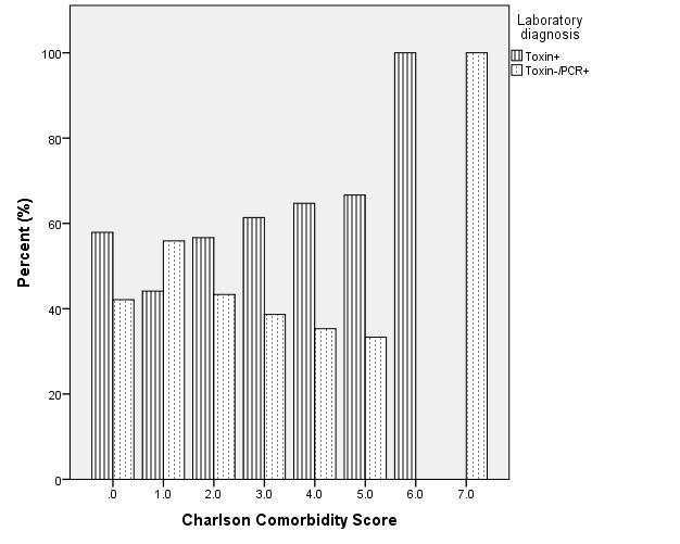
94 A. B. B. 81
: A) across categories of 30 days mortality risk (lower strata indicates higher risk of mortality)(test of trend, p=0.")
95 C. Figure 4.7. Distribution of study participants by laboratory diagnosis (percent): A) across categories of 30 days mortality risk (lower strata indicates higher risk of mortality)(test of trend, p=0.06) B) across categories of Charlson Comorbidity Score (higher strata indicates more morbidity)(test of trend, p= 0.22), C) across categories of CDI severity (test of trend, p=0.11). 82
EDUCATIONAL COMMENTARY CLOSTRIDIUM DIFFICILE UPDATE
 EDUCATIONAL COMMENTARY CLOSTRIDIUM DIFFICILE UPDATE Educational commentary is provided through our affiliation with the American Society for Clinical Pathology (ASCP). To obtain FREE CME/CMLE credits click
Clostridium difficile infection (CDI) Week 52 (Ending 30/12/2017)
 Week 52 (Ending 30/12/2017)") Clostridium difficile infection (CDI) Week 52 (Ending 30/12/2017) What is Clostridium difficile? Clostridium difficile is a Gram-positive anaerobic spore forming bacillus. It is ubiquitous in nature and
Clostridium difficile infection (CDI) Week 52 (Ending 30/12/2017) What is Clostridium difficile? Clostridium difficile is a Gram-positive anaerobic spore forming bacillus. It is ubiquitous in nature and
ABSTRACT PURPOSE METHODS
 ABSTRACT PURPOSE The purpose of this study was to characterize the CDI population at this institution according to known risk factors and to examine the effect of appropriate evidence-based treatment selection
Clostridium difficile Infection: Diagnosis and Management
 Clostridium difficile Infection: Diagnosis and Management Brian Viviano D.O. Case study 42 year old female with history of essential hypertension and COPD presents to ED complaining of 24 hours of intractable,
Diagnosis, Management, and Prevention of Clostridium difficile infection in Long-Term Care Facilities: A Review
 Diagnosis, Management, and Prevention of Clostridium difficile infection in Long-Term Care Facilities: A Review October 18, 2010 James Kahn and Carolyn Kenney, MSIV Overview Burden of disease associated
Division of GIM Lecture Series Case Presentation David A. Erickson, M.D October 9th, 2013
 Division of GIM Lecture Series Case Presentation David A. Erickson, M.D October 9th, 2013 Financial Disclosures No financial disclosures Objectives Review a case of recurrent Clostridium difficile infection
The Epidemiology of Clostridium difficile DANIEL SAMAN, DRPH, MPH RESEARCH SCIENTIST ESSENTIA INSTITUTE OF RURAL HEALTH
 The Epidemiology of Clostridium difficile DANIEL SAMAN, DRPH, MPH RESEARCH SCIENTIST ESSENTIA INSTITUTE OF RURAL HEALTH Some history first Clostridium difficile, a spore-forming gram-positive (i.e., thick
ENGLISH FOR PROFESSIONAL PURPOSES UNIT 3 HOW TO DEAL WITH CLOSTRIDIUM DIFFICILE
 ENGLISH FOR PROFESSIONAL PURPOSES UNIT 3 HOW TO DEAL WITH CLOSTRIDIUM DIFFICILE The diagnosis of CDI should be based on a combination of clinical and laboratory findings. A case definition for the usual
Clostridium difficile: An Overview
 Clostridium difficile: An Overview CDI Webinar July 11, 2017 PUBLIC HEALTH DIVISION Acute and Communicable Disease Prevention Section Outline Background Microbiology Burden Pathogenesis Diagnostic testing
Incidence of and risk factors for communityassociated Clostridium difficile infection
 University of Iowa Iowa Research Online Theses and Dissertations 2010 Incidence of and risk factors for communityassociated Clostridium difficile infection Jennifer Lee Kuntz University of Iowa Copyright
9/18/2018. Clostridium Difficile: Updates on Diagnosis and Treatment. Clostridium difficile Infection (CDI) Clostridium difficile Infection (CDI)
 Clostridium difficile Infection (CDI)") Clostridium Difficile: Updates on Diagnosis and Treatment Elizabeth Hudson, DO, MPH 9/25/18 Antibiotic-associated diarrhea and colitis were well established soon after widespread use of antibiotics In
Clostridium Difficile: Updates on Diagnosis and Treatment Elizabeth Hudson, DO, MPH 9/25/18 Antibiotic-associated diarrhea and colitis were well established soon after widespread use of antibiotics In
March 3, To: Hospitals, Long Term Care Facilities, and Local Health Departments
 March 3, 2010 To: Hospitals, Long Term Care Facilities, and Local Health Departments From: NYSDOH Bureau of Healthcare Associated Infections HEALTH ADVISORY: GUIDANCE FOR PREVENTION AND CONTROL OF HEALTHCARE
Clostridium difficile Essential information
 Clostridium difficile Essential information Clostridium difficile Origins Clostridium difficile (C. diff) is a Gram positive, spore forming, anaerobic bacterium with a rod structure. It was first identified
The incubation period is unknown. However; the onset of clinical disease is typically 5-10 days after initiation of antimicrobial treatment.
 C. DIFFICILE Case definition CONFIRMED CASE A patient is defined as a case if they are one year of age or older AND have one of the following requirements: A laboratory confirmation of a positive toxin
DETECTION OF TOXIGENIC CLOSTRIDIUM DIFFICILE
 CLINICAL GUIDELINES For use with the UnitedHealthcare Laboratory Benefit Management Program, administered by BeaconLBS DETECTION OF TOXIGENIC CLOSTRIDIUM DIFFICILE Policy Number: PDS 021 Effective Date:
Updated Clostridium difficile Treatment Guidelines
 Updated Clostridium difficile Treatment Guidelines Arielle Arnold, PharmD, BCPS Clinical Pharmacist Saint Alphonsus Regional Medical Center September 29 th, 2018 Disclosures Nothing to disclose Learning
Stony Brook Adult Clostridium difficile Management Guidelines. Discontinue all unnecessary antibiotics
 Stony Brook Adult Clostridium difficile Management Guidelines Summary: Use of the C Diff Infection (CDI) PowerPlan (Adult) Required Patient with clinical findings suggestive of Clostridium difficile infection
Clostridium Difficile Infection: Applying New Treatment Guidelines and Strategies to Reduce Recurrence Rate
 Clostridium Difficile Infection: Applying New Treatment Guidelines and Strategies to Reduce Recurrence Rate Objectives Summarize the changing epidemiology and demographics of patients at risk for Clostridium
Clostridium difficile Infection (CDI) Management Guideline
 Management Guideline") Clostridium difficile Infection (CDI) Management Guideline Do not test all patients with loose or watery stools for CDI o CDI is responsible for
Clostridium difficile Infection (CDI) Management Guideline Do not test all patients with loose or watery stools for CDI o CDI is responsible for
DUKE ANTIMICROBIAL STEWARDSHIP OUTREACH NETWORK (DASON) Antimicrobial Stewardship News. Volume 3, Number 6, June 2015
 Antimicrobial Stewardship News. Volume 3, Number 6, June 2015") DUKE ANTIMICROBIAL STEWARDSHIP OUTREACH NETWORK (DASON) Antimicrobial Stewardship News Volume 3, Number 6, June 2015 Diagnostic Testing for Clostridium difficile Infection Background Clostridium difficile
DUKE ANTIMICROBIAL STEWARDSHIP OUTREACH NETWORK (DASON) Antimicrobial Stewardship News Volume 3, Number 6, June 2015 Diagnostic Testing for Clostridium difficile Infection Background Clostridium difficile
CLOSTRIDIUM DIFICILE. Negin N Blattman Infectious Diseases Phoenix VA Healthcare System
 CLOSTRIDIUM DIFICILE Negin N Blattman Infectious Diseases Phoenix VA Healthcare System ANTIBIOTIC ASSOCIATED DIARRHEA 1978: C diff first identified 1989-1992: Four large outbreaks in the US caused by J
ESCMID Online Lecture Library. by author
 ECDIS-NET: Update on Clostridium difficile epidemiology in Europe 1 E d J. K u i j p e r, S o f i e v a n D o r p a n d D a a n N o t e r m a n s. D e p a r t m e n t o f M e d i c a l M i c r o b i o
Molecular epidemiology of Clostridium difficile infection in British Columbia, Canada
 Molecular epidemiology of Clostridium difficile infection in British Columbia, Canada Agatha Jassem, PhD Senior Scientist, BCCDC Public Health Laboratory Objectives Molecular typing methods for C. difficile
DISCLOSURE Relevant relationships with commercial entities Wyeth (received advisory board & speaker honoraria) Potential for conflicts of interest wit
 Potential for conflicts of interest wit") GASTROENTERITIS DISCLOSURE Relevant relationships with commercial entities Wyeth (received advisory board & speaker honoraria) Potential for conflicts of interest within this presentation fidaxomicin (which
GASTROENTERITIS DISCLOSURE Relevant relationships with commercial entities Wyeth (received advisory board & speaker honoraria) Potential for conflicts of interest within this presentation fidaxomicin (which
Probiotics for Primary Prevention of Clostridium difficile Infection
 Probiotics for Primary Prevention of Clostridium difficile Infection Objectives Review risk factors for Clostridium difficile infection (CDI) Describe guideline recommendations for CDI prevention Discuss
Case 1. Which of the following would be next appropriate investigation/s regarding the pts diarrhoea?
 Case 1 21 yr old HIV +ve, Cd4-100 HAART naïve Profuse diarrhoea for 3/52. Stool MC&S ve Which of the following would be next appropriate investigation/s regarding the pts diarrhoea? Repeat stool MC&S Stool
Clostridium Difficile Associated Disease. Edmund Krasinski, Jr., D.O., F.A.C.G. Southwest Conference on Medicine 2011
 Clostridium Difficile Associated Disease Edmund Krasinski, Jr., D.O., F.A.C.G. Southwest Conference on Medicine 2011 Introduction Which of the following is more common in community hospitals in the Southeast
Clostridium difficile Infection (CDI)
") 18.09.10 월요집담회 Clostridium difficile Infection (CDI) R4 송주혜 Clostridium difficile infection (CDI) Anaerobic gram (+), spore-forming, toxin(tcda&tcdb)-producing bacillus Transmitted among humans through
18.09.10 월요집담회 Clostridium difficile Infection (CDI) R4 송주혜 Clostridium difficile infection (CDI) Anaerobic gram (+), spore-forming, toxin(tcda&tcdb)-producing bacillus Transmitted among humans through
ACG Clinical Guideline: Diagnosis, Treatment, and Prevention of Clostridium difficile Infections
 ACG Clinical Guideline: Diagnosis, Treatment, and Prevention of Clostridium difficile Infections Christina M. Surawicz, MD 1, Lawrence J. Brandt, MD 2, David G. Binion, MD 3, Ashwin N. Ananthakrishnan,
CDI The Impact. Disclosures. Acknowledgments. Objectives and Agenda. What s in the Name? 11/14/2012. Lets Talk Numbers
 Disclosures No conflict of interest to declare Acknowledgments Objectives and Agenda Society for Healthcare Epidemiology of America (SHEA) and the Infectious Diseases Society of America (IDSA) Guidelines
Objectives Clostridium difficile Infections, So Many Tests, Which One to Choose?
 Objectives Clostridium difficile Infections, So Many Tests, Which One to Choose? March 9, 0 http://www.slh.wisc.edu/outreach-data/event-detail.php?id=03 Raymond P. Podzorski, Ph.D., D(ABMM) Clinical Microbiologist
L. Clifford McDonald, MD. Senior Advisor for Science and Integrity September 16, 2015
 Controversies and Current Issues in Diagnosis, Surveillance, and Treatment of Clostridium difficile infeciton L. Clifford McDonald, MD Senior Advisor for Science and Integrity September 16, 2015 Division
Clostridium difficile CRISTINA BAKER, MD, MPH INFECTIOUS DISEASE PARK NICOLLET/HEALTH PARTNERS 11/9/2018
 Clostridium difficile CRISTINA BAKER, MD, MPH INFECTIOUS DISEASE PARK NICOLLET/HEALTH PARTNERS 11/9/2018 Disclosures None Objectives Highlight important changes in the management of Clostridium difficile
Clostridium difficile infection surveillance: Applying the case definition
 Clostridium difficile infection surveillance: Applying the case definition PICNet Conference March 3 rd 2016 Presented by: Tara Leigh Donovan, MSc Managing Consultant (Former Epidemiologist) 1 Disclaimer
Clostridium difficile
 Clostridium difficile Care Homes IPC Study Day Sue Barber Infection Prevention & Control Lead AV & Chiltern CCG s Clostridium difficile A spore forming Bacterium. Difficult to grow in the laboratory hence
The epidemiology of Clostridium difficile infection (CDI) in hospitals, longterm care and the community. J Scott Weese DVM DVSc DipACVIM
 in hospitals, longterm care and the community. J Scott Weese DVM DVSc DipACVIM") The epidemiology of Clostridium difficile infection (CDI) in hospitals, longterm care and the community J Scott Weese DVM DVSc DipACVIM C. difficile Gram positive anaerobic sporeforming bacterium first
The epidemiology of Clostridium difficile infection (CDI) in hospitals, longterm care and the community J Scott Weese DVM DVSc DipACVIM C. difficile Gram positive anaerobic sporeforming bacterium first
Sherwood L. Gorbach, MD Professor of Public Health, Medicine, and Microbiology Tufts University School of Medicine
 Sherwood L. Gorbach, MD Professor of Public Health, Medicine, and Microbiology Tufts University School of Medicine Chief Scientific Officer, Optimer Pharmaceuticals, Inc. Conflicts: Chief Scientific Officer,
What s New for Clostridium difficile John Lynch MD MPH Harborview Medical Center University of Washington
 What s New for Clostridium difficile 2013 John Lynch MD MPH Harborview Medical Center University of Washington Pathogenic Mechanisms of Diarrhea Toxins: Preformed: S aureus, C perfringens, B cereus Formed
Long-Term Care Updates
 Long-Term Care Updates April 2018 By Austin Smith, PharmD Candidate and Lindsay Slowiczek, PharmD is the most common healthcare-acquired infection (HAI) in the United States. 1,2 A 2014 prevalence survey
Journey to Decreasing Clostridium Difficile and the Unexpected Twist. Jackie Morton, Infection Prevention Cortney Swiggart, Medication Safety Officer
 Journey to Decreasing Clostridium Difficile and the Unexpected Twist Jackie Morton, Infection Prevention Cortney Swiggart, Medication Safety Officer 4/13/2018 Objectives Discuss the organism and clinical
C Difficile - The Ultimate Challenge: Controlling the Spread
 C Difficile - The Ultimate Challenge: Controlling the Spread Linda R. Greene, RN, MPS, CIC Manager of Infection Prevention Highland Hospital Rochester, NY University of Rochester Medical Center linda_greene@urmc.rochester.edu
Le infezioni da Clostridium difficile, gravi, ricorrenti e complicate Nicola Petrosillo
 Le infezioni da Clostridium difficile, gravi, ricorrenti e complicate Nicola Petrosillo Istituto Nazionale per le Malattie Infettive «lazzaro Spallanzani», IRCCS-Roma The infectious cycle of transmission
6/14/2012. Welcome! PRESENTATION OUTLINE CLOSTRIDIUM DIFFICILE PREVENTION. Teaming Up to Prevent Infections! 1) Impact. 2) Testing Recommendations
 Impact. 2) Testing Recommendations") CLOSTRIDIUM DIFFICILE PREVENTION Beth Goodall, RN, BSN Board Certified in Infection Prevention and Control DCH Health System Epidemiology Director Welcome! Teaming Up to Prevent Infections! CLOSTRIDIUM
CLOSTRIDIUM DIFFICILE PREVENTION Beth Goodall, RN, BSN Board Certified in Infection Prevention and Control DCH Health System Epidemiology Director Welcome! Teaming Up to Prevent Infections! CLOSTRIDIUM
SMT19969: A Selective Therapy for C. difficile Infection
 SMT19969: A Selective Therapy for C. difficile Infection One Bug, One Drug 25 th September 2012 SMT19969: A Selective Therapy for CDI SMT19969 is a novel antibiotic for the specific treatment of Clostridium
Pros and Cons of Alternative Diagnostic Testing Strategies for C. difficile Infection
 Pros and Cons of Alternative Diagnostic Testing Strategies for C. difficile Infection Christopher R. Polage, MD, MAS Associate Professor of Pathology and Infectious Diseases UC Davis Disclosures Test materials
Los Angeles County Department of Public Health: Your Partner in CDI Prevention
 Los Angeles County Department of Public Health: Your Partner in CDI Prevention Dawn Terashita, MD, MPH Acute Communicable Disease Control Los Angeles County Department of Public Health dterashita@ph.lacounty.gov
Patient Safety Summit 2014
 Patient Safety Summit 2014 The War on C Diff Mark Mellow, MD + C Diff The Organism Gram + bacillus Anaerobic Spore forming Intestinal flora (up to 35% hospitalized patients, 3% of healthy adults) Leading
JMSCR Vol 05 Issue 07 Page July 2017
 www.jmscr.igmpublication.org Impact Factor 5.84 Index Copernicus Value: 83.27 ISSN (e)-2347-176x ISSN (p) 2455-0450 DOI: https://dx.doi.org/10.18535/jmscr/v5i7.15 Rapid Diagnosis of Toxigenic Clostridium
Annex C: - CDI What s the diff? 4 th Annual Outbreak Management Workshop September 19, 2013 Naideen Bailey & Grace Volkening
 Annex C: - CDI What s the diff? 4 th Annual Outbreak Management Workshop September 19, 2013 Naideen Bailey & Grace Volkening There s an updated Annex C Annex C is an extension to the PIDAC Infection Prevention
Clostridium difficile: Can you smell the new updates?
 Clostridium difficile: Can you smell the new updates? Sunish Shah, Pharm.D. PGY-2 Infectious Disease Pharmacy Resident Yale-New Haven Hospital sshah1741@mail.usciences.edu Learning objectives Recognize
Clostridium difficile Diagnostic and Clinical Challenges
 Papers in Press. Published December 11, 2015 as doi:10.1373/clinchem.2015.243717 The latest version is at http://hwmaint.clinchem.org/cgi/doi/10.1373/clinchem.2015.243717 Clinical Chemistry 62:2 000 000
Clostridium difficile Infection (CDI) Guideline Update:
 Guideline Update:") Clostridium difficile Infection (CDI) Guideline Update: Understanding the Data Behind the Recommendations Erik R. Dubberke, MD, MSPH A Webinar for HealthTrust Members Professor of Medicine September 24,
Clostridium difficile Infection (CDI) Guideline Update: Understanding the Data Behind the Recommendations Erik R. Dubberke, MD, MSPH A Webinar for HealthTrust Members Professor of Medicine September 24,
This is a repository copy of Clostridium difficile infection: advances in epidemiology, diagnosis and transmission.
 This is a repository copy of Clostridium difficile infection: advances in epidemiology, diagnosis and transmission. White Rose Research Online URL for this paper: http://eprints.whiterose.ac.uk/93246/
Clostridium difficile Infection in the Department of Defense (DOD):
:") 1 1 Clostridium difficile Infection in the Department of Defense (DOD): 2007-2013 NMCPHC-EDC- TR-271-2015 By Charlotte Neumann and Uzo Chukwuma February 2015 Approved for public release. Distribution is
1 1 Clostridium difficile Infection in the Department of Defense (DOD): 2007-2013 NMCPHC-EDC- TR-271-2015 By Charlotte Neumann and Uzo Chukwuma February 2015 Approved for public release. Distribution is
Bacterial Enteric Pathogens: Clostridium difficile, Salmonella, Shigella, Escherichia coli, and others
 GUIDE TO INFECTION CONTROL IN THE HOSPITAL CHAPTER 48 Bacterial Enteric Pathogens: Clostridium difficile, Salmonella, Shigella, Escherichia coli, and others Authors Olivier Vandenberg, MD, PhD Michèle
Each copy of any part of a JSTOR transmission must contain the same copyright notice that appears on the screen or printed page of such transmission.
 Clinical Practice Guidelines for Clostridium difficile Infection in Adults: 2010 Update by the Society for Healthcare Epidemiology of America (SHEA) and the Infectious Diseases Society of America (IDSA)
A Pharmacist Perspective
 Leveraging Technology to Reduce CDI A Pharmacist Perspective Ed Eiland, Pharm.D., MBA, BCPS (AQ-ID) Clinical Practice and Business Supervisor Huntsville Hospital System Huntsville Hospital 881 licensed
more intense treatments are needed to get rid of the infection.
 What Is Clostridium Difficile (C. Diff)? Clostridium difficile, or C. diff for short, is an infection from a bacterium that can grow in your intestines and cause bad GI symptoms. The main risk of getting
Modern approach to Clostridium Difficile Infection
 Modern approach to Clostridium Difficile Infection Pseudomembranous Colitis: Principles for diagnosis and treatment Aggelos Stefos Internist, Infectious diseases Specialist Department of Medicine and Research
Clostridium Difficile Infection in Adults Treatment and Prevention
 Clostridium Difficile Infection in Adults Treatment and Prevention Definition: Clostridium Difficile colonizes the human intestinal tract after the normal gut flora has been altered by antibiotic therapy
! MQ is a 44 year old woman that I first saw in Sept ! In MVA in Jan 2003 requiring spinal surgery
 Case MQ is a 44 year old woman that I first saw in Sept 2006 UPDATE ON CLOSTRIDIUM DIFFICILE DISEASE Richard A. Jacobs, M.D.,PhD In MVA in Jan 2003 requiring spinal surgery Subsequently developed fecal
True Pathogens of the Enterobacteriaceae: Salmonella, Shigella & Yersinia Salmonella
 Lec. 6 Oral Microbiology Dr. Chatin True Pathogens of the Enterobacteriaceae: Salmonella, Shigella & Yersinia Salmonella General Characteristics of Salmonella جامعة تكريت كلية طب االسنان Coliform bacilli
Chain of Infection Agent Mode of transmission Contact (direct, indirect, droplet spread) Airborne Common-vehicle spread Host
 Airborne Common-vehicle spread Host") Goals Microbiology of Healthcare-associated Infections William A. Rutala, Ph.D., M.P.H. Director, Statewide Program for Infection Control and Epidemiology and Research Professor of Medicine, University
Goals Microbiology of Healthcare-associated Infections William A. Rutala, Ph.D., M.P.H. Director, Statewide Program for Infection Control and Epidemiology and Research Professor of Medicine, University
Bezlotoxumab (Zinplava) as Adjunct Treatment for Clostridium difficile. Janel Liane Cala, RPh Medical Center Hospital
 as Adjunct Treatment for Clostridium difficile. Janel Liane Cala, RPh Medical Center Hospital") Bezlotoxumab (Zinplava) as Adjunct Treatment for Clostridium difficile Janel Liane Cala, RPh Medical Center Hospital Objectives Review pathophysiology, risk factors, prevention, and treatment options of
Bezlotoxumab (Zinplava) as Adjunct Treatment for Clostridium difficile Janel Liane Cala, RPh Medical Center Hospital Objectives Review pathophysiology, risk factors, prevention, and treatment options of
Foundations in Microbiology
 Foundations in Microbiology Fifth Edition Talaro Chapter 13 Microbe Human Interactions: Infection and Disease Chapter 13 2 3 Infection a condition in which pathogenic microbes penetrate host defenses,
C. DIFF QUIK CHEK COMPLETE. Get the complete diagnostic picture with just one test. TAP HERE TO SEE THE NEXT PAGE
 C. DIFF QUIK CHEK COMPLETE Get the complete diagnostic picture with just one test. TAP HERE TO SEE THE PAGE CLINICIAN Would actionable C. difficile test results in less than 30 minutes improve patient
Clostridium difficile
 Clostridium difficile Alex Aspinall MD, PhD, FRCPC Clinical Assistant Professor, University of Calgary Division of Gastroenterology and Hepatology, South Health Campus www.seacourses.com 1 Learning Points
Historical Perspective
 The Diagnosis of C. difficile Infections: Does Molecular Make it More or Less difficile? Stephen M. Brecher Ph.D. VA Boston Health Care System BU School of Medicine The opinions expressed in this presentation
C. difficile infection
 C. difficile infection Most common cause of infectious diarrhoea in hospital patients 2 major virulence factors: PaLoc toxin A (an enterotoxin) toxin B (a cytotoxin) 3 rd binary toxin Bartlett JG Clin
Clostridium Difficile colitismore
 Clostridium Difficile colitismore virulent than ever ECHO- February 18, 2016 Charles Krasner, M.D. UNR School of Medicine Sierra NV Veterans Affairs Hospital Growing problem of pseudomembranous colitis
Clostridium DifficileInfection & Readmissions: An ounce of prevention is worth a pound of cure
 Clostridium DifficileInfection & Readmissions: An ounce of prevention is worth a pound of cure Brian S. Zuckerbraun, MD, FACS Henry T. Bahnson Professor of Surgery University of Pittsburgh Chief, Trauma
New Molecular Diagnostic Assays for Solana Performance Assessment, Workflow Analysis, and Clinical Utility
 New Molecular Diagnostic Assays for Solana Performance Assessment, Workflow Analysis, and Clinical Utility Gerald A. Capraro, Ph.D., D(ABMM) Director, Clinical Microbiology Carolinas Pathology Group Atrium
Probiotics: Their Role in Medicine Today. Objectives. Probiotics: What Are They? 11/3/2017
 Probiotics: Their Role in Medicine Today Viki Barr Pharm.D., BCPS AQ ID Assistant Professor, Pharmacy Practice Rosalind Franklin University of Medicine and Science Clinical Pharmacist, Infectious Diseases
Comparison of Clinical Features Between Inpatient and Outpatient Cases of Clostridium difficile Infection
 Comparison of Clinical Features Between Inpatient and Outpatient Cases of Clostridium difficile Infection Marcus J. Belger A thesis submitted in partial fulfillment of the requirements for the degree of
INFECTION CONTROL AND HOSPITAL EPIDEMIOLOGY MAY 2010, VOL. 31, NO. 5
 INFECTION CONTROL AND HOSPITAL EPIDEMIOLOGY MAY 2010, VOL. 31, NO. 5 SHEA-IDSA GUIDELINE Clinical Practice Guidelines for Clostridium difficile Infection in Adults: 2010 Update by the Society for Healthcare
Long-Term Care Updates
 Long-Term Care Updates April 2017 Bezlotoxumab to Prevent Recurrent Infection By Amy Wilson, PharmD and Zara Risoldi Cochrane, PharmD, MS, FASCP Introduction The Gram-positive bacteria is a common cause
Corporate Medical Policy Fecal Microbiota Transplantation
 Corporate Medical Policy Fecal Microbiota Transplantation File Name: Origination: Last CAP Review: Next CAP Review: Last Review: Fecal_microbiota_transplantation 7/2014 11/2017 11/2018 11/2017 Description
Clostridium difficile infections Crappy Options
 Clostridium difficile infections Crappy Options Alexandru Petre David MD Infectious Diseases April 21, 2015 Objectives Microbiology Epidemiology Who is at risk? Pathogenesis Diagnosis Treatment Historical
Update on Clostridium difficile infection.
 Update on Clostridium difficile infection. K. Honein Gastroenterologist, HDF Associate Professor Head of Medicine Department St Joseph University-Beirut. Introduction Gram+anaerobic bacillus responsible
Laboratory diagnosis of Clostridium difficile infection
 Review Article Page 1 of 5 Laboratory diagnosis of Clostridium difficile infection Katherine Kendrick Mercer University School of Medicine (SOM), Navicent Health, Macon, GA, USA Correspondence to: Katherine
Duodenal infusion of donor feces for recurrent Clostridium difficile infection A French experience
 Duodenal infusion of donor feces for recurrent Clostridium difficile infection A French experience Benoit Guery Unité des Maladies Infectieuses CHRU - Faculté de Médecine Lille Conflicts of interest Conferences,
Does Extending Clostridium Difficile Treatment In Patients Who Are Receiving Concomitant Antibiotics Reduce The Rate Of Relapse?
 ISPUB.COM The Internet Journal of Infectious Diseases Volume 15 Number 1 Does Extending Clostridium Difficile Treatment In Patients Who Are Receiving Concomitant Antibiotics Reduce The Rate Of Relapse?
ECHO-Antibiotic Stewardship Program
 ECHO-Antibiotic Stewardship Program More Interesting Recent Literature Updates April 20, 2017 Charles Krasner, M.D. University of NV, Reno School of Medicine Sierra NV Veterans Affairs Medical Center Antibiotic
C. difficile: The Changing Epidemiology Evaluations Clostridium difficile Thank You to our Sponsors
 C. difficile: The Changing Epidemiology Ghinwa Dumyati, MD University of Rochester Monroe County Department of Public Health Thank You to our Sponsors Evaluations School of Public Health, University at
Clostridioides difficile Infection: What s the BIG Stink?
 Clostridioides difficile Infection: What s the BIG Stink? GLORIA MATHIS, RN, BSN, CIC 29 TH ANNUAL MEDICAL SURGICAL CONFERENCE APRIL 4, 2019 SAN FRANCISCO, CA I am sre my patient has C. diff. I cold smell
500,000 29,000. New 2015 Data. Lessa et al, N Eng J Med 2015: 34.2% of CDI cases were considered community-acquired
 Cost-effective Treatment of Clostridium difficile Infection in the ICU Kevin W. Garey, PharmD, MS. Professor and Chair University of Houston College of Pharmacy New 2015 Data 500,000 29,000 Lessa et al,
The Potential For Microbiome Modification In Critical Illness. Deborah Cook
 The Potential For Microbiome Modification In Critical Illness Deborah Cook To review Objectives The microbiome & concepts about its modification during critical illness Interventions Predisposition to
Antimicrobial prophylaxis in liver transplant A multicenter survey endorsed by the European Liver and Intestine Transplant Association
 Antimicrobial prophylaxis in liver transplant A multicenter survey endorsed by the European Liver and Intestine Transplant Association Els Vandecasteele, Jan De Waele, Dominique Vandijck, Stijn Blot, Dirk
National Center for Emerging and Zoonotic Infectious Diseases The Biggest Antibiotic Resistance Threats
 National Center for Emerging and Zoonotic Infectious Diseases The Biggest Antibiotic Resistance Threats Jean B. Patel, PhD, D(ABMM) Science Lead, Antibiotic Resistance and Coordination Unit Centers for
Rapid-VIDITEST C. difficile Ag (GDH) Card/Blister
 Card/Blister") Li StarFish S.r.l. Via Cavour, 35-20063 Cernusco S/N (MI), Italy Tel. +39-02-92150794 - Fax. +39-02-92157285 info@listarfish.it -www.listarfish.it Rapid-VIDITEST C. difficile Ag (GDH) Card/Blister One
Li StarFish S.r.l. Via Cavour, 35-20063 Cernusco S/N (MI), Italy Tel. +39-02-92150794 - Fax. +39-02-92157285 info@listarfish.it -www.listarfish.it Rapid-VIDITEST C. difficile Ag (GDH) Card/Blister One
Mechanisms of Pathogenicity
 Mechanisms of Pathogenicity The Microbes Fight Back Medically important bacteria Salmonella Bacillus anthracis Shigella dysenteriae Campylobacter Shigella sonnei Clostridium botulinum Staphylococcus aureus
3/23/2012. Impact of Laboratory Testing on Detection and Treatment of Healthcare Associated Infection: The Case of CDI
 Impact of Laboratory Testing on Detection and Treatment of Healthcare Associated Infection: The Case of CDI L. Clifford McDonald, MD Lance R. Peterson, MD March 27, 2012 1 Potential COI L. Clifford McDonald,
The Bristol Stool Scale and Its Relationship to Clostridium difficile Infection
 JCM Accepts, published online ahead of print on 16 July 2014 J. Clin. Microbiol. doi:10.1128/jcm.01303-14 Copyright 2014, American Society for Microbiology. All Rights Reserved. 1 The Bristol Stool Scale
Clinical. Clostridium Difficile: Standard Operating Procedure. Document Control Summary. Contents
 Clinical Clostridium Difficile: Standard Operating Procedure Document Control Summary Status: Version: Author/Title: Owner/Title: Approved by: Ratified: Related Trust Strategy and/or Strategic Aims Implementation
Clostridium difficile Asymptomatic Carriers The Hidden Part of the Iceberg?
 Clostridium difficile Asymptomatic Carriers The Hidden Part of the Iceberg? Disclosures Merck Canada, BD Diagnostics, AMD Medical, Canadian Institute for Health Research Merck Canada, Pfizer OBJECTIVES
2/3/2011. Adhesion of Bifidobacterium lactis HN019 to human intestinal
 PROBIOTICS LEARNING THE WHY AND WHEN PROBIOTICS DEFINITION live micro-organisms organisms that are beneficial to the host organism WHO: Live organisms which, when administered in adequate amounts, confer
Understanding C. diff. atomalliance.org/cdifftraining
 Understanding C. diff atomalliance.org/cdifftraining This booklet is a printed guide of the online educational resource atomalliance.org/cdifftraining Target Audience Understanding C. diff This educational
Gastroenteritis and viral infections
 Gastroenteritis and viral infections A Large number of viruses are found in the human gut; these include some that are associated with gastroenteritis Rotaviruses Adenoviruses 40/41 Caliciviruses Norwalk-like
GUIDELINE FOR THE MANAGEMENT OF ANTIBIOTIC- ASSOCIATED DIARRHOEA IN ADULTS
 GUIDELINE FOR THE MANAGEMENT OF ANTIBIOTIC- ASSOCIATED DIARRHOEA IN ADULTS Version 3.0 Date ratified May 2008 Review date May 2010 Ratified by NUH Antibiotic Guidelines Committee NUH Drugs and Therapeutics
Disclosure. Objectives. Assessment Questions. History. Clinical Case 2/27/2015. Clostridium difficile update and new therapies
 Disclosure Clostridium difficile update and new therapies Corey Frederick, PharmD PGY-2 Pharmacy Resident, Infectious Diseases Jackson Memorial Hospital Miami, Florida I do not have a vested interest in