PERFORATED CLICK X Augmentable pedicle screws for osteoporotic bone
|
|
|
- Claude Hardy
- 5 years ago
- Views:
Transcription


1 PERFORATED CLICK X Augmentable pedicle screws for osteoporotic bone Instruments and implants approved by the AO Foundation. This publication is not intended for distribution in the USA. SURGICAL TECHNIQUE
2 Image intensifier control Warning This description alone does not provide sufficient background for direct use of DePuy Synthes products. Instruction by a surgeon experienced in handling these products is highly recommended. In addition, dedicated education on the bone cement instrumentation, associated risks and technique is mandatory. Processing, Reprocessing, Care and Maintenance For general guidelines, function control and dismantling of multi-part instruments, please contact your local sales representative or refer to: For general information about reprocessing, care and maintenance of Synthes reusable devices, instrument trays and cases, as well as processing of Synthes non-sterile implants, please consult the Important Information leaflet (SE_023827) or refer to:
3 Table of Contents Introduction Perforated Click X 2 Indications and Contraindications 4 Surgical Technique Preoperative Planning 5 Surgical Technique 6 Notes and Warnings 12 Product Information Implants and Instruments 14 Perforated Click X Surgical Technique DePuy Synthes 1


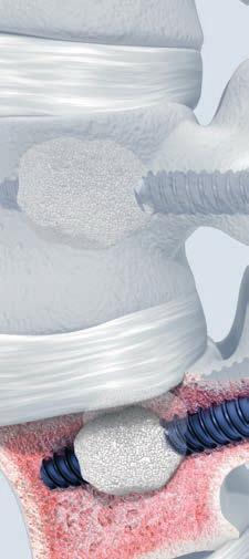
4 Perforated Click X. Augmentable pedicle screws for osteoporotic bone. Improved anchoring and shear stability in osteoporotic bone Screw anchoring and vertebral body support thanks to cement cloud Six radial screw openings for 360 cement distribution Controlled cement injection, performed simultaneously or alternated at different levels Augmentation after final screw positioning 2 DePuy Synthes Perforated Click X Surgical Technique


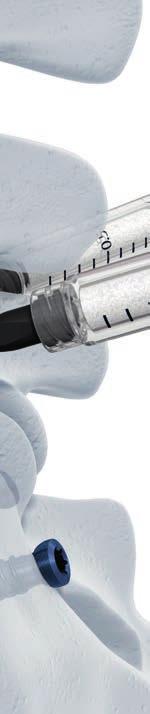
5 Syringe Syringe provides tactile feedback during injection Adapter Adapter with Luer lock fits any commercial syringe Press-fit connection for leak-proof cement injection Perforated dual-core pedicle screw Cylindrical large core with low and broad thread flanks for higher strength in dense cortical bone shell of pedicle Cylindrical small core with high and thin thread flanks for cancellous bone in vertebral body Six radial openings for 360 cement distribution and optimal screw anchoring Large cannulation diameter for low cement injection resistance Kirschner wire cannulation for guided screw insertion over 1.6 mm Kirschner wire Minimal cement outflow from screw tip Perforated Click X Surgical Technique DePuy Synthes 3
6 Indications and Contraindications The Perforated Click X system is a transpedicular screw/rod system intended for the posterior stabilization of the thoracolumbar and lumbar spine. The Perforated Click X screws are cannulated. However, they may be inserted like standard solid Click X screws (see Click X System surgical technique) or like Cannulated Click X screws by means of Kirschner wire guidance (see Cannulated Click X surgical technique). The lateral perforations allow direct cement augmentation of the screw in the osteoporotic bone. General indications The implants can be used for the following indications in the area of the lower thoracic and lumbar spine Degenerative instabilities Instabilities following decompression Type A1 fractures and related types of the B and C group Type A2 and A3 fractures as well as similar fractures of the C group, if combined with an anterior intervention Tumors without anterior defect Osteoporosis when used concurrently with bone cement indicated for internal spinal fixation supplementation Contraindications Deformities In fractures and tumors with severe anterior vertebral body disruption, an additional anterior support is required Osteoporosis when used without cement augmentation Severe osteoporosis Note: Refer to the manufacturer's directions accompanying the bone cement for specific information on its use, indications, contraindications, precautions, warnings and side effects. 4 DePuy Synthes Perforated Click X Surgical Technique
7 Preoperative Planning Preoperative planning includes evaluation and assessment of the patient with regard to the specifications of the bone cement used for augmenting perforated Click X screws. Proper imaging equipment must be used to determine correct implant dimensions in relation to the anatomy. The decision whether or not to augment Perforated Click X screws can be taken intraoperatively, based on tactile feedback upon pedicle preparation and screw insertion. If screws are augmented, bilateral screw augmentation is recommended. Note: Do not reinforce more than six vertebrae with bone cement in one session. The injected cement pushes bone marrow into the blood circulation and can cause fat embolization. The amount of cement that is injected in one session should therefore be limited to about 25 ml or even less if a patient shows severely compromised cardiopulmonary function. Note: Handling knowledge of bone cement is required. Image intensifier control is highly recommended while injecting bone cement. Perforated Click X Surgical Technique DePuy Synthes 5
8 Surgical Technique This surgical technique contains complementary instructions on handling Perforated Click X screws. For the standard surgical Click X procedure consult the Click X System surgical technique. Notes on working with Kirschner wires If Perforated Click X screws are inserted over Kirschner wires also consult the technique guide on handling Cannulated Click X screws. Under image intensifier control ensure that each Kirschner wire is in the appropriate position for screw insertion. Especially the tip of the Kirschner wire should be radiologically monitored to confirm that it does not penetrate the anterior wall of the vertebral body and thus risking damage of the vessels in front of it. Kirschner wires are for single use. 1 Prepare pedicles and insert screws Instruments Click X Basic Instrument Set in Vario Case Screwdriver Shaft Stardrive, T25, cannulated, with Hexagonal Coupling, B 6.0 mm Optional instrument Cannulated Instruments for Pedicle Preparation, in Vario Case Open and prepare the pedicles and insert the screws as defined in the Click X System surgical technique. Use the cannulated screwdriver shaft. Do not attach the 3-D heads yet. 6 DePuy Synthes Perforated Click X Surgical Technique


9 Assess the cortical shell for perforations. Note: In case of any perforation, special caution is required when bone cement is applied. Cement leakage and its related risks may compromise the physical condition of the patient. Select appropriate screw lengths. Choose screws with the maximum possible diameter and length to achieve maximum stability. 80% Adequate length. Radial openings well located in vertebral body. The Perforated Click X screw must enter in approximately 80% of the vertebral body. Notes on screw length If screws are too short, the bone cement risks being injected too close to the pedicle. If screws are too long, or placed bicortically, the anterior cortical wall may be penetrated and cement leakage may occur. Screw too short. Radial openings too close to pedicle. Precaution: It is imperative that the screw perforations are located in the vertebral body, close to the anterior cortical wall. For this reason 35 mm screws should only be placed in the sacrum. Perforated Click X Surgical Technique DePuy Synthes 7

10 Surgical Technique 2 Assess proper screw placement Use a B 2.0 mm Kirschner wire to clean the cannula for proper cement injection. 3 Prepare bone cement Prepare the bone cement according to the manufacturer s direction. Use syringes with Luer-Lock for injecting the bone cement. 8 DePuy Synthes Perforated Click X Surgical Technique
11 4 Inject bone cement Place the C-Arm in a lateral position to monitor the extrusion of the cement into the vertebral body. Additional image intensifier control in the AP projection is recommended. Note: Do not augment more than four screws at once. Note: Ensure that there is a firm connection between the adapter and the screw recess. Perforated Click X Surgical Technique DePuy Synthes 9


 e.g. with the screwdriver shaft.")
12 Surgical Technique 1. Inject approximately 0.5 ml cement per screw until cement extrudes from the perforations. Note: Ensure that no cement leakage occurs outside the intended area. Immediately stop the injection in the event of leakage. 2. Following the same order of screws, add another ml of cement to each screw. The cement extrusion needs to be monitored under continuous image intensifier control. A growing cloud pattern should form. If a spider web-like pattern forms, wait approx. 30 to 45 seconds or proceed with another screw and return to the present screw later. 3. If more cement is needed or the injection pressure is too high, switch to the 1 ml syringes and inject more cement. Start again with the first screw. Augmentation is complete when each screw has been augmented with a total volume of approximately 2 to 3 ml. Note: Do not remove or replace syringes immediately after injection. This avoids contaminating the screw drive and the patient s soft tissues with cement drips. The longer the syringe remains connected to the screw, the lower the risk of contamination. Warning: Any cement remaining in the screw drive must be removed while it is still soft (or has not hardened) e.g. with the screwdriver shaft. This will ensure that future revision surgeries remain possible. Warning: The cement flow follows the path of least resistance. Therefore it is mandatory, during the whole injection procedure, to maintain real-time image intensifier control in the lateral projection. If the filling pattern does not behave as expected or if the cement is not clearly visible, the injection must be stopped. Wait until the cement has cured before removing adapters and continuing with the instrumentation. 11 DePuy Synthes Perforated Click X Surgical Technique

13 5 Attach Click X 3-D heads and assemble construct Prior to attaching the 3-D heads ensure that the pedicle screw heads are clean and not contaminated with any bone cement. Remove any cement from the soft tissues. Attach the 3-D heads and continue with the following steps according to the Click X System surgical technique. Perforated Click X Surgical Technique DePuy Synthes 11
14 Notes and Warnings Cement leakage The inherent risk of this technique is cement leakage. Risks can be minimized and complications rate reduced by observing the above mentioned technique. The second challenge that needs to be emphasized is fat embolism since the injected cement will push bone marrow into the blood circulation the amount of cement that is injected in one session should be limited to about 25 ml and less if a patient shows severely compromised cardiovascular function. Furthermore, systemic reactions during cement injection can occur as a consequence of cement monomer release. If significant leakage occurs, stop the procedure. Return your patient to the ward and perform a clinical assessment in order to clarify the neurological situation. If the neurological state is not compromised, there is no need for further surgical measures. If neurological function is severely compromised, an emergency CT scan should be performed in order to assess the amount and location of the extravasation. If applicable, open surgical decompression and cement removal may be performed as an emergency procedure. Extravasation In order to minimize the risk of extravasation, it is strongly recommended to follow the described surgical technique, i.e.: Use a Kirschner wire for pedicle screw placement Use a high-quality C-arm in lateral position Use highly viscous and highly radiopaque bone cement Additional image intensifier control in the AP projection is recommended. Leakage outside the vertebra What action is required if leakage occurs? If you recognize leaking outside the vertebra, stop the injection immediately. Wait for 45 seconds. Then slowly continue with the injection. Due to the faster curing in the vertebral body, the cement occludes the small vessels and the filling can be accomplished. It is possible to recognize amounts of cement as small as 0.2 ml. If filling cannot be performed as described, stop the procedure. 11 DePuy Synthes Perforated Click X Surgical Technique
15 Leakage into the spinal canal What action is required if leakage into the spinal canal occurs? Stop the injection. If the leak is very small, you may proceed as described in step 6. Fracture The risk of a fracture at adjacent levels appears to be increased after cement reinforcement. Patients and their doctors should therefore be made aware that if new pain occurs, a new fracture may have occurred. Radiological control should be performed and, if necessary, further reinforcement should be considered in such cases also including the adjacent vertebrae. All patients with osteoporotic fractures should be evaluated and treated by an osteologist or their family doctor and, if applicable, receive systemic treatment with vitamin D and bisphosphonates. Pregnancy Refer to the manufacturer s directions accompanying the bone cement indicated for internal spinal fixation supplementation for indications, contraindications, precautions, warnings and side effects (e.g. infertility, teratogenic effects). What screw size is recommended? Preoperative planning and selection of the appropriate screw length and diameter is important. In the average lumbar spine B 6.2 mm screws are recommended, as scientific papers report a higher rate of pedicle perforation with B 7.0 mm screws. Placement of pedicle screw What is the optimal position of the pedicle screw in the vertebral body? A pedicle screw must enter in approximately 80% of the vertebral body (see step 1). Perforated Click X Surgical Technique DePuy Synthes 11
16 Implants and Instruments Perforated Click X pedicle screws S 227S Click X pedicle screw B 5.2 mm, perforated, with Dual Core, TAN, length mm, sterile S 237S Click X pedicle screw B 6.2 mm, perforated, with Dual Core, TAN, length mm, sterile S 247S Click X pedicle screw B 7.0 mm, perforated, with Dual Core, TAN, length mm, sterile All screw lengths available in 5 mm increments. Precaution: It is imperative that the screw perforations are located in the vertebral body, close to the anterior cortical wall. For this reason 35 mm screws should only be placed in the sacrum Screwdriver Shaft Stardrive, T25, cannulated, with Hexagonal Coupling, B 6.0 mm Kirschner Wire B 2.0 mm with trocar tip, length 150 mm, Titanium Alloy (TAV) 14 DePuy Synthes Perforated Click X Surgical Technique


17 Augmentation material S Adapter for Stardrive T25, with Luer-Lock, 2 pieces, sterile S Screw Cementation Kit, sterile S Vertecem V+ Cement Kit, sterile 1 Vertecem V+ Mixer prefilled with cement powder 1 Monomer glass ampoule S Vertecem V+ Syringe Kit Content: 8 blue 1 cc syringes 5 white 2 cc syringes 1 one-way stop-cock Perforated Click X Surgical Technique DePuy Synthes 11
18 Implants and Instruments Click X Vario Cases Click X Basic Instrument Set in Vario Case (make sure ratchet or is included) Click X Implants in Vario Case Cannulated Pedicle Preparation Set Cannulated Instruments for Pedicle Preparation in Vario Case 11 DePuy Synthes Perforated Click X Surgical Technique

19 Click X implants* * Click X Locking Cap Stardrive, T25, Titanium Alloy (TAN) * Click X 3D-Head for Pedicle Screw, posterior open, Titanium Alloy (TAN) * Rod B 6.0 mm, soft, curved, 180* lengths mm, Pure Titanium * 143* * 154* Rod B 6.0 mm, straight, soft, TiCP, lengths mm * Available non-sterile and sterile packed. Add suffix S to article number to order sterile product. Perforated Click X Surgical Technique DePuy Synthes 11
20 DSEM/SPN/1215/ /16 Synthes GmbH Eimattstrasse 3 This publication is not intended for distribution in the USA Oberdorf Switzerland Not all products are currently available in all markets. Tel: Fax: All surgical techniques are available as PDF files at DePuy Synthes Spine, a division of Synthes GmbH All rights reserved
Augmentable Pedicle Screws for Osteoporotic Bone. Perforated Click X. Surgical Technique
 Augmentable Pedicle Screws for Osteoporotic Bone Perforated Click X Surgical Technique Image intensifier control This description alone does not provide sufficient background for direct use of DePuy Synthes
USS II Poly Perforated
 Treatment solution for pedicle screws in osteoporotic bone USS II Poly Perforated Surgical Technique Image intensifier control This description alone does not provide sufficient background for direct use
TSLP Thoracolumbar Spine Locking Plate
 Anterior thoracolumbar spine locking plate TSLP Thoracolumbar Spine Locking Plate Surgical Technique Image intensifier control This description alone does not provide sufficient background for direct use
MATRIX Spine System Perforated
 The total solution for simple and complex spine pathology MATRIX Spine System Perforated Surgical Technique Image intensifier control Warning This description alone does not provide sufficient background
VECTRA-T SURGICAL TECHNIQUE. The Translational Anterior Cervical Palate System. This publication is not intended for distribution in the USA.
 VECTRA-T The Translational Anterior Cervical Palate System This publication is not intended for distribution in the USA. SURGICAL TECHNIQUE Image intensifier control This description alone does not provide
The Versatile Polyaxial Solution for the Universal Spine Systems. USS II Polyaxial. Surgical Technique
 The Versatile Polyaxial Solution for the Universal Spine Systems USS II Polyaxial Surgical Technique Image intensifier control This description alone does not provide sufficient background for direct use
TELEFIX SURGICAL TECHNIQUE. Implant system for the anterior stabilization of the thoracolumbar spine
 TELEFIX Implant system for the anterior stabilization of the thoracolumbar spine Instruments and implants approved by the AO Foundation. This publication is not intended for distribution in the USA. SURGICAL
SYNEX The vertebral body replacement with ratchet mechanism
 SYNEX The vertebral body replacement with ratchet mechanism Instruments and implants approved by the AO Foundation. This publication is not intended for distribution in the USA. SURGICAL TECHNIQUE Image
USS II ILIO-SACRAL Modular System for Stable Fixation in the Sacrum and Illium
 USS II ILIO-SACRAL Modular System for Stable Fixation in the Sacrum and Illium Instruments and implants approved by the AO Foundation. This publication is not intended for distribution in the USA. TECHNIQUE
CSLP-Cervical Spine Locking Plate
 For anterior, cervical fixation CSLP-Cervical Spine Locking Plate Surgical Technique Image intensifier control This description alone does not provide sufficient background for direct use of DePuy Synthes
PROXIMAL FEMORAL NAIL REMOVAL SET
 PROXIMAL FEMORAL NAIL REMOVAL SET for PFN, TFN and PFNA/PFNA-II Instruments and Implants approved by the AO Foundation. This publication is not intended for distribution in the USA. SURGICAL TECHNIQUE
LCP Metaphyseal Plates. For extra-articular fractures.
 LCP Metaphyseal Plates. For extra-articular fractures. Surgical Technique This publication is not intended for distribution in the USA. Instruments and implants approved by the AO Foundation. Image intensifier
ARCH Laminoplasty System
 Dedicated System for Open-door Laminoplasty ARCH Laminoplasty System Surgical Technique Image intensifier control This description alone does not provide sufficient background for direct use of DePuy Synthes
Collinear Reduction Clamp
 For minimally invasive fracture reduction Collinear Reduction Clamp Surgical Technique Image intensifier control This description alone does not provide sufficient background for direct use of DePuy Synthes
LCP Low Bend Medial Distal Tibia Plates 3.5 mm. Anatomic plates with low profile head for intra- and extraarticular fractures.
 LCP Low Bend Medial Distal Tibia Plates 3.5 mm. Anatomic plates with low profile head for intra- and extraarticular fractures. Surgical Technique This publication is not intended for distribution in the
Interbody fusion cage for the transforaminal approach. Travios. Surgical Technique
 Interbody fusion cage for the transforaminal approach Travios Surgical Technique Image intensifier control This description alone does not provide sufficient background for direct use of DePuy Synthes
Mini External Fixator.
 Mini External Fixator. Assembly and Surgical Technique This publication is not intended for distribution in the USA. Instruments and implants approved by the AO Foundation. Image intensifier control Warning
3.0/3.5/4.0/4.5/6.5/7.0/7.3. Cannulated Screws. Surgical Technique
 3.0/3.5/4.0/4.5/6.5/7.0/7.3 Cannulated Screws Surgical Technique Image intensifier control This description alone does not provide sufficient background for direct use of DePuy Synthes products. Instruction
ECD EXPANDABLE CORPECTOMY DEVICE Continuously Expandable Vertebral Body Replacement for Tumour Cases
 ECD EXPANDABLE CORPECTOMY DEVICE Continuously Expandable Vertebral Body Replacement for Tumour Cases Instruments and implants approved by the AO Foundation. This publication is not intended for distribution
ARCH Laminoplasty System. Dedicated System for Open-door Laminoplasty.
 ARCH Laminoplasty System. Dedicated System for Open-door Laminoplasty. Surgical Technique This publication is not intended for distribution in the USA. Instruments and implants approved by the AO Foundation.
The Calcaneal Plate. The Synthes non-locking solution for the Calcaneus.
 The Calcaneal Plate. The Synthes non-locking solution for the Calcaneus. Surgical Technique This publication is not intended for distribution in the USA. Instruments and implants approved by the AO Foundation.
2.4 mm Variable Angle LCP Volar Extra-Articular Distal Radius System. For fragment-specific fracture fixation with variable angle locking technology.
 2.4 mm Variable Angle LCP Volar Extra-Articular Distal Radius System. For fragment-specific fracture fixation with variable angle locking technology. Surgical Technique This publication is not intended
OBSOLETED. LCP Medial Distal Tibia Plate, without Tab. The Low Profile Anatomic Fixation System with Angular Stability and Optimal Screw Orientation.
 LCP Medial Distal Tibia Plate, without Tab. The Low Profile Anatomic Fixation System with Angular Stability and Optimal Screw Orientation. Surgical Technique LCP Small Fragment System This publication
Minimally Invasive Posterior Instruments
 A Comprehensive Set Supporting Both Direct and Transforaminal Posterior Approaches to the Lumbar Spine Minimally Invasive Posterior Instruments Support mini-open procedures and ideal for use with Minimally
VA-Locking Intercarpal Fusion System. Variable angle locking technology for mediocarpal partial arthrodesis.
 VA-Locking Intercarpal Fusion System. Variable angle locking technology for mediocarpal partial arthrodesis. Surgical Technique This publication is not intended for distribution in the USA. Image intensifier
The vertebral body replacement with ratchet mechanism. Synex. Surgical Technique
 The vertebral body replacement with ratchet mechanism Synex Surgical Technique Image intensifier control This description alone does not provide sufficient background for direct use of DePuy Synthes products.
Surgical Technique & Product Catalogue. Guide for Open & MIS Procedures
 Surgical Technique & Product Catalogue Guide for Open & MIS Procedures INTRODUCTION The VIPER Cortical Fix Fenestrated Screw System is the first pedicle screw implant to offer enhanced fixation in both
3.5 mm Locking Attachment Plate
 For Treatment of Periprosthetic Fractures 3.5 mm Locking Attachment Plate Surgical Technique Table of Contents Introduction 3.5 mm Locking Attachment Plate 2 Indications 4 Surgical Technique Preparation
Cannulated Angled Blade Plate 3.5 and 4.5, 90.
 Cannulated Angled Blade Plate 3.5 and 4.5, 90. Technique Guide This publication is not intended for distribution in the USA. Instruments and implants approved by the AO Foundation. Table of Contents Introduction
SynCage-C short. Surgical Technique. This publication is not intended for distribution in the USA.
 SynCage-C short Surgical Technique This publication is not intended for distribution in the USA. Instruments and implants approved by the AO Foundation. Table of contents Implants 2 Indications/contra-indications
Thoracolumbar Spine Locking Plate (TSLP) System. A low-profile plating system for anterior stabilization of the thoracic and lumbar spine.
 System. A low-profile plating system for anterior stabilization of the thoracic and lumbar spine.") Thoracolumbar Spine Locking Plate (TSLP) System. A low-profile plating system for anterior stabilization of the thoracic and lumbar spine. Technique Guide Instruments and implants approved by the AO Foundation
Thoracolumbar Spine Locking Plate (TSLP) System. A low-profile plating system for anterior stabilization of the thoracic and lumbar spine. Technique Guide Instruments and implants approved by the AO Foundation
Technique Guide. Locking Attachment Plate. For treatment of periprosthetic fractures.
 Technique Guide Locking Attachment Plate. For treatment of periprosthetic fractures. Table of Contents Introduction Locking Attachment Plate 2 Indications 4 Surgical Technique Patient Positioning 5 Preparation
XRL A modular expandable radiolucent vertebral body replacement system
 XRL A modular expandable radiolucent vertebral body replacement system This publication is not intended for distribution in the USA. SURGICAL TECHNIQUE Table of Contents Introduction XRL 2 AO Spine Principles
PFNA. With Augmentation Option.
 PFNA. With Augmentation Option. Superior anchor in osteoporotic bone Simple and reproducible procedure through standardized technique Intraoperative decision for augmentation This publication is not intended
The minimally invasive Schanz Screw system for complete spinal fracture reduction. USS Fracture MIS. Surgical Technique
 The minimally invasive Schanz Screw system for complete spinal fracture reduction USS Fracture MIS Surgical Technique Image intensifier control This description alone does not provide sufficient background
VIPER Cortical Fix Fenestrated Screw System. Surgical Technique for OPEN & MIS Procedures
 VIPER Cortical Fix Fenestrated Screw System Surgical Technique for OPEN & MIS Procedures Image intensifier control This description alone does not provide sufficient background for direct use of DePuy
LCP DISTAL TIBIA PLATE
 LCP DISTAL TIBIA PLATE Instruments and implants approved by the AO Foundation. This publication is not intended for distribution in the USA. SURGICAL TECHNIQUE Image intensifier control This description
CERVIFIX Modular tension band system for posterior fixation of the occipito cervical spine, upper and lower cervical spine, and upper thoracic spine
 CERVIFIX Modular tension band system for posterior fixation of the occipito cervical spine, upper and lower cervical spine, and upper thoracic spine PRODUCT OBSOLETED 30th June 2017 DSEM/SPN/1215/0387(1)
LCP Medial Distal Tibia Plate, without Tab. The Low Profile Anatomic Fixation System with Angular Stability and Optimal Screw Orientation.
 LCP Medial Distal Tibia Plate, without Tab. The Low Profile Anatomic Fixation System with Angular Stability and Optimal Screw Orientation. Technique Guide LCP Small Fragment System Table of Contents Introduction
Technique Guide. VA-Locking Intercarpal Fusion System. Variable angle locking technology for mediocarpal partial arthrodesis.
 Technique Guide VA-Locking Intercarpal Fusion System. Variable angle locking technology for mediocarpal partial arthrodesis. Table of Contents Introduction VA-Locking Intercarpal Fusion System 2 Indications
LCP Medial Proximal Tibial Plate 4.5/5.0. Part of the Synthes LCP periarticular plating system.
 LCP Medial Proximal Tibial Plate 4.5/5.0. Part of the Synthes LCP periarticular plating system. Technique Guide This publication is not intended for distribution in the USA. Instruments and implants approved
USS Variable Axis Screw
 USS Variable Axis Screw Polyaxial side-opening pedicle screw Surgical technique Original Instruments and Implants of the Association for the Study of Internal Fixation AO/ASIF USS Variable Axis Screw
Technique Guide. USS Fracture MIS. The minimally invasive Schanz Screw system for complete spinal fracture reduction.
 Technique Guide USS Fracture MIS. The minimally invasive Schanz Screw system for complete spinal fracture reduction. Image intensifier control Warning This description alone does not provide sufficient
Button Plate. Reinforcement for transosseous fixations.
 Button Plate. Reinforcement for transosseous fixations. Product Information This publication is not intended for distribution in the USA. Instruments and implants approved by the AO Foundation. Image intensifier
Mandible External Fixator II. Provides treatment for fractures of the maxillofacial area.
 Mandible External Fixator II. Provides treatment for fractures of the maxillofacial area. Technique Guide This publication is not intended for distribution in the USA. Instruments and implants approved
MatrixNEURO. The next generation cranial plating system.
 MatrixNEURO. The next generation cranial plating system. Technique Guide CMF Matrix This publication is not intended for distribution in the USA. Instruments and implants approved by the AO Foundation
LCP Medial Proximal Tibial Plate 3.5. Part of the Synthes small fragment Locking Compression Plate (LCP) system.
 system.") LCP Medial Proximal Tibial Plate 3.5. Part of the Synthes small fragment Locking Compression Plate (LCP) system. Technique Guide This publication is not intended for distribution in the USA. Instruments
LCP Medial Proximal Tibial Plate 3.5. Part of the Synthes small fragment Locking Compression Plate (LCP) system. Technique Guide This publication is not intended for distribution in the USA. Instruments
In-Space. Interspinous distraction through a mini-open, posterior, unilateral approach.
 In-Space. Interspinous distraction through a mini-open, posterior, unilateral approach. Surgical Technique Posterior Approach PRODUCT OBSOLETED 30th September 2017 DSEM/SPN/0915/0348(1) This publication
Technique Guide. Compact 2.0 LOCK Mandible. The locking system for the mandible.
 Technique Guide Compact 2.0 LOCK Mandible. The locking system for the mandible. Table of Contents Introduction Compact 2.0 LOCK Mandible 2 AO Principles 4 Indications and Contraindications 5 Surgical
VA LOCKING CALCANEAL PLATES 2.7
 VA LOCKING CALCANEAL PLATES 2.7 Instruments and Implants approved by the AO Foundation. This publication is not intended for distribution in the USA. SURGICAL TECHNIQUE Image intensifier control Warning
Wrist Fusion Instrument and Implant Set.
 Wrist Fusion Instrument and Implant Set. Surgical Technique Discontinued December 2016 DSEM/TRM/0815/0479(2) This publication is not intended for distribution in the USA. Instruments and implants approved
SynCage. Surgical Technique. This publication is not intended for distribution in the USA. Instruments and implants approved by the AO Foundation.
 SynCage Surgical Technique This publication is not intended for distribution in the USA. Instruments and implants approved by the AO Foundation. Image intensifier control Warning This description alone
Alignment Rod. For intraoperatively confirming correction of the mechanical leg axis.
 Alignment Rod. For intraoperatively confirming correction of the mechanical leg axis. Easy to use Accuracy of surgery Reduces X-ray exposure Table of Contents Introduction Alignment Rod 2 Handling Technique
LCP Medial Proximal Tibial Plate 3.5. Part of the Synthes small fragment Locking Compression Plate (LCP) system.
 system.") LCP Medial Proximal Tibial Plate 3.5. Part of the Synthes small fragment Locking Compression Plate (LCP) system. Surgical Technique This publication is not intended for distribution in the USA. Instruments
LCP Medial Proximal Tibial Plate 3.5. Part of the Synthes small fragment Locking Compression Plate (LCP) system. Surgical Technique This publication is not intended for distribution in the USA. Instruments
CSLP-CERVICAL SPINE LOCKING PLATE For anterior, cervical fixation
 CSLP-CERVICAL SPINE LOCKING PLATE For anterior, cervical fixation Instruments and implants approved by the AO Foundation. This publication is not intended for distribution in the USA. SURGICAL TECHNIQUE
SYNFLATE SYSTEM SURGICAL TECHNIQUE
 SYNFLATE SYSTEM Vertebral augmentation system for the reduction of fractures and/or creation of a void in cancellous bone in the spine SURGICAL TECHNIQUE TABLE OF CONTENTS INTRODUCTION SynFlate 2 AO Principles
Technique Guide. NFlex. Semi-rigid rods for posterior lumbar stabilization.
 Technique Guide NFlex. Semi-rigid rods for posterior lumbar stabilization. Table of Contents Introduction NFlex 2 Indications and Contraindications 4 NFlex Principles 6 Surgical Technique Preoperative
Technique Guide. ArcoFix. Anterior-only reduction plate.
 Technique Guide ArcoFix. Anterior-only reduction plate. Table of Contents Introduction ArcoFix 2 AO Principles 4 Indications and Contraindications 5 Surgical Technique Preoperative Planning 6 Insert ArcoFix
3.5 mm LCP Extra-articular Distal Humerus Plate
 Part of the DePuy Synthes Locking Compression Plate (LCP ) System 3.5 mm LCP Extra-articular Distal Humerus Plate Surgical Technique Table of Contents Introduction 3.5 mm LCP Extra-articular Distal Humerus
LOW PROFILE NEURO. This publication is not intended for distribution in the USA. SURGICAL TECHNIQUE
 LOW PROFILE NEURO This publication is not intended for distribution in the USA. SURGICAL TECHNIQUE TABLE OF CONTENTS INTRODUCTION Low Profile Neuro Plating System 2 Intended Use, Indications, Contraindications
Humerus Block. Discontinued December 2016 DSEM/TRM/0115/0296(1) Surgical Technique. This publication is not intended for distribution in the USA.
 Surgical Technique. This publication is not intended for distribution in the USA.") Humerus Block Surgical Technique Discontinued December 2016 DSEM/TRM/0115/0296(1) This publication is not intended for distribution in the USA. Instruments and implants approved by the AO Foundation. Contents
Humerus Block Surgical Technique Discontinued December 2016 DSEM/TRM/0115/0296(1) This publication is not intended for distribution in the USA. Instruments and implants approved by the AO Foundation. Contents
The Top-loading Pedicle Screw and Rod System Designed for the Posterior Stabilization of the Lower Back. Click X System. Surgical Technique
 The Top-loading Pedicle Screw and Rod System Designed for the Posterior Stabilization of the Lower Back Click X System Surgical Technique Image intensifier control This description alone does not provide
Distal Radius Plate 2.4/2.7 dorsal and volar
 Distal Radius Plate 2.4/2.7 dorsal and volar Surgical Technique This publication is not intended for distribution in the USA. Instruments and implants approved by the AO Foundation. Distal Radius Plate
Technique Guide. Orthodontic Bone Anchor (OBA) System. Skeletal implants for the orthodontic movement of the teeth.
 System. Skeletal implants for the orthodontic movement of the teeth.") Technique Guide Orthodontic Bone Anchor (OBA) System. Skeletal implants for the orthodontic movement of the teeth. Table of Contents Introduction Orthodontic Bone Anchor (OBA) System 2 Indications and
Technique Guide Orthodontic Bone Anchor (OBA) System. Skeletal implants for the orthodontic movement of the teeth. Table of Contents Introduction Orthodontic Bone Anchor (OBA) System 2 Indications and
In-Space. Percutaneous interspinous distraction.
 In-Space. Percutaneous interspinous distraction. Surgical Technique PRODUCT OBSOLETED 30th September 207 DSEM/SPN/095/0344() This publication is not intended for distribution in the USA. Instruments and
MEFiSTO. Monolateral External Fixation System for Trauma and Orthopaedics.
 MEFiSTO. Monolateral External Fixation System for Trauma and Orthopaedics. Surgical Technique This publication is not intended for distribution in the USA. Instruments and implants approved by the AO Foundation.
Ref: Q400-09T1 EBI Spine. September 05/VS02. c/o BIOMET Spain Orthopaedics, S.L.
 Ref: Q400-09T1 EBI Spine. September 05/VS02 c/o BIOMET Spain Orthopaedics, S.L. www.ebimedical.com EBI Omega 21 TM LP Since its introduction in 1996, and with thousands of patients treated so far, the
LCP Proximal Radius Plates 2.4. Plates for radial head rim and for radial head neck address individual fracture patterns of the proximal radius.
 LCP Proximal Radius Plates 2.4. Plates for radial head rim and for radial head neck address individual fracture patterns of the proximal radius. Surgical Technique This publication is not intended for
3.5 mm LCP Olecranon Plates
 Part of the DePuy Synthes Locking Compression Plate (LCP ) System 3.5 mm LCP Olecranon Plates Surgical Technique Table of Contents Introduction 3.5 mm LCP Olecranon Plates 2 AO Principles 3 Indications
LCP Proximal Radius Plates 2.4. Plates for radial head rim and for radial head neck address individual fracture patterns of the proximal radius.
 LCP Proximal Radius Plates 2.4. Plates for radial head rim and for radial head neck address individual fracture patterns of the proximal radius. Surgical Technique This publication is not intended for
External Distal Radius Fixator. Supplement to the 8 mm rod fixator system
 External Distal Radius Fixator. Supplement to the 8 mm rod fixator system Surgical technique This publication is not intended for distribution in the USA. Instruments and implants approved by the AO Foundation
LCP Superior Clavicle Plate. The anatomically precontoured fixation system with angular stability for clavicle shaft and lateral clavicle.
 LCP Superior Clavicle Plate. The anatomically precontoured fixation system with angular stability for clavicle shaft and lateral clavicle. Surgical Technique This publication is not intended for distribution
LCP Percutaneous Aiming System 3.5 for PHILOS. For less invasive surgery at the proximal humerus.
 LCP Percutaneous Aiming System 3.5 for PHILOS. For less invasive surgery at the proximal humerus. Surgical Technique This publication is not intended for distribution in the USA. Instruments and implants
The Titanium Tibial Nail System
 The Titanium Tibial Nail System Solid Tibial Nails (UTN) and Cannulated Tibial Nails (CTN) Surgical Technique This publication is not intended for distribution in the USA. Instruments and implants approved
Technique Guide. 3.5 mm LCP Olecranon Plates. Part of the Synthes locking compression plate (LCP) system.
 system.") Technique Guide 3.5 mm LCP Olecranon Plates. Part of the Synthes locking compression plate (LCP) system. Table of Contents Introduction 3.5 mm LCP Olecranon Plates 2 AO Principles 3 Indications 3 Clinical
Technique Guide 3.5 mm LCP Olecranon Plates. Part of the Synthes locking compression plate (LCP) system. Table of Contents Introduction 3.5 mm LCP Olecranon Plates 2 AO Principles 3 Indications 3 Clinical
LCP Distal Fibula Plates. Part of the Synthes locking compression plate (LCP) system.
 system.") LCP Distal Fibula Plates. Part of the Synthes locking compression plate (LCP) system. Surgical Technique This publication is not intended for distribution in the USA. Instruments and implants approved
LCP Distal Fibula Plates. Part of the Synthes locking compression plate (LCP) system. Surgical Technique This publication is not intended for distribution in the USA. Instruments and implants approved
LCP Condylar Plate 4.5/5.0. Part of the LCP Periarticular Plating System.
 LCP Condylar Plate 4.5/5.0. Part of the LCP Periarticular Plating System. Surgical Technique This publication is not intended for distribution in the USA. Instruments and implants approved by the AO Foundation.
Contact Fusion Cage. Surgical Technique
 Contact Fusion Cage Surgical Technique Image intensifier control This description alone does not provide sufficient background for direct use of DePuy Synthes products. Instruction by a surgeon experienced
LCP Condylar Plate 4.5/5.0. Part of the LCP Periarticular Plating System.
 LCP Condylar Plate 4.5/5.0. Part of the LCP Periarticular Plating System. Surgical Technique This publication is not intended for distribution in the USA. Instruments and implants approved by the AO Foundation.
Technique Guide. LCP Distal Fibula Plates. Part of the Synthes locking compression plate (LCP) system.
 system.") Technique Guide LCP Distal Fibula Plates. Part of the Synthes locking compression plate (LCP) system. Table of Contents Introduction LCP Distal Fibula Plates 2 AO Principles 4 Indications 5 Surgical Technique
Technique Guide LCP Distal Fibula Plates. Part of the Synthes locking compression plate (LCP) system. Table of Contents Introduction LCP Distal Fibula Plates 2 AO Principles 4 Indications 5 Surgical Technique
3. Insert Tocar Sleeves Insert the NCB tissue protection sleeve assembly 1.6 to 10mm through a skin incision (Fig. 38).
.") NCB Proximal Humerus Plating System Surgical Technique 19 2. Temporary Plate Fixation The plate can be temporary fixed to the bone with 1.6mm K-wire through the proximal cannulated fixation screw of the
NCB Proximal Humerus Plating System Surgical Technique 19 2. Temporary Plate Fixation The plate can be temporary fixed to the bone with 1.6mm K-wire through the proximal cannulated fixation screw of the
Femoral Neck System. Surgical Technique
 Femoral Neck System Surgical Technique Image intensifier control This description alone does not provide sufficient background for direct use of DePuy Synthes products. Instruction by a surgeon experienced
LCP Periarticular Proximal Humerus Plate 3.5. The anatomic fixation system with anterolateral shaft placement.
 LCP Periarticular Proximal Humerus Plate 3.5. The anatomic fixation system with anterolateral shaft placement. Surgical Technique This publication is not intended for distribution in the USA. Instruments
PHILOS and PHILOS Long. The anatomic fixation system for the proximal humerus.
 PHILOS and PHILOS Long. The anatomic fixation system for the proximal humerus. Surgical Technique This publication is not intended for distribution in the USA. Instruments and implants approved by the
LCP Distal Fibula Plates. Part of the Synthes locking compression plate (LCP) system.
 system.") LCP Distal Fibula Plates. Part of the Synthes locking compression plate (LCP) system. Surgical Technique This publication is not intended for distribution in the USA. Instruments and implants approved
LCP Distal Fibula Plates. Part of the Synthes locking compression plate (LCP) system. Surgical Technique This publication is not intended for distribution in the USA. Instruments and implants approved
LCP Proximal Tibial Plate 4.5/5.0 with Periarticular Aiming Arm Instruments
 LCP Proximal Tibial Plate 4.5/5.0 with Periarticular Aiming Arm Instruments Surgical Technique This publication is not intended for distribution in the USA. Instruments and implants approved by the AO
2.4 mm Variable Angle LCP Intercarpal Fusion System. Variable angle locking technology for mediocarpal partial arthrodesis.
 2.4 mm Variable Angle LCP Intercarpal Fusion System. Variable angle locking technology for mediocarpal partial arthrodesis. Technique Guide Instruments and implants approved by the AO Foundation Table
Surgical Technique. This publication is not intended for distribution in the USA. Instruments and implants approved by the AO Foundation.
 LCP Extra-articular Distal Humerus Plate. The anatomically shaped and angular stable fixation system for extraarticular fractures of the distal humerus. Surgical Technique This publication is not intended
Surgical technique. synex. The vertebral body replacement with ratchet mechanism.
 Surgical technique synex. The vertebral body replacement with ratchet mechanism. Table of contents Indications and contraindications 2 Implants 3 Surgical technique 4 Cleaning of instruments 10 Optional
SYNCAGE EVOLUTION. This publication is not intended for distribution in the USA. SURGICAL TECHNIQUE
 SYNCAGE EVOLUTION This publication is not intended for distribution in the USA. SURGICAL TECHNIQUE Image intensifier control Warning This description alone does not provide sufficient background for direct
VA-LCP Anterior Clavicle Plate. The anatomically precontoured fixation system with angular stability for clavicle shaft and lateral clavicle.
 VA-LCP Anterior Clavicle Plate. The anatomically precontoured fixation system with angular stability for clavicle shaft and lateral clavicle. Surgical Technique This publication is not intended for distribution
ACIS Anterior Cervical Interbody Spacer
 Implants and Instruments for Interbody Fusion Available in both PEEK and ProTi 360º Titanium Integrated Technology ACIS Anterior Cervical Interbody Spacer Surgical Technique Image intensifier control This
LCP Superior Anterior Clavicle Plate. The anatomically precontoured fixation system with angular stability for clavicle shaft and lateral clavicle.
 LCP Superior Anterior Clavicle Plate. The anatomically precontoured fixation system with angular stability for clavicle shaft and lateral clavicle. Surgical Technique This publication is not intended for
LCP Distal Humerus Plates
 The anatomic fixation system for the distal humerus with angular stability Surgical technique LCP Locking Compression Plate Contents Indications and contraindications 2 Implants 3 Instruments 5 Preparation
USS Variable Axis Screw (VAS) System. For posterior fixation of the lumbar spine.
 System. For posterior fixation of the lumbar spine.") USS Variable Axis Screw (VAS) System. For posterior fixation of the lumbar spine. Technique Guide Instruments and implants approved by the AO Foundation Table of Contents Introduction USS Variable Axis
USS Variable Axis Screw (VAS) System. For posterior fixation of the lumbar spine. Technique Guide Instruments and implants approved by the AO Foundation Table of Contents Introduction USS Variable Axis
MEFiSTO. Monolateral External Fixation System for Trauma and Orthopaedics.
 MEFiSTO. Monolateral External Fixation System for Trauma and Orthopaedics. Surgical Technique This publication is not intended for distribution in the USA. Instruments and implants approved by the AO Foundation.
PFNA-II. Proximal Femoral Nail Antirotation.
 PFNA-II. Proximal Femoral Nail Antirotation. Surgical Technique This publication is not intended for distribution in the USA. Instruments and implants approved by the AO Foundation. PFNA-II. Proximal Femoral
Variable Angle LCP Forefoot/Midfoot System 2.4/2.7. Procedure specific plates for osteotomies, arthrodeses and fractures of the foot.
 Variable Angle LCP Forefoot/Midfoot System 2.4/2.7. Procedure specific plates for osteotomies, arthrodeses and fractures of the foot. Compression technology Variable angle locking technology Anatomic and