CS 2.0. Product information. Intelligent innovations for a better life.
|
|
|
- Shanon Moore
- 5 years ago
- Views:
Transcription
1 CS 2.0 Product information Intelligent innovations for a better life.
2 Syntellix AG MAGNEZIX Compression Screw 2.0 Introduction CAUTION This product description is not sufficient or adequate to allow immediate use of the instruments and the implant described. Instruction must be given by authorized personnel prior to use of these instruments and implants! Since the implants are designed for single use only, reuse of MAGNEZIX implant devices is grossly negligent and can result in an increased risk of infection and loss in implant stability. In general, re-sterilization alters the implant s functionality in an unpredictable way. In the case of concurrent use of third party implants it must borne in mind that steel, titanium and cobalt-chromium alloys may not remain in direct contact with a MAGNEZIX implant at the intervention site (i.e. no physical contact of implants). The image shown on the cover is a CAD image. The actual implant may differ in appearance.
3 INTRODUCTION THE MATERIAL MAGNEZIX INTENDED USE INDICATIONS CONTRAINDICATIONS WARNINGS ADVANTAGES AND FEATURES SURGICAL TECHNIQUE PRODUCT OVERVIEW IMPLANTS INSTRUMENTS
4 Syntellix AG MAGNEZIX Compression Screw 2.0 Introduction MAGNEZIX THE MATERIAL MAGNEZIX MAGNEZIX is the name given to the world s first bioabsorbable implant material: it is a metallic alloy that has a CE Mark approval for medical applications within Europe. MAGNEZIX is a magnesium-based alloy with the properties of a metal but which is nonetheless fully absorbable in the body where it is replaced by body-own tissue. The biomechanical properties of MAGNEZIX are very similar to those of human bone. Some studies have also demonstrated that magnesium alloys have osteoconductive properties. 1 Advantages for users and patients Î Complete bioabsorption of the implant makes later removal of the product obsolete. Î The mechanical properties are significantly better than those of conventional resorbable implants. Î The implant is completely and homogenously transformed into body-own tissue. Î Histological investigations show bone formation at the implant s surface and bone growth into previously bioabsorbed implant sites. Î The use of MAGNEZIX implants does not lead to so-called stress shielding (bone degradation) due to bone-like biomechanical properties. Î In terms of applicability, MAGNEZIX implants hardly differ from conventional implants made of steel or titanium. This is ensured by the adapted design, which takes the material and bioabsorbable properties into account. Î MAGNEZIX implants are visible radiographically, MR conditional and do cause marginal artifacts only (please refer to the Instructions for Use). 1 Revell et al. (2004) The effect of magnesium ions on bone bonding to hydroxyapatite coating on titaniumalloy implants. Key Eng Mater Vol , Liu et al. (1988) Magnesium directly stimulates osteoblast proliferation. J Bone Miner Res (3), 104. Zreiqat et al. (2002) Mechanisms of magnesium-stimulated adhesion of osteoblastic cells to commonly used orthopaedic implants. J Biomed Mater Res 62 (2),


 In vivo study of a biodegradable orthopedic screw (MgYREZr-alloy) in a rabbit")
5 Figure left: Histological evaluations in an animal study verified full and complete transformation of the metallic implant after a 12-month implant period. Studies demonstrated new bone formation with direct implant contact and presence of osteoblasts and osteoclasts. Orthopädische Klinik der MHH SAGE Publications Ltd. All rights reserved. Waizy H, Diekmann J, Weizbauer A et al. (2014) In vivo study of a biodegradable orthopedic screw (MgYREZr-alloy) in a rabbit model for up to 12 months. J Biomater Appl 28 (5),
6 Syntellix AG MAGNEZIX Compression Screw 2.0 Introduction INTENDED USE CONTRAINDICATIONS MAGNEZIX CS bioabsorbable compression screws serve the purpose of re-establishing bone continuity after fractures and osteotomies (osteosynthesis) as well as for treatment of pseudarthroses (re-osteotomies). The objective when using the MAGNEZIX CS device is specifically anatomic retention by way of surgical splinting of assembled bone fractions after prior repositioning until bony healing. The implants are designed for single use only. INDICATIONS The indications for MAGNEZIX CS implants are reconstructive procedures after fractures, malpositions and/or other pathological bone alterations of the human skeleton. The surgeon must in all cases determine the extent of the injuries or the bony alterations and the scope of the necessary surgical intervention and select the appropriate operating procedure and the appropriate implant. This applies in particular when using bioabsorbable MAGNEZIX implants. The surgeon is always responsible for the decision to use the implant. According to its respective dimension, MAGNEZIX CS can be used for adaption- and exercise-stable fixation of bones and bone fragments in children, adolescents and adult persons. Relevant medical literature and guidelines must be observed when determining the dimensions of screws to be used. The MAGNEZIX CS 2.0 is for example suitable for the following: Î Intra-articular and extra-articular fractures of small bones and bony fragments Î Arthrodeses, osteotomies and pseudarthroses of small bones and joints Î Small bony avulsions of ligaments and tendons Including: Î Phalanges, metacarpalia Î Processus styloideus radii et ulnae Î Capitulum and caput radii Î Osteochondrosis dissecans In specific clinical situations the use of MAGNEZIX implants may be prohibited (absolute contraindication) or use may be planned subject to certain considerations (relative contraindication). Absolute contraindications Î Insufficient bone substance to anchor the implant Î Evidence or suspicion of septic-infectious operating area Î Known allergies and/or known foreign body reactions Î Application in the area of the epiphyseal plates Î Load-stable osteosyntheses Î Arthrodeses of medium-sized and large joints Î Use in the spinal column Relative contraindications Î Options for conservative treatment Î Acute sepsis Î Osteoporosis Î Alcohol and/or drug misuse Î Epilepsy Î Limited skin/soft tissue conditions Î Non co-operative patient or limited mental state of patient Î No possibility for providing adequate post-operative follow-up (e.g. temporary load relief) WARNINGS In the case of concurrent use of third party implants it must borne in mind that steel, titanium and cobalt-chromium alloys may not remain in direct contact with a MAGNEZIX implant at the intervention site (i.e. no physical contact of implants). Since the implants are designed for single use only, reuse of MAGNEZIX implant devices is grossly negligent and can result in an increased risk of infection and loss in implant stability. In general, resterilization alters the implant s functionality in an unpredictable way.

7
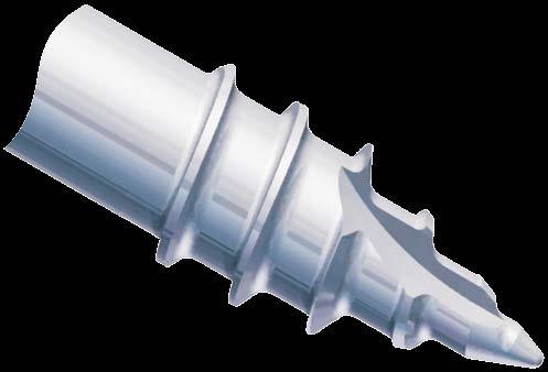
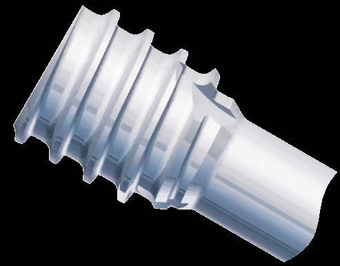
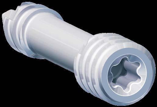
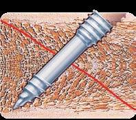
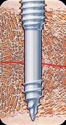
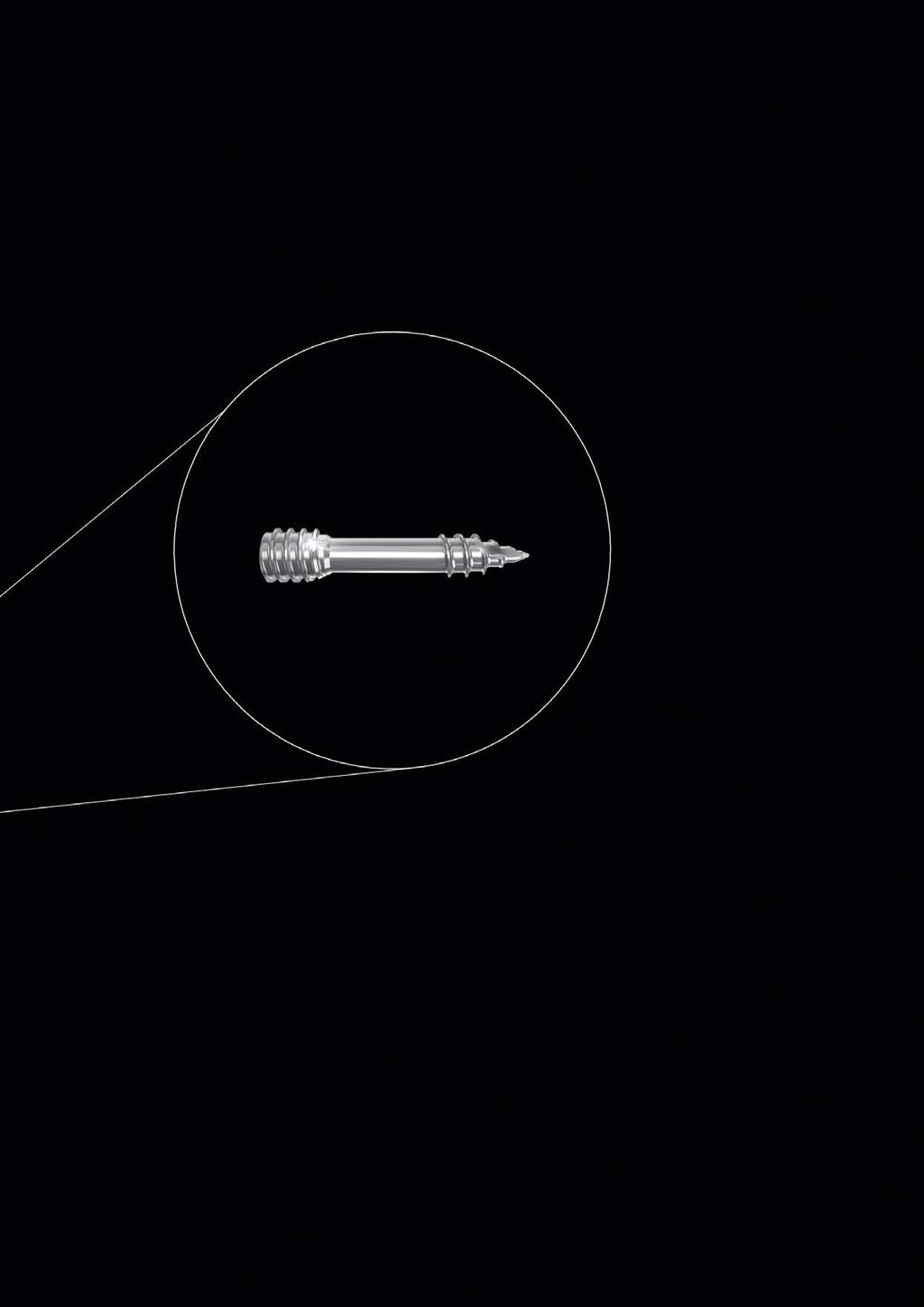
8 Syntellix AG MAGNEZIX Compression Screw Advantages and Features ADVANTAGES AND FEATURES BIOABSORBABLE MAGNESIUM ALLOY The use of MAGNEZIX makes subsequent removal of the implant obsolete: furthermore MAGNEZIX promotes the bone healing process. MAG- NEZIX is bioabsorbable, biocompatible and non-toxic in a biological environment. The use of the innovative bioabsorbable MAGNEZIX metal alloy allows the screw to be implanted using standard techniques. Self-tapping screw tip The self-tapping properties of the screw tip reduce the operation time and simplify the surgical application technique. Self-tapping head thread The self-tapping design of the screw head simplifies insertion and countersinking of the screw head. Different thread pitches The threads of the head and the shaft have different thread pitches. This adapted design of the screw generates compressive forces and supports the intended interfragmentary compression. Self-holding screwdriver The head of the screw is of T4 (ISO ) design. The advantages of this ISO standardized technology are: Î Enlarged contact area Î Improved self-retaining mechanism Î Improved torque transmission


9
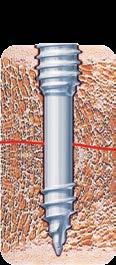
10 Syntellix AG MAGNEZIX Compression Screw Surgical Technique SURGICAL TECHNIQUE SURGICAL TECHNIQUE MAGNEZIX CS STEP BY STEP Prior to implanting a MAGNEZIX CS 2.0 screw it is necessary to ensure repositioning and temporary stabilization of the fracture or the osteotomy. Although the MAGNEZIX CS 2.0 screw has a self-cutting tip, a pilot hole must always be predrilled. The pilot hole also allows precise selection of the correct screw length. Step 1: Drilling the pilot hole Position the double drill guide through the soft tissue to the bone. Insert the drill bit through the double drill guide and into the bone, possibly monitoring with the image intensifier until it is at the required depth. Important If no pilot hole is drilled, the precise screw length cannot be correctly determined. Pre-drilling with an incorrect alignment can lead to malfunction of the screw. Instruments used: ➀ Double Drill Guide, Ø 2.2/1.5 mm ➀ Drill Bit, Ø 1.5 mm Step 2: Determination of screw length The length of the screw is determined by means of the depth gauge to determine the depth of the pre-drilled pilot hole in the bone. (18 mm in the figure). Important When selecting the length of the screw one has to ensure proper compression of the fracture gap. Instruments used: ➂ Depth gauge for screws


11 ➁ ➀ STEP 1 ➂ STEP 2
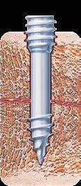
12 Syntellix AG MAGNEZIX Compression Screw Surgical Technique Step 3: Countersinking In order to simplify insertion of the screw head the head-side of the intended implant position is now reamed using the countersink. Important If the screw is positioned perpendicular to the bone surface, countersinking to the first ring marking (RM 1) is required in order to achieve adequate countersinking of the screw head. If the screw is positioned at an angle of 45 to the bone surface, countersinking to the second ring marking (RM 2) is required in order to achieve adequate countersinking of the screw head. Instruments used: ➀ Double Drill Guide, Ø 2.2/1.5 mm ➁ Countersink Ø 2.2/1.5 mm, for quick coupling


13 ➁ STEP 3 ➀ RM 2 RM 1 0 ➁ RM 2 ➀ 45
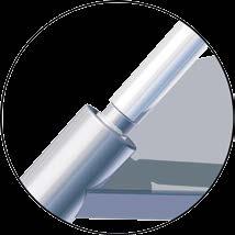
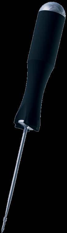
14 Syntellix AG MAGNEZIX Compression Screw Surgical Technique Step 4: Inserting the screw The MAGNEZIX Compression Screw 2.0 of the previously determined length (step 2) is now screwed into place. Important Bear in mind that the shaft thread could pull out of the distal bone fragment if the induced compression forces when screwing-in the screw are excessive. If the selected screw is too short the shaft thread might cross the fracture or osteotomy gap. If this situation results no compression will be generated. Therefore, to ensure the correct position of the threaded shaft it is recommended to check the position using an image intensifier. If one finds the thread crossing the fracture or osteotomy gap the screw must be removed and a longer screw has to be selected in order to generate compression. When doing this and in the case of a hard (dense) bone situation, it might be necessary to repeat the pre-drilling process as described in step 1 to further deepen the pre-drilled pilot hole for the selected screw with an adequate length. Instruments used ➀ Screwdriver T4, One-Piece Handle Optional Screwdriver T4, Multi-Part Handle

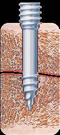
15 STEP 4 ➀
16 Syntellix AG MAGNEZIX Compression Screw Product Overview MAGNEZIX CS 2.0 IMPLANTS Art. No. Threaded shaft length Screw length [mm] SG [mm] L mm SG L Ø 2.5 mm Head thread Ø 1.6 mm Shaft diameter Ø 2.0 mm Shaft thread All implants are individually sterile packaged. It is not possible to re-sterilize the implants.
17 MAGNEZIX CS 2.0 INSTRUMENTS* Art. No. Description Screwdriver T4, One-Piece Handle, consisting of: One-Piece Handle for Screwdriver Screwdriver Blade T Screwdriver T4, Multi-Part Handle, consisting of: Multi-Part Handle for Screwdriver Screwdriver Blade T Drill Bit Ø 1.5 mm, length 88/63 mm, for quick coupling Countersink Ø 2.2/1.5 mm, for quick coupling Double Drill Guide, Ø 2.2/1.5 mm Depth gauge for screws *The figures are not to scale. Not shown: Sterilizing Tray for MAGNEZIX CS Ø 2.0 mm, without contents Lid for Sterilizing Tray, for MAGNEZIX CS Ø 2.0 mm

18 METALLIC AND BIOABSORBABLE. A MEDICAL SENSATION. MAGNEZIX
19
20 Presented by: Syntellix AG Schiffgraben Hannover Germany T F info@syntellix.com Implants are manufactured in Germany in cooperation with Königsee Implantate GmbH /15
CS 2.0. Product information. Intelligent innovations for a better life. Presented by: Syntellix AG Schiffgraben Hannover Germany
 Presented by: CS 2.0 Product information Syntellix AG Schiffgraben 11 30159 Hannover Germany T +49 511 270 413 50 F +49 511 270 413 79 info@syntellix.com www.syntellix.com Implants are manufactured in
CS 2.7. Product information. Intelligent innovations for a better life. Presented by: Syntellix AG Aegidientorplatz 2a Hannover Germany
 Presented by: CS 2.7 Product information Aegidientorplatz 2a 30159 Hannover Germany T +49 511 270 413 50 F +49 511 270 413 79 info@syntellix.com www.syntellix.com Implants are manufactured in Germany in
Compression Screw 3.2 Bioabsorbable. Product information
 Compression Screw 3.2 Bioabsorbable Product information Caution The current product description is not sufficient for the immediate application of instruments and implants. Instructional training by an
A NEW STANDARD OF IMPLANTS!
 A NEW STANDARD OF IMPLANTS! MAGNEZIX PRODUCT OVERVIEW Intelligent innovations for a better life. www.syntellix.com THE BENEFITS AT A GLANCE Similar stability to comparable titanium implants. Greater stability
Syntellix AG MAGNEZIX CS /3.2 Product information. Intelligent innovations for a better life.
 Syntellix AG Introduction 1 CS 2.0 2.7/3.2 Product information Intelligent innovations for a better life. 02 Syntellix AG Introduction INTRODUCTION............................................................03
THE IMPLANT OF TOMORROW SPECIAL PROPERTIES UNIQUE BENEFITS: MAGNEZIX
 THE IMPLANT OF TOMORROW SPECIAL PROPERTIES UNIQUE BENEFITS: MAGNEZIX Intelligent innovations for a better life. www.syntellix.com STABILITY, RESORBABILITY AND OSTEOCONDUCTIVITY DEFINE A NEW STANDARD OF
STABLE, TRANSFORMABLE, UNIVERSALLY APPLICABLE
 STABLE, TRANSFORMABLE, UNIVERSALLY APPLICABLE THE NEW MAGNEZIX CBS OFFERS NEW ADVANTAGES! Intelligent innovations for a better life. www.syntellix.com The advantages are clear to see at a glance Unique
STABILITY WITHOUT COMPROMISES MAGNEZIX PIN: AT FIRST METAL THEN BONE
 STABILITY WITHOUT COMPROMISES MAGNEZIX PIN: AT FIRST METAL THEN BONE UP TO 5x MORE STABLE THAN POLYMER PINS! Intelligent innovations for a better life. www.syntellix.com The benefits at a glance Metallic
Distal Ulnar Locking Plate
 INDEX Indications Patient Position Surgical Technique - Step 1 Approach - Step 2 Plate Contouring - Step 3 Fracture Reduction - Step 4 Distal Plate Fixation - Step 5 Confirm Proper Reconstruction - Step
Headless Compession Screw 2.5 / 3.0
 SURGICAL TECHNIQUE Headless Compession Screw 2.5 / 3.0 Titanium or Stainless Steel Cannulated Headless Design Multiple Thread Options Torx Driver Sterile and Non-Sterile Options Simple Instrumentation
RapidSorb Resorbable Tacks. Resorbable Fixation System.
 RapidSorb Resorbable Tacks. Resorbable Fixation System. Fast Safe Resorbable Drill Press Fixed Table of Contents Introduction Overview 2 Indications and Contraindications 4 RapidSorb 5 Surgical Technique
LCP Medial Distal Tibia Plate, without Tab. The Low Profile Anatomic Fixation System with Angular Stability and Optimal Screw Orientation.
 LCP Medial Distal Tibia Plate, without Tab. The Low Profile Anatomic Fixation System with Angular Stability and Optimal Screw Orientation. Technique Guide LCP Small Fragment System Table of Contents Introduction
Distal Radius Plate 2.4/2.7 dorsal and volar
 Distal Radius Plate 2.4/2.7 dorsal and volar Surgical Technique This publication is not intended for distribution in the USA. Instruments and implants approved by the AO Foundation. Distal Radius Plate
2.4 mm Variable Angle LCP Volar Extra-Articular Distal Radius System. For fragment-specific fracture fixation with variable angle locking technology.
 Technique Guide 2.4 mm Variable Angle LCP Volar Extra-Articular Distal Radius System. For fragment-specific fracture fixation with variable angle locking technology. Table of Contents Introduction 2.4
Headless Compession Screw 4.5 / 6.5 SURGICAL TECHNIQUE. Titanium or Stainless Steel. Cannulated Headless Design. Multiple Thread Options.
 Headless Compession Screw SURGICAL TECHNIQUE 4.5 / 6.5 Titanium or Stainless Steel Cannulated Headless Design Multiple Thread Options Torx Driver Sterile and Non-Sterile Options Simple Instrumentation
NCB Distal Femur System. Surgical Technique
 NCB Distal Femur System Surgical Technique NCB Distal Femur System Surgical Technique 3 Surgical Technique NCB Distal Femur System Table of Contents Introduction 4 Indications 8 Preoperative Planning
low ProfIle neuro PlaTIng system
 low ProfIle neuro PlaTIng system surgical TeChnIque Table of Contents Introduction Low Profile Neuro Cranial Plating System 2 Surgical Technique Technique 5 Product Information Low Profile Neuro Plates
2.4 mm LCP Volar Column Distal Radius Plates. Part of the 2.4 mm LCP Distal Radius System.
 2.4 mm LCP Volar Column Distal Radius Plates. Part of the 2.4 mm LCP Distal Radius System. 12 anatomically shaped volar plates Multiple screw options for fixedangle support to articular surface Combi holes
LCP Medial Proximal Tibial Plate 4.5/5.0. Part of the Synthes LCP periarticular plating system.
 LCP Medial Proximal Tibial Plate 4.5/5.0. Part of the Synthes LCP periarticular plating system. Technique Guide This publication is not intended for distribution in the USA. Instruments and implants approved
LCP Medial Proximal Tibial Plate 3.5. Part of the Synthes small fragment Locking Compression Plate (LCP) system.
 system.") LCP Medial Proximal Tibial Plate 3.5. Part of the Synthes small fragment Locking Compression Plate (LCP) system. Technique Guide This publication is not intended for distribution in the USA. Instruments
LCP Medial Proximal Tibial Plate 3.5. Part of the Synthes small fragment Locking Compression Plate (LCP) system. Technique Guide This publication is not intended for distribution in the USA. Instruments
Technique Guide. 3.5 mm LCP Low Bend Medial Distal Tibia Plates. Part of the Synthes locking compression plate (LCP) system.
 system.") Technique Guide 3.5 mm LCP Low Bend Medial Distal Tibia Plates. Part of the Synthes locking compression plate (LCP) system. Table of Contents Introduction 3.5 mm LCP Low Bend Medial Distal Tibia Plates
Technique Guide 3.5 mm LCP Low Bend Medial Distal Tibia Plates. Part of the Synthes locking compression plate (LCP) system. Table of Contents Introduction 3.5 mm LCP Low Bend Medial Distal Tibia Plates
MetaFix Ludloff Plate
 Merete MetaFix Ludloff Plate Low Profile Locking Bone Plate System Surgical Technique and Ordering Information - Content - Content 1. Description.................................................. 3 2.
Elbow Plating System Surgical Technique
 Locking Compression Technology by aap 1 Disclaimer This surgical technique is exclusively intended for medical professionals, especially physicians, and therefore may not be regarded as a source of information
Low Profile Neuro Plating System. Surgical Technique
 Low Profile Neuro Plating System Surgical Technique TABLE OF CONTENTS INTRODUCTION Low Profile Neuro Plating System 2 SURGICAL TECHNIQUE Technique 5 PRODUCT INFORMATION Low Profile Neuro Plates 10 Low
Lapidus Arthrodesis System Instructions for Use
 Lapidus Arthrodesis System Instructions for Use Description The AlignMATE Lapidus Arthrodesis System consists of bone plates and bone screws (locking, non-locking and interfragmentary), which are intended
Thoracolumbar Spine Locking Plate (TSLP) System. A low-profile plating system for anterior stabilization of the thoracic and lumbar spine.
 System. A low-profile plating system for anterior stabilization of the thoracic and lumbar spine.") Thoracolumbar Spine Locking Plate (TSLP) System. A low-profile plating system for anterior stabilization of the thoracic and lumbar spine. Technique Guide Instruments and implants approved by the AO Foundation
Thoracolumbar Spine Locking Plate (TSLP) System. A low-profile plating system for anterior stabilization of the thoracic and lumbar spine. Technique Guide Instruments and implants approved by the AO Foundation
SMV Scientific Bone Plate and Screw System Surgical Technique
 SMV Scientific Bone Plate and Screw System Surgical Technique Description: The SMV Scientific Bone Plate and Screw System consists of non-locking plates and bone screw fasteners in a variety of lengths,
IT HELPS YOU GET FIT AGAIN, FASTER AND SAFER
 IT HELPS YOU GET FIT AGAIN, FASTER AND SAFER THE IMPLANT THAT TURNS INTO BONE MAGNEZIX OBVIATES THE NEED FOR A SECOND OPERATION TO REMOVE METAL IMPLANTS Intelligent innovations for a better life. www.syntellix.com
Technique Guide. 2.4 mm Variable Angle LCP Distal Radius System. For fragment-specific fracture fixation with variable angle locking technology.
 Technique Guide 2.4 mm Variable Angle LCP Distal Radius System. For fragment-specific fracture fixation with variable angle locking technology. Table of Contents Introduction 2.4 mm Variable Angle LCP
Aesculap Orthopaedics Targon DR
 Aesculap Orthopaedics Targon DR Intramedullary nail for the distal radius Stability from within Targon DR stability from within PD Dr. med. Georg Gradl, Dept. for Accident and Reconstructive Surgery at
2.4 mm Variable Angle LCP Volar Extra-Articular Distal Radius System. For fragment-specific fracture fixation with variable angle locking technology.
 2.4 mm Variable Angle LCP Volar Extra-Articular Distal Radius System. For fragment-specific fracture fixation with variable angle locking technology. Surgical Technique This publication is not intended
Technique Guide. 3.5 mm LCP Olecranon Plates. Part of the Synthes locking compression plate (LCP) system.
 system.") Technique Guide 3.5 mm LCP Olecranon Plates. Part of the Synthes locking compression plate (LCP) system. Table of Contents Introduction 3.5 mm LCP Olecranon Plates 2 AO Principles 3 Indications 3 Clinical
Technique Guide 3.5 mm LCP Olecranon Plates. Part of the Synthes locking compression plate (LCP) system. Table of Contents Introduction 3.5 mm LCP Olecranon Plates 2 AO Principles 3 Indications 3 Clinical
MatrixNEURO. The next generation cranial plating system.
 MatrixNEURO. The next generation cranial plating system. Technique Guide CMF Matrix This publication is not intended for distribution in the USA. Instruments and implants approved by the AO Foundation
3. Insert Tocar Sleeves Insert the NCB tissue protection sleeve assembly 1.6 to 10mm through a skin incision (Fig. 38).
.") NCB Proximal Humerus Plating System Surgical Technique 19 2. Temporary Plate Fixation The plate can be temporary fixed to the bone with 1.6mm K-wire through the proximal cannulated fixation screw of the
NCB Proximal Humerus Plating System Surgical Technique 19 2. Temporary Plate Fixation The plate can be temporary fixed to the bone with 1.6mm K-wire through the proximal cannulated fixation screw of the
OBSOLETED. LCP Medial Distal Tibia Plate, without Tab. The Low Profile Anatomic Fixation System with Angular Stability and Optimal Screw Orientation.
 LCP Medial Distal Tibia Plate, without Tab. The Low Profile Anatomic Fixation System with Angular Stability and Optimal Screw Orientation. Surgical Technique LCP Small Fragment System This publication
TSLP Thoracolumbar Spine Locking Plate
 Anterior thoracolumbar spine locking plate TSLP Thoracolumbar Spine Locking Plate Surgical Technique Image intensifier control This description alone does not provide sufficient background for direct use
LCP Metaphyseal Plates. For extra-articular fractures.
 LCP Metaphyseal Plates. For extra-articular fractures. Surgical Technique This publication is not intended for distribution in the USA. Instruments and implants approved by the AO Foundation. Image intensifier
3.0/3.5/4.0/4.5/6.5/7.0/7.3. Cannulated Screws. Surgical Technique
 3.0/3.5/4.0/4.5/6.5/7.0/7.3 Cannulated Screws Surgical Technique Image intensifier control This description alone does not provide sufficient background for direct use of DePuy Synthes products. Instruction
SpeedTip CCS 2.2, 3.0
 PRODUCT INFORMATION SpeedTip CCS 2.2, 3.0 Cannulated Compression Screws APTUS 2 SpeedTip CCS 2.2, 3.0 Cannulated Compression Screws SpeedTip CCS * 2.2, 3.0 Cannulated Compression Screws A new generation
2.4 mm LCP Radial Head Plates. Part of the Synthes LCP Distal Radius Plate System.
 2.4 mm LCP Radial Head Plates. Part of the Synthes LCP Distal Radius Plate System. Technique Guide Instruments and Implants approved by the AO Foundation Table of Contents Introduction 2.4 mm LCP Radial
LCP Distal Humerus Plates
 The anatomic fixation system for the distal humerus with angular stability Surgical technique LCP Locking Compression Plate Contents Indications and contraindications 2 Implants 3 Instruments 5 Preparation
Long Volar Plates for Diaphyseal-Metaphyseal Radius Fractures LCP. Dia-Meta Volar Distal Radius Plates. Surgical Technique
 Long Volar Plates for Diaphyseal-Metaphyseal Radius Fractures LCP Dia-Meta Volar Distal Radius Plates Surgical Technique Table of Contents Introduction LCP Dia-Meta Volar Distal Radius Plates 2 AO Principles
ORION Screw System PRODUCT INFORMATION
 ORION Screw System PRODUCT INFORMATION 2 ORION Screw System www.hnmtotal.com ORION SCREW SYSTEM CONTENTS ORION 2.5/3.0 Screw Set Headed Screws Headless Screws Instrumentation 3 5 7 ORION 4.5/6.5 Screw
Instrument and Implant for wrist fracture
 Instrument and Implant for wrist fracture Jansri Janpanya Product specialist The Bangkok Unitrade Co,.ltd. Objectives Type of LCP for distal radius Fx. The new LCP design for distal radius Fx. Have knowledge
LCP Distal Tibia Plate
 Surgical Technique LCP Locking Compression Plate Original Instruments and Implants of the Association for the Study of Internal Fixation AO/ASIF Table of contents Indications 3 Implants/Instruments 5 Surgical
Technique Guide. LCP Proximal Femoral Hook Plate 4.5/5.0. Part of the LCP Periarticular Plating System.
 Technique Guide LCP Proximal Femoral Hook Plate 4.5/5.0. Part of the LCP Periarticular Plating System. Table of Contents Introduction Features and Benefits 2 AO ASIF Principles 4 Indications 5 Surgical
3.5 mm LCP Extra-articular Distal Humerus Plate
 Part of the DePuy Synthes Locking Compression Plate (LCP ) System 3.5 mm LCP Extra-articular Distal Humerus Plate Surgical Technique Table of Contents Introduction 3.5 mm LCP Extra-articular Distal Humerus
LOW PROFILE NEURO. This publication is not intended for distribution in the USA. SURGICAL TECHNIQUE
 LOW PROFILE NEURO This publication is not intended for distribution in the USA. SURGICAL TECHNIQUE TABLE OF CONTENTS INTRODUCTION Low Profile Neuro Plating System 2 Intended Use, Indications, Contraindications
PediLoc 3.5mm and 4.5mm Contour Femur Plate Surgical Technique
 PediLoc 3.5mm and 4.5mm Contour Femur Plate Surgical Technique Surgical Technique Contour Femur Plate The technique description herein is made available to the healthcare professional to illustrate the
Surgical Technique. Cannulated Angled Blade Plate 3.5 and 4.5, 90
 Surgical Technique Cannulated Angled Blade Plate 3.5 and 4.5, 90 Cannulated Angled Blade Plate 3.5 and 4.5, 90 Table of contents Indications/Contraindications 2 Implants 3 Surgical technique 5 Implant
PROXIMAL TIBIAL PLATE
 SURGICAL NÁSTROJE TECHNIQUE PRO ARTROSKOPII PROXIMAL INSTRUMENTS TIBIAL FOR PLATE ARTHROSCOPY Proximal Tibial Plate Description of medical device The Proximal Tibial Plate is used in epyphyseal and metaphyseal
Technique Guide. Compact 2.0 LOCK Mandible. The locking system for the mandible.
 Technique Guide Compact 2.0 LOCK Mandible. The locking system for the mandible. Table of Contents Introduction Compact 2.0 LOCK Mandible 2 AO Principles 4 Indications and Contraindications 5 Surgical
Technique Guide. 6.5 mm Midfoot Fusion Bolt. For intramedullary fixation of the medial column of the foot.
 Technique Guide 6.5 mm Midfoot Fusion Bolt. For intramedullary fixation of the medial column of the foot. Table of Contents Introduction 6.5 mm Midfoot Fusion Bolt 2 AO Principles 4 Indications 5 Surgical
Low Bend Distal Tibia Plates
 Part of the DePuy Synthes Locking Compression Plate (LCP ) System 3.5 mm LCP Low Bend Medial Distal Tibia Plates Surgical Technique Table of Contents Introduction 3.5 mm LCP Low Bend Medial Distal Tibia
LCP Low Bend Medial Distal Tibia Plates 3.5 mm. Anatomic plates with low profile head for intra- and extraarticular fractures.
 LCP Low Bend Medial Distal Tibia Plates 3.5 mm. Anatomic plates with low profile head for intra- and extraarticular fractures. Surgical Technique This publication is not intended for distribution in the
A locking plate system that expands a surgeon s options in trauma surgery. Zimmer NCB Plating System
 A locking plate system that expands a surgeon s options in trauma surgery Zimmer NCB Plating System The Power of Choice The power of having true intraoperative options is at your fingertips. Using standard
Locking Compression Technology by aap
 Minimally Invasive Locking Compression Technology by aap Minimally Invasive 1 Disclaimer This surgical technique is exclusively intended for medical professionals, especially physicians, and therefore
Complex process High costs
 MTP With a non sterile standard kit Calling on medical staff Constraints Complex traceability Contracted out sterilization Suppliers deadline Complex process High costs 1 Delivery 2 Storage 3 Unpacking
Olecranon Locking Plate II
 INDEX Indications Patient Position Fracture Reduction and Fixation Surgical Technique Step 1 Surgical Approach Step 2 Implantation Step 3 Proximal Locking Screw Insertion Step 4 Distal Screw Insertion
LCP Proximal Radius Plates 2.4. Plates for radial head rim and for radial head neck address individual fracture patterns of the proximal radius.
 Technique Guide LCP Proximal Radius Plates 2.4. Plates for radial head rim and for radial head neck address individual fracture patterns of the proximal radius. Table of Contents Introduction LCP Proximal
3.5 mm LCP Olecranon Plates
 Part of the DePuy Synthes Locking Compression Plate (LCP ) System 3.5 mm LCP Olecranon Plates Surgical Technique Table of Contents Introduction 3.5 mm LCP Olecranon Plates 2 AO Principles 3 Indications
Technique Guide. 2.7 mm/3.5 mm LCP Distal Fibula Plates. Part of the Synthes locking compression plate (LCP) system.
 system.") Technique Guide 2.7 mm/3.5 mm LCP Distal Fibula Plates. Part of the Synthes locking compression plate (LCP) system. Table of Contents Introduction 2.7 mm/3.5 mm LCP Distal Fibula Plates 2 AO Principles
Technique Guide 2.7 mm/3.5 mm LCP Distal Fibula Plates. Part of the Synthes locking compression plate (LCP) system. Table of Contents Introduction 2.7 mm/3.5 mm LCP Distal Fibula Plates 2 AO Principles
LCP Medial Proximal Tibial Plate 3.5. Part of the Synthes small fragment Locking Compression Plate (LCP) system.
 system.") LCP Medial Proximal Tibial Plate 3.5. Part of the Synthes small fragment Locking Compression Plate (LCP) system. Surgical Technique This publication is not intended for distribution in the USA. Instruments
LCP Medial Proximal Tibial Plate 3.5. Part of the Synthes small fragment Locking Compression Plate (LCP) system. Surgical Technique This publication is not intended for distribution in the USA. Instruments
The Calcaneal Plate. The Synthes non-locking solution for the Calcaneus.
 The Calcaneal Plate. The Synthes non-locking solution for the Calcaneus. Surgical Technique This publication is not intended for distribution in the USA. Instruments and implants approved by the AO Foundation.
Correction System. Surgical Technique
 Nextra Hammertoe Correction System Surgical Technique Maximized Bone Purchase* Stable and Secure Phalanx Optimized Screw Design Adjustable Bone-to-Bone Apposition Progressive Ratchet Tightening Mechanism
Small Fragment Plating System. Securing optimal fixation through locked and compression plating technology
 Small Fragment Plating System Securing optimal fixation through locked and compression plating technology Contents Design Rationale Introduction Interfragmentary Fixation Insertion of a 3.5 mm Cortical
Small Fragment Plating System
 Small Fragment Plating System Securing optimal fixation through locked and compression plating technology SURGICAL TECHNIQUE RECOVERY FUNCTION SURVIVORSHIP DePuy believes in an approach to trauma surgery
WINSTA-C. Clavicle Plating System
 Clavicle Plating System Clinical Advisor Michael Kurer FRCS FRCS (Orth) Consultant Orthopaedic and Shoulder Surgeon North Middlesex University Hospital NHS Trust Table of Contents Introduction Indication
Flower Medium Headless & Cannulated Screws
 Flower Medium Headless & Cannulated Screws PROCEDURE GUIDE www.flowerortho.com The Flower Foot & Ankle Application NC FUSION PLATE 2-HOLE COMPRESSION PLATE TMT FUSION PLATE LAPIDUS FUSION PLATE COMPRESSION
Surgical Technique. CONQUEST FN Femoral Neck Fracture System
 Surgical Technique CONQUEST FN Femoral Neck Fracture System Table of Contents Introduction... 3 Indications... 3 Product Overview... 4 Surgical Technique... 5 Patient Positioning... 5 Reduce the Fracture...
Technique Guide. Small Fragment Locking Compression Plate (LCP) System. Stainless steel and titanium.
 System. Stainless steel and titanium.") Technique Guide Small Fragment Locking Compression Plate (LCP) System. Stainless steel and titanium. Table of Contents Introduction Small Fragment Locking Compression Plate (LCP) System 2 AO Principles
Technique Guide Small Fragment Locking Compression Plate (LCP) System. Stainless steel and titanium. Table of Contents Introduction Small Fragment Locking Compression Plate (LCP) System 2 AO Principles
Technique Guide. PHILOS and PHILOS Long. The anatomic fixation system for the proximal humerus.
 Technique Guide PHILOS and PHILOS Long. The anatomic fixation system for the proximal humerus. Table of Contents Introduction PHILOS and PHILOS Long 2 AO Principles 4 Indications 5 Surgical Technique
Clavicle Hook Locking Plate
 990210003 Clavicle Hook Locking Plate Clavicle Hook Locking Plate Clavicle Hook Locking Plate INDEX Indications Patient Position Surgical Technique Step 1 Approach Step 2 Reduction Step 3 Temporary Fixation
PEDUS-L. Locking Plantar Lapidus Plate
 PEDUS-L Locking Plantar Lapidus Plate Page 1 PEDUS-L - Locking Plantar Lapidus Plate Table of Contents Implants 3 System 4 Operation manual 5 Approach 5 Identification of the TMT 1 joint with a cannula
Small Fragment Locking Compression Plate (LCP ) System
 System") Stainless Steel and Titanium Small Fragment Locking Compression Plate (LCP ) System Surgical Technique Table of Contents Introduction Small Fragment Locking Compression Plate (LCP) System 2 AO Principles
Stainless Steel and Titanium Small Fragment Locking Compression Plate (LCP ) System Surgical Technique Table of Contents Introduction Small Fragment Locking Compression Plate (LCP) System 2 AO Principles
ALIANS ELBOW & OLECRANON. INNOVATION means MOTION. Distal humerus and olecranon plating system Polyaxial locking technology Precontoured implants
 INNOVATION means MOTION ALIANS ELBOW distal humerus & OLECRANON POLYAXIAL LOCKING FIXATION DUALTEC SYSTEM II Distal humerus and olecranon plating system Polyaxial locking technology Precontoured implants
AxSOS Locking Plate System
 AxSOS Locking Plate System Operative Technique Small Fragment Basic Fragment 1 2 Contents Page 1. Introduction 4 2. Features & Benefits 5 4 and 5mm Compression Plates 5 Reconstruction and 1/3 Tubular Locking
SURGICAL TECHNIQUE GUIDE: JONES FRACTURE USING THE PRECISION JONES FRACTURE SCREW SYSTEM
 PRODUCT DESCRIPTION The PRECISION Jones Fracture Screw System offers extensive options of Type II Anodized Titanium screws. System-specific instrumentation is designed to address procedural challenges
WINSTA-R. Distal Radius System
 Distal Radius System Table of Contents Introduction WINSTA-R System 2 Indication 2 Surgical Technique Palmar Access for Radius Plate 3 Dorsal Access for Radius Plate 3 Positioning of the Radius Plate
Zimmer Anterior Buttress Plate System. Surgical Technique
 Zimmer Anterior Buttress Plate System Surgical Technique 2 Zimmer Anterior Buttress Plate System Surgical Technique Zimmer Anterior Buttress Plate System Surgical Technique Description, Indications & Contraindications...
VariAx Compression Plating System. Operative technique
 VariAx Compression Plating System Operative technique VariAx Compression Plating System Operative technique VariAx Compression Plating System Contents 1. Introduction... 3 2. Indications, MR safety information
Complex process High costs
 With a non sterile standard kit Calling on medical staff Constraints Complex traceability Contracted out sterilization Suppliers deadline Complex process High costs 1 Delivery 2 Storage 3 Unpacking 4 Control
Femur Condylar Plate System Procedural Steps.
 Femur Condylar Plate System Procedural Steps www.carbo-fix.com 1 Table of Contents Introduction..3 Instrumentation Set... 8 Procedural Steps:...... 12 Ordering Information 19 2 Introduction The CarboFix
CHARLOTTE. 3.0 and 4.3 Multi-Use Compression Screws SURGIC A L T ECHNIQUE
 CHARLOTTE 3.0 and 4.3 Multi-Use Compression Screws SURGIC A L T ECHNIQUE CHARLOTTE 3.0 and 4.3 Multi-Use Compression Screws SURGICAL TECHNIQUE Surgical Technique as described by: Robert Anderson, MD Bruce
Surgical Technique. Distal Humerus Locking Plate
 Surgical Technique Distal Humerus Locking Plate PERI-LOC Locked Plating System Distal Humerus Locking Plate Surgical Technique Table of Contents Introduction...2 Indications...3 Plate Features...3 Patient
2.7 mm/3.5 mm LCP Distal Fibula Plate System. Part of the Synthes locking compression plate (LCP) system.
 system.") 2.7 mm/3.5 mm LCP Distal Fibula Plate System. Part of the locking compression plate (LCP) system. Anatomically contoured Multiple screw options for fixedangle support Coaxial holes minimize screw head
2.7 mm/3.5 mm LCP Distal Fibula Plate System. Part of the locking compression plate (LCP) system. Anatomically contoured Multiple screw options for fixedangle support Coaxial holes minimize screw head
Technique Guide. Midface Distractor System. For the temporary stabilization and gradual lengthening of the cranial or midfacial bones.
 Technique Guide Midface Distractor System. For the temporary stabilization and gradual lengthening of the cranial or midfacial bones. Table of Contents Introduction Midface Distractor System 2 Indications
Distal Radius Sterile Kit. Optimization And Efficiency You Can Rely On
 Distal Radius Sterile Kit Optimization And Efficiency You Can Rely On Introducing The Distal Radius Sterile Kit Improved OR Workflow The Distal Radius Sterile Kit provides high-quality single-use implants
UNDERSTANDING INNOVATIONS A CLOSER LOOK AT THE SPECIAL PROPERTIES OF MAGNEZIX
 UNDERSTANDING INNOVATIONS A CLOSER LOOK AT THE SPECIAL PROPERTIES OF MAGNEZIX Intelligent innovations for a better life. www.syntellix.com STABILITY, DISSOLUTION AND OSTEOCONDUCTIVITY DEFINE A NEW STANDARD
4.5 mm LCP Medial Proximal Tibia Plates
 Part of the DePuy Synthes LCP Periarticular Plating System 4.5 mm LCP Medial Proximal Tibia Plates Surgical Technique Table of Contents Introduction 4.5 mm LCP Medial Proximal Tibia Plates 2 AO Principles
Distal Radius Plate Instrument and Implant Set. Discontinued December 2017 DSUS/TRM/0916/1063(1)
") Distal Radius Plate Instrument and Implant Set Surgical Technique Discontinued December 2017 DSUS/TRM/0916/1063(1) The Distal Radius Plates Indications For fixation of fractures and osteotomies, including
Distal Radius Plate Instrument and Implant Set Surgical Technique Discontinued December 2017 DSUS/TRM/0916/1063(1) The Distal Radius Plates Indications For fixation of fractures and osteotomies, including
Table of Contents.
 surgical technique The Ambassador TM Anterior Cervical Plate System is a versatile system of implants and instruments with a variety of sizes to provide optimal anatomic compatibility. The integrated cam
Biomimetic Product catalogue
 Biomimetic Product catalogue 20 Pol. Industrial Santa Anna I Apartado 20 08251 Santpedor (Barcelona) España T. (+34) 902 38 38 48 F. (+34) 93 827 38 73 www.avinent.com avinent@avinent.com 1 CORAL is the
Technique Guide. VA-Locking Intercarpal Fusion System. Variable angle locking technology for mediocarpal partial arthrodesis.
 Technique Guide VA-Locking Intercarpal Fusion System. Variable angle locking technology for mediocarpal partial arthrodesis. Table of Contents Introduction VA-Locking Intercarpal Fusion System 2 Indications
2.7 mm/3.5 mm LCP Distal Fibula Plate
 Part of the DePuy Synthes Locking Compression Plate (LCP ) System 2.7 mm/3.5 mm LCP Distal Fibula Plate Surgical Technique Table of Contents Introduction 2.7 mm/3.5 mm LCP Distal Fibula Plates 2 AO Principles
Surgical Technique. Olecranon Locking Plate
 Surgical Technique Olecranon Locking Plate PERI-LOC Locked Plating System Olecranon Locking Plate Surgical Techniquealog Infor Table of Contents Introduction...2 Indications...3 Plate Features...3 Patient
VA-LCP Olecranon Plates 2.7/3.5. The fracture-specific low-profile fixation system with variable angle locking technology.
 VA-LCP Olecranon Plates 2.7/3.5. The fracture-specific low-profile fixation system with variable angle locking technology. Surgical Technique This publication is not intended for distribution in the USA.
Technique Guide. Locking Attachment Plate. For treatment of periprosthetic fractures.
 Technique Guide Locking Attachment Plate. For treatment of periprosthetic fractures. Table of Contents Introduction Locking Attachment Plate 2 Indications 4 Surgical Technique Patient Positioning 5 Preparation