Practical Examination Handbook
|
|
|
- Kelley Greer
- 5 years ago
- Views:
Transcription


1 Practical Examination Handbook 1
2 Copyright January 2018 This work is copyright Copyright is held by the Australian Dental Council Ltd. It may not be reproduced for commercial use or sale. Reproduction requires a licence or written permission which may be obtained from: Australian Dental Council Ltd Po Box Law Courts Victoria 8010 Australia Tel +61 (0)

3 Australian Dental Council Contents INTRODUCTION... 5 VENUE ALLOCATIONS AND REQUIREMENTS... 5 REGISTRATION AND OTHER DENTAL BOARD OF AUSTRALIA REQUIREMENTS... 6 CANDIDATE CONTACT INFORMATION... 6 WITHDRAWING FROM AN EXAMINATION... 6 CONTENT AND FORMAT... 7 EXAMINATION SCHEDULE... 7 EXAMINATION REGISTRATION... 8 EQUIPMENT, INSTRUMENTS AND SUPPLIES... 8 EXAMINATION INSTRUCTIONS... 9 Tasks... 9 Teeth... 9 Infection Control Breaks Incidents POLICIES Examination Conduct Policy Time Extension Policy Adverse Incident Policy ASSESSMENT TASKS ASSESSMENT CRITERIA Final Result Grade Derivation RESULTS
4 VERIFICATION, REVIEW AND APPEAL REPEAT EXAMINATIONS APPENDIX A: ASSESSMENT CRITERIA Class III Composite Resin Cavity Preparation Class II Amalgam Cavity Preparation Class II Composite Resin Cavity Preparation Full Gold Crown Preparation Metal-ceramic Crown Preparation Endodontic Access Preparation Provisional Crown Restoration Class II Composite Resin Restoration Class IV Composite Resin Restoration Class II Amalgam Restoration Radiographic exercise Rubber Dam Application Record of Procedures Infection Control Clinical Communication
5 Introduction Under the provisions of the Health Practitioner Regulation National Law Act 2009 the Australian Dental Council (ADC) has been assigned the accreditation functions of the Dental Board of Australia. One of the key accreditation functions is the assessment of the knowledge, judgement, clinical skills and professional attributes of overseas qualified dentists who are seeking registration with the Dental Board of Australia to practise in Australia and whose qualifications are not otherwise approved for registration. In addition, the ADC is the national assessment authority appointed by the Department of Immigration and Border Protection to assess professional skills for migration purposes. The ADC assessment and examination procedure consists of the following steps: 1. Initial Assessment of Professional Qualifications in Dentistry 2. Written Examination 3. Practical Examination The format of the ADC examination process has been approved for the purposes of registration in Australia. The ADC cannot grant exemptions from the requirements of the examinations. Venue Allocations and Requirements Practical Examinations are held over two days at various venues in Australia. They are scheduled to be held twice each year, generally in June and November, but additional examinations may be scheduled according to demand and the availability of examination venues. You can apply to sit for any available examination for which you are eligible by submitting an application form and the relevant examination fee during the appropriate application period. After successful submission of an application form and payment, you will receive confirmation of the examination to which you have been allocated, and also receive an information pack relevant to that particular venue. The venues in which the ADC examinations are held are usually clinics within dental hospitals or university dental schools. Each venue has its own requirements that you will need to satisfy and comply with. 5
6 Registration and other Dental Board of Australia Requirements As the examination format does not involve the treatment of patients, you are not required to provide evidence of immune status when sitting your examination but you will be required to have ascertained this before being registered to practise in Australia. If you are successful in the Practical Examination the ADC will advise the DBA that you have been awarded an ADC Certificate (General Dentist). Candidate Contact Information You must notify the ADC of any change in mailing address immediately in writing. Information may not be reissued if you fail to advise the ADC or do not employ a suitable mail redirection service from your previous address. Timetables and urgent information may be communicated to you through your nominated address. You must ensure the address you provide is reliable and checked regularly. If you use free webmail services (Gmail, Yahoo, Hotmail, etc.) you should properly maintain your mailboxes. The ADC will not accept responsibility for non-receipt of correctly addressed s. Withdrawing from an Examination Withdrawal from an ADC examination can be made by submitting a Notification of withdrawal from an ADC examination form directly to the ADC, advising that you wish to withdraw. If the date of the examination is imminent, you may submit your intention to withdraw via and subsequently post your withdrawal form and supporting documentation to the ADC. The ADC considers your withdrawal as confirmed upon receipt of all required documentation. The ADC will respond to you in writing. If you withdraw from an examination before the closing date for applications for that examination session you will be eligible to receive an 80% refund of the examination fee. If you withdraw after the closing date for the examination you will be eligible to receive a 50% refund of the examination fee. If you withdraw within four weeks of the examination date you are not entitled to a refund unless your withdrawal is due to illness and is supported by a medical certificate reporting inability to undertake the examination, in which case you may be eligible for a 60% refund. Please visit the ADC website for more details of the withdrawal process and withdrawal fees. Examinations cannot be rescheduled, i.e. application forms and examination fees cannot be transferred to alternate examination dates. If a candidate wishes to sit the examination at a later date they must withdraw from the current examination session and submit a new application and payment. Failure to undertake the examination because of an inability to obtain necessary visas or to arrange travel etc. will be considered a withdrawal and the refunds, as above, will apply. 6
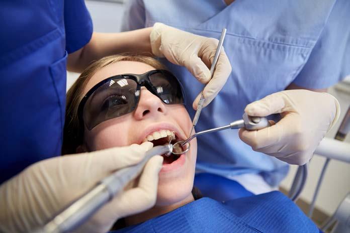
7 Content and Format All Public Sector Dental Workforce Scheme Candidates will be required to sit the Transitional Written Examination. This component of the examinations assesses concepts, knowledge and clinical problemsolving previously assessed in the Clinical Examination. The Transitional Written Examination is a two hour examination paper consisting of scenario-based and stand-alone multiple choice questions. Candidates who pass the Transitional Written Examination but fail the Practical Examination will not be required to re-sit the Transitional Written Examination. All other candidates will be required to have a valid Written Examination. The two-day Practical Examination will evaluate your performance of dental procedures on simulated patients (manikins) in a clinical setting. At the start of each day you will be given: 1. a set of dental models (upper and lower), labelled with your Candidate ID and mounted in a manikin on a dental chair, 2. a task list, detailing all the procedures that you will be required to undertake for that day, including tooth numbers and surfaces and a designated practice tooth and 3. a timetable with attendance times for the Rubber Dam, Communication and Radiology tasks. Examination Schedule The following is an indicative schedule for the Practical Examination. More detailed information will be provided to candidates who enrol for the examination. TRANSITIONAL WRITTEN EXAM (Friday before the Practical Examination) Examination registration Transitional written examination 9:30 a.m a.m. 10:00 a.m. - 12:00 noon PRACTICAL EXAMINATION ORIENTATION (Friday before the Saturday examinations commence) Orientation lecture 2:00 p.m. 3:00 p.m. PRACTICAL EXAMINATION DAY 1 Examination Registration 7 7:30 a.m. - 8:00 a.m.
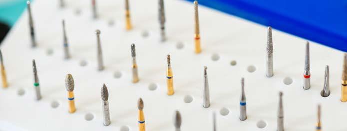
8 Orientation and instructions Bay set-up and model check Completion of task list 8:00 a.m. - 8:30 a.m. 8:30 a.m. - 9:00 a.m. 9:00 a.m. - 4:30 p.m. PRACTICAL EXAMINATION DAY 2 Examination Registration Bay set-up and model check Completion of task list 7:30 a.m. - 8:00 a.m. 8:00 a.m. - 8:30 a.m. 8:30 a.m. - 4:00 p.m. Examination Registration At the start of each day (Saturday and Sunday) you will be issued your Candidate ID badge at the Examination Registration Desk on presentation of current government photographic identification (i.e. a current passport or driver s licence) that includes your signature. The name on the government photographic identification must also match the name you used when you registered with the ADC for the Practical Examination. Your face will be matched to your photograph and your signature will be confirmed. If you do not provide the identification requested you will not be admitted into the examination. You must wear your Candidate ID Badge at all times. At the end of each day you must hand in your ID badge as directed. If you fail to hand in your ID badge, you may receive a fail grade for all requirements in the Examination. Equipment, Instruments and Supplies We will provide all candidates with the equipment and materials necessary for the examination. Equipment and materials may vary slightly between venues but will be appropriate for successful completion of examination tasks. You are not permitted to supply your own instruments or handpieces. Detailed instrument information will be provided to you as part of your venue pack. Please note that dental burs will not be provided. You are required to bring all relevant burs to the examination. You may bring your own materials to the examination (e.g. restorative materials and rubber dam sheets) although this is not encouraged as we cannot guarantee that equipment needed to dispense such materials will be available at the Examination Venue. You must place your burs and materials into a single small container to bring into the examination area. This container must not be larger than 30cm/15cm/20cm. You will not be permitted to enter the examination if you do not meet these requirements. The dental clinic at the Examination Venue will NOT be accessible prior to the Assessment. 8
9 You are advised to bring a small clock or analogue watch to the examination. You will not be permitted to use phones, laptop computers, tablets or other electronic devices to monitor the time. Examination Instructions Tasks You will be given a task list detailing all the procedures that you will be required to undertake at the start of each day. Three of the tasks will be timetabled Rubber Dam, Radiography and Communication. You will be allocated 30 minutes for the Rubber Dam task on one day, and will be allocated 15 minutes each for the Radiography task and the Communication task on the other day. You may undertake the other tasks for the day in any order. The FDI two digit tooth numbering system is used during the examination If you start or complete a procedure not listed on the task sheet, or start or complete a task on an incorrect tooth you will receive a fail grade for that task. Although tasks are conducted on manikins, you must demonstrate that you are competent to perform procedures in a clinical environment, including undertaking tasks in positions appropriate for both the operator and the patient. Examiners will ask you to correct inappropriate positions, such as having the manikin s head or neck in a position that would be uncomfortable for a patient or contact with the manikin that would be considered inappropriate in a clinical situation. If you continue to work on the manikin in an inappropriate position you may be dismissed from the examination. You must wear eye protection, masks, gowns and gloves whilst undertaking any simulated clinical activity. The venue will supply gowns, masks and gloves, but you must provide your own eye protection. You must be appropriately and professionally attired. You should wear suitable, closed shoes and long hair should be appropriately controlled. You may use magnification aids. This does not include hand-held magnifiers. You are responsible for your own materials and belongings. The ADC and the Examination Venue will not be held responsible for personal supplies left unattended. You will be financially responsible for any damage caused to any supplied equipment. Teeth The ADC will provide special prepared typodont models to be used for the restorative exercises. These models will include combinations of teeth and dental tissues of the following types o o Plain Ivorine teeth that are of uniform colour and consistency. Simulated enamel that is white in colour and is made of composite resin that is harder than the simulated dentine and simulated caries. The teeth have been manufactured so that they can be prepared with a dental bur using normal pressure and, if desired, preparations can be finished using normal pressure with sharp hand instruments. 9
10 o o Simulated dentine that is yellow in colour and is softer than the simulated enamel Simulated caries: Currently the ADC is using two forms of teeth that have simulated caries present. Some have simulated caries placed by the manufacturers and some are pre-prepared and filled with Cavit TM to simulate caries. For those enamel/dentine teeth with simulated caries the manufacturing process ensures that the caries depth is standardized for each tooth used. The simulated caries in dentine is grey in colour and is softer than the simulated enamel but of similar hardness to the simulated dentine. In some anterior teeth, there is also a cavity in the simulated enamel on the proximal surface(s). This cavity extends through the simulated enamel into the simulated dentine and must be included as part of the preparation. The manufacturing process for teeth with simulated caries ensures that caries depth is standardized for each tooth used as part of the Assessment. As a result of the manufacturing process, there may be a small cement-filled space between the simulated enamel and the simulated dentin which may appear grey in colour. This is not simulated caries. Simulated pulp chamber and canals: The simulated dental pulp chamber and canals are hollow spaces lined with red colouring. Please Note: The use of metal hand instruments in cavity preparations will leave a grey stain. Infection Control Candidates must perform all tasks as if they were being performed on a live patient and follow standard infection control procedures with some minor modifications to cater for an examination environment. Candidates should assume that all instruments are sterile on Day One and that they are treating the same patient for both days. At the start of each day of the examination you will be allocated 30 minutes to set up your cubicle. During set-up time you: should assume that your cubicle is disinfected at the start of each day are NOT required to use barriers may arrange instruments, materials and handpieces without using standard infection control procedures are required to observe the designated clean and dirty areas within your bays Once set-up time is over and the examination has started you should treat the cubicle as a standard clinical area. All benches, instrument storage areas and materials outside your cubicle should be considered potentially contaminated. You may use transfer tweezers whilst gloved to retrieve items from a clean area. You will be provided with designated transfer tweezers. Standard infection control procedures are modified for this examination to allow candidates to continue wearing gloves in the following specific circumstances only: when adjusting the manikin head position, patient chair controls and overhead light 10
11 when using the amalgamators. If you drop an instrument or treatment material during a task, you must notify an examiner before retrieving the article. As part of the assessment of infection control you will also be observed in relation to: Breaks your management of potentially hazardous materials such as amalgam or sharps appropriate attire and use of personal protective equipment appropriate manikin positioning and handling. There will be a mandated 45 minute lunch break scheduled for each day. During the break you will be required to leave the examination area. You may take additional breaks whenever needed during the day except during timetabled tasks. Incidents If there are problems with any of the teeth, models or manikins, you should bring this to the attention of an examiner or the examination convenor as soon as possible so that they can be rectified if necessary. If you experience what you consider to be an adverse incident during the examination, it is your responsibility to notify one of the examiners immediately. The ADC will not be able to take into consideration any adverse incidents that are reported to the ADC after the examination. Policies Examination Conduct Policy The Practical Examination is conducted under the following conditions. You may enter the Examination Venue only under the direction of your Convenor or their delegate. You are not permitted to enter examination areas (including the dental clinic) outside of examination hours. You must be punctual for both days of the examination. If you arrive late for an examination you will not be given any extra time to complete the examination. Space at the Examination Venue is provided for candidates only. Family and friends will not be admitted. You must be appropriately and professionally attired. You should wear suitable, closed shoes and long hair should be appropriately controlled. If you choose to wear your own clinical attire, you will be required to wear the venue protective gowns over your clothing. During the examination all personal items must be left in the area designated by your Convenor. You must not bring the Practical Examination Handbook, dental textbooks or other reference material (including teeth) into the examination area. 11
12 You may not take the following types of personal items into your examination bay: mobile phones, hand-held computers, personal digital assistants (PDAs) or other electronic devices, pagers, watches, wallets, purses, hats (or other non-religious head coverings), bags, coats, books, and notes. Electronic devices must be turned off prior to placing them in the designated area. The ADC is not responsible for lost, stolen, or misplaced personal items. You may not bring food and/or drink (including water) into your examination bay unless a special accommodation for medical reasons has been granted by the ADC office. A written request to the ADC office must include a medical certificate signed by a doctor indicating the accommodation required. You will be given a task list detailing all the procedures that you will be required to undertake at the start of each day. The rubber dam, communication and radiography tasks will be timetabled for each candidate. You may perform the remaining tasks for the day in any order. Standard equipment and materials will be provided that are suitable for the examination exercises. You are able to supply your own dental materials, but you will not be able to supply your own handpieces or instruments (e.g. material dispensing devices, hand instruments, rubber dam clamps or matrices). Please note that dental burs will not be provided. You are required to bring all necessary burs to the examination. You will be required to complete tasks on simulated patients (manikins) in a clinical setting. You must position and handle your manikin appropriately and wear eye protection, masks and gloves as if you were treating patients. You may use appropriate magnification aids. This does not include hand-held magnifiers. You must manage sharps and excess amalgam appropriately and ensure that your work areas are clean and safe. You may not remove teeth from models, nor may you remove models from the manikins. You may not remove Task Sheets or ID badges from the examination area. If you start or complete a procedure not listed on the task sheet, or if you start or complete a task on an incorrect tooth you will receive a fail grade for that task. Your ability to read, interpret and comply with instructions and other written material is part of the examination. Examination supervisors and invigilators will not answer questions involving content of the Assessment. During the mandated 45-minute lunch break you will be required to leave the examination area. You may take additional breaks whenever needed during the day except during timetabled tasks. You must stop working and leave the examination room at the indicated ending time. If you refuse to leave the examination room at the indicated ending time you will be given a fail grade for all requirements that day. If you consider that you have been disadvantaged by an adverse incident beyond your control, occurring either immediately before or during the examination, you must inform 12
13 the Examination Convenor immediately and can later request that the ADC void the results of the examination. The ADC cannot accept advice of an adverse incident after the examination. You may be held financially responsible for damage caused to any supplied equipment. You are required to read the current version of the ADC Practical Examination Handbook. You must not engage in examination misconduct. This includes, but is not limited to, copying or looking at the work of another candidate, sharing or discussing the tasks used in the examination, the use of unauthorised resources, removal of examination resources from the examination or behaviour that affects other candidates at any stage of the practical examination process. Failure to comply with this policy will constitute a breach of examination procedures and may result in action being taken against you. You will be required to provide a signature prior to the examination indicating that you have read and understood the Examination Conduct Policy, and agree to abide by the conditions of the examination. Time Extension Policy If you experience problems with equipment, you should ask a member of staff for assistance. If there is a problem with equipment that the ADC or Examination Venue has provided, and you lose more than 30 minutes of assessment time, you can ask to complete a Time Extension form to request an extension. It is important to note that the examination already has an extra 30 minutes of time built-in, and generally delays amounting to less than 30 minutes will not be granted a time extension. You may be moved to another bay in order to solve problems with non-functioning equipment. You will not be granted a time extension for problems that arise with anything that you have supplied for the examination. Adverse Incident Policy If you experience what you consider to be an adverse incident during the examination, you must bring it to the attention of one of the examiners immediately. The Convenor will then assess the situation and complete an Adverse Incident form if warranted. The ADC will not be able to take into consideration any adverse incidents that are reported after the examination. Adverse incidents include situations that are beyond your control and likely to affect your performance in the examination, for example chair malfunction or a broken manikin. Personal illness and minor incidents that are readily rectified (i.e. a loose tooth that is tightened before work commences) would not usually warrant the completion of an Adverse Incident form. You may not request special consideration for tasks as a result of personal illness during an examination. 13
14 If you wish to request any special accommodations for pre-existing medical conditions (e.g. taking medications into the examination room) you should contact the ADC with your request prior to the examination. 14
15 Assessment Tasks You will be required to perform 12 tasks from the following list. The tasks will be set for each examination by the ADC. The ADC reserves the right to amend or substitute tasks without notice. Restorations of pre-prepared teeth o o o Preparations o o o o Class II composite resin Class IV composite resin Class II amalgam Class III composite resin Class II amalgam OR Class II composite resin Full gold crown Metal-ceramic (porcelain fused to metal) crown preparation. Endodontic access on a molar tooth. Teeth with simulated enamel, dentine and pulp will be provided for the Endodontic Access Preparation Fabrication of a provisional crown for a tooth prepared for a metal-ceramic (porcelain fused to metal) crown. Clinical communication. Taking nominated radiographs in a manikin Applying a rubber dam Record keeping In addition to the 12 tasks, Infection Control will be assessed throughout the examination. The rubber dam, communication and infection control tasks are assessed by examiners on-site. The assessment of the other tasks from all examination venues is undertaken at a central site after the examination. Assessment Criteria You will receive a score for each task that will be determined by the Assessment Criteria for that task. If the task is scored as "Borderline" for any of the three criteria then the overall score will be "Borderline" Similarly, if the task is scored as "Unsatisfactory" for any of the criteria then the overall score will be Unsatisfactory" 15
16 The "Assessment Criteria" for each task are provided in "Appendix A" for your information. Final Result Grade Derivation To obtain an overall "PASS" in the Practical Examination, requires: Nine or more Satisfactory grades and no more than one Unsatisfactory grade, or Eight or more Satisfactory grades and no Unsatisfactory grades Results Candidates will be advised of their result in the Practical Examination via the ADC Candidate Portal. To access the Portal you will be provided with your own unique login and password prior to the release of results. Written Examination results will be released approximately six weeks after the date of the examination. You should not contact the ADC before this time has elapsed Verification, Review and Appeal You are referred to the ADC Appeal Policy for information regarding verification, review and appeal processes for the Practical Examination Repeat Examinations If you do not pass the Practical Examination, you are permitted to apply again to repeat it, provided that your Written Examination test result is still valid. There are no Supplementary Examinations for the Practical Examination, and the examination must be taken in full and passed in a single attempt. No credits or exemptions will be given for previous attempts at the Practical Examination. 16
17 Appendix A: Assessment Criteria The following Assessment Criteria are used by the examiners to assess you performance on each of assessment tasks. The Criteria have been developed so that: IDEAL SATISFACTORY BORDERLINE identifies the attributes of the task that will be considered and defines what you should aim to achieve identifies minor deviations from the ideal that could be easily corrected, or would not significantly compromise the clinical outcome, and might reasonably occur on occasions when a task is undertaken by a competent operator identifies additional, more major deviations from the ideal that should where possible have been corrected during the procedure, or would compromise the clinical outcome to a minor extent, and should not often occur when a task is undertaken by a competent operator UNSATISFACTORY identifies additional, major deviations from the ideal that cannot be corrected, or would significantly compromise the clinical outcome, and should not occur when a task is undertaken by a competent operator 17
18 Class III Composite Resin Cavity Preparation External form Ideal Satisfactory Borderline Unsatisfactory - Optimal extension based on extent and location of caries - Gingival margin supragingival - No damage to gingiva, adjacent teeth or to the assessment tooth beyond preparation Internal form - No unnecessary removal of tooth structure - Optimal resistance and retention form based on extent of caries - All internal line angles rounded Finish - Smooth cavo-surface margin - No debris or caries (infected dentine) - Minor over-preparation - Minor damage to adjacent tooth not requiring enameloplasty - Minor damage to assessment tooth beyond preparation margin not requiring further adjustment - Inadequate convenience form - under-extended by <0.5mm - Over-extended by <0.5mm - Minor, correctable damage to adjacent tooth - Minor, correctable damage to assessment tooth beyond preparation margin - Minor damage to gingiva - Moderate unsupported enamel - Minor over-preparation - Inadequate resistance and retention form - Moderate over-preparation pulpally - Unnecessary removal of internal tooth structure - Sharp line angles - Minor roughness - Absence of debris - Unacceptable roughness - Presence of debris - Insufficient convenience form - Overextended by > 0.5mm - Cavitation not included in preparation - Major damage to adjacent tooth requiring restoration - Major damage to assessment tooth beyond preparation margin - Major damage to gingiva - Significant unsupported enamel - Unacceptable resistance and retention form - Major over-preparation generally - Major over-preparation risking pulpal exposure - Caries remaining in cavity - Gross roughness 18
19 Class II Amalgam Cavity Preparation Ideal Satisfactory Borderline Unsatisfactory External form - Optimal extension based on extent and location of caries - Gingival margin supra-gingival - No damage to gingiva, adjacent teeth or to the assessment tooth beyond preparation - Proximal and/or gingival margins clear adjacent teeth by <0.5mm - Buccal and lingual/palatal walls are 90 o to cavosurface Internal form - Minor over-preparation - Minor damage to adjacent tooth not requiring further adjustment - Minor damage to assessment tooth beyond preparation margin not requiring further adjustment - Inadequate convenience form - under-extended by <0.5mm - Over-extended by <0.5mm - Minor, correctable damage to adjacent tooth - Minor, correctable damage to assessment tooth beyond preparation margin - Minor damage to gingiva - Moderate unsupported enamel - Insufficient convenience form - Overextended by > 0.5mm - Cavitation not included in preparation - Major damage to adjacent tooth requiring restoration - Major damage to assessment tooth beyond preparation - Major damage to gingiva - Significant unsupported enamel - No unnecessary removal of tooth structure - Optimal resistance and retention form based on extent of caries - All internal line angles rounded - Gingival and occlusal floors parallel to occlusal plane - Minor over-preparation axially (and/or in occlusal extension if present) - Minor under-preparation axially or occlusally - Minor over-preparation pulpally - Inadequate resistance and retention form - Unnecessary removal of internal tooth structure - Sharp line angles - Divergent walls - Moderate over-preparation pulpally - Unacceptable resistance and retention form - Excessive over-preparation - Major over-preparation pulpally risking pulpal exposure Finish - Smooth cavo-surface margin - No debris or caries (infected dentine) - Minor roughness - Absence of debris - Unacceptable roughness - Presence of debris - Caries remaining in cavity - Gross roughness - All unsupported enamel removed 19
20 Class II Composite Resin Cavity Preparation Candidates are advised to refer to the following publications for guidance on appropriate cavity design for this task: Pickard's Manual of Operative Dentistry: Ninth edition. Banerjee, Avijit; Watson, Timothy F. 9 ed. Oxford: Oxford University Press, p. J. Sabbagh, et al., Posterior composites: Update on cavities and filling techniques, Journal of Dentistry (2017), Ideal Satisfactory Borderline Unsatisfactory External and Internal form - Optimal extension based on extent and location of caries in the following four dimensions: o Occlusal outline o Bucco-lingual box width o Axial depth o Proximal depth - Occlusal outline has no more than a discrete dovetail - All internal line angles rounded - Gingival floor clears contact - No damage to gingiva, adjacent teeth or to the assessment tooth beyond the preparation Finish - Minor over-preparation of sound tooth structure (<0.5mm) in any dimension - Some internal line angles not well-rounded - Gingival floor clears contact - Minor damage to adjacent tooth or assessment tooth not requiring further adjustment - Cavity over-extended by between 0.5 and 1.5 mm in up to two dimensions - Some sharp internal line angles - Gingival floor partly clears contact - Minor, correctable damage to adjacent tooth or to assessment tooth beyond preparation margin - Minor damage to gingiva - Cavity over-extended by more than 1.5mm in any dimension, or between 0.5 and 1.5mm in more than two dimensions - Cavitation not included in preparation - Internal line angles generally sharp - Gingival floor does not clear contact - Major damage to adjacent tooth, or to assessment tooth beyond preparation requiring restoration - Major damage to gingiva - Smooth cavo-surface margin - No caries - No debris - Minor roughness of cavosurface margin - Minor presence of fragile, unsupported enamel - Absence of caries - Absence of debris - Moderate roughness of cavosurface margin - Moderate presence of fragile, unsupported enamel - Margins bevelled (gingival, axial or occlusal) - Minor caries remaining at one location - Presence of debris - Gross roughness of cavo-surface margin - Significant presence of fragile, unsupported enamel - Margins heavily bevelled (gingival, axial or occlusal) - Extensive caries remaining - Gross amount of debris 20
21 Full Gold Crown Preparation Ideal Satisfactory Borderline Unsatisfactory Path of insertion and taper of preparation - Optimal path of insertion for final restoration - No undercuts - Preparation taper 12 o Preservation of tooth vitality - Optimal preparation to allow for fabrication of a functional restoration - No damage to adjacent teeth - Reduction axially mm; occlusally 1.5mm - Uniform reduction reflects anatomy - Finish and margins - Margin is 0.5mm supragingival - Margin is smooth, continuous, identifiable and 0.5mm wide - No debris - No damage to gingiva - Path of insertion requires no modification of adjacent teeth - Preparation taper Preparation axially mm and/or occlusally mm - Minor damage to adjacent tooth not requiring further adjustment - Maintenance of occlusal anatomy - - Margin is generally supragingival 0-1.0mm - No damage to gingiva - Margin is mm wide - Minor undercuts in the preparation that can be managed by laboratory - Preparation taper or <6 0 - Path of insertion for final crown would require minor modification of an adjacent tooth - Inadequate reduction of tooth structure compromising final crown - Over-preparation axially mm and/or occlusally mm - Minor, correctable damage to adjacent tooth - Sharp line angles or cusps compromising final crown - Moderate loss of occlusal anatomy - Margin is generally sub-gingival < 1.0mm, or supragingival mm - Presence of debris - Indistinct and/or rough margins - Minor damage to gingiva - Presence of unsupported enamel at the margin - Margin is mm wide - Undercuts present that cannot be managed by the laboratory - Preparation taper > Path of insertion for final crown would require major modification of adjacent tooth/teeth - Inadequate reduction of tooth structure preventing final crown production - Over-preparation axially >2.5mm or occlusally >3.0mm compromising success of final restoration - Major damage to adjacent tooth requiring restoration - Sharp line angles or cusps preventing final crown production - Complete loss of occlusal anatomy - Incorrect tooth prepared - Margin is generally >2.0mm supragingivally or >1.0mm sub-gingivally - Margin not appropriate to crown material - Grossly indistinct or rough margin - Grossly unsupported enamel at margins - Major damage to gingiva - Margin is at contact point - Margin is >1.5mm wide 21
22 Metal-ceramic Crown Preparation Ideal Satisfactory Borderline Unsatisfactory Path of insertion and taper of preparation - Optimal path of insertion for final restoration - No undercuts - Preparation taper of 12 0 Preservation of tooth vitality and strength - Optimal preparation to allow for fabrication of an aesthetic and functional restoration - No damage to adjacent teeth - Full occlusal ceramic coverage - Uniform reduction reflects anatomy - Reduction of 1.2mm (buccal, mesial and distal), mm lingual, 2.0mm occlusal/incisal Finish and margins - Margin is 0.5mm supra-gingival - Margin is smooth, continuous, identifiable - Margin is 1.2mm wide on the buccal margin and 0.5mm wide on the lingual margin - No debris - No damage to gingiva - Path of insertion requires no modification of adjacent teeth - Preparation taper Reduction of: mm (buccal, mesial, distal) mm (lingual) mm (occlusal, incisal) - Minor damage to adjacent tooth not requiring further adjustment - Margin is equigingival or <1.0mm supra-gingival - No damage to gingiva - Margin is mm wide on buccal, mm on lingual - No debris - No damage to gingiva - Minor undercuts in the preparation that can be managed by the laboratory - Preparation taper or <6 0 - Path of insertion for final crown would require minor modification of an adjacent tooth - Reduction of: mm (buccal, mesial, distal) mm (lingual) mm (occlusal, incisal) - Inadequate reduction of tooth structure compromising final crown - Sharp line angles or cusps compromising final crown - Minor, correctable damage to adjacent tooth - Margin is mm supra-gingival or <1.0mm sub-gingival - Margin is mm wide on buccal and/or mm on lingual - Presence of unsupported enamel at the margins - Indistinct and/or rough margins - Presence of debris - Minor damage to gingiva - Undercuts present that cannot be managed by the laboratory - Preparation taper > Path of insertion for final crown would require major modification of adjacent tooth/teeth - Reduction compromising success of final restoration - >2.5mm (buccal, mesial, distal) - >2.0mm (lingual) - >3.5mm (occlusal, incisal) - Inadequate reduction of tooth structure preventing final crown production - Sharp line angles or cusps preventing final crown production - Major damage to adjacent tooth requiring restoration - Incorrect tooth prepared - Margin is >2.0mm supra-gingival or >1.0mm sub-gingivally - Margin is >2.0mm wide on buccal, >1.5mm on lingual - Margin not appropriate to crown material - Grossly indistinct or rough margin - Grossly unsupported enamel at margins - Major damage to gingiva 22
23 Endodontic Access Preparation Ideal Satisfactory Borderline Unsatisfactory External form - Optimal outline form to provide appropriate removal of pulp horns and allow access to all canals - Optimal removal of unsupported tooth structure - Over- or under-extension at access outline by <1.0mm - Access cavity has appropriate shape and positioning - Over- or under-extension at access outline by mm - Access cavity shape and/or positioning compromising access to canals - Over- or under-extension at access outline by >2.0mm - Access shape and/or positioning preventing access to canals - Chamber not accessed - Part of the roof of pulp chamber still present Internal form - Optimal internal form to allow straight-line access to all canals - Optimally tapered preparation walls Finish - Minor removal of internal tooth structure beyond optimal preparation - Slightly over-tapered internal walls - Unobstructed access to all canals - Moderately excessive removal of internal tooth structure - Gouging of internal tooth walls - Marginal ridge undermined - Obstructed access to canals - Grossly excessive removal of internal tooth structure - Flared internal walls - Excessive gouging of internal tooth walls - Perforation - Canal(s) not located - Smooth walls and cavosurface - No debris left on walls of access cavity - Minor roughness - Minimal debris left on walls of access cavity - Unacceptable roughness - Moderate debris left on walls of access cavity - Excessive roughness - Debris obscuring chamber and/or canal orifices 23
24 Provisional Crown Restoration Ideal Satisfactory Borderline Unsatisfactory Marginal contour and adaptation - Optimal marginal fit - Preparation margin undamaged - No excess temporary crown material in/on adjacent soft tissues or hard tissues Morphology - Margin of temporary crown is overextended by <0.5mm - Margin of temporary crown is overextended by mm - Marginal defects of crown <0.5mm - Excess temporary crown material on adjacent soft or hard tissues - Minor damage to preparation margin - Margin of temporary crown is overextended by >1.0mm - Marginal defects >0.5mm - Gross amount of temporary crown material on adjacent soft or hard tissues - Major damage to preparation margin - Optimal emergence profile - Optimal interproximal contacts - Optimal occlusal contacts - Temporary crown can be removed Finish - Over-contoured < 0.5mm with respect to adjacent teeth - Under-contoured by <0.5mm with respect to adjacent teeth - Infra-occlusion <0.5mm - Supra-occlusion <0.5mm - Over-contoured by mm with respect to adjacent teeth - Under-contoured by mm with respect to adjacent teeth - Proximal contact too light or too tight - Infra-occlusion by mm - Supra-occlusion by mm - Over-contoured by >1.0mm with respect to adjacent teeth - Under-contoured by >1.0mm with respect to adjacent teeth - No proximal contact - Broken or cracked temporary crown - Temporary crown cannot be removed - Infra-occlusion by >1.0mm - Supra-occlusion by >1.0mm - Smooth finish - Optimal polish - No porosities - Adequate polish - No porosities - Roughness manageable by further polishing - Porosities - Minor damage to adjacent soft tissues or teeth - Gross roughness or porosities requiring remake - Major damage to adjacent soft tissues or teeth 24
25 Class II Composite Resin Restoration Note: shade-matching is not part of the evaluation. Ideal Satisfactory Borderline Unsatisfactory Restoration integrity and finish - Uniform smoothness - Highly polished - No stains, porosities or incremental lines Margins - Some minor polishing required - Minor stains, porosities or incremental lines present that do not affect the durability or aesthetics - Generalised roughness - Porosities present that affect durability or aesthetics - Excessive roughness - Excessive stains, porosities and/or incremental lines requiring replacement of restoration - Uncured resin present - Junction of tooth/restoration not detectable with probe - No excess resin composite past preparation margin - No damage to adjacent teeth, gingiva or assessment tooth Contour and function - Junction of restoration/tooth slightly detectable by probe - Minor amount of resin beyond preparation margin - Minor damage to adjacent teeth and/or assessment tooth not requiring further adjustment - Distinct deficiency or void at margins <0.5mm wide - Excessive resin beyond preparation margin requiring correction - Moderate, correctable damage to adjacent teeth and/or assessment tooth - Minor damage to gingiva - Deficiency or void at margins >0.5mm wide - Gross amount of excess resin beyond preparation margin - Major damage to adjacent teeth and/or assessment tooth requiring restoration - Major damage to gingiva - Optimal contours on occlusal and proximal surfaces - Optimal proximal contact - No excess resin composite on adjacent soft tissues and/or teeth - Under- or over-contoured by <0.5mm - Proximal contact slightly occlusally or gingivally placed - Under- or over-contoured by mm - Poorly defined morphology - Light proximal contact - Marginal ridge height discrepancy of <0.5mm - Poor contour of proximal contact - Excess resin on adjacent soft tissues and/or other teeth - Under- or over-contoured by >1.0mm - Lack of appropriate morphology - Marginal ridge height discrepancy of >0.5mm - Proximal contact absent - Restoration fractured - Gross amount of excess resin on adjacent soft tissues and/or other teeth 25
26 Class IV Composite Resin Restoration Note: shade-matching is not part of the evaluation. Ideal Satisfactory Borderline Unsatisfactory Restoration integrity and finish - Optimal smoothness and polish - Absence of porosities, stains or incremental lines Margins - Some minor polishing required - Minor stains, porosities or incremental lines present that do not affect the durability or aesthetics - Major areas of roughness or scratches - Major areas of stains, incremental lines or porosities that require correction - Excessive roughness - Excessive stains, porosities and/or incremental lines requiring replacement of restoration - Junction of tooth/restoration not detectable by probe - No excess resin past preparation margin - No damage to adjacent teeth, gingiva or assessment tooth Contour and function - Junction of restoration/tooth slightly detectable by probe - Minor amount of resin composite beyond preparation margin - Minor damage to adjacent teeth and/or assessment tooth not requiring further adjustment - - Distinct deficiency or void at margins <0.5mm wide - Excessive resin beyond preparation margin requiring correction - Minor, correctable damage to adjacent teeth and/or assessment tooth that is correctable by enameloplasty - Minor damage to gingiva - Deficiency or void at margins >0.5mm wide - Gross amount of excess resin beyond preparation margin - Major damage to adjacent teeth and/or assessment tooth requiring restoration - Major damage to gingiva - Excessive modification of existing preparation - Morphology of tooth restored - Optimal proximal contact restored - Optimal occlusal contour - Under- or over-contoured by <0.5mm - Proximal contact slightly occlusally or gingivally placed - Under- or over-contoured by mm - Poorly defined morphology - Light proximal contact - Poor morphology of proximal contact - Excess resin on adjacent soft tissues and/or other teeth - Under- or over-contoured by >1.0mm - Proximal contact absent - Lack of appropriate morphology - Gross amount of excess resin on adjacent soft tissues and/or other teeth 26
27 Class II Amalgam Restoration Ideal Satisfactory Borderline Unsatisfactory Restoration integrity and finish - Optimal smoothness - Some areas of surface roughness - No porosities Margins - Junction of restoration and tooth not detectable by probe - No damage to adjacent tooth, assessment tooth or gingiva Contour and function - Optimal restoration of morphologic tooth contours, including cusp height and position - Optimal proximal contact restored - Junction of restoration/tooth slightly detectable by probe - Minor damage to adjacent tooth and/or assessment tooth not requiring further adjustment - Restoration morphology resembles original anatomy - Cusp height over/under-contoured by <0.5mm - Cusp position is in functional occlusion - Proximal contact slightly occlusally or gingivally placed - Proximal contact slightly broad - Some areas of roughness requiring correction - Minor porosities - Excess amalgam at margin <0.5mm - Distinct deficiency or void at margins <0.5mm wide - Moderate, correctable damage to adjacent tooth and/or assessment tooth - Minor damage to adjacent gingiva - Poorly defined morphology - Cusp height over/under-contoured by mm - Cusp is incorrectly positioned but can be corrected to functional occlusion - Marginal ridge height discrepancy of <0.5mm - Increased depth of occlusal contouring compromising function - Light proximal contact - Poor contour of proximal contact - Excessive roughness that cannot be polished - Deep or excessive porosities on surface - Excess amalgam at margin >0.5mm - Deficiency or void at margins >0.5mm wide - Major damage to adjacent tooth and/or assessment tooth requiring restoration - Major damage to adjacent gingiva - Absence of tooth morphology - Cusp height over/under-contoured by >1.0mm - Cusp is incorrectly positioned and cannot be corrected to functional occlusion - Marginal ridge height discrepancy >0.5mm - Excessive depth of occlusal contouring requiring replacement - Proximal contact absent - Restoration fractured or loose 27
28 Radiographic exercise Ideal Satisfactory Borderline Unsatisfactory - Appropriate film selected and oriented - Optimal positioning of film so that assessment tooth is centred in the image (Periapical) - Occlusal plane is centred and parallel to film borders (Bitewing) - No cone cut - No horizontal overlapping of proximal surfaces for assessment tooth - No foreshortening or elongation of image - Apex and 2.0mm of adjacent area of assessment tooth visible on image (periapical) - Interproximal areas from distal of 4s to mesial 7s visible (bitewing) - Assessment tooth is slightly off centre but still visible on image - Cone cut <10% and not affecting nominated area - Overlapping of proximal areas <1/2 enamel width - Slight foreshortening or elongation of image - Apex and adjacent area of assessment tooth visible on image (periapical) - All nominated interproximal areas visible (bitewing) - Inappropriate film orientation - Assessment tooth not completely visible on image - Significant cone cut but still not affecting nominated area - Overlapping of occlusal or proximal areas >1/2 enamel width - Moderate foreshortening or elongation - Apex of assessment tooth is visible on image but no periapical bone (periapical) - One nominated interproximal area not visible (bitewing) - Inappropriate film selected - Film reversed - Film artifact appears in area of nominated tooth - Assessment tooth not visible on image - Cone cut affecting nominated area - Overlapping of occlusal or proximal areas involving dentine - Major foreshortening or elongation - Apex of assessment tooth not visible on image - >1 nominated interproximal area not visible (bitewing) 28
Practical examination handbook for dentists
 Practical examination handbook for dentists Australian Dental Council Ltd Address: Level 6, 469 La Trobe Street Melbourne VIC 3000 Australia Postal address: PO Box 13278, Law Courts, VIC, 8010 Tel +61
Assessment of Clinical Skills June 2018 Protocol
 Assessment of Clinical Skills June 2018 Protocol Assessment of Clinical Skills 2018 Protocol Table of Contents Schedule... 3 Check-in... 3 Format... 4 Assessment Requirements... 4 Restorative and Endodontic
Hands-on Posterior Tooth Preparation. Practical Skills Courses, SWL, 25/11/2016
 Hands-on Posterior Tooth Preparation Practical Skills Courses, SWL, 25/11/2016 Hands-On Didactic Teaching A Tooth-Friendly-Approach - Hands-on Tooth Preparation Course - Dental Simulation to include: Posterior
أ.م. هدى عباس عبد اهلل CROWN AND BRIDGE جامعة تكريت كلية. Lec. (2) طب االسنان
 طب االسنان") Lec. (2) CROWN AND BRIDGE أ.م. هدى عباس عبد اهلل Patient selection and examination A thorough diagnosis must first be made of the patient's dental condition, considering both hard and soft tissues. this
Lec. (2) CROWN AND BRIDGE أ.م. هدى عباس عبد اهلل Patient selection and examination A thorough diagnosis must first be made of the patient's dental condition, considering both hard and soft tissues. this
Lec. 3-4 Dr. Saif Alarab Clinical Technique for Class I Amalgam Restorations The outline form
 Lec. 3-4 Dr. Saif Alarab Clinical Technique for Class I Amalgam Restorations Class I refers to -Restorations on the occlusal surfaces of posterior teeth, - The occlusal two thirds of facial and lingual
Indications The selection of amalgam as a restorative material for class V cavity should involve the following considerations:
 1 Lec.7 د.عبد املنعم اخلفاجي CLASS V CAVITY PREPARATION FOR AMAGLAM Indications The selection of amalgam as a restorative material for class V cavity should involve the following considerations: 1- Caries:
Dr.Adel F.Ibraheem Partial Veneer Crown(Three quarter crown) Three quarter (¾ )crown: Uses: Indications ---- For posterior teeth ;
 Three quarter (¾ )crown: Uses: Indications ---- For posterior teeth ;") Lecture.9 Dr.Adel F.Ibraheem Partial Veneer Crown(Three quarter crown) *It is a cast metal crown restoration that cover only a part of the clinical crown, most commonly used type of partial veneer crown
Lecture.9 Dr.Adel F.Ibraheem Partial Veneer Crown(Three quarter crown) *It is a cast metal crown restoration that cover only a part of the clinical crown, most commonly used type of partial veneer crown
Direct composite restorations for large posterior cavities extended range of applications for high-performance materials
 Direct composite restorations for large posterior cavities extended range of applications for high-performance materials A case study by Ann-Christin Meier, Dr. med. dent., Stapelfeld, Germany When large
Advance Dental Simulation Module on Crown Preparation
 Advance Dental Simulation Module on Crown Preparation Ranier M. Adarve, DMD, MS, MHPE Prosthodontist and Instructional Designer Introduction Welcome! This module is developed to guide dental students in
GENERAL INFORMATION PRECLINICAL FIXED PROSTHODONTICS OBJECTIVES
 GENERAL INFORMATION PRECLINICAL FIXED PROSTHODONTICS OBJECTIVES To prepare the dental student to master technical skills and knowledge necessary for beginning clinical practice of Fixed Prosthodontics.
روپ قداص رتکد ناشخرد یاھدادعتسا رتفد
 CLASS II (pp 658-664 664, 696-708 708) دکتر صادق پور دفتر استعدادھای درخشان Seven Steps of Cavity Preparation 1) Establish the outline form 2) Establish resistance form 3) Establish retention form 4) Provide
CLASS II CAVITY PREPARATION CHARACTERISTICS OF AN IDEAL CLASS II
 CLASS II CAVITY PREPARATION CHARACTERISTICS OF AN IDEAL CLASS II Contact area carious lesion Proximal view Vertical section - Buccal view Class II carious lesions are diagnosed using bitewing radiographs.
CLASS II AMALGAM RESTORATIONS. Amalgam restorations that restore one or both of the proximal surfaces of the tooth
 CLASS II AMALGAM RESTORATIONS Amalgam restorations that restore one or both of the proximal surfaces of the tooth Initial Tooth Preparation Occlusal outline form (occlusal step).the occlusal outline form
The Future of Teaching Clinical Simulation in the Virtual Reality
 Consortium of Operative Dentistry Educators Annual Meeting. Chicago, IL February 23, 2017 The Future of Teaching Clinical Simulation in the Virtual Reality Sandra Farah-Franco Brent Fung Brian Chui Robert
The width of the MCXL step bur is 1.4 mm wide and has a blunt end. As the bur approaches the inside of
 As I teach first year dental students how to prep a tooth for a full gold crown, get an impression, pour and mount models, wax-up, cast and polish, they are almost always amazed at all the required steps
THE ROYAL COLLEGE OF SURGEONS OF ENGLAND FACULTY OF GENERAL DENTAL PRACTICE (UK) DIPLOMA IN RESTORATIVE DENTISTRY. Case 3 Mrs JG
 DIPLOMA IN RESTORATIVE DENTISTRY. Case 3 Mrs JG") THE ROYAL COLLEGE OF SURGEONS OF ENGLAND FACULTY OF GENERAL DENTAL PRACTICE (UK) DIPLOMA IN RESTORATIVE DENTISTRY Case 3 Mrs JG Andrew Shelley October 2007 Table of Contents 1 One Page Case summary...1
THE ROYAL COLLEGE OF SURGEONS OF ENGLAND FACULTY OF GENERAL DENTAL PRACTICE (UK) DIPLOMA IN RESTORATIVE DENTISTRY Case 3 Mrs JG Andrew Shelley October 2007 Table of Contents 1 One Page Case summary...1
Educational Training Document
 Educational Training Document Table of Contents Part 1: Resource Document Disclaimer Page: 2 Part 2: Line Item Grade Sheets Page: 3 Release: 11/2016 Page 1 of 6 Part 1: Resource Document Disclaimer The
Technical Report. Assessment of Clinical Skills
 Technical Report Assessment of Clinical Skills - 2016 Table of Contents Introduction... 3 Part A - Background and Overview... 4 History... 4 Purpose... 4 Structure... 4 Certification... 5 Competencies
Objective Structured Clinical Examination 2016 Protocol
 Objective Structured Clinical Examination 2016 Protocol Objective Structured Clinical Examination Table of Contents Content and Format... 1 Schedule... 2 Validation of Registration... 2 Instructions...
Stainless Steel Crowns
 Stainless Steel Crowns Objectives Indications for use of stainless steel crowns Technique used in preparing and placing a stainless steel crown restoration on a primary molar. Indications for SSC Restoration
Filtek LS Low Shrink Posterior Restorative System Case 1: Cusp build-up
 Case 1 This case study focuses on the restoration of an upper molar. Filtek LS Low Shrink Posterior Restorative System Case 1: Cusp build-up The existing, inadequate restoration led to the development
Dr.Zainab. second. step): The. proximal
: The. proximal") Class II Amalgam Restorations Dr.Zainab This section introduces the principles and techniques of a Class II tooth preparation for an amalgam restoration involving a carious lesion on one proximal surface.
Class II Amalgam Restorations Dr.Zainab This section introduces the principles and techniques of a Class II tooth preparation for an amalgam restoration involving a carious lesion on one proximal surface.
DENTAL PLAN QUICK FACTS AND QUICK LINKS
 DENTAL PLAN QUICK FACTS AND QUICK LINKS A Quick Look at the Dental Plan Dental Service TakeCare Network Dentists Only Annual Maximum Benefit $1,500 per covered person per calendar year Diagnostic & Preventive
Assessment of Clinical Judgement 2016 Protocol
 Assessment of Clinical Judgement 2016 Protocol Assessment of Clinical Judgement Table of Contents Content and Format... 1 Schedule... 2 Validation of Registration... 2 Instructions... 3 Sample Questions...
Preparation and making fillings Class V., III., IV.
 Preparation and making fillings Class V., III., IV. Class V. Cervical defects - Dental caries - Non carious lesions (erosion, abrasion, V shaped defects) Types of defects Caries Erosion Abrasion V shaped
Principles of. By: Dr. Ahmad Rabah
 Principles of By: Dr. Ahmad Rabah 1. Utilize what's present: Whenever possible, select a design that fits the teeth and soft tissues, rather than choosing one that requires tissue alteration. When minimal
Comprehensive Aesthetic and Restorative Dentistry
 MIZRAHI DENTAL TEACHING Knowledge, Skills and Inspiration to Elevate Your Dentistry Specialist Teaching in Aesthetic and Restorative Dentistry by Dr Basil Mizrahi since 2006 Comprehensive Aesthetic and
PROPAEDEUTICS OF CONSERVATIVE DENTISTRY
 MEDICAL UNIVERSITY VARNA FACULTY OF DENTAL MEDICINE DEPARTMENT OF CONSERVATIVE DENTISTRY AND ORAL PATHOLOGY QUESTIONNAIRE OF PROPAEDEUTICS OF CONSERVATIVE DENTISTRY Academic year 2016/2017 Questions for
WaveOne Gold reciprocating instruments: clinical application in the private practice: Part 2
 C L I N I C A L WaveOne Gold reciprocating instruments: clinical application in the private practice: Part 2 Peet van der Vyver 1 and Martin Vorster 2 1 Department of Odontology, School of Dentistry, University
CAD/CAM PREPARATION GUIDELINES & TISSUE MANAGEMENT TECHNIQUES RECOMMENDATIONS FOR OPTIMAL SCANNING, DESIGNING, AND MILLING
 CAD/CAM PREPARATION GUIDELINES & TISSUE MANAGEMENT TECHNIQUES RECOMMENDATIONS FOR OPTIMAL SCANNING, DESIGNING, AND MILLING CROWN PREPARATION GUIDELINES IDEAL CROWN PREPARATIONS POSTERIOR RESTORATIONS Rounded
Clinical report. Drs Paul and Alexandre MIARA and F. CONNOLLY COMPOSITE POSTERIOR FILLINGS. How to control. layering? 8 - Dentoscope n 124
 COMPOSITE POSTERIOR FILLINGS How to control layering? 8 - Dentoscope n 124 CV FLASH Dr Paul MIARA Dental surgeon Dr Alexandre MIARA Dental surgeon Dr F. CONNOLLY Dental surgeon Thanks to continuous improvements
POS Perkins Statewide Articulation Agreement Documentation Coversheet
 POS Perkins Statewide Articulation Agreement Documentation Coversheet Student Name: Secondary School Name: Secondary School Address: CTE Program of Study: CIP # CIP Program Name Grade 9 1. CAREER AND TECHNICAL
SCD Case Study. Background
 SCD Case Study Background A female aged over 70 presented with an unremarkable medical history seeking a comprehensive examination as the last dental examination was over four years ago. The patient is
Primary Teeth Chapter 18. Dental Anatomy 2016
 Primary Teeth Chapter 18 Dental Anatomy 2016 Primary Teeth - Introduction Synonyms deciduous teeth, baby teeth, temporary teeth, milk teeth. There are 20 primary teeth, designated as A thru T in the Universal
ÆLITE Composites. Bisco. Instructions for Use. Light- Cured. U.S. Patent: 6,709,271
 Bisco ÆLITE Composites 0459 Light- Cured Instructions for Use U.S. Patent: 6,709,271 IN-131R6 Rev. 4/16 BISCO, Inc. 1100 W. Irving Park Road Schaumburg, IL 60193 U.S.A. 847-534-6000 1-800-247-3368 Caution:
HOW TO USE THIS BOOK. The instruments in this guide are not to scale, and during photography some colours may have been altered.
 SBGPR 3/25/06 9:35 AM Page vi HOW TO USE THIS BOOK As the dental profession evolves, there is an increasing demand for supplementary material that can keep up with advancing trends. Hands on practical
FRACTURES AND LUXATIONS OF PERMANENT TEETH
 FRACTURES AND LUXATIONS OF PERMANENT TEETH 1. Treatment guidelines and alveolar bone Followup Procedures INFRACTION Clinical findings Radiographic findings Treatment Follow-Up Favorable Outcome Unfavorable
On successful completion of the Unit the learner will be able to:
 Higher National Unit specification General information Unit code: H9R8 34 Superclass: PF Publication date: September 2015 Source: Scottish Qualifications Authority Version: 03 Unit purpose This Unit is
BASCD Trainers Pack for Caries Prevalence Studies. Updated: June 2014 for UK Training & Calibration exercise for the Deciduous Dentition
 BASCD Trainers Pack for Caries Prevalence Studies Updated: June 2014 for UK Training & Calibration exercise for the Deciduous Dentition Prepared by Helen Paisley, Cynthia Pine and Girvan Burnside Administrative
QUALITY ASSURANCE MANUAL. July 2017
 QUALITY ASSURANCE MANUAL July 2017 1 INDEX Page I. Guidelines 3 II. Standards of Care 5 III. Protocols of Care by Discipline IV. Protocols Dental Hygiene 6 Endodontics 7 Fixed and Removable Prosthodontics
Class II lesion selection NERB exam
 View2Learn Class II lesion selection NERB exam Howard E. Strassler, DMD Eligible Class II restorations tooth selected must have cusp-fossa occlusal relationship; there must be occlusion on the tooth selected
D.T. LIGHT-POST. Instructions for Use. Bisco 0459 D.T. LIGHT-POST X-RO. Radiopaque Translucent Fiber Post System ILLUSION
 Bisco 0459 D.T. LIGHT-POST -RO D.T. LIGHT-POST ILLUSION Radiopaque Translucent Fiber Post System Instructions for Use RTD Patents: US8298973, US7726971. IN-165R6 Rev. 10/16 BISCO, Inc. 1100 W. Irving Park
Endodontics Cracked Tooth: How to manage it in daily practice
 Calogero Bugea Endodontics Cracked Tooth: How to manage it in daily practice 5 Feb 2016 Tooth Fractures are not rare, surface cracks, or craze lines, are relatively common in teeth. In most of cases they
Cutting instruments. Instruments
 Instruments أﻧﺲ اﻟﻌﺒﯿﺪي. د The removal and shaping of tooth structure are essential aspects of restorative dentistry. Initially this was a difficult process accomplished entirely by the use of - Hand instruments.
Collin College - Continuing Education Course Syllabus
 Collin College - Continuing Education Course Syllabus Course Title: Dental Assistant Training Course Number: DENT5285 Course Description: A stimulating fast-paced dental assisting course designed to prepare
Contouring vs. Orthodontics. Contouring to Eliminate Fractures and Enhance Proportions
 Contouring vs. Orthodontics Photo 1 Maxillary central incisors are overlapped. Patient chose rapid tooth movement instead of contouring. Photo 2 Maxillary central incisors after six months of orthodontic
Restoring Deep Cavity Preparations
 Restoring Deep Cavity Preparations Townies share techniques and discuss restoring posterior teeth with composites Dentaltown.com > Message Boards > Cosmetic Dentistry > Restoring Deep Cavity Preparations
IFURH0017.V04 NOV2017 INSTRUCTIONS FOR USE CLINICAL PROCEDURE
 IFURH0017.V04 NOV2017 H Y B R I D INSTRUCTIONS FOR USE CLINICAL PROCEDURE OVC PROCEDURE 1. OVC Selection Determine the mesial-distal (M-D) distance of the affected tooth using the appropriate Selector
COURSE CURRICULUM FOR AESTHETIC DENTISTRY
 COURSE CURRICULUM FOR AESTHETIC DENTISTRY Esthetic Dentistry is actually the fourth dimension in clinical dentistry. In addition to biologic, Physiologic, and mechanical factors, all of which must be understood
Part II National Board Review Operative Dentistry. Module 3D General Questions Answers in BOLD (usually the first answer)
") Part II National Board Review Operative Dentistry Module 3D General Questions Answers in BOLD (usually the first answer) Howard E. Strassler, DMD University of Maryland Dental School With special acknowledgements
Part II National Board Review Operative Dentistry Module 3D General Questions Answers in BOLD (usually the first answer) Howard E. Strassler, DMD University of Maryland Dental School With special acknowledgements
Essentials of. Dental Assisting. Edition 6. Debbie S. Robinson Doni L. Bird
 Essentials of Dental Assisting Edition 6 Debbie S. Robinson Doni L. Bird CHAPTER21 Restorative Procedures http://evolve.elsevier.com/robinson/essentials/ LEARNING OBJECTIVES KEY TERMS 1. Pronounce, define,
Objective Structured Clinical Examination 2017 Protocol
 Objective Structured Clinical Examination 2017 Protocol Objective Structured Clinical Examination Table of Contents Content and Format... 1 Schedule... 2 Validation of Registration... 2 Instructions...
Device Technical File
 Device Technical File ContactPro Matrix System Instructions for Use 1.0 Product Description A. For placement of restorations in the posterior area of the oral cavity, the Microbrush ContactPro Sectional
FEE SCHEDULE. Complete Dental Plan is a discount plan offered and administered by our organization at:
 FEE SCHEDULE Complete Dental Plan is a discount plan offered and administered by our organization at: 7801 CORAL WAY SUITE # 106, MIAMI, FL 33144 (786) 326-6873 F (305) 6979785 COMPLETE DENTAL PLAN HIGHLIGHTS
Faculty of Dentistry Regulations for:- Diploma of Primary Care Dentistry Membership of the Faculty of Dentistry Membership in General Dental Surgery
 Royal College of Surgeons in Ireland Coláiste Ríoga na Máinleá in Éirinn Faculty of Dentistry Regulations for:- Diploma of Primary Care Dentistry Membership of the Faculty of Dentistry Membership in General
SURVEYING OF REMOVABLE PARITAL DENTURES FEB, 11, 2015
 SURVEYING OF REMOVABLE PARITAL DENTURES FEB, 11, 2015 Dental Surveyor: It is a mechanical device used to determine the relative parallelism of the teeth surfaces and the undercuts areas in relation to
Types of prostetic appliances Dr. Barbara Kispélyi
 Semmelweis University Faculty of Dentistry Department of Prosthodontics Types of prostetic appliances Dr. Barbara Kispélyi Types of prostetic appliances Types of the fixed prostetic appliances According
D.T. LIGHT-POST. Bisco. Instructions for Use. Radiopaque Translucent Fiber Post System. Double Taper
 Bisco D.T. LIGHT-POST Double Taper 0459 Radiopaque Translucent Fiber Post System Instructions for Use IN-083R7 Rev. 8/16 BISCO, Inc. 1100 W. Irving Park Rd. Schaumburg, IL 60193 U.S.A. 847-534-6000 1-800-247-3368
Syllabus for International Dental Assistants. Prepare and maintain the dental surgery, instruments, and equipment for clinical dental procedures
 Syllabus for International Dental Assistants UNIT1 Prepare and maintain the dental surgery, instruments, and equipment for clinical dental procedures This section concerns the preparation and maintenance
Fundamental & Preventive Curvatures of Teeth and Tooth Development. Lecture Three Chapter 15 Continued; Chapter 6 (parts) Dr. Margaret L.
 Dr. Margaret L.") Fundamental & Preventive Curvatures of Teeth and Tooth Development Lecture Three Chapter 15 Continued; Chapter 6 (parts) Dr. Margaret L. Dennis Proximal contact areas Contact areas are on the mesial and
Fundamental & Preventive Curvatures of Teeth and Tooth Development Lecture Three Chapter 15 Continued; Chapter 6 (parts) Dr. Margaret L. Dennis Proximal contact areas Contact areas are on the mesial and
synocta Meso abutment for cement-retained restorations PROSTHETICS Step-by-step instructions
 synocta Meso abutment for cement-retained restorations PROSTHETICS Step-by-step instructions INTRODUCTION synocta Meso abutment for cement-retained restorations 1 The synocta Meso abutments, available
CLASS II AMALGAM RESTORATION 36MO
 INTERNATIONAL PEER REVIEW CLASS II AMALGAM RESTORATION 36MO Leung Kwan Yu, Steve BDS II - Class 2019, The University of Hong Kong instruments Mirror Periodontal probe Hatchets Gingival margin trimmers
Shadeguides Ceramic Veneers: Tooth Preparation for Enamel Preservation
 Ajay Juneja Shadeguides Ceramic Veneers: Tooth Preparation for Enamel Preservation 1 Feb 2016 The rationale of having minimal preparation and having ceramic veneers cemented to enamel in order for the
Summary of Benefits Dental Coverage - New Dental Option
 Summary of Benefits Dental Coverage - New Dental Option Managed Dental Plan MET225 - Texas Code Description Co-Payment Diagnostic Treatment D0120 Periodic Oral Evaluation established patient $0 D0150 Comprehensive
Fixed Partial Denture
 University of Minnesota School of Dentistry Fixed Partial Denture Self-guided Instructional Material for Dental Students Authors Ranier M. Adarve, DMD, MS, MHPE Lisa Fedor James, DDS, Cert. Pros. Fixed
Referral of Patients. to the. Community Dental Referral Service. Hillingdon
 Referral of Patients to the Community Dental Referral Service In Hillingdon June 2012 1 Contents Page Background 3 Best use of Resources 3 Process for Referral 3-4 Acceptance Criteria for Specialist Treatment
Class I Cavity Preparation. Alaa Sabrah, BDS, MSD, PhD Nov,
 بسم اهلل الرحمن الرحيم Class I Cavity Preparation Alaa Sabrah, BDS, MSD, PhD Nov, 2015 15 Goals and Objectives?Define class I carious lesion.1 Describe the clinical technique for treating class I?carious
Patient s Presenting Complaint V.C. presented with discomfort and mobility from the crowned maxillary left central incisor tooth. Fig 1.
 Patient s Presenting Complaint V.C. presented with discomfort and mobility from the crowned maxillary left central incisor tooth. Fig 1. A longitudinal root fracture was suspected and confirmed when the
Senior Dental Insurance Scheduled Allowance
 Senior Dental Insurance Scheduled Allowance LIST OF COVERED DENTAL SERVICES The following is a complete list of those dental services which will be considered for payment by The American Progressive Life
COURSE: Dental Radiography
 COURSE: Dental Radiography Course Overview Units of study HLTDEN007 Apply the principles of radiation biology and protection in dental practice HLTDEN008 Prepare to expose a prescribed dental radiographic
Practice Impact Questionnaire
 Practice Impact Questionnaire Your practitioner identifier is: XXXXXXXX It is very important that ONLY YOU complete this questionnaire because your responses will be compared to responses that you provided
how to technique How to treat a cracked, but still inact, cusp. Disadvantages. 1 Issue Full coverage crown. >>
 Issue 37 2013 How to treat a cracked, but still inact, cusp. 1 Full coverage crown. >> Advantages. A full crown can splint all sections of the tooth together. 1 Disadvantages. 1 Encircles crown and acts
FIRST YEAR RDS 111 DENTAL ANATOMY, MORPHOLOGY AND INTRODUCTION TO OPERATIVE DENTISTRY. Course Director (DUC) PROF. ALI M. EL-SAHN
 PROF. ALI M. EL-SAHN") FIRST YEAR RDS 111 DENTAL ANATOMY, MORPHOLOGY AND INTRODUCTION TO OPERATIVE DENTISTRY Course Director (DUC) PROF. ALI M. EL-SAHN Course Contributors: Dr. M. Benhameurlaine, Dr. T. Al Qunaian, Dr. Abdullah
FIRST YEAR RDS 111 DENTAL ANATOMY, MORPHOLOGY AND INTRODUCTION TO OPERATIVE DENTISTRY Course Director (DUC) PROF. ALI M. EL-SAHN Course Contributors: Dr. M. Benhameurlaine, Dr. T. Al Qunaian, Dr. Abdullah
Dental Data Checklist. UNIDENTIFIED PERSON FILE Data Collection Entry Guide. City, State, and ZIP. Street Address. FAX Number.
 Investigating Agency Agency Case Number Street Address City, State and Zip Telephone Number FAX Number Medical Examiner/Coroner Medical Examiner/Coroner Case Number Street Address City, State, and ZIP
COOPERATIVE AGREEMENT FOR DENTAL SERVICES
 COOPERATIVE AGREEMENT FOR DENTAL SERVICES (Community Dental Provider) This agreement dated by and between (Name of Head Start/Early Head Start Program) (Address of Head Start/Early Head Start Program)
Treatment Options for the Compromised Tooth
 New Edition Treatment Options for the Compromised Tooth A Decision Guide American Association of Endodontists www.aae.org/treatmentoptions TREATMENT PLANNING CONSIDERATIONS The Treatment Options for the
The Professional Relationships Associated with the Practice of Dental Therapy Practice Standard
 The Professional Relationships Associated with the Practice of Dental Therapy Practice Standard December 2006 I:\Communications\C7 - Website\Practice standards rebranded Aug15\Dental therapy working relationship
WASHINGTON STATE COUNCIL OF COUNTY AND CITY EMPLOYEES AFSCME AFL-CIO DENTAL PLAN VIII
 WASHINGTON STATE COUNCIL OF COUNTY AND CITY EMPLOYEES AFSCME AFL-CIO DENTAL PLAN VIII HOW TO OBTAIN PLAN BENEFITS To obtain benefits see the Payment of Claims provision. Forward Your completed claim form
LIST OF COVERED DENTAL SERVICES
 LIST OF COVERED DENTAL SERVICES The following is a complete list of those dental Services which will be considered for payment by Constitution Life Insurance Company after the expiration of any applicable
Selection and arrangement of teeth in rpd
 Selection and arrangement of teeth in rpd upon completion of the articulator mounting and a thorough assessment of the occlusal requirements, the practitioner should be able to perform the proper arrangement
Jet Carbides. Enduring esthetics. Effortless efficiency. Precisely.
 Jet Carbides Enduring esthetics. Effortless efficiency. Precisely. Your practice inspires Kerr to bring the highest quality, most innovative products to the dental community. Shank Type Code Shank Type
COVERED SERVICES DIAGNOSTIC AND PREVENTATIVE SERVICES: CO-PAY
 PLAN DENTAL 1-2 TIJUANA AV PASEO TIJUANA #406 THIRD FLOOR SIMNSA BUILDING TIJUANA B.C. Tel: (664) 231-4739 Monday Friday: 8 A.M. 8 P.M. Saturday: 8 A.M. 4 P.M. Sunday: 10 A.M. 2 P.M. MEXICALI CALLE E #123
Excellent temporaries
 Excellent temporaries User report on Structur 2 SC and Structur 2 Premium from VOCO T he use of acrylic and composite type materials for the provisionalisation of crown and bridge work has been well established
Crowns, Veneers and Temporaries Hands-on Course
 MIZRAHI DENTAL TEACHING Knowledge, Skills and Inspiration to Elevate Your Dentistry Specialist Teaching in Aesthetic and Restorative Dentistry by Dr Basil Mizrahi since 2006 Crowns, Veneers and Temporaries
MINIMAL INTERVENTION DENTISTRY THE PENN COMPOSITE STENT
 SCD Case Study MINIMAL INTERVENTION DENTISTRY THE PENN COMPOSITE STENT The Penn Composite Stent is a treatment technique following the principles of Minimal Intervention Dentistry. What is Minimal Intervention
2. Gap closure and replacement of the missing tooth 35 with directly modelled bridge region 34-36
 GrandTEC Test Kit Dear User, This Test Kit has been put together to enable you to test GrandTEC on the model before using it in a clinical situation. GrandTEC is a resin-impregnated glass fibre strip.
The University of Jordan Faculty of Dentistry Department of Conservative Dentistry and Prosthodontics 2014/2015
 The University of Jordan Faculty of Dentistry Department of Conservative Dentistry and Prosthodontics 014/01 Course Title: Conservative Dentistry 4 Crown & Bridge (practical) Course Code: 13040 Semester:
Dowel restorations Treatment with a post and core
 Dowel restorations Treatment with a post and core A post and core is a dental restoration used to sufficiently buildup tooth structure for future restoration with a crown when there is not enough tooth
Course Syllabus Wayne County Community College District DA 202 Expanded Functions for the Dental Assistant
 Course Syllabus Wayne County Community College District DA 202 Expanded Functions for the Dental Assistant CREDIT HOURS: 3.00 CONTACT HOURS:.00 COURSE DESCRIPTION: This lecture/laboratory course is designed
KING SAUD UNIVERSITY College of Dentistry. Department of Restorative Dental Sciences DIVISION OF ENDODONTICS COURSE OUTLINE 323 RDS
 KING SAUD UNIVERSITY College of Dentistry Department of Restorative Dental Sciences DIVISION OF ENDODONTICS COURSE OUTLINE 323 RDS Pre-Clinical Endodontics Three (3) Credit Hours Third Year 2014-2015 Prepared
ASAR. Australian Sonographer Accreditation Registry. Form 1-2. Application guide for entry onto the register of Accredited Student Sonographers
 ASAR Limited (02) 8850 1144, registry@asar.com.au, www.asar.com.au Form 1-2 Application guide for entry onto the register of Accredited Student Sonographers FS520622 Limited GPO Box 7109 Sydney NSW 2001
TOOTH PREPARATION. (Boucher's Clinical Dental Terminology, 4th ed, p239)
") Budapest, 2017 TOOTH PREPARATION The selected form given to a natural tooth when it is reduced by instrumentation to receive a prosthesis (e.g. artificial crown or a retainer for a fixed or removable prosthesis)
Budapest, 2017 TOOTH PREPARATION The selected form given to a natural tooth when it is reduced by instrumentation to receive a prosthesis (e.g. artificial crown or a retainer for a fixed or removable prosthesis)
Entry Level Assessment Blueprint Dental Assisting
 Entry Level Assessment Blueprint Test Code: 4226 / Version: 01 Specific Competencies and Skills Tested in this Assessment: Introduction to the Profession Identify career roles, functions, obligations,
GDA Coronal Polishing Enrollment Packet
 GDA Coronal Polishing Saturday, March 30, 2019 8 am to 5 pm 8200 Roberts Drive GDA Coronal Polishing Enrollment Packet Thank you for requesting information on the Georgia Dental Association s Coronal Polishing
Dentists. Schedule of Dental Services and Fees for Ontario Works Adults
 Dentists Schedule of Dental Services and Fees for Ontario Works Adults 2017 2017 Ontario Works Adults - Schedule of Dental Services and Fees PURPOSE OF THE PROGRAM Halton Region does not intend to provide
Teaming Students Together To Evaluate Each Other's Cavity Preparation Using CAD/CAM Technology
 Teaming Students Together To Evaluate Each Other's Cavity Preparation Using CAD/CAM Technology Michele M. Harutunian DDS, Angela Debartolo DDS, Gerald Klaczany DDS, Denise Estafan DDS,MS Mark Wolff DDS,
Collin College - Continuing Education Course Syllabus
 Course Title: Dental Assistant Training Course Number: XHLT 3104 Collin College - Continuing Education Course Syllabus Hours: 100 hours of classroom training, and 50 clinical hours (to be assigned by instructor).
PLATE 1. Outline Form
 PLATE 1 Outline Form A. A standard radiograph (left) in buccolingual projection provides only a two-dimensional view of what is actually a three-dimensional problem. If a mesiodistal x-ray projection could
Protemp Crown Temporization Material
 Clinical Case Studies Introduction from 3M ESPE is the world s first preformed, malleable temporary crown, providing speed and simplicity in temporization like never before. With this revolutionary light-curable