Wildlife Ophthalmology D R. H E A T H E R R E I D T O R O N T O W I L D L I F E C E N T R E T O R O N T O, O N C A N A D A
|
|
|
- Arabella Hines
- 6 years ago
- Views:
Transcription
1 Wildlife Ophthalmology D R. H E A T H E R R E I D T O R O N T O W I L D L I F E C E N T R E T O R O N T O, O N C A N A D A

2 Why understand eyes? Wildlife need to have excellent vision to survive in the wild Eye related problems are common in wildlife admitted to rehabilitation centers

3 What we will cover Anatomy of the eye Differences between birds and mammals The eye exam Recognizing common problems Prognosis Treatment options When to see the vet


4 Anatomy Around the Eye: Muscles & nerves Skin Eye lids Nictitating eyelid Conjunctiva & sclera Tear glands & ducts Ossicles (birds)


5 Anatomy Front of the Eye: Cornea Iris Pupil Ciliary body Anterior Chamber Aqueous humor
 Posterior Chamber Vitreous")
6 Anatomy Back of the Eye: Lens Retina Optic nerve Choroid Pecten (birds) Posterior Chamber Vitreous humor


7 Fundus of the Eye Mammal Eye Bird Eye

8 The Avian Eye - Differences Small eye size in most birds and small pupil size makes it hard to examine Can control the size of their pupil Lower eyelid more developed The nictitating membrane spreads the tears allowing birds to blink less Moves horizontally across eye


9 The Avian Eye - Differences Eyes are not as protected by skull Less muscles around eye so less eye movement Boney ossicles support the eye Three main eye shapes; flat, globose & tubular

10 The Avian Eye - Differences Four different color receptors compared to the three in mammals means better color detail Can see in the ultraviolet range Higher flicker rate can detect lights that flicker at more than 100 flashes per second (humans detect at 50)


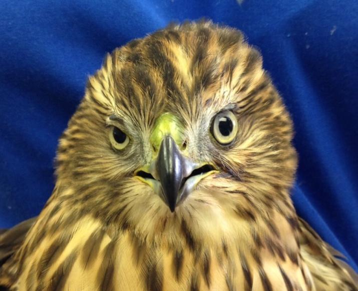
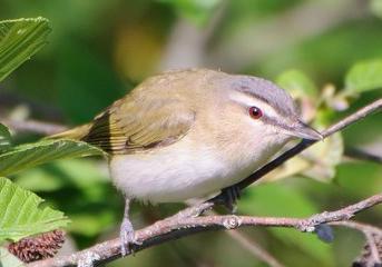
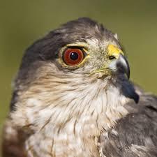
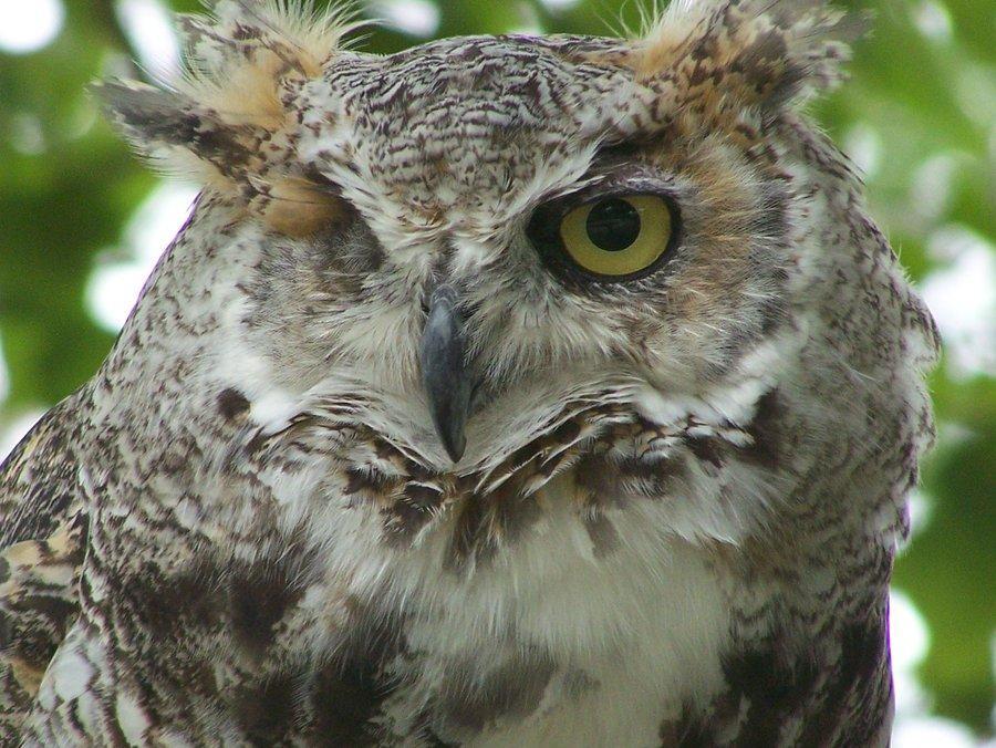
11 The Avian Eye - Differences In some species the eye color changes with age Red Eyed Vireo Red Tailed Hawk Great Horned Owl


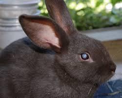
12 Mammal Eyes Tapetum lucidum Many animals have a special layer at the back of their eye that helps reflect light Improves night vision, helping them see better in the dark Some animals do not have a tapetum such as squirrels, birds, rabbits and primates
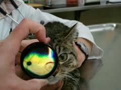
13 Injuries to the cornea are common Can range from scratches and abrasions to punctures into the eye Can determine the depth of the injury by using fluorescein stain The outer layer (epithelium) repels the stain The middle layer (stroma) absorbs it, appearing green Cornea - Structure


14 Cornea - Structure Flourescein stain Corneal ulcer detected

15 Eye Exam and Vision Assessment Observe animal Physical exam; include area around eyes Assess reflexes; PLR, palpebral reflexes, menace response Direct ophthalmoscope to assess structures in the front and back of the eyes

16 Eye Exam and Vision Assessment Pupillary Light Reflex - PLR A reflex that controls the size of the pupil Allows animal to adapt to different levels of light Pupil gets smaller when bright light shines on the retina In mammals there is a consensual PLR opposite eye also constricts

17 Eye Exam and Vision Assessment Pupillary Light Reflex - PLR Eyes very sensitive to light one pupil can be different size than other Birds have some control over their pupil size In birds there is no consensual PLR PLR assesses the function of the retina and the brain.


18 Eye Exam and Vision Assessment Measure tear production Measure intraocular pressure
19 Eye Exam and Vision Assessment Take sample for culture and sensitivity if needed Then stain eyes with fluorescein stain Flush with sterile saline before assessing Use cobalt blue light to better appreciate any stain uptake


20 Eye Exam and Vision Assessment Slit lamp biomicroscopy Indirect ophthalmoscopy
21 Eye Exam and Vision Assessment Assessment or Test Physical Exam, examine area around the eyes Pupillary Light Reflex Other reflexes; e.g. palpepral reflexes, menace Culture or cytology (if required) Schwimmwer Tear Test Intraocular Pressure Fluorescein stain Direct ophthalmoscopy Indirect ophthalmoscopy Slit lamp biomicroscopy X-ray skull and orbital area Ultrasound eyes Electroretinography Vision check e.g. obstacle course, live prey testing Can you do it? Yes Yes Yes Yes Maybe No Yes Yes Maybe No Maybe No No Yes
22 Eye Exam and Vision Assessment Your exam should consist of the below tests as a minimum Can make an exam sheet to use during your eye exam Complicated cases will need to be referred to your veterinarian for further testing Assessment or Test Physical Exam, examine area around the eyes Pupillary Light Reflex Other reflexes; e.g. palpepral reflexes, menace Fluorescein stain Direct ophthalmoscopy Culture or cytology (if required) Vision check e.g. obstacle course, live prey testing Can you do it? Yes Yes Yes Yes Yes Yes Yes
23 Problems with the Eye The painful eye squinting, swollen, tearing The white or cloudy eye whole eye, partial, front of eye, back of eye The red eye entire eye, around eye, part of eye, front of eye, back of eye Other conditions; trauma around the eye, the missing eye, the blind eye

24 The Painful Eye
25 The Painful Eye Quick Differentials: Injury to cornea ulcer Injury to structures around the eye Increased pressure within the eye glaucoma Severe uveitis inflammation within the eye

26 The Painful Eye Corneal Ulcer Superficial erosion damage to the surface layer Partial thickness ulceration variable depth into stroma Descemetocoele deep ulcer, to the level of Descemet s membrane Perforation right through all layers
27 The Painful Eye Corneal Ulcer Three questions to answer: Timeline for healing: 1) Why is there an ulcer? 2) How deep is the ulcer? 3) Is it healing as expected? Uncomplicated superficial ulcers should heal within 3-5 days with treatment Deeper ulcers should stop taking up stain within 5-7 days although defect may be visible for longer

28 The Painful Eye Corneal Ulcer Treatment Corneal ulcers are very painful = pain medication e.g. oral meloxicam There is a risk that it will become deeper and rupture = prevent infection with topical antibiotics Drops vs. ointment

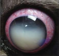
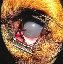
29 The White or Cloudy Eye
30 The White or Cloudy Eye Quick Differentials Corneal edema from injury to cornea or uveitis Scar tissue on cornea from injury to cornea Hypopyon = pus in the front of the eye Cataracts lenses are not clear Uveitis inflammation in the front of the eye

31 The White or Cloudy Eye Corneal edema Cornea becomes leaky and water enters Causes include corneal ulcers, trauma, uveitis, glaucoma Treat the underlying cause!


32 The White or Cloudy Eye Cataracts Four main causes: Congenital Trauma Geriatric Disease


33 The White or Cloudy Eye - Cataracts Treatment is surgery to remove the lens In wildlife it is very difficult to replace the lens so vision is compromised Solution for animals that don t rely on perfect vision

34 The White or Cloudy Eye - Cataracts Exceptions: Very young darkeyed owls have naturally bluish lenses that become clear as they age Dehydrated baby squirrels may have cloudy lenses that resolve once rehydrated

35 The White or Cloudy Eye - Uveitis Inflammation of the front of the eye Symptoms: pain, squinting, redness, aqueous flare, small pupil size, changes to iris color
36 The White or Cloudy Eye - Uveitis Causes Infection viral, bacterial, fungal Blunt trauma can lead to leaky vessels Other cancer, problem with the immune system, unknown cause, breed related (dogs) Principle of therapy Treat the underlying cause e.g. topical antibiotics Treat the inflammation topical or oral steroids Treat the pain topical or oral pain medication
37 The White or Cloudy Eye - Uveitis Use steroids with caution! ALWAYS use fluorescein stain to make sure there is no corneal ulcer present Steroids interfere with ulcer healing so DON T USE if an ulcer is present Can use topical NSAIDs instead Many antibiotic & steroid combinations available
 with atropine, tropicamide or phenylephrine drops.")
38 The White or Cloudy Eye - Uveitis Open up the pupil Pupil is often very small because the iris muscle is in spasm Painful and interferes with normal fluid movement in the eye affecting the eye pressure Dilate the pupil (in mammals) with atropine, tropicamide or phenylephrine drops. More drastic solution is a subconjuctival injection of these agents (by veterinarian)

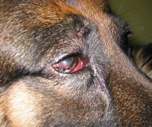
39 The Red Eye
40 The Red Eye Quick Differentials Hyphema = blood in the front of the eye Retinal hemorrhage blood in the back of the eye Conjunctivitis inflamed conjunctiva around eye Glaucoma / uveitis inflamed tissue in and around the eye


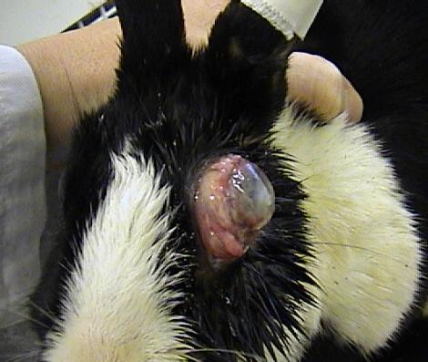
41 The Red Eye Hyphema Blood in the Front Eye Blood in the anterior chamber Most common cause is trauma May be difficult to see all structures in eye May need to wait for clot to form and resolve Ultrasound is an option

42 The Red Eye Retinal Hemorrhage Blood in the Back Eye Blood in back of eye in the posterior chamber Causes include trauma, clotting disorders, infection, tumor Often see bleeding around pecten in birds after trauma Danger of retinal detachment
43 The Red Eye Blood in the Eye - Treatment Topical steroids if corneal ulcer not present (stain eye!) Topical NSAIDs if ulcer is present Topical atropine (mammals) if uveitis present and normal eye pressure Poor prognosis if bleeding continues or recurs, in cases of glaucoma, or if retina detaches


44 The Red Eye Conjunctivis Conjunctiva is the tissue that lines the inside of the eyelids and the white part of the eye Conjunctivitis is an inflammation of these tissues Swelling is also present Irritating but not painful

45 The Red Eye Conjunctivis - Bacterial Purulent discharge from eyes, can crust shut if extreme Treatment: topical antibiotic NO steroids can make infection worse Culture if not responding to treatment

46 The Red Eye Finch Conjunctivis Common infectious disease of finches Caused by Mycoplasma gallisepticum Primarily a respiratory problem Treatment is topical antibiotics - ciprofloxacin and oral tylosin in the drinking water Concern about carriers

47 The Red Eye Conjunctivis Distemper Virus Infectious viral disease of canids, mustelids, skunks and raccoons Caused by Canine Distemper Virus No treatment once infected Prevent with vaccination Poor prognosis
48 The Red Eye - Glaucoma Increased intra-ocular pressure - EMERGENCY Symptoms extremely painful condition, red eye, blindness Causes secondary to intra-ocular inflammation, tumor, inherited disease (dogs) Affects the flow of fluid within the eye.


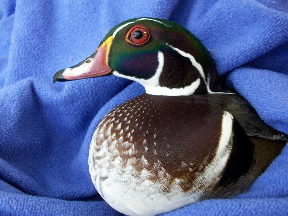
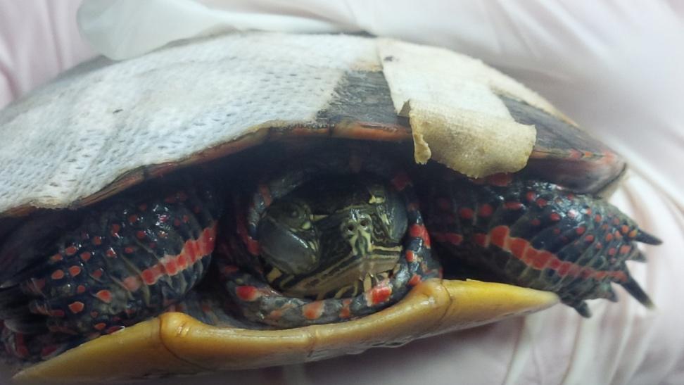
49 The Red Eye - Normal!


50 Other Conditions Pox Infections Viral infection that causes nodular lesions If nodules are close to the eye can cause irritation to the eye including corneal ulcers In extreme cases lose vision in eye Can recover with supportive care


51 Other Conditions Wounds Wounds to structures around eyes lids, conjunctiva Concern if the wound affects the function of the eye - e.g. can no longer close eye Surgery has the best outcome

52 Other Conditions Mange Parasitic infection with Sarcoptes mange mites Skin becomes thickened and crusty Skin around the eye is often affected Can cause corneal irritation or ulceration Treat for secondary bacterial infection


53 Other Conditions Species specific Overweight Virginia opossums have fat deposits that build up around their eyes so they look crosseyed Aquatic animals, like beavers, muskrats, and water birds can develop crusty eyes when kept out of water. Owls that are blind in one eye have been noted to have tuft down on that side
54 Other Conditions Missing Eye Irreparably Damaged Eye

55 Conditions you can treat Treatment by the Wildlife Rehabilitator Simple corneal ulcers Hyphema from trauma Retinal hemorrhage from trauma Trauma around eye that doesn t affect lids, glands, etc. Conjunctivitis


56 Conditions for your Veterinarian Deep corneal ulcers Non-healing ulcers Complicated eyes Uveitis Suspected glaucoma Lacerations & punctures Hypopyon Anything you aren t sure about!
57 Equipment and Medications to have: Bright pen light Direct ophthalmoscope Fluorescein strips or drops + saline flush Culture swabs Exam sheet +/- Tonopen +/- STT strips Topical antibiotics Topical steroids Topical NSAIDs Topical antibiotic + steroid combinations Atropine Topical anesthetic Oral pain medication Oral antibiotics

58 Assess Visual Function Evaluate behavior from a distance Food intake can it find food? Can it hunt? Movement in enclosure - reluctant to climb, jump, swim, fly? Head posture is there one eye used more than the other? Startle reaction is there an exaggerated response?
 631-0662 x 3206")
59 Dr. Heather Reid Toronto Wildlife Centre 60 Carl Hall Road, Unit 4 Toronto, ON M3K 2C1 (416) x 3206 Veterinary@torontowildlifecentre.com
Around The Globe in 60 Minutes
 Around The Globe in 60 Minutes Around the GLOBE in Sixty Minutes Basic Ocular Anatomy, Examination, and Diagnostic Techniques Introduction Focusing on canine and feline ocular anatomy and basic examination
Eye Examination Techniques in Horses
 Eye Examination Techniques in Horses Dennis E. Brooks DVM, PhD Dip ACVO University of Florida brooksd@mail.vetmed.ufl.edu Basic Instruments How to tell the potential of vision? PLRs (retina, CN 2, chiasm,
CORNEAL CONDITIONS CORNEAL TRANSPLANTATION
 GENERAL INFORMATION CORNEAL CONDITIONS CORNEAL TRANSPLANTATION WHAT ARE CORNEAL CONDITIONS? The cornea is the clear outer layer of the eye. Shaped like a dome, it helps to protect the eye from foreign
Acute Eyes for ED. Enis Kocak. The Alfred Ophthalmology
 Acute Eyes for ED Enis Kocak The Alfred Ophthalmology The problem with eyes Things to cover Ocular anatomy Basic assessment Common presentations Eye first aid and procedures Ophthalmic emergencies What
Ocular and Periocular Trauma. Tina Rutar, MD. Assistant Professor of Ophthalmology and Pediatrics. Director, Visual Center for the Child
 Ocular and Periocular Trauma Tina Rutar, MD Assistant Professor of Ophthalmology and Pediatrics Director, Visual Center for the Child University of California, San Francisco Phone: 415-353-2560 Fax: 415-353-2468
Ocular and periocular trauma
 Ocular and periocular trauma No financial disclosures. Tina Rutar M.D. Assistant Professor of Clinical Ophthalmology and Pediatrics Director, Visual Center for the Child University of California San Francisco
Scrub In. What is the function of vitreous humor? What does the pupil do when exposed to bright light? a. Maintain eye shape and provide color vision
 Scrub In What is the function of vitreous humor? a. Maintain eye shape and provide color vision b. Maintain eye shape and refract light rays c. Provide night vision and color vision d. Provide night vision
Ocular Urgencies and Emergencies
 Ocular Urgencies and Emergencies Pam Boyce, O.D., F.A.A.O. Boyce Family Eye Care, Ltd. 528 Devon Ave. Park Ridge, IL 60068 847-518-0303 Somebody s going to lose an eye Epidemiology 2.4 million ocular and
EYE TRAUMA: INCIDENCE
 Introduction EYE TRAUMA: INCIDENCE 2.5 million eye injuries per year in U.S. 40,000 60,000 of eye injuries lead to visual loss Introduction Final visual outcome of many ocular emergencies depends on prompt,
Corneal Ulceration. Client Information Sheet Copyright Bilton Veterinary Centre All rights Reserved. What is the cornea?
 What is the cornea? Corneal Ulceration The cornea is the central clear part of the eye that is surrounded by the white of the eye called the Sclera. Looking through the cornea, you can see the coloured
Anterior Uveitis in Dogs
 Customer Name, Street Address, City, State, Zip code Phone number, Alt. phone number, Fax number, e-mail address, web site Anterior Uveitis in Dogs (Inflammation of the Front Part of the Eye, Including
EYE INJURIES OBJECTIVES COMMON EYE EMERGENCIES 7/19/2017 IMPROVE ASSESSMENT OF EYE INJURIES
 EYE INJURIES BRITTA ANDERSON D.O. DMC PRIMARY CARE SPORTS MEDICINE ASSOCIATE TEAM PHYSICIAN DETROIT TIGERS OBJECTIVES IMPROVE ASSESSMENT OF EYE INJURIES UNDERSTAND WHAT IS CONSIDERED AN EMERGENCY DEVELOP
Specialist Referral Service Willows Information Sheets. Cataract surgery
 Specialist Referral Service Willows Information Sheets Cataract surgery An operating microscope in use A total cataract - the normally black pupil is bluish white Cataract surgery These notes do not cover
Glaucoma. Cornea. Iris
 Glaucoma Introduction Glaucoma is a group of eye diseases that can lead to blindness if not treated. Openangle glaucoma, the most common form of glaucoma, affects about 3 million Americans. Half of those
Case Study: Fuzz April 18th
 Case Study: Fuzz April 18th 33 year old Quarter Horse Had been battling corneal ulcer for several weeks before seeing us No foreign debris found Culture and cytology were taken. Started on topical antibiotics,
Ulcerative Keratitis (Type of Inflammation of the Cornea) Basics
 Basics") Ulcerative Keratitis (Type of Inflammation of the Cornea) Basics OVERVIEW Keratitis is inflammation of the cornea; the cornea is the clear outer layer of the front of the eye The corneal epithelium is
Ulcerative Keratitis (Type of Inflammation of the Cornea) Basics OVERVIEW Keratitis is inflammation of the cornea; the cornea is the clear outer layer of the front of the eye The corneal epithelium is
OPHTHALMOLOGY REFERRAL GUIDE FOR GPS
 OPHTHALMOLOGY REFERRAL GUIDE FOR GPS A guidebook to support general practitioners in the management and referral of a range of common eye problems. Contents 3 Introduction 4 Ophthalmic Workup 6 Acute Visual
Conjunctivitis in Cats
 Customer Name, Street Address, City, State, Zip code Phone number, Alt. phone number, Fax number, e-mail address, web site Conjunctivitis in Cats (Inflammation of the Moist Tissues of the Eye) Basics OVERVIEW
Paediatric acute ophthalmology. Harry Bradshaw
 Paediatric acute ophthalmology Harry Bradshaw Approach Red eye Leukocoria Neurological Trauma Visual loss Red eye Orbital Eyelid Conjunctiva Cornea Uvea Orbital Orbit fixed volume Contiguous with sinuses,
Test Bank for Medical Surgical Nursing An Integrated Approach 3rd Edition by White
 Test Bank for Medical Surgical Nursing An Integrated Approach 3rd Edition by White Link full download : http://testbankair.com/download/test-bank-for-medical-surgical-nursing-anintegrated-approach-3rd-edition-by-white/
Vision I. Steven McLoon Department of Neuroscience University of Minnesota
 Vision I Steven McLoon Department of Neuroscience University of Minnesota 1 Eye Cornea Sclera Conjunctiva 2 Eye The conjunctiva lines the inner surface of the eyelids and outer surface of the sclera. 3
UC SF. g h. Eye Trauma. Martha Neighbor, MD Emergency Services San Francisco General Hospital University of California
 UC SF Eye Trauma sf g h Martha Neighbor, MD Emergency Services San Francisco General Hospital University of California Goals Recognize vision threatening eye emergencies Treat them when we can Know when
THE RED EYE Cynthia McNamara, MD Week 25
 THE RED EYE Cynthia McNamara, MD Week 25 Educational Objectives: 1. Know the differential diagnosis and presentation of specific etiologies of the red eye 2. Be able to evaluate patients presenting with
Dry Eye Assessment and Management Study ELIGIBILITY OCULAR EVALUATION FORM
 Page 1 of 13 BEFORE COMPLETING THE OCULAR EXAMINATION, YOU MUST BE ABLE TO ANSWER YES TO THE FOLLOWING QUESTIONS: Have you done MMP9? (SVonly) The Following are done at Baseline: Have you done Tear Osmolarity?
Conjunctivitis in dogs
 Conjunctivitis in dogs Overview Conjunctivitis means inflammation (swelling) of the conjunctiva and can be caused by many different conditions. The conjunctiva is a very thin layer of tissue that covers
Focus on Ophthalmology Inside the Eye of the Horse
 www.ivis.org Proceedings of the American Association of Equine Practitioners - Focus Meeting Focus on Ophthalmology Inside the Eye of the Horse Raleigh, NC, USA 2012 Next Focus Meetings: August 4-6, 2013
UNDERSTAND MORE ABOUT UVEITIS UVEITIS
 UNDERSTAND MORE ABOUT UVEITIS UVEITIS Uveitis What is uveitis? Uveitis is inflammation of the uvea, the middle layer of your eye. The eye is shaped much like a tennis ball, with three different layers
Table of Contents 1 Orbit 3 2 Eyelids 7
 Table of Contents Preface, x List of abbreviations xi Glossary xii Section I Atlas 1 1 Orbit 3 Clinical signs associated with orbital neoplasia 3 Clinical signs associated with orbital cellulitis 3 Enophthalmos
Uveitis. Pt Info Brochure. Q: What is Uvea?
 Pt Info Brochure Uveitis Q: What is Uvea? A: Uvea is the middle layer of the eye. It is the most vascular structure of the eye. It provides nutrition to the other parts of the eye. The uvea is made up
 measure of your overall performance. An isolated glucose test is helpful to let you know what your sugar level is at one moment, but it doesn t tell you whether or not your diabetes is under adequate control
OOGZIEKTEN VOOR DE HUISARTS F. GOES, JR.
 OOGZIEKTEN VOOR DE HUISARTS F. GOES, JR. HET RODE OOG F. GOES, JR. Condition Signs Symptoms Causes Conjunctivitis Viral Normal vision, normal pupil size Mild to no pain, diffuse Adenovirus (most common),
Differential diagnosis of the red eye. Carol Slight Nurse Practitioner Ophthalmology
 Differential diagnosis of the red eye Carol Slight Nurse Practitioner Ophthalmology The red eye Conjunctivitis HSV Keratitis Acute angle closure glaucoma Anterior Uveitis Red eye Scleritis Subconjunctival
Some of the ophthalmic surgeries
 Some of the ophthalmic surgeries Some of the ophthalmic surgeries performed at the DMV Center. This document presents some types of the surgeries performed by the ophthalmology service at the DMV veterinary
Ophthalmic Trauma Update
 Ophthalmic Trauma Update Richard S. Davidson, M.D. Professor of Ophthalmology Vice Chair for Quality and Clinical Affairs UCHealth Eye Center University of Colorado School of Medicine August 5, 2017 Financial
OUR EYES & HOW WE SEE
 OUR EYES & HOW WE SEE UNDERSTAND MORE ABOUT OUR EYES & HOW WE SEE Our Eyes & How We See The eye is our visual gateway to the world. Within it, an array of delicate components labour away to give us the
Injury. Contusion Lamellar Laceration Laceration Rupture. Penetrating IOFB. Perforating
 Mechanical Ocular Trauma Došková Hana, MD. Department of Ophthalmology Medicine Faculty of Masaryk University Brno General Considerations Ocular trauma constitude about 6% of all injuries, but eyes set
MRI masterfile Part 5 WM Heme Strokes.ppt 1
 Ocular and Orbital Trauma Eye Trauma: Incidence 1.3 million eye injuries in the US per year. 40,000 of these injuries lead to blindness in the US. Patrick Sibony, MD March 23, 2013 Ophthalmic Emergencies
UVEITIS IN GENERAL. Information for patients UVEITIS CLINIC WHAT IS UVEITIS? MAIN CATEGORIES OF UVEITIS
 Information for patients UVEITIS CLINIC UVEITIS IN GENERAL WHAT IS UVEITIS? The uvea is a name given to the pigmented layer of tissue inside the eye. When all or part of the uvea becomes inflamed, the
Assessment and Management of Ocular Trauma. Disclosure I have no direct financial interests in today s subject matter. 3/25/2019. Normal Eye Anatomy
 Assessment and Management of Ocular Trauma Samiksha Fouzdar Jain, MD,FRCS Department of Ophthalmology & Visual Sciences Truhlsen Eye Institute Disclosure I have no direct financial interests in today s
Proceedings of the World Small Animal Veterinary Association Sydney, Australia 2007
 Proceedings of the World Small Animal Sydney, Australia 2007 Hosted by: Next WSAVA Congress MANAGEMENT OF CORNEAL ULCERS IN SMALL ANIMALS Robin G Stanley, BVSc(Hons), FACVSc-Ophthalmology Animal Eye Care
FACING YOUR FUNDIC FEARS: EXAMINATION OF THE OCULAR FUNDUS J. Seth Eaton, VMD, DACVO Cornell University Veterinary Specialists
 FACING YOUR FUNDIC FEARS: EXAMINATION OF THE OCULAR FUNDUS J. Seth Eaton, VMD, DACVO Cornell University Veterinary Specialists The goal of a thorough fundus examination is to clinically evaluate the structures
Conjunctivitis in Dogs
 Customer Name, Street Address, City, State, Zip code Phone number, Alt. phone number, Fax number, e-mail address, web site Conjunctivitis in Dogs (Inflammation of the Moist Tissues of the Eye) Basics OVERVIEW
2/5/2018. Trauma. Subdivided into two main categories: Closed globe Open Globe
 1 2 3 4 5 Ocular Trauma Guide for Eye Care Office Staff Winter Thaw 2018 Aaron Yatskevich OD Definition A broad term used to describe a physical or chemical wound to the eye or eye socket. Ocular trauma
Flashers and Floaters
 Flashers and Floaters Introduction Sometimes people see small, moving spots or specks in their field of vision. These sensations are called floaters. About 7 out of 10 people experience floaters at some
INSIGHT INTO RABBIT EYE DISEASES
 Vet Times The website for the veterinary profession https://www.vettimes.co.uk INSIGHT INTO RABBIT EYE DISEASES Author : ELISABETTA MANCINELLI Categories : Vets Date : August 12, 2013 ELISABETTA MANCINELLI
By Darlene Jones, Nurse. May 2017
 By Darlene Jones, Nurse May 2017 Disclosure of potential conflict of interest Darlene Jones, Nurse I have no conflict of interest Course objectives Become familiar with the different pathologies in ophthalmology
Alert signs of ocular diseases in pets
 Alert signs of ocular diseases in pets Some ocular diseases can progress very quickly and lead to the loss of the vision and/or the loss of the eye. This document reviews the signs that should be an alert
Retinal Tear and Detachment
 Retinal Tear and Detachment Introduction The retina is the layer of tissue in the back of the eye that is responsible for vision. It is attached to the choroid tissue, which supplies the retina with blood.
02/03/2014. Average Length: 23mm (Infant ~16mm) Approximately the size of a quarter Volume: ~5mL
 Approximately the size of a quarter Volume: ~5mL") Identify the anatomy of the eye. Explain the basic physiology of the parts of the eye. Briefly discuss various surgeries related to different parts of the anatomy. Average Length: 23mm (Infant ~16mm) Approximately
Identify the anatomy of the eye. Explain the basic physiology of the parts of the eye. Briefly discuss various surgeries related to different parts of the anatomy. Average Length: 23mm (Infant ~16mm) Approximately
Clinical Practice Guide for the Diagnosis, Treatment and Management of Anterior Eye Conditions. April 2018
 Clinical Practice Guide for the Diagnosis, Treatment and Management of Anterior Eye Conditions This Clinical Practice Guide provides evidence-based information about current best practice in the management
Advanced Examination of the Retina: Scleral Indentation & Retinal 3-Mirror
 Advanced Examination of the Retina: Scleral Indentation & Retinal 3-Mirror Meredith Whiteside, OD, FAAO Nimesh Patel, OD, FAAO John Shan, OD, FAAO Please silence all mobile devices. Unauthorized recording
PAINFUL PAINLESS Contact lens user BOV
 Common Causes Allergies Infections Ocular Cornea, uveitis, endophthalmitis Orbital Orbital cellulitis Inflammation Uveitis Scleritis / episcleritis Glaucomas Trauma Foreign bodies Chemical injuries History
Examining Children s Eyes
 Paediatric Ophthalmology What to refer & when? Aims Tips for assessing a child s eyes in general practice Common paediatric ophthalmology symptoms and signs What needs to be referred and when? MISS FARIHA
Eye conditions in Samoyeds
 Eye conditions in Samoyeds Information for breeders and pet owners Melbourne EyeVet Mulgrave Essendon Bundoora Frankston Geelong Bendigo Wodonga Traralgon Darwin Extra eyelashes/distichiasis Distichia
Ocular warning signs in GP practice: Paediatric Eye Pointers
 Ocular warning signs in GP practice: Paediatric Eye Pointers Dr Benjamin Chang MB, BCh, BAO, MMedSci, FRCS(Irel), FRCS(Edin), FRCOphth(Lond) Senior Consultant Ophthalmology and Visual Sciences Khoo Teck
What are some common conditions that affect the cornea?
 What are some common conditions that affect the cornea? Injuries After minor injuries or scratches, the cornea usually heals on its own. Deeper injuries can cause corneal scarring, resulting in a haze
Frequently Asked Questions about General Ophthalmology:
 1. Normal Eye Structure The eye is a slightly asymmetrical globe, about an inch in diameter. The parts of the eye include: Cornea (a clear dome over the iris), Iris (the pigmented part); Pupil (the black
Assisting in Ophthalmology. Copyright 2011, 2007, 2003, 1999 by Saunders, an imprint of Elsevier Inc. All rights reserved.
 Assisting in Ophthalmology Learning Objectives Define, spell, and pronounce the terms listed in the vocabulary. Apply critical thinking skills in performing patient assessment and care. Explain the differences
The Human Eye. Cornea Iris. Pupil. Lens. Retina
 The Retina Thin layer of light-sensitive tissue at the back of the eye (the film of the camera). Light rays are focused on the retina then transmitted to the brain. The macula is the very small area in
CONSENT FOR CATARACT SURGERY REQUEST FOR SURGICAL OPERATION / PROCEDURE AND ANAESTHETIC
 CONSENT FOR CATARACT SURGERY REQUEST FOR SURGICAL OPERATION / PROCEDURE AND ANAESTHETIC Your doctor has indicated that the condition of your eye appears stable and your cataract surgery and/or implantation
LECTURE # 7 EYECARE REVIEW: PART III
 LECTURE # 7 EYECARE REVIEW: PART III HOW TO TRIAGE EYE EMERGENCIES STEVE BUTZON, O.D. EYECARE REVIEW: HOW TO TRIAGE EYE EMERGENCIES FOR PRIMARY CARE PHYSICIANS Steve Butzon, O.D. Member Director IDOC President
Ocular Emergencies. What is an emergency to the patient is not necessarily an emergency to the staff
 OCULAR EMERGENCIES Ophthalmic Photographers Society November 15, 2013 Michael A. DellaVecchia MD PhD FACS Wills Eye Emergency Department Philadelphia PA Ocular Emergencies What is an emergency to the patient
Glaucoma. Glaucoma. Glaucoma. Trevor Arnold, MS, DVM, DACVO
 Glaucoma Trevor Arnold, MS, DVM, DACVO Glaucoma Physiology of Aqueous Humor Produced in the ciliary body Flows out the iridocorneal angle and ciliary cleft High intraocular pressures are caused by a decreased
NEPTUNE RED BANK BRICK
 NEPTUNE RED BANK BRICK Diabetes & The Eye Diabetics are more likely to develop Cataracts at a younger age. Diabetics are twice as likely to develop Glaucoma when compared to non-diabetics. The primary
DO YOU SEE WHAT I SEE? OPHTHALMIC EXAM BASICS
 DO YOU SEE WHAT I SEE? OPHTHALMIC EXAM BASICS Shelby Reinstein, DVM, MS, DACVO Veterinary Specialty and Emergency Center 301 Veterans Highway Levittown, PA 19057 Ophthalmic History Obtaining a thorough
PENETRATING EYE INJUIRES
 PENETRATING EYE INJUIRES King Harold receives a mortal penetrating injury to the eye at the Battle of Hastings 1066, Detail Bayeux Tapestry, Eleventh century. Then Earl William came from Normandy into
THE 35 GOLDEN EYE RULES
 THE 35 GOLDEN EYE RULES The Sense of Sight, from La Dame a la Licorne, The Lady and the Unicorn Tapestries, Late 15th Century Flemish Tapestry in wool and silk, Musée Nationale du Moyen Age, Paris. 1.
For further reading we recommend the following excellent textbooks:
 FURTHER READING Intravitreal Injections Downloaded from www.worldscientific.com For further reading we recommend the following excellent textbooks: Clinical Anatomy of the Eye by Richard S Snell and Michael
Focusing on A&E. By Sandy Cooper, (Ophthalmic Nurse Practitioner), Tel
, Tel") Focusing on A&E By Sandy Cooper, (Ophthalmic Nurse Practitioner), Tel 01752 439331 Email sandra.cooper5@nhs.net sandracooper041@btinternet.com THINGS TO WORRY ABOUT WITH ANY EYE PROBLEM CHANGES IN VISION
Focusing on A&E By Sandy Cooper, (Ophthalmic Nurse Practitioner), Tel 01752 439331 Email sandra.cooper5@nhs.net sandracooper041@btinternet.com THINGS TO WORRY ABOUT WITH ANY EYE PROBLEM CHANGES IN VISION
Screening for Uveitis in Children
 Information for patients and parents Manchester Royal Eye Hospital Paediatric Uveitis Service Screening for Uveitis in Children What is uveitis? Uveitis is inflammation of a layer of the eye, called the
FROM CATARACTS TO CLARITY
 Cathy Cataracts FROM CATARACTS TO CLARITY If you re 55 or older, you may have cataracts and not even know it. What You Need to Know Seeing Beyond the Symptoms Cataracts are one of the leading causes of
Ocular Anatomy for the Paraoptometric
 Ocular Anatomy for the Paraoptometric Minnesota Optometric Association Paraoptometric CE Friday September 30, 2016 Lindsay A. Sicks, OD, FAAO Assistant Professor, Illinois College of Optometry lsicks@ico.edu
Cataract. What is a Cataract?
 Cataract What is a Cataract? We all have a lens in our eye. This is positioned just behind the iris, which is the coloured ring in the eye that gives your eye its colour. The lens function is to focus
OPHTHALMOLOGIC PEARLS FOR THE NON- OPHTHALMOLOGIST. David G. Gross D.O. Deen-Gross Eye Centers Merrillville-Hobart Deengrosseye.
 OPHTHALMOLOGIC PEARLS FOR THE NON- OPHTHALMOLOGIST David G. Gross D.O. Deen-Gross Eye Centers Merrillville-Hobart Deengrosseye.com A FEW OF THE AREAS WE WILL DISCUSS Red Eye Glaucoma Neuro ophthalmic tid
Glaucoma What You Should Know
 Glaucoma What You Should Know U.S. DEPARTMENT OF HEALTH AND HUMAN SERVICES National Institutes of Health National Eye Institute The National Eye Institute (NEI) conducts and supports research that leads
Uveitis. What is Uveitis?
 Uveitis What is Uveitis? Uveitis [u-vee-i-tis] is a term for inflammation of the eye. It can occur in one eye or both eyes and affects the layer of the eye called the uvea [u-vee-uh]. It also can be associated
Identify the choice that best completes the statement or answers the question.
 Chapter 5. The Eye Multiple Choice Identify the choice that best completes the statement or answers the question. 1. The most common type of eye disorder is: A. Refractive errors B. Macular conditions
Specialist Referral Service Willows Information Sheets. Recurrent corneal erosions (indolent ulcers)
") Specialist Referral Service Willows Information Sheets Recurrent corneal erosions (indolent ulcers) A rabbit s cornea undergoing debridement under topical anaesthesia Recurrent corneal erosions (indolent
Specialist Referral Service Willows Information Sheets Recurrent corneal erosions (indolent ulcers) A rabbit s cornea undergoing debridement under topical anaesthesia Recurrent corneal erosions (indolent
Cataract. What is a Cataract?
 Cataract What is a Cataract? We all have a lens in our eye. This is positioned just behind the iris, which is the coloured ring in the eye that gives your eye its colour. The lens s function is to focus
10 EYE EMERGENCIES. Who goes, who you better not send! Brant Slomovic, MD, FRCPC University Health Network
 10 EYE EMERGENCIES Who goes, who you better not send! Brant Slomovic, MD, FRCPC University Health Network DISCLOSURES I have none PVD CASE 1 WHAT IS A PVD? a process of aging (45-55) liquefaction of vitreous
Visian ICL (Implantable Collamer Lens) For Nearsightedness. Facts You Need To Know About STAAR Surgical s Visian ICL SURGERY
 For Nearsightedness. Facts You Need To Know About STAAR Surgical s Visian ICL SURGERY") Visian ICL (Implantable Collamer Lens) For Nearsightedness Facts You Need To Know About STAAR Surgical s Visian ICL SURGERY PATIENT INFORMATION BOOKLET For Nearsightedness (Myopia) between 3 to 20 Diopters
Visian ICL (Implantable Collamer Lens) For Nearsightedness Facts You Need To Know About STAAR Surgical s Visian ICL SURGERY PATIENT INFORMATION BOOKLET For Nearsightedness (Myopia) between 3 to 20 Diopters
A LITTLE ANATOMY. three layers of eye: 1. outer: corneosclera. 2. middle - uvea. anterior - iris,ciliary body. posterior - choroid
 GLAUCOMA A LITTLE ANATOMY three layers of eye: 1. outer: corneosclera 2. middle - uvea anterior - iris,ciliary body posterior - choroid connection at the pars plana between post and ant uvea 3. retina
RETINAL CONDITIONS RETINAL CONDITIONS
 GENERAL INFORMATION RETINAL CONDITIONS RETINAL CONDITIONS WHAT ARE RETINAL CONDITIONS? Retinal conditions affect the light-sensitive tissue at the back of eye known as the retina. They include diseases
EYE CARE PROTOCOL FOR PATIENTS IN ITU
 EYE CARE PROTOCOL FOR PATIENTS IN ITU Back to contents Developed by SUE LIGHTMAN PROFESSOR OF CLINICAL OPHTHALMOLOGY/CONSULTANT OPHTHALMOLOGIST MOORFIELDS EYE HOSPITAL Amended for UCLU ICU by Caroline
4/22/16. Eye. External Anatomy of Eye. Accessory Structures. Bio 40B Dr. Kandula
 Eye Bio 40B Dr. Kandula External Anatomy of Eye Accessory Structures l Eyebrows l Levator Palpebrae Superioris - opens eye l Eyelashes l Ciliary glands modified sweat glands l Small sebaceous glands l
DEFINITION Corneal abrasion is a defect in the corneal surface epithelium due to scraping or rubbing of the corneal epithelium.
 DEFINITION Corneal abrasion is a defect in the corneal surface epithelium due to scraping or rubbing of the corneal epithelium. IMMEDIATE CONSULTATION REQUIRED IN THE FOLLOWING SITUATIONS Dendritic pattern
Ocular Injuries in Sports. Rance McClain, D.O. Associate Dean, Clinical Sciences William Carey University FM/NMM-OMM/Sports Medicine
 Ocular Injuries in Sports Rance McClain, D.O. Associate Dean, Clinical Sciences William Carey University FM/NMM-OMM/Sports Medicine http://sudc.org/vienna/ Learning Objectives 1. Know the sport classification
Glaucoma Basics OVERVIEW GENETICS SIGNALMENT/DESCRIPTION OF PET
 Glaucoma Basics OVERVIEW Glaucoma is a disease of the eye, in which the pressure within the eye is increased (pressure within the eye is known as intraocular pressure or IOP) High intraocular pressure
Ears. Mouth. Jowls 6 Major Bones of the Face Nasal bone Two
 1 2 3 4 5 Chapter 25 Injuries to the Face, Neck, and Eyes Injuries to the Face and Neck Face and neck are to injury Relatively unprotected positions on body Some injuries are life-threatening. trauma to
Chapter 7, Section 1 Review Questions. Directions: Place the letter of the best definition next to each key term. Name PER Date
 Name PER Date Chapter 7, Section 1 Review Questions Directions: Place the letter of the best definition next to each key term. A. the middle layer of the wall of the eye B. the structure between the choroid
Goals. Glaucoma PARA PEARL TO DO. Vision Loss with Glaucoma
 Glaucoma Janet R. Fett, OD Drs. Kincaid, Fett and Tharp So Sioux City, NE eyewear21@hotmail.com Goals Understand Glaucoma Disease process Understand how your data (objective and subjective) assists in
SCHEDULING STATUS Schedule 4 PROPRIETARY NAME AND DOSAGE FORM
 Page 1 of 5 SCHEDULING STATUS Schedule 4 PROPRIETARY NAME AND DOSAGE FORM FML Liquifilm Sterile Eye Suspension COMPOSITION FML Liquifilm Sterile Eye Suspension contains: Fluorometholone 1,0 mg/ml Liquifilm
Sepideh Tara Rousta, MD FAAO Robert Wood Johnson University Hospital Saint Peter s University Hospital Wills Eye Hospital
 Sepideh Tara Rousta, MD FAAO Robert Wood Johnson University Hospital Saint Peter s University Hospital Wills Eye Hospital 14 mo old w R eye cross (parents) 9 mo old R eye crossing getting worse for past
Pediatric Ocular Sonography
 Pediatric Ocular Sonography Cicero J Torres A Silva, MD Associate Professor of Radiology 2016 SPR Pediatric Ultrasound Course Yale University School of Medicine None Disclosures Objectives of Presentation
Retinal Detachment PATIENT EDUCATION
 Retinal Detachment PATIENT EDUCATION What is Retinal Detachment (RD)? Retina is the light-sensitive layer at the back of the eye that converts light images into nerve impulses that are relayed to the brain
The Sense Organs 10/13/2016. The Human Eye. 1. Sclera 2. Choroid 3. Retina. The eye is made up of three layers:
 The human body gathers information from the outside world by using the five senses of: The Sense Organs 12.3 Sight Hearing Taste Smell Touch This information is essential in helping the body maintain homeostasis.
The Orbit. The Orbit OCULAR ANATOMY AND DISSECTION 9/25/2014. The eye is a 23 mm organ...how difficult can this be? Openings in the orbit
 The eye is a 23 mm organ...how difficult can this be? OCULAR ANATOMY AND DISSECTION JEFFREY M. GAMBLE, OD COLUMBIA EYE CONSULTANTS OPTOMETRY & UNIVERSITY OF MISSOURI DEPARTMENT OF OPHTHALMOLOGY CLINICAL
ASSESSING THE EYES. Structures. Eyelids Extraocularmuscles Eyelashes Lacrimal glands: Lacrimal ducts Cornea Conjunctiva Sclera Pupils Iris.
 ASSESSING THE EYES Structures External Eyelids Extraocularmuscles Eyelashes Lacrimal glands: Lacrimal ducts Cornea Conjunctiva Sclera Pupils Iris 1 2 Structures Internal Optic disc Physiological cup Retinal
Recurrent intraocular hemorrhage secondary to cataract wound neovascularization (Swan Syndrome)
") Recurrent intraocular hemorrhage secondary to cataract wound neovascularization (Swan Syndrome) John J. Chen MD, PhD; Young H. Kwon MD, PhD August 6, 2012 Chief complaint: Recurrent vitreous hemorrhage,
Recurrent intraocular hemorrhage secondary to cataract wound neovascularization (Swan Syndrome) John J. Chen MD, PhD; Young H. Kwon MD, PhD August 6, 2012 Chief complaint: Recurrent vitreous hemorrhage,