DISSERTATION ON A PROSPECTIVE STUDY OF ANATOMICAL VARIATION OF OSTEOMEATAL COMPLEX IN CHRONIC SINUSITIS PATIENTS
|
|
|
- Allyson Charles
- 5 years ago
- Views:
Transcription

1 DISSERTATION ON A PROSPECTIVE STUDY OF ANATOMICAL VARIATION OF OSTEOMEATAL COMPLEX IN CHRONIC SINUSITIS PATIENTS Submitted in partial fulfillment of the requirements for M.S. DEGREE BRANCH -IV OTORHINOLARYNGOLOGY of THE TAMILNADU DR. M.G.R. MEDICAL UNIVERISTY, UPGRADED INSTITUTE OF OTORHINOLARYNGOLOGY MADRAS MEDICAL COLLEGE CHENNAI MARCH 2009
2 CERTIFICATE This is to certify that this dissertation entitled A PROSPECTIVE STUDY OF ANATOMICAL VARIATION OF OSTEOMEATAL COMPLEX IN CHRONIC SINUSITIS PATIENTS submitted by Dr. R.KARTHIKEYAN, appearing for M.S. E.N.T.. Branch IV Degree examination in March 2009 is a bonafide record of work done by him under my direct guidance and supervision in partial fulfillment of regulations of the Tamil Nadu Dr. M.G.R. Medical University, Chennai. I forward this to the Tamil Nadu Dr.M.G.R. Medical University, Chennai, Tamil Nadu, India. DIRECTOR & PROFESSOR, Upgraded Institute of Otorhinolaryngology, Madras Medical College, Government General Hospital, Chennai
3 CERTIFICATE This is to certify that this dissertation entitled A PROSPECTIVE STUDY OF ANATOMICAL VARIATION OF OSTEOMEATAL COMPLEX IN CHRONIC SINUSITIS PATIENTS submitted by Dr. R.KARTHIKEYAN, appearing for M.S. E.N.T. Branch IV Degree examination in March 2009 is a bonafide record of work done by him under my direct guidance and supervision in partial fulfillment of regulations of the Tamil Nadu Dr. M.G.R. Medical University, Chennai. I forward this to the Tamil Nadu Dr.M.G.R. Medical University, Chennai, Tamil Nadu, India. The DEAN, Madras Medical College, Government General Hospital, Chennai
4 DECLARATION I solemnly declare that the dissertation entitled A PROSPECTIVE STUDY OF ANATOMICAL VARIATION OF OSTEOMEATAL COMPLEX IN CHRONIC SINUSITIS PATIENTS is done by me at the Madras Medical College and Government General Hospital, Chennai during under the guidance and supervision of Prof. S. KULASEKARAN, M.S., D.L.O. This dissertation is submitted to The Tamilnadu Dr. M.G.R Medical University, towards partial fulfillment of regulation for the award of M.S. DEGREE IN Otorhinolaryngology. (BRANCH IV). Place: Date: DR.R.KARTHIKEYAN M.S. E.N.T. post graduate, Upgraded Institute of Otorhinolaryngology, Madras Medical College, Government General Hospital, Chennai
5 ACKNOWLEDGEMENT I am immensely grateful to Prof. S. Kulasekaran, M.S. D.L.O., The Director, Upgraded Institute of Otorhinolaryngology, for his valuable guidance, suggestions, encouragement and help in conducting this study. I am greatly indebted to Prof. K. Balakumar M.S., D.L.O., Professor, Upgraded Institute of Otorhinolaryngology, who encouraged and helped me throughout this study. I express my sincere gratitude to Ex-Director and Professor Late Dr. A. K. Sukumaran M.S., D.L.O., for his valuable support in conducting the study. I would like to express my sincere gratitude to Prof.T.P.KALANITI, M.D., The DEAN, Madras Medical College, for having permitted me to use the hospital material in this study.
6 I express my sincere thanks to all the Assistant Professors, for their thoughtful guidance throughout the work. I thank the Secretary and Chairman of Institutional Ethical Committee, Government General Hospital and Madras Medical College, Chennai. I thank all the Professors, Assistant Professors and post graduates of the Department of RADIOLOGY for their valuable support. I thank all my colleagues and friends for their constant encouragement and valuable criticism. Last but not least, I express my gratitude for the generosity shown by all the patients who participated in the study. I am extremely thankful to my family members for their continuous support. Above all I thank God Almighty for His immense blessings.
7 CONTENTS S.no Contents Page no. 1. INTRODUCTION 1 2. AIMS OF THE STUDY 4 3. REVIEW OF THE LITERATURE 5 4. MATERIALS AND METHODS RESULTS AND ANALYSIS DISCUSSION SUMMARY CONCLUSION BIBLIOGRAPHY 10. PROFORMA 11. MASTER CHART 12. ETHICAL COMMITTEE CERTIFICATE
8 INTRODUCTION Sinusitis is one of the leading health care problems nowadays, increasing in both incidence and prevalence. Various articles have been published regarding aetiopathogenesis, microbiology, anatomical variations and management aspects. DEFINITION: American Academy of Otorhinolaryngology Head and Neck Surgery formulated certain working definitions. Clinically, Sinusitis is defined as the condition manifest by an inflammatory response of the mucous membrane of the nasal cavity and para nasal sinuses, fluid within the cavity, and / or underlying bone. Also defined as a group of disorders characterized by inflammation of the mucosa of the nose and para nasal sinuses lasting for atleast 12 weeks. 1 At present diagnostic nasal endoscopic evaluation of nose and para nasal sinuses is a routine component for evaluating patients with evidence of suspected nose and para nasal sinus disease. Arrival of the endoscopes has
9 helped us in many ways to recognize the lesion or changes that are hidden from the naked eye or even from inspection under microscopes. With this, provisional diagnosis may be confirmed, expanded or revised. Also it helps the Otorhinolaryngologist in deciding the mode of treatment. All the patients who have significant findings in diagnostic nasal endoscopy are subject to CT Scan Para nasal sinus evaluation. CT Scan Para nasal sinus is not routinely indicated in acute sinusitis except in the state of complication or no improvement with medical therapy. Whatever may be the diagnosis, CT Scan Para nasal sinus is mandatory before performing sinus surgery. This is to avoid inadvertent complications while performing the procedure. In patients with a history of chronic sinusitis over a period of months or years, and not responding to medical therapy, computed tomography should be performed. As a number of lateral nasal wall diseases cannot be recognized and identified by endoscope, we perform tomography even when the diagnostic nasal endoscopic finding is insignificant, provided that the history and clinical findings suggest the presence of some disease. The anatomical variations of lateral nasal wall and Para nasal sinuses are surgically and patho-physiologically important because they narrow the drainage pathway of the para nasal sinuses, which in turn lead on to
10 stagnation of secretions, then infection and inflammation of the mucosa lining the sinuses. Diseases in extensively pneumatised sinuses lead on to exposure of important structures like Optic nerve and Internal Carotid artery, to infection and inflammation, and also increases risk during surgical procedure. Hence, Endoscopic evaluation and CT Scan evaluation of nose and Para nasal sinuses is mandatory in chronic sinusitis patients, to evaluate the detailed anatomy (normal anatomy, anatomical variation and the extent of the disease process) that are commonly encountered in the osteomeatal complex and lateral nasal wall per se. This will help the endoscopic surgeon in pre-operative assessment and planning of the surgery, in complete eradication of the disease and to reduce the intra operative and post operative complications.
11 AIMS OF THE STUDY 1. To study the incidence of anatomical variations of osteomeatal complex common in chronic sinusitis patients. 2. To study the anatomical variation commonly associated with each paranasal sinus inflammation. 3. To study the importance of pre-operative computed tomography evaluation of the osteomeatal complex variation in chronic sinusitis patients.
12 REVIEW OF LITERATURE EMBRYOLOGY: Classic anatomic treatises attribute initial paranasal sinus development to lateral nasal wall ridges called ethmoturbinals. 2 A series of five to six ridges first appear during the eighth week of development; through regression and fusion, however, three to four ridges ultimately persist. The first ethmoturbinal regresses during development; its ascending portion forms the agger nasi, while its descending portion forms the uncinate process. The second ethmoturbinal ultimately forms the middle turbinate, the third ethmoturbinal forms the superior turbinate, and the fourth and fifth ethmoturbinals fuse to form the supreme turbinate. These structures are all considered to be ethmoid in their origin. An additional ridge, the maxilloturbinal, arises inferior to these structures. This ridge ultimately forms the inferior turbinate but is not considered ethmoid in its embryologic origin. The primary furrows that lie between the ethmoturbinals form the various nasal meati and recesses. The first primary furrow is located
13 between the first and second ethmoturbinals. Its descending aspect forms the ethmoidal infundibulum, hiatus semilunaris, and middle meatus, while its ascending aspect can contribute to the formation of the frontal recess. The primordial maxillary sinus develops from the inferior aspect of the ethmoidal infundibulum. 3-5 The second primary furrow forms the superior meatus and the third primary furrow forms the supreme meatus. In their development from the lateral wall, the ethmoturbinals form bony structures that traverse the ethmoid complex to attach to the lamina papyracea of the orbit and skull base. The furrows develop into the various pre-recesses and recesses and contribute to the extensive and complex pneumatization of the ethmoid bone. As development progresses, secondary evaginations and invaginations emerge from the lateral nasal wall between the maxillo- and ethmoturbinal. 6,7 The nomenclature ascribed to these structures varies among authors and includes secondary concha or accessory concha of the middle nasal meatus for the evaginations and secondary furrows or accessory meati of the middle nasal meatus for the invaginations. 6,7 Given the fact that the precise mechanism of ridge and furrow development, hence the nomenclature, is subject to some interpretation, the primordial ethmoid bulla appears to arise as a secondary lateral nasal wall evagination, and the primordial supra and retrobullar
 The anterior ostiomeatal unit is not a discrete anatomic structure but refers collectively to several middle meatal structures: the uncinate")
14 recesses (sinus lateralis) appear to arise from the secondary furrows that form above and behind the primordial ethmoid bulla. ANATOMY : Fig : 1 ANTERIOR OSTIOMEATAL COMPLEX : (Fig : 2) The anterior ostiomeatal unit is not a discrete anatomic structure but refers collectively to several middle meatal structures: the uncinate process, the ethmoid infundibulum, anterior ethmoid cells, and ostia of the anterior ethmoid, maxillary, and frontal sinuses.the ostiomeatal unit is a functional

15 rather than an anatomic designation, coined by Naumann in discussing the pathophysiology of sinusitis. 9 He emphasized that a small amount of obstruction in this critical region could lead to significant disease in the larger frontal and maxillary sinuses. Fig : 2 POSTERIOR OSTIOMEATAL COMPLEX: (Fig : 3) The relationship between the aerated portion of the sphenoid sinus and the posterior ethmoid sinus needs to be accurately perceived by the surgeon so as to avoid complications during surgery. This morphology is best displayed in the axial plane and with three-dimensional imaging. In most patients, aeration of the posterior ethmoid sinus is wider and somewhat
, the sphenoid sinus is situated more inferiorly, and the most posterosuperior air space is the posterior ethmoid sinus.")
16 higher than that of the sphenoid sinus. Usually in the paramedian sagittal plane, the sphenoid sinus is the most superior and posterior air space. More laterally (1.5 to 2 cm from the nasal septum), the sphenoid sinus is situated more inferiorly, and the most posterosuperior air space is the posterior ethmoid sinus. Fig : 3 AGGER NASI On anterior rhinoscopy, a prominence can be easily appreciated at and just anterior to the middle turbinate s insertion into the lateral nasal wall.


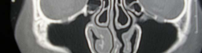
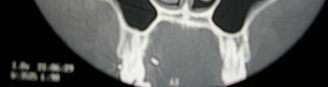
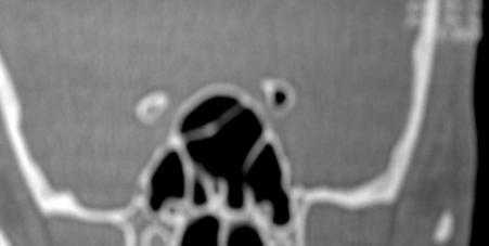
17 Fig : 4 Fig : 5 Chronic sinusitis patient - CT scan para nasal sinuses. X-Ray paranasal sinus- Chronic sinusitis patient Fig : 6 Fig : 7 Pansinusitis Purulent discharge from accessory ostium of posterior frontanel in chronic sinusitis patients
18 This region was designated as the agger nasi, taken from the Latin agger, meaning mound or eminence, and nasi, meaning nose. In many but not all cases, the agger nasi region is pneumatized by an anterior ethmoid cell, referred to as the agger nasi cell. This cell usually takes its origin from the superior aspect of the infundibulum or the frontal recess. 2,9,10 The agger nasi cell is bordered, Anteriorly by the frontal process of the maxilla, Superiorly by the frontal recess/sinus, Anterolaterally by the nasal bones, Inferomedially by the uncinate process of the ethmoid bone, and Inferolaterally by the lacrimal bone. The intimate relationship of the cell to the lacrimal bone readily explains the finding of epiphora in select patients with sinus disease. The agger nasi can also be important in frontal sinusitis and its treatment. The superior aspect of the cell serves as the anteromedial floor of the frontal sinus and a significant portion of the anterior border of the frontal recess. This is relevant for understanding the pathophysiology of frontal sinusitis and the surgical treatment of the frontal sinus. The agger nasi can pneumatize inferomedially to pneumatize the uncinate process. In a small
19 percentage of patients, the pneumatization can be significant, and bulla formation of the uncinate may occur. 11,12 The agger nasi cell is situated below the frontal sinus. It reaches the lacrimal fossa inferolaterlly and anterolaterally and is arched by the nasal bones. These cells usually border the primary floor of the frontal sinus ostium, lying anterior, lateral, and inferior to the frontal recess. In a study conducted by the author and others, 13 the agger nasi cell was found to occur in nearly all members of the population. This finding is echoed by Bolger et al., 14 who found the agger nasi cell to present in 98.5% of his study population. These percentages would suggest that the agger nasi cell is a part of normal anatomy; however, some investigators reported the incidence of agger nasi cells to be as low as 3 to 23.6%. 15 This discrepancy could be due to the varying definitions of agger nasi cells; some authors consider only large agger nasi cells extending beyond the parameters described above, while others consider any structure fitting the above description of an air chamber below the frontal sinus to be an agger nasi cell. The method of analysis used to determine the prevalence of agger nasi cells could also be a reason for varying reports. 14 When CT scanning is used, the cell is easily detected, even when small. In anatomic dissection, however, agger nasi cells
20 are more difficult to detect, and reports using this method of analysis vary widely in the reported prevalence of the variation. UNCINATE PROCESS The uncinate process is most easily appreciated by viewing a sagittal gross anatomic specimen after deflecting the middle turbinate superiorly. This ethmoid structure is nearly sagittally oriented, nearly paralleling the ethmoidal bulla. It is approximately 3 to 4 mm wide and 1.5 to 2 cm in length. Through most of its course, its posterior margin is free as it has no bony attachments. The hiatus semilunaris lies directly behind the posterior margin of the uncinate. Anteriorly and superiorly, it attaches to the ethmoidal crest of the maxillae, just inferior to the lateral attachment of the anterior aspect of the middle turbinate and agger nasi. Directly inferior to this, it fuses with the posterior aspect of the lacrimal bone. Its anterior inferior aspect does not have a bony attachment Posteriorly and inferiorly, the uncinate attaches to the ethmoidal process of the inferior turbinate bone. The attachment here is thick, and the uncinate often splits or widens in this region to fuse with the stouter inferior turbinate bone.
21 At its posterior and superior limit, the uncinate also gives off a small bony projection to attach to the lamina perpendicularis of the palatine bone. 16 The uncinate has no bony attachment anterior and posterior to its attachment to the inferior turbinate bone. Here, the lateral nasal wall is made not of bone but rather middle meatal mucosa, a small layer of intervening connective tissue, and sinus mucosa. These areas are referred to as the anterior and posterior fontanelles.. Accessory ostia are frequently encountered in the posterior fontanelle region, occurring in approximately 20 to 25% of patients. 17 Returning to its superior aspect, the uncinate projects posterior and superior to the middle turbinate attachment and most commonly bends laterally to insert on the lamina papyracea of the orbit. Inferior and lateral to this portion of the uncinate lies the superior aspect of the infundibular air space, the recessus terminalis. Superior and medial to this portion of the uncinate (most commonly) lies the floor of the frontal recess. Alternatively, the uncinate can attach centrally to the skull base or medially to the superior aspect of the vertical lamella of the middle turbinate near the turbinate s insertion to the cribriform plate. 18
22 In select cases, the uncinate is displaced medially to such an extent that it recurves on itself and has been misinterpreted as a duplication of the middle turbinate. Additionally, in a small percentage of cases, the uncinate process can be pneumatized. 19,20 An appreciation of uncinate variability is important. If lateral displacement of the uncinate with accompanying atelectasis of the infundibulum is not appreciated during infundibulotomy incision, inadvertent orbital injury can occur. 21 ETHMOID BULLA: The ethmoid bulla is one of the most constant and largest of the anterior ethmoid air cells. It is located within the middle meatus directly posterior to the uncinate process and anteriorto the basal lamella of the middle turbinate. The cell is based on the lamina papyracea and projects medially into the middle meatus. The cell has the appearance of a bulla, that is, a hollow, thin-walled, rounded, bony prominence. Superiorly, the anterior wall of the ethmoid bulla can extend to the skull base and form the posterior limit of the frontal recess. Posteriorly, the bulla can blend with the ground lamella. Anatomic variations can occur in the ethmoid bulla. When highly pneumatized, the ethmoid bulla can be one of the largest ethmoid air cells
23 and can lie in the lower aspect of the middle meatus. In select cases, a lowlying bulla can potentially narrow the ethmoidal infundibulum and impair mucociliary transport and ventilation. The ethmoid bulla is formed by pneumatization of, and behind, the second basal lamella or bulla lamella. When unpneumatized, a bony projection from the lamina papyracea results and is referred to as the torus lateralis. 22 It is estimated that this occurs in approximately 8% of subjects. HIATUS SEMILUNARIS : (Latin: hiatus, -a gap, cleft or passageway, and semilunaris, - crescent-shaped.) Indeed, the hiatus semilunaris is a crescent-shaped gap between the posterior-free margin of the uncinate processand the anterior wall of the ethmoid bulla. It is through this two-dimensional, sagittally oriented cleft or passageway that the middle meatus communicates with the ethmoid infundibulum. This cleft is further designated as the hiatus semilunaris inferior by Grunwald to distinguish it from the smaller, less defined crescent- shaped cleft, the hiatus semilunaris superior. 22 The hiatus semilunaris superior is the cleft formed between the posterior wall of the ethmoid bulla and the basal lamellae of the middle turbinate and is the
24 passageway through which the middle meatus communicates with the lateral sinus (retro and suprabullar recess). ETHMOIDAL INFUNDIBULUM: (Latin root, infundibulum, - funnel or funnel-shaped structure or passage). 24 The ethmoidal infundibulum is the funnel-shaped passage through which the secretions from various anterior ethmoid cells, the maxillary sinus, and, in some cases, the frontal sinus are transported or channeled into the middle meatus. The ethmoidal infundibulum is a three-dimensional space located in the anterior ethmoid region, bordered, medially by the mucosa-covered uncinate process, laterally by the lamina papyracea, and anteriorly and superiorly by the frontal process of the maxilla and lacrimal bone superolaterally. 23 When viewed from a sagittal perspective, the ethmoidal infundibulum is curved in shape, corresponding to the course of the uncinate process and anterior wall of the ethmoid bulla. 22 The anterior wall of the ethmoid bulla forms the posterior border of the ethmoidal infundibulum. The ethmoidal infundibulum communicates with the middle meatus through the hiatus
25 semilunaris, a two-dimensional cleft between the uncinate process and ethmoid bullae. When viewed in a coronal perspective just above the level of the maxillary ostia, the medial boundary of the infundibulum is the uncinate process, the lateral boundary is the lamina papyracea, and the maxillary ostium forms the inferior boundary. The superior boundary is formed by the anteriorethmoidal Infundibulum anterior wall of the ethmoid bulla, recognizing that the superior medial boundary is formed by the hiatus semilunaris. The superior aspect of the infundibulum is important as it is intimately related to the frontal recess. The relationship of the infundibulum and frontal recess is largely determined by the attachment of the uncinate process. Most commonly, the uncinate bends laterally to attach to the lamina papyracea and forms the superior boundary of the ethmoidal infundibulum, the recessus Terminalis. The frontal recess will drain medial to the uncinate, if the uncinate attaches to the lamina papyracea laterally.alternatively, the uncinate can attach to the ethmoid roof or insert into the middle turbinate. In these cases, the frontal recesses will be contiguous with the ethmoidal infundibulum.. Tremendous variations in the uncinate attachment and the relationship of the infundibulum and frontal recess exist. The inferior aspect
26 of the infundibulum is also important, notably for its relationship to the maxillary ostium.the natural ostium of the maxillary sinus is most commonly located in the posteroinferior one-third of the ethmoid infundibulum. The most inferoposterior portion of the infundibulum terminates as it empties into the middle meatus and blends with the posterior fontanelle mucosa. SINUS LATERALIS (SUPRABULLAR AND RETROBULLAR RECESSES) The sinus lateralis is a variable air space that lies behind and nabove the ethmoid bulla and is also referred to as the suprabullar and retrobullar recesses 22. This space, initially described by Grunwald, can be highly developed, and in such cases, it is bordered by the ethmoid roof superiorly, the lamina papyracea laterally, the ethmoid bulla roof and posterior wall inferiorly and anteriorly, and the basal lamella of the middle turbinate posteriorly. As the sinus lateralis lies anterior to the basal lamella of the middle turbinate, it is anterior ethmoid in its location. It is not, however, considered an anterior ethmoid cell as it does not have a single ostia for ventilation and drainage. Rather, it is considered a recess or space that can communicate with the middle meatus via the hiatus semilunaris superior.
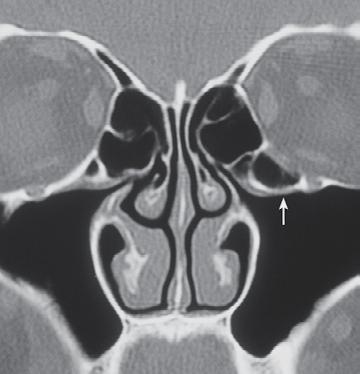
27 Stammberger points out that if the ethmoid bulla does not extend to the skull base to form the posterior wall of the frontal recess, the sinus lateralis can communicate with the frontal recess and the hiatus semilunaris inferior. 22,23 The ethmoid bulla often opens posteriorly into the sinus lateralis. PNEUMATISED SEPTUM: Not infrequently, air cells will be present within the posterior nasal septum.the ostia of these air cells are always in communication with the sphenoid sinus. These cells can also be infected by inflammatory disease and as their ostia close, they can evolve into a mucocele. One must be careful and prudent in distinguishing this pathologic entity from neoplasm or encephaloceles. MRI may prove to be indispensable in clearly defining these entities. HALLER CELLS (INFRAORBITAL RECESS CELLS): (Fig : 18) Haller cells are pneumatized ethmoid air cells that project along the medial roof of the maxillary sinus and the most inferior portion of the lamina papyracea, below the ethmoid bulla and lateral to the uncinate process. Most often, they arise from the anterior ethmoid cells and are closely relatedto the infundibulum. Less frequently, Haller cells may arise from posterior ethmoid
28 cells; in this case, however, they are less likely to compromise the infundibulum. 25 These cells contribute to the narrowing of the infundibulum and may also compromise the adjacent ostium of the maxillary sinuses. Consequently, many authors 26,27 cite Haller cells as a factor in recurrent maxillary sinusitis. A statistical analysis conducted by Bolger et al. 33 found no statistical difference between the prevalence of Haller s cells noted in patients presenting with recurrent maxillary sinusitis and those asymptomatic patients evaluated, leading these investigators to suggest evaluating the role of Haller s cells in disease on an individual basis, depending on the size, placement, evidence of inflammation, and mucosal contact evident in the cell. Wanamaker reported on a rare case 29 in which inflammation within the Haller s cell itself caused severe headache due to secondary obstruction of the infundibulum. 29 He also noted that the cells were found as part of diffuse sinus disease and identified them as predisposing factors for recurrent maxillary sinusitis. Tonai and Baba, 30 however, did not report any correlation between Haller s cells and the recurrence of disease in their population.
29 ENLARGED ETHMOID BULLA: (Fig : 14) The ethmoid bulla is the air cell directly above and posterior to the infundibulum and hiatus semilunaris. Although it is the largest ethmoidal air cell, its excessive growth and pneumatization can impair sinus ventilation and drainage. Very large ethmoid bullae may encroach upon the infundibulum and middle meatus, sometimes growing to such size as to completely fill the sinus of the middle turbinate bone. An excessively enlarged ethmoid bulla is established as a potential cause of sinus infection, especially if pus, polyps, or cysts are present; however, Lloyd 31 reports this variation as one that is difficult to assess and suggests that it may be over diagnosed. His study also found that, when compared with other variations, enlarged ethmoid bulla had a slightly lower incidence of correlation with sinus disease. UNCINATE PROCESS DEVIATION AND PNEUMATIZATION : Two main types of variation deviation(fig : 17) and pneumatization (Fig : 15) are associated with the uncinate process. The structure may deviate laterally, compromising the infundibulum, or medially, compromising the middle meatus. Any deviation of the uncinate process is recognized by most authors as a detriment to the normal mucociliary


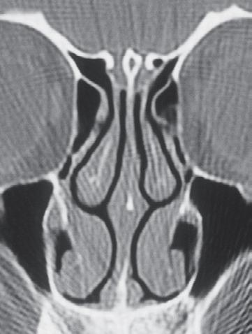
30 Fig : 8 Concha Bullosa Fig : 9 Concha Bullosa


31 Fig : 10 Agger nasai cell Fig :11 Agger nasai cell
32 drainage of the anterior ethmoid, frontal, and maxillary sinuses. The uncinate process may also be pneumatized. Bolger states that pneumatization of this structure occurs when an agger nasi cell extends into the most anterosuperior region of the uncinate process; 28 however, there is uncertainty expressed in the literature concerning the exact physiology behind this phenomenon. Pneumatization of the uncinate process is particularly likely to narrow the infundibulum, hampering sinus ventilation and drainage. PARADOXICAL MIDDLE TURBINATE: (Fig : 12, 13) Paradoxical middle turbinate is identified when the curve of the middle turbinate projects laterally, toward the nasal septum. Usually, the turbinate curves more medially, toward the lateral sinus wall. Some paradoxical middle turbinates have been found to compromise the middle meatus and compress the infundibulum; because of this tendency, most authors agree that paradoxical middle turbinates can be a contributing factor to sinusitis.


33 Fig : 12 Fig : 13 Paradoxical middle turbinate Paradoxical middle turbinate Fig : 14 Fig : 15 Prominent bulla ethmoidalis and concha bullosa. Concha bullosa and pneumatised uncinate process
34 CONCHA BULLOSA: Any pneumatization of the middle turbinate is technically referred to as a concha bullosa. However, there is great variability among subjects as to the degree of pneumatization. If the vertical portion or lamella of the middle turbinate is pneumatized, the cell that is formed is referred to as the nterlamellar cell. The term interlamellar cell distinguishes this pattern of middle turbinate pneumatization from pneumatization that includes the more inferiorly located bulbous portion of the turbinate. This is more commonly referred to as a concha bullosa. The term concha bullosa is more clearly understood by examining the Latin roots: bulla (which has already been discussed), which is present within the concha, or turbinate. A large concha bullosa is an anatomic variation that can narrow the middle meatus and reduce mucociliary clearance and ventilation. NASAL SEPTAL DEVIATION: In normal anatomy, the cartilaginous and bony portions of the nasal septum form a straight wall. A common variation, however, is the bowing of this structure, especially at the junctions of the cartilaginous portions of the septum and the bony perpendicular ethmoid plate and vomer. It is widely recognized that this bowing can compress the middle turbinate laterally and compromise the middle meatus. Swollen membranes, secondary
35 inflammation, and infection of the middle meatus have also been reported as a result of this variation. Bony spurring at the above-mentioned junctions is also commonly observed; excessive spurring may further narrow the nasal and ethmoidal air passages. ONODI CELLS: (Fig : 19) The sphenoethmoid (Onodi) cell has received much attention throughout the past century as various sinus surgeries evolved. An Onodi cell is defined as the pneumatization of the most posterior ethmoid air cell, where a bulge of the optic canal into the posterior ethmoid is apparent. When an Onodi cell exists, this pneumatization causes the optic nerve to be closer to the posterior ethmoid sinus than is usually observed. Understanding the relationship between the optic nerve, the posterior ethmoid sinus, and sphenoid sinus is critical in completing a safe operative procedure and avoiding optic nerve injury during FESS. Onodi cells are also a potential cause for incomplete sphenoidectomy. If a surgeon is operating in an Onodi cell, he or she may recognize landmarks traditionally associated with the sphenoid sinus (internal carotid artery, optic nerve) and therefore may mistakenly conclude that the sphenoid sinus has been entered and consider the operation completed, when in fact it is not. Studies performed


36 Fig : 16 Fig : 17 Intumescentia septi nasi anterior (ISNA): Medialised uncinate process Fig : 18 Fig : 19 Haller cell Onodi cell
37 to determine the prevalence of Onodi cells in the population have used a variety of analytical methods; consequently, their results vary widely. Habal et al. 34 and Maniscalco and Habal 35 found a prevalence of 25% using transorbital dissection, and Kainz and Stammberger 36 reported a 42% prevalence using endoscopic dissection. Gross anatomic and radiographic evaluation studies reported much lower prevalences, ranging anywhere from 1 to 11.6% 37,38 and 1.3 to 8% 39 respectively. Driben et al. 40 showed that coronal CT scans identified sphenoethmoid cells in 3 of 41 (7%) of their patient population. However, anatomic dissection identified Onodi cells in 16 of 41 (39%) of the subjects. Endoscopic evaluation reveals that the sphenoethmoid cell is a more common anatomic variation than previously appreciated. Awareness of this fact may help reduce the risk of optic nerve trauma during FESS. FRONTAL CELL : Frontal cells are a less common type of anterior ethmoid air cell. The recognition and subsequent definition of the appearance and etiology of frontal cells was initiated by J. Parson Schaeffer. During the course of his observations of the embryonic development of the sinuses, he discovered that it was possible, although unusual, for one cell to aerate each half of the
38 frontal bone, each with a separate communication to the frontal recess. Schaeffer coined the term frontal cell to describe this phenomenon. 42,43 Van Alyea 44,45,46 subsequently defined the frontal cell as a cell encroaching on the frontal recess or frontal sinus. He considered supraorbital ethmoid, agger nasi, and intersinus septal cells as well those limited to the frontal recess as frontal cells. Bent et al. 41 have defined frontal cells more specifically as belonging to one of four categories, detailed in Table. They also state that all frontal cells derive from the anterior ethmoid sinus behind the agger nasi cell and pneumatize the frontal recess above the agger nasi cell. Each type of frontal cell may obstruct the nasofrontal communication or the frontal sinus itself. Type I Type II Type III Single frontal recess cell above agger nasi cell Tier of cells in frontal recess above agger nasi cell Single massive cell pneumatizing cephalad into frontal sinus Type IV Single isolated cell within the frontal sinus Intumescentia septi nasi anterior (ISNA): 47 (Fig: 16) Intumescentia septi nasi anterior is a common anatomical variation that is not routinely noticed by surgeons or radiologists. ISNA is a mucosal
39 bulging located on each side of the anterior part of the septum. 47 In this study, computerized tomography (CT) scans of the paranasal sinus were obtained from 595 patients who had symptoms of chronic sinusitis. Among 595 subjects, ISNA was found in 332 (55.79 per cent) of subjects. It was found more frequently in males than females in every age group. Although ISNA is a common anatomical variation, it is generally overlooked. This is the first report on ISNA in different age groups and sexes. FRONTAL RECESS AND SINUS: The frontal sinus drains into the middle meatus and nasal cavity through a complex passage. Several authors describe a nasofrontal duct that forms the nasofrontal connection. Anatomic dissection reveals that a true duct, that is, a tubular structure conducting any fluid, does not exist. In an attempt to refine the nomenclature and more accurately characterize the anatomy, the term frontal recess has been recommended. The frontal recess is the most anterosuperior aspect of the anterior ethmoid sinus that forms the connection with the frontal sinus. 48 The boundaries of the frontal recess are the lamina papyracea laterally, the middle turbinate medially, the posterosuperior wall of the agger nasi cell (when present) anteriorly, and the anterior wall of the ethmoid bulla posteriorly. If the anterior wall of the
40 ethmoid bulla does not reach the skull base and form a complete posterior wall, the frontal recess may communicate with the suprabullar recess. 48 The frontal recess tapers as it approaches the superiorly located internal os of the frontal sinus; above the os, it again widens, as the anterior and posterior tables diverge to their respective positions. An hourglass-like appearance is evident, with the narrowest portion being the frontal ostium. There is tremendous variation with respect to the pattern of the nasofrontal connection. The anatomic complexity of this region is better understood when the effect of the surrounding ethmoid cells, such as the agger nasi cell, frontal cells, and supraorbital ethmoid cells, are considered. An intimate relationship therefore exists between the agger nasi cell and the frontal recess. Secretions from the frontal sinus destined for the nasal cavity usually follow a path through the frontal recess and over the posterior and medial surface of the agger nasi cell. If the agger nasi cell is extensively pneumatized, the frontal recess can be relatively narrowed, and hence the patient may be predisposed to frontal sinusitis. In surgery, an extensively pneumatized agger nasi can be mistaken for the frontal recess or sinus. If a large agger nasi cell is opened and mistaken for a frontal sinus, the residual superoposterior wall of the agger nasi cell can scar posteriorly to the
41 ethmoid roof, and iatrogenic stenosis or obstruction of the nasofrontal connection can occur. 50 In addition to the agger nasi cell, there are other ethmoid cells that have an intimate relationship with the frontal recess. Van Alyea reported that approximately 50% of anatomic specimens had anterior ethmoid cells that encroached into the frontal sinus, and that one-third of these encroached into the area of the frontal ostium.he termed these cells frontal cells. Schaeffer pointed out that anterior ethmoid cells could pneumatize sufficiently into the frontal sinus to give the appearance of duplication of the sinus. Stammberger points out that from the frontal recess, anterior ethmoid cells can develop into the frontal bone along side the frontal sinus. These were called the bulla frontalis by Zuckerkandl. 2 The supraorbital ethmoid cell is another anatomic variation in the region of the frontal recess. Supraorbital ethmoid cells commonly occur from pneumatization of the orbital plate of the frontal bone by ethmoid air cells. Kasper felt that these cells originated in the third and fourth frontal furrow regions, from which they pneumatized laterally and superiorly over the orbit into the orbital plate of the frontal bone. Pneumatization of the orbital plate of the frontal bone can also occur, however, from the frontal sinus proper.
42 MAXILLARY SINUS: The maxillary sinus is usually a single chamber, with its limits being the orbital roof superiorly, the hard palate, alveolus and dental portion of the maxilla inferiorly, the zygomatic process laterally, a thin plate of bone separating the cavity from the infratemporal and pterygopalatine fossa posteriorly, and the uncinate process, fontanelles, and inferior turbinate medially. The maxillary sinus ostium is located within the most posteroinferior one-third of the infundibulum in 71.8% 51 The most common anatomic variation in the maxillary sinus region is the infraorbital ethmoidal cells or Haller s cells. Haller, an eighteenth century anatomist, first described the ethmoidal cell which excavates the os planum and os maxillare, outwardly continuing from the ethmoid labyrinth capsule. 52 The cell is an ethmoid cell that pneumatizes into the floor of the orbit-roof of the maxillary sinus, inferolateral to the ethmoidal bulla, intimately related to the ethmoidal infundibulum and maxillary sinus ostium. Haller s cells are felt to arise from the anterior ethmoid in 88% and the posterior ethmoid in 12%. 53 A variety of terms have been used to refer to Haller s cells, including the maxillo-orbital cells, the maxilloethmoidal cells, and orbitoethmoidal cells. 54,55 Recommendations have recently been made to refine the nomenclature and refer to Haller s cells as the infraorbital ethmoidal cell.
43 This term is more exact, specifying the location of the cell and its origin and distinguishing it from the supraorbital cell that arises from the frontal recess or suprabullar recess. 48 Another anatomic variation is hypoplasia or atelectasis of the maxillary sinus. 56,57 In this variation, the maxillary sinus is smaller, the surrounding maxillary bone is thicker, and the uncinate process is hypoplastic and lies against the inferomedial orbit; hence, the infundibulum is atelectatic. Uncinectomy can be more difficult in these patients due to the lateral displacement of the structure and the risk of inadvertent orbital entry due to the lateral displacement of the uncinate against the orbit. POSTERIOR ETHMOID SINUS: The posterior ethmoid sinus is a collection of one to five ethmoid cells that drain into the superior and supreme meati as they are developmentally derived from the second and third primary furrows. The posterior ethmoid sinus is bounded anteriorly by the basal lamella of the middle turbinate, posteriorly by the anterior wall of the sphenoid sinus, laterally by the lamina papyracea, medially by the vertical portions of the superior and supreme turbinates and their accompanying meati, and superiorly by the ethmoid roof. An intimate understanding of the boundaries and limits of the posterior
44 ethmoid sinuses is important if surgeons operating in this region are to avoid intraoperative complications. The posterior ethmoids have specific surgical significance due to their proximity to the skull base and optic nerve. Anatomic variation in the posterior ethmoid is particularly important for surgeons to appreciate. Onodi performed detailed investigations of the variability in posterior ethmoid anatomy, and he specifically highlighted the relationship the most posterior ethmoid cell could have with the optic nerve.40 Onodi described 38 variations in the relationship of the most posterior ethmoids to the optic nerves, putting them into 12 major groups. He stressed that when the most posterior ethmoid cell was highly pneumatized, it could extend posteriorly along the lamina papyracea into the anterior wall of the sphenoid sinus. If this occurred, the optic nerve, usually considered to border the lateral and superior aspects of the sphenoid sinus, would actually be adjacent to the posterior ethmoid cell. Dissection in the posterior ethmoid could result in trauma to the optic nerve and blindness, if the anatomic variation was not appreciated. Modern endoscopic surgeons began to refer to this anatomic variation as an Onodi cell; however, the term sphenoethmoidal cell has been recommended, as it is more descriptive and anatomically precise. 48 If the sphenoethmoidal cell is large, the carotid canal can bulge into the posterior ethmoid sinus as well.onodi tried many times to
45 dissuade the otolaryngologists of his time from the commonly held belief that the sphenoid sinus was situated directly behind the posterior ethmoid. He cautioned surgeons not to assume that to reach the sphenoid, one simply extended the dissection through the limit of the posterior ethmoid. Dissection within the posterior ethmoids should be oriented in an inferomedial direction, rather than a superolateral direction, to avoid cranial or orbital injury. SPHENOID SINUS : The sphenoid sinus is usually embedded into the clivus and bordered superoposteriorly by the sella turcica. Its ostium is anterosuperior to the nasal septum and is optimally demonstrated with a paramedian sagittal reconstruction. This ostium (as well as the posterior ethmoid air cells situated behind the basal lamella of the middle turbinate) drains into the sphenoethmoid recess via the superior meatus, supreme meatus, or other tiny ostia located just under the superior turbinate.the sphenoethmoid recess lies between the anterior wall of the sphenoid sinus and the posterior ethmoid sinus cells. Septations in the sphenoid sinus assume a vertical direction.it is important to note the number of these septations and whether these bony structures adhere to the carotid canal and optic canal. In such instances, the
46 surgeon operating in the sphenoid sinus must be extremely careful not to infringe on this relationship. Doing so could result in a carotid artery puncture or optic nerve injury. Horizontally situated septations are actually bony separations between the posterior ethmoid sinus and the sphenoid sinus and do not represent true septations in the sphenoid sinus. Horizontally oriented bony structures in the sphenoid sinus demonstrated on a coronal CT scan indicate posterior extension of the posterior ethmoid sinus above the aerated sphenoid sinus. Sphenoid sinus shows great variability in size and shape. It is of three types, Type I : Conchal sinus It is most primitive and rarest type. 2-3% of incidence. Hence the sella turcica is totally surrounded by bone and remains totally unexposed to the aerated sinus Type II : Pre sellar pneumatisation About an incidence of %. Here posterior half is surrounded by bone, while anterior part is pneumatised. Type III : Sellar pneumatisation Most commonest type with an incidence of 86 %.
47 CHRONIC SINUSITIS: Clinically sinusitis is defined as the condition manifest by an inflammatory response of the following, the mucous membrane of the nasal cavity and para nasal sinuses, fluid within the cavity, and / or underlying bone. (Fig : 4,6,7) Also defined as group of disorders characterized by inflammation of the mucosa of the nose and para nasal sinuses lasting for atleast 12 weeks. CRITERIA FOR CHRONIC RHINOSINUSITIS : (sinus and allergy health partnership in January 2002) 1 1. Continuous symptom that persist for 12 consecutive weeks or longer and physical findings of chronic sinusitis on examination or radiographic sinus imaging. 2. One of these signs of inflammation must be present and identified in association with on going symptoms consistent wit chronic sinusitis, discolored nasal drainage arising from the nasal passage, nasal polyp or polypoidal swelling on physical examination.
48 Edema / Erythema of middle meatus of ethmoid bulla as identified on endoscopy. Generalized or localized erythema or edema or granulation tissue, if the middle meatus or bulla ethmoidalis not involved. Radiological imaging is required to confirmed the diagnosis. Imaging modalities for confirming the diagnosis, 1 CT Scan demonstrating isolated or diffuse mucosal thickening, bone changes, air fluid level. (Fig : 4,6) Plain sinus radiograph (waters view) revealing mucous membrane thickening of 5 mm or greater or complete opacification one or more sinuses. (Fig : 5) MRI Scan is not recommended as an alternative to CT Scan for routine diagnosis of chronic sinusitis because of its excessive high sensitivity and lack of specificity. Factors associated with diagnosis of Chronic Rhinusinusitis: MAJOR: 1. Facial pain / pressure 2. Nasal obstruction / blockage 3. Nasal discharge / purulence 4. Discolored post nasal discharge
49 5. Hyposmia / anosmia 6. Purulence in nasal cavity on examination MINOR: 1. Head ache 2. Fever 3. Halitosis 4. Fatigue 5. Dental pain 6. Cough 7. Ear pain / pressure / fullness The factors responsible for sinusitis are, Ostial patency Ciliary activity Quality of mucous About 2 liters of mucous secreated every day in para nasal sinuses. Its physical property (viscosity) is more important than its biochemical constituents. Normal mucous is 98% water and rest is composed of Ig A, lysozymes, mast cells, polymorphs, eosinophils, albumin and globulin. Ciliary activity is highly directional and independent of the body position. It moves the mucous at the rate of 1 cm / minute. In the
50 maxillary sinus mucocilliary movement originates from the floor of the sinus and radiates along the walls of the sinus superiorly to reach the ostium. This upward movement is maintained even in the presence of the more inferior surgical nasoantral window. The mucous blanket normally contains mast cells, polymorphs, eosinophils, lysozyme, and immunoglobulin A. the upper layer (gel layer) is highly viscous, which enables the cilia to move the blanket forward. The system captures 80 % of the inspired particles larger than 3-5 microns and 60% of those larger than 2 microns and exposes them to mast cells, polymorphs, etc., while sweeping them into the pharynx to be swallowed. In the frontal sinus ciliary clearance proceeds along the septal wall to the roof and medially along the floor to reach the ostium. There is also some recirculation in the frontal recess. Ciliary activity in sphenoid and ethmoid air cells is towards their respective ostium. Quinlan (1969), puchelle (1981) and sakakura (1985) described the relation between sinonasal dysfunction and impaired mucociliary clearance. Anatomical variations can compromise the ostia and drainage channels of the para nasal sinuses. When there is superadded inflammation it leads to mucosal swelling and apposition, which causes ostial occlusion.
51 This impairs the ventilation and drainage of the sinus leading to decreased po2, increased pco2, increased PH and retained secretions. This environment decreases ciliary motility and bacterial over growth resulting in viscid secretions, bacterial exotoxins are also released, further decreasing the ciliary activity resulting in a vicious cycle, which ends in sinusitis. Hence come the basic concept of preserving normal ventilation and drainage of sinuses, to assist the diseased mucosa to recover and regenerate. NASAL CYCLE: Thickening and congestion of the nasal mucosa is a cyclical phenomenon occurring normally. Cycle may repeat in every 50 minutes 6 hour period. It is controlled by the suprachiasmatic nucleus in the hypothalamus and this control decrease with age. Thickening is seen along the nasal septum, turbinates, and ethmoid sinus, sparing the maxillary, frontal and sphenoid sinuses. Hence during interpretation of CT scan, unilateral thickening upto 3mm in those areas should be considered as physiological and not misinterpreted as pathological thickening.
52 MATERIALS AND METHODS Patients selection criteria: Adult patients > 17 yrs with complaints suggestive of chronic sinusitis for a period more than 12 weeks including those with acute exacerbation of chronic sinusitis. Patients who fulfill the criteria for chronic sinusitis clinically. Persistent chronic sinusitis despite medical therapy ( requiring surgical management ) Exclusion criteria : Patients with acute sinusitis Patient with mass or polyp obstructing the nasal cavity. Patients who where previously operated. Patient with facial anomalies The age of the patients selected ranged from 17yrs to 55 yrs of which 63 were male and 37 were female patients.
53 After selecting the patients they were subject to endoscopic nasal evaluation. 100 patients were studied during the period of December 2006 to October 2008 in our institute. All these patients are then evaluated with CT scan para nasal sinus. Patients preparation before CT Scan : A course of antibiotics, nasal decongesants and antihistaminics given for a period of 4 weeks Nasal decongesants (xylometazoline) 15 minutes prior to CT scan. Patient asked to blow the nose forcefully just prior to CT scan. CT scan was performed in a Toshiba CT scanner in Barnard Institute of Radiology, Madras Medical College, Chennai. Direct coronal sections were done in all patients. Limited axial scans parallel to the orbitomeatal line, with the patients in supine position, were also done whenever required. All films are taken without contrast. No intravenous contrast was used.
Radiological anatomy of frontal sinus By drtbalu
 2009 Radiological anatomy of frontal sinus By drtbalu Anatomy of frontal sinus is highly variable. Precise understanding of these variables will help a surgeon to avoid unnecessary complications during
Boundaries Septum Turbinates & Meati Lamellae Drainage Pathways Variants
 The Fastest 20 Minutes in Michelle A. Michel, MD Professor of Radiology and Otolaryngology Medical College of Wisconsin, Milwaukee Overview Nasal cavity Anterior skull base Ostiomeatal complex Frontal
Three-Dimensional Volumetric Display of the Nasal Ostiomeatal Channels and Paranasal Sinuses
 Downloaded from www.ajronline.org by 37.44.202.192 on 12/22/17 from IP address 37.44.202.192. Copyright RRS. For personal use only; all rights reserved Three-Dimensional Volumetric Display of the Nasal
Chapter Five. 1 of 8 11/3/2008 2:52 PM.
 1 of 8 11/3/2008 2:52 PM Email : myousefmian@hotmail.com Chapter Five FRONT COVER Introduction Acknowledgement CHAPTERS Chapter One Chapter Two Chapter Three Chapter Four Chapter Five Chapter Six Chapter
Primary Sinus Surgery May 2009
 TITLE: Primary Sinus Surgery SOURCE: Grand Rounds Presentation, The University of Texas Medical Branch, Department of Otolaryngology DATE: May 29, 2009 RESIDENT PHYSICIAN: Francisco G. Pernas, MD DISCUSSANT:
SINUS ANATOMY AND FUNCTION
 EMBRYOLOGY AND DEVELOPMENT SINUS ANATOMY AND FUNCTION -4 th week gestation: -frontonasal process identified, arises over developing forebrain -ectodermal -contributes to nasal capsule -9 th and 10 th week
CT OF THE PARANASAL SINUSES : NORMAL ANATOMY, VARIANTS AND PATHOLOGY
 Journal of Optoelectronics and Biomedical Materials Vol.2 Issue 4, October-December 2010, p. 281 289 CT OF THE PARANASAL SINUSES : NORMAL ANATOMY, VARIANTS AND PATHOLOGY AMIT N D DWIVEDI *, KAPIL KUMAR
Anatomical Variations in Osteomeatal Complex among Patients undergoing Functional Endoscopic Sinus Surgery
 V Narendrakumar, V Subramanian Original article 10.5005/jp-journals-10013-1259 Anatomical Variations in Osteomeatal Complex among Patients undergoing Functional Endoscopic Sinus Surgery 1 V Narendrakumar,
Morphological Changes of the Ethmoid and Maxillary Cavities after Endoscopic Sinus Surgery, A Quantitative Digital Analysis.
 Morphological Changes of the Ethmoid and Maxillary Cavities after Endoscopic Sinus Surgery, A Quantitative Digital Analysis Thesis Submitted for fulfillment of M.D. degree in Otorhinolaryngology By: Hisham
Skull Base Danger Zones in FESS
 Skull Base Danger Zones in FESS Poster No.: C-2278 Congress: ECR 2014 Type: Educational Exhibit Authors: L. Renza Lozada, R. Carreño Gonzalez, G. Quintana Sanchez, 1 2 1 1 1 2 R. E. Figueroa ; Malaga/ES,
Surgical Anatomy 2 of the Paranasal Sinuses
 Chapter 2 Surgical Anatomy 2 of the Paranasal Sinuses Zoukaa B. Sargi, Roy R. Casiano Core Messages There are learned anatomical landmarks that can help surgeons perform safe endoscopic sinus surgery.
Nasal region. cartilages: septal cartilage (l); lateral nasal cartilage (2); greater alar cartilages (2); lesser alar cartilages (?
; lateral nasal cartilage (2); greater alar cartilages (2); lesser alar cartilages (?") Nasal region skull bones: nasal and frontal processes of maxilla cartilages: septal cartilage (l); lateral nasal cartilage (2); greater alar cartilages (2); lesser alar cartilages (?) 1 Nasal cavity Roof
Nasal region skull bones: nasal and frontal processes of maxilla cartilages: septal cartilage (l); lateral nasal cartilage (2); greater alar cartilages (2); lesser alar cartilages (?) 1 Nasal cavity Roof
FESS imaging - the role of MDCT
 FESS imaging - the role of MDCT Poster No.: C-0179 Congress: ECR 2013 Type: Educational Exhibit Authors: J. Plascak, K. Makaruha, B. Klasic, L. Kavur, V. Vidjak; Zagreb/HR Keywords: Image verification,
We are IntechOpen, the world s leading publisher of Open Access books Built by scientists, for scientists. International authors and editors
 We are IntechOpen, the world s leading publisher of Open Access books Built by scientists, for scientists 3,350 108,000 1.7 M Open access books available International authors and editors Downloads Our
The Frontal Sinus Drainage Pathway and Related Structures
 Pictorial Essay The Frontal Sinus Drainage Pathway and Related Structures David L. Daniels, Mahmood F. Mafee, Michelle M. Smith, Timothy L. Smith, Thomas P. Naidich, W. Douglas Brown, William E. Bolger,
Transnasal Endoscopic Sinonasal Surgery
 Reda kamel, Cadaveric dissection 1 Transnasal Endoscopic Sinonasal Surgery Cadaver Dissection Guide For Endoscopic Sinus Surgery Cairo University Egypt Reda Kamel Professor of Rhinology Cairo University
Communication issue - What should the radiologist report before functional endoscopic sinus surgery
 Communication issue - What should the radiologist report before functional endoscopic sinus surgery Poster No.: C-0509 Congress: ECR 2015 Type: Educational Exhibit Authors: A. M. Dobra 1, C. A. Badiu 1,
Endoscopic versus Traditional Craniofacial Resection of Sinonasal Tumors
 Endoscopic versus Traditional Craniofacial Resection of Sinonasal Tumors Thesis submitted for partial fulfillment of M.Sc. Degree in Otorhinolaryngology By Osama Mohamed AbdEl Hamid Hassan (M.B.B.Ch.)
Reasons for Failure and Surgical Revisions. Stil Kountakis, MD, PhD Professor and Chief, Division of Rhinology
 Reasons for Failure and Surgical Revisions Stil Kountakis, MD, PhD Professor and Chief, Division of Rhinology Medical College of Georgia of Georgia Regents University Department of Otolaryngology / Head
International Journal of Biological & Medical Research
 Int J Biol Med Res.2015;6(1):4775-4781 Contents lists available at BioMedSciDirect Publications International Journal of Biological & Medical Research Journal homepage: www.biomedscidirect.com BioMedSciDirect
Anatomical Analysis of the Frontal Recess Cells in Endoscopic Sinus Surgery An Indian Perspective
 ORIGINAL ARTICLE Anatomical Analysis of the Frontal Recess Cells in Endoscopic Sinus Surgery An Indian Perspective 1 Dhingra Shruti, 2 Agarwal AK, 3 Passey JC, 4 Kaul JM 1 Resident, Department of Otolaryngology
Spheno-Ethmoidectomy
 Diagnostic and Therapeutic Endoscopy, Vol. 5, pp. 1-8 Reprints available directly from the publisher Photocopying permitted by license only (C) 1998 OPA (Overseas Publishers Association) N.V. Published
FRONTAL SINUPLASTY P R E P A R E D A N D P R E S E N T E D B Y : D R. Y A H Y A F A G E E H R 4 16/ 12/ 2013
 FRONTAL SINUPLASTY P R E P A R E D A N D P R E S E N T E D B Y : D R. Y A H Y A F A G E E H R 4 16/ 12/ 2013 ANATOMY: FRONTAL SINUS Not present at birth Starts developing at 4 years Radiographically visualized
The cribriform plate. ethmoid bone. Ethmoid bone consists from: 1) A horizontal cribriform plate. 2) A perpendicular plate. 3) Two lateral labyrinths.
 A horizontal cribriform plate. 2) A perpendicular plate. 3) Two lateral labyrinths.") ethmoid bone Ethmoid bone consists from: 1) A horizontal cribriform plate. 2) A perpendicular plate. 3) Two lateral labyrinths. The cribriform plate 1) Connect the two labyrinths to the perpendicular plate.
ethmoid bone Ethmoid bone consists from: 1) A horizontal cribriform plate. 2) A perpendicular plate. 3) Two lateral labyrinths. The cribriform plate 1) Connect the two labyrinths to the perpendicular plate.
Computed tomography road map of the paranasal sinuses for treatment planning
 Computed tomography road map of the paranasal sinuses for treatment planning Poster No.: C-2607 Congress: ECR 2013 Type: Educational Exhibit Authors: N. Schembri, A. S. Gatt, D. Ellul, J. Brunton; Dundee/UK
A Computer-Assisted Anatomical Study of the Nasofrontal Region
 The Laryngoscope Lippincott Williams & Wilkins, Inc., Philadelphia 2001 The American Laryngological, Rhinological and Otological Society, Inc. A Computer-Assisted Anatomical Study of the Nasofrontal Region
ORIGINAL ARTICLE RELATIONSHIP OF CONCHA BULLOSA WITH OSTEOMEATAL UNIT BLOCKAGE. TOMOGRAPHIC STUDY IN 200 PATIENTS.
 RELATIONSHIP OF CONCHA BULLOSA WITH OSTEOMEATAL UNIT BLOCKAGE. TOMOGRAPHIC STUDY IN 200 PATIENTS. Shrikrishna B H 1, Jyothi A C 2, Sanjay G 3, Sandeep Samson G 4. 1. Associate Professor, Department of
CT anatomy of paranasal sinuses.
 CT anatomy of paranasal sinuses. Poster No.: C-2117 Congress: ECR 2017 Type: Educational Exhibit Authors: O. Dib, H. Chahinez, B. Asma, C. abdelouahab, M. Ourrad El, 1 2 1 1 1 2 1 3 3 B. Nacereddine ;
Bones Ethmoid bone Inferior nasal concha Lacrimal bone Maxilla Nasal bone Palatine bone Vomer Zygomatic bone Mandible
 splanchnocranium - Consists of part of skull that is derived from branchial arches - The facial bones are the bones of the anterior and lower human skull Bones Ethmoid bone Inferior nasal concha Lacrimal
Professor Dr.Muhammad Ajmal Dr.Tehmina Nazir. HOLY FAMILY HOSPITAL Rawalpindi
 Professor Dr.Muhammad Ajmal Dr.Tehmina Nazir HOLY FAMILY HOSPITAL Rawalpindi SCHEME OF PRESENTATION PLAIN X-RAYS CT SCAN MRI CONCLUSION IMAGING MODALITIES PLAIN X-RAYS CT SCAN MRI OCCIPITOMENTAL/WATER
Imaging of the Paranasal Sinuses
 14. Sommerschule Imaging of the Paranasal Sinuses Bettlach 24.08.2018 Christoph Schlegel Conventional Radiology NNH-Status: okzipito-frontal: frontal sinus, anterior ethmoid okzipito-nasal : maxillary
2. Endoscopic Anatomy of the Paranasal Sinuses. The Nasal cavity and its Endoscopic Landmarks
 2. Endoscopic Anatomy of the Paranasal Sinuses Anatomical textbooks and atlases offer very accurate descriptions of the structure and topography of the nose and the paranasal sinuses, but the details have
Bones of the skull & face
 Bones of the skull & face Cranium= brain case or helmet Copyright The McGraw-Hill Companies, Inc. Permission required for reproduction or display. The cranium is composed of eight bones : frontal Occipital
ROLE OF ANATOMICAL OBSTRUCTION IN THE PATHOGENESIS OF CHRONIC SINUSITIS
 From the SelectedWorks of Balasubramanian Thiagarajan July 1, 2012 ROLE OF ANATOMICAL OBSTRUCTION IN THE PATHOGENESIS OF CHRONIC SINUSITIS Balasubramanian Thiagarajan Available at: https://works.bepress.com/drtbalu/51/
Computed tomographic evaluation of anatomical variations of paranasal sinus region
 International Journal of Research in Medical Sciences Gupta S et al. Int J Res Med Sci. 2016 Jul;4(7):2909-2913 www.msjonline.org pissn 2320-6071 eissn 2320-6012 Research Article DOI: http://dx.doi.org/10.18203/2320-6012.ijrms20161975
The Relation between Anatomical Variations of Osteomeatal Complex & Nasal Structures and Chronic Sinusitis by Computed Tomography
 International Journal of Medical Imaging 2015; 3(2): 16-20 Published online March 6, 2015 (http://www.sciencepublishinggroup.com/j/ijmi) doi: 10.11648/j.ijmi.20150302.12 ISSN: 2330-8303 (Print); ISSN:
Conventional Sinus Surgery Vs Fess
 IOSR Journal of Dental and Medical Sciences (IOSR-JDMS) e-issn: 2279-0853, p-issn: 2279-0861.Volume 16, Issue 7 Ver. III (July. 2017), PP 44-51 www.iosrjournals.org Conventional Sinus Surgery Vs Fess *
The Nose and Sinuses. Ophir Ilan, MD, PhD Department of Otolaryngology/Head&Neck surgery Hadassah University Hospital
 The Nose and Sinuses Ophir Ilan, MD, PhD Department of Otolaryngology/Head&Neck surgery Hadassah University Hospital Nasal Mucociliary System Function of the Nasal Mucosa warming and humidifying the
Katya A. Shpilberg 1 Simon C. Daniel 1 Amish H. Doshi 1 William Lawson 2 Peter M. Som 1. Neuroradiology/Head and Neck Imaging Original Research
 Neuroradiology/Head and Neck Imaging Original Research Shpilberg et al. CT of Paranasal Sinuses and Nasal Cavity Neuroradiology/Head and Neck Imaging Original Research Katya A. Shpilberg 1 Simon C. Daniel
A radiological study of anatomical variations in ostiomeatal complex in patients with chronic rhinosinusitis
 International Journal of Otorhinolaryngology and Head and Neck Surgery Rajneesh et al. Int J Otorhinolaryngol Head Neck Surg. 2017 Jul;3(3):528-533 http://www.ijorl.com pissn 2454-5929 eissn 2454-5937
A Targeted Endoscopic Approach to Chronic Isolated Frontal Sinusitis
 Otolaryngology Head and Neck Surgery (2006) 134, 28-32 ORIGINAL RESEARCH A Targeted Endoscopic Approach to Chronic Isolated Frontal Sinusitis Roee Landsberg, MD, Yoram Segev, MD, Michael Friedman, MD,
International Journal of Health Sciences and Research ISSN:
 International Journal of Health Sciences and Research www.ijhsr.org ISSN: 2249-9571 Original Research Article Anatomical Study of the Middle Meatus with Emphasis to the Maxillary Ostium and Their Clinical
Diagnostic Performance of Multidetector Computed Tomography (MDCT) in Diagnosis of Sinus Variations
 in Diagnosis of Sinus Variations") Signature: Pol J Radiol, 2017; 82: 713-725 DOI: 10.12659/PJR.903684 ORIGINL RTICLE Received: 2017.02.08 ccepted: 2017.02.23 Published: 2017.11.17 uthors Contribution: Study Design Data Collection C Statistical
Signature: Pol J Radiol, 2017; 82: 713-725 DOI: 10.12659/PJR.903684 ORIGINL RTICLE Received: 2017.02.08 ccepted: 2017.02.23 Published: 2017.11.17 uthors Contribution: Study Design Data Collection C Statistical
A Study of Anatomical Variations in Patients with Chronic Rhinosinusitis.
 DOI: 10.2127/aimdr.201..2.EN1 Original Article ISSN (O):239-222; ISSN (P):239-21 A Study of Anatomical Variations in Patients with Chronic Rhinosinusitis. Smruti Swain 1 1 Associate Professor, Department
JMSCR Vol 04 Issue 05 Page May 2016
 www.jmscr.igmpublication.org Impact Factor 5.244 Index Copernicus Value: 5.88 ISSN (e)-2347-176x ISSN (p) 2455-0450 DOI: http://dx.doi.org/10.18535/jmscr/v4i5.25 Radiologic Variations of Nose and Paranasal
Chapter Six. 1 of 6 11/3/2008 2:21 PM.
 1 of 6 11/3/2008 2:21 PM Email : myousefmian@hotmail.com Chapter Six FRONT COVER Introduction Acknowledgement CHAPTERS Chapter One Chapter Two Chapter Three Chapter Four Chapter Five Chapter Six Chapter
PROBLEM RECOMMENDATION
 PREVENTION (MINIMIZING) IN ENDOSCOPIC Steven D. Schaefer, MD Professor and Chair Department of Otolaryngology PREVENTION AND Intraoperative Hemorrhage Loss of Orientation Inability to Identify/Preserve
Anatomic Relations Summary. Done by: Sohayyla Yasin Dababseh
 Anatomic Relations Summary Done by: Sohayyla Yasin Dababseh Anatomic Relations Lecture 1 Part-1 - The medial wall of the nose is the septum. - The vestibule lies directly inside the nostrils (Nares). -
Incidence of accessory ostia in patients with chronic maxillary sinusitis
 International Journal of Otorhinolaryngology and Head and Neck Surgery Ghosh P et al. Int J Otorhinolaryngol Head Neck Surg. 2018 Mar;4(2):443-447 http://www.ijorl.com pissn 2454-5929 eissn 2454-5937 Original
Unique Journal of Medical and Dental Sciences Available online: Research Article
 ISSN 2347-5579 Unique Journal of Medical and Dental Sciences Available online: www.ujconline.net Research Article STUDY OF ANATOMICAL VARIATIONS OF LATERAL WALL OF NOSE BY ENDOSCOPE Kolvekar VD 1*, Kazi
Relationship of the Optic Nerve to the Posterior Paranasal Sinuses: A CT Anatomic Study
 Relationship of the Optic Nerve to the Posterior Paranasal Sinuses: A CT Anatomic Study Mark C. DeLano, F. Y. Fun, and S. James Zinreich PURPOSE: To delineate the relationship between the optic nerves
*in general the blood supply of the nose comes from branches of the internal and external carotid arteries.
 In the previous lecture we talked about the anatomy of the nasal cavity, today we will talk about its blood supply, venous drainage, innervations, and finally about the paranasal sinuses. When we describe
Anatomy #1; Respiratory Nose and the Nasal Cavity December 1st, 2013
 Note #1: the doctor skipped some slides in the lecture. Those slides are not included in this sheet and so you will have to review the slides to study them. The reason they were not included is because
Imaging Anatomy in Revision Sinus Surgery
 Chapter 1 Imaging Anatomy in Revision Sinus Surgery Ramon E. Figueroa 1 Core Messages An intimate knowledge of sinus anatomy and a clear understanding of the baseline postsurgical anatomy are required
JMSCR Vol 05 Issue 09 Page September 2017
 www.jmscr.igmpublication.org Impact Factor 5.84 Index Copernicus Value: 71.58 ISSN (e)-2347-176x ISSN (p) 2455-0450 DOI: https://dx.doi.org/10.18535/jmscr/v5i9.52 Relationship of Agger Nasi Cell and Uncinate
Pathological consequences of anatomical variations in the sino-nasal region: how can radiologists help clinicians?
 Pathological consequences of anatomical variations in the sino-nasal region: how can radiologists help clinicians? Poster No.: C-0735 Congress: ECR 2016 Type: Educational Exhibit Authors: M. E. Laino,
Omran Saeed. Luma Taweel. Mohammad Almohtaseb. 1 P a g e
 2 Omran Saeed Luma Taweel Mohammad Almohtaseb 1 P a g e I didn t include all the photos in this sheet in order to keep it as small as possible so if you need more clarification please refer to slides In
Chapter 7 Part A The Skeleton
 Chapter 7 Part A The Skeleton Why This Matters Understanding the anatomy of the skeleton enables you to anticipate problems such as pelvic dimensions that may affect labor and delivery The Skeleton The
Dr. Sami Zaqout, IUG Medical School
 The skull The skull is composed of several separate bones united at immobile joints called sutures. Exceptions? Frontal bone Occipital bone Vault Cranium Sphenoid bone Zygomatic bones Base Ethmoid bone
The advent of high-resolution computerized tomography
 An anatomic classification of the ethmoidal bulla REUBEN C. SETLIFF, III, MD, PETER J. CATALANO, MD, FACS, LISA A. CATALANO, MPH, and CHAD FRANCIS, BA, Sioux Falls, South Dakota, and Burlington, Massachusetts
Bisection of Head & Nasal Cavity 頭部對切以及鼻腔. 解剖學科馮琮涵副教授 分機
 Bisection of Head & Nasal Cavity 頭部對切以及鼻腔 解剖學科馮琮涵副教授 分機 3250 E-mail: thfong@tmu.edu.tw Outline: The structure of nose The concha and meatus in nasal cavity The openings of paranasal sinuses Canals, foramens
Anatomical variants of the uncinate process CT scan imaging study
 Romanian Journal of Rhinology, Vol. 2, No. 7, July - September 2012 original Study Anatomical variants of the uncinate process CT scan imaging study Vasilica Baldea 1, Mihail Dan Cobzeanu 2, Florina Mihalcea
Frontal sinus disease continues to be one of the great
 Unilateral transnasal endoscopic approach to frontal sinuses: Draf IIc Mohammed K. Al Komser, M.D., M.A.S. and Andrew N. Goldberg, M.D., M.S.C.E. ABSTRACT For chronic sinusitis surgery, the Draf III approach
Infratemporal fossa: Tikrit University college of Dentistry Dr.Ban I.S. head & neck Anatomy 2 nd y.
 Infratemporal fossa: This is a space lying beneath the base of the skull between the lateral wall of the pharynx and the ramus of the mandible. It is also referred to as the parapharyngeal or lateral pharyngeal
Towards the Understanding of Sinonasal Anatomical Variations A Cadaveric Study
 ORIGINAL ARTICLE Towards the Understanding of Sinonasal Anatomical Variations A Cadaveric Study Towards the Understanding of Sinonasal Anatomical Variations A Cadaveric Study 1 Daisy Sahni, 2 Rupa Mehta,
Review Article The Prevalence of Concha Bullosa and Nasal Septal Deviation and Their Relationship to Maxillary Sinusitis by Volumetric Tomography
 Hindawi Publishing Corporation International Journal of Dentistry Volume 2010, Article ID 404982, 5 pages doi:10.1155/2010/404982 Review Article The Prevalence of and Nasal Septal Deviation and Their Relationship
Dr. Sami Zaqout Faculty of Medicine IUG
 The Nose External Nose Nasal Cavity External Nose Blood and Nerve Supplies of the External Nose Blood Supply of the External Nose The skin of the external nose Branches of the ophthalmic and the maxillary
EVALUATION OF PATIENT'S OUTCOME AFTER ENDOSCOPIC SINUS SURGERY
 Basrah Journal Of Surgery Bas J Surg, September, 16, 2010 EVALUATION OF PATIENT'S OUTCOME AFTER ENDOSCOPIC SINUS SURGERY Hiwa A Abdulkareem MB.ChB, FICMS, CABS(ENT); Teaching Hospital; University of Sulaimania.
Chapter 7: Head & Neck
 Chapter 7: Head & Neck Osteology I. Overview A. Skull The cranium is composed of irregularly shaped bones that are fused together at unique joints called sutures The skull provides durable protection from
A COMPUTERIZED TOMOGRAPHIC STUDY OF UNCINATE PROCESS OF ETHMOID BONE
 Original Article A COMPUTERIZED TOMOGRAPHIC STUDY OF UNCINATE PROCESS OF ETHMOID BONE N. Vinay Kumar * 1, E. Kamala 2, T. S. Guga Priya 3, S. D. NalinaKumari 4. *1,2 Assistant professor, Department of
 www.oralradiologists.com CONE BEAM CT REPORT CASE XXXX Patient information Patient Name: - Referring Doctor: - Patient DOB: - Scan Date: [Start date] Reason for Exam: Maxillary facial pain Doctor Notes:
Nose & Mouth OUTLINE. Nose. - Nasal Cavity & Its Walls. - Paranasal Sinuses. - Neurovascular Structures. Mouth. - Oral Cavity & Its Contents
 Dept. of Human Anatomy, Si Chuan University Zhou hongying eaglezhyxzy@163.com Nose & Mouth OUTLINE Nose - Nasal Cavity & Its Walls - Paranasal Sinuses - Neurovascular Structures Mouth - Oral Cavity & Its
Chronic Frontal Rhinosinusitis: Diagnosis and Management
 Chapter Chronic Frontal Rhinosinusitis: Diagnosis and Management Core Messages Despite significant advances in surgical techniques, technology, and knowledge of pathophysiology, management of chronic frontal
The orbit-1. Dr. Heba Kalbouneh Assistant Professor of Anatomy and Histology
 The orbit-1 Dr. Heba Kalbouneh Assistant Professor of Anatomy and Histology Orbital plate of frontal bone Orbital plate of ethmoid bone Lesser wing of sphenoid Greater wing of sphenoid Lacrimal bone Orbital
OPEN ACCESS ATLAS OF OTOLARYNGOLOGY, HEAD & NECK OPERATIVE SURGERY
 OPEN ACCESS ATLAS OF OTOLARYNGOLOGY, HEAD & NECK OPERATIVE SURGERY INFERIOR MAXILLECTOMY Tumours of the hard palate and superior alveolus may be resected by inferior maxillectomy (Figure 1). A Le Fort
Nasal Polyposis. DEPARTMENT OF ENT K.S.Hegde Medical Academy Deralakatte, Mangalore
 Nasal Polyposis DEPARTMENT OF ENT K.S.Hegde Medical Academy Deralakatte, Mangalore Def: INTRODUCTION Chronic inflammatory disease of the mucous membrane in the nose & PNS, presenting as pedunculated smooth
Chapter 7. Skeletal System
 Chapter 7 Skeletal System 1 Skull A. The skull is made up of 22 bones: 8 cranial bones, 13 facial bones, and the mandible. B. The Cranium encloses and protects the brain, provides attachments for muscles,
Skeletal System -Axial System. Chapter 7 Part A
 Skeletal System -Axial System Chapter 7 Part A Skeleton Learn: Names of the s. Identify specific landmarks that allow: Bones to fit into each other, Organs to fit into the cavities, Muscles to attach,
A DISSECTION COURSE ON ENDOSCOPIC ENDONASAL SINUS SURGERY. Werner HOSEMANN Jochen FANGHÄNEL
 A DISSECTION COURSE ON ENDOSCOPIC ENDONASAL SINUS SURGERY Werner HOSEMANN Jochen FANGHÄNEL A DISSECTION COURSE ON ENDOSCOPIC ENDONASAL SINUS SURGERY Prof. Werner HOSEMANN. M.D. Department of Otolaryngology
The sebaceous glands (glands of Zeis) open directly into the eyelash follicles, ciliary glands (glands of Moll) are modified sweat glands that open
 open directly into the eyelash follicles, ciliary glands (glands of Moll) are modified sweat glands that open") The Orbital Region The orbits are a pair of bony cavities that contain the eyeballs; their associated muscles, nerves, vessels, and fat; and most of the lacrimal apparatus upper eyelid is larger and more
The Orbital Region The orbits are a pair of bony cavities that contain the eyeballs; their associated muscles, nerves, vessels, and fat; and most of the lacrimal apparatus upper eyelid is larger and more
Dr.Ban I.S. head & neck anatomy 2 nd y جامعة تكريت كلية طب االسنان مادة التشريح املرحلة الثانية أ.م.د. بان امساعيل صديق 6102/6102
 جامعة تكريت كلية طب االسنان مادة التشريح املرحلة الثانية أ.م.د. بان امساعيل صديق 6102/6102 Pterygopalatine fossa: The pterygopalatine fossa is a cone-shaped depression, It is located between the maxilla,
Chronic Sinusitis. Acute Sinusitis. Sinusitis. Anatomy of the Paranasal Sinuses. Sinusitis. Medical Topics - Sinusitis
 1 Acute Chronic is the inflammation of the inner lining of the parnasal sinuses due to infection or non-infectious causes such as allergies or environmental pullutants. If the inflammation lasts more than
Biology 218 Human Anatomy. Adapted from Martini Human Anatomy 7th ed. Chapter 6 The Skeletal System: Axial Division
 Adapted from Martini Human Anatomy 7th ed. Chapter 6 The Skeletal System: Axial Division Introduction The axial skeleton: Composed of bones along the central axis of the body Divided into three regions:
S INONASAL imaging has progressed methodically as each new generation
 David M. Yousem, MD Imaging of Sinonasal Inflammatory Disease Changes in imaging sinonasal inflammatory disease have paralleled changes in the treatment of chronic sinusitis. As functional endoscopic sinus
The Study of the Agger Nasi Cell
 The Study of the Agger Nasi Cell The agger nasi (from agger meaning "mound or heap") is a small ridge on the lateral side of the nasal cavity. It is located midway at the anterior edge of the middle nasal
AXIAL SKELETON SKULL
 AXIAL SKELETON SKULL CRANIAL BONES (8 total flat bones w/ 2 paired) 1. Frontal forms forehead & upper portion of eyesocket (orbital) 2. Parietal paired bones; form superior & lateral walls of cranium 3.
STUDY OF SINONASAL VARIATIONS BY CT SCAN AND NASAL ENDOSCOPY IN CHRONIC SINUSITIS: A PROSPECTIVE CLINICAL STUDY
 STUDY OF SINONASAL VARIATIONS BY CT SCAN AND NASAL ENDOSCOPY IN CHRONIC SINUSITIS: A PROSPECTIVE CLINICAL STUDY A. V. S. Hanumantha Rao 1, B. Vijay Kumar 2, J. Suresh Babu 3 1Associate Professor, Department
This Page Intentionally Left Blank
 This Page Intentionally Left Blank ISBN: 0-8247-0743-5 Marcel Dekker, Inc., and the author make no warranty with regard to the accompanying software, its accuracy, or its suitability for any purpose other
Skull-2. Norma Basalis Interna Norma Basalis Externa. Dr. Heba Kalbouneh Associate Professor of Anatomy and Histology
 Skull-2 Norma Basalis Interna Norma Basalis Externa Dr. Heba Kalbouneh Associate Professor of Anatomy and Histology Norma basalis interna Base of the skull- superior view The interior of the base of the
Sinonasal Imaging. Mamdouh Mahfouz MD Professor of Radiology Cairo University. ssregypt.com
 Sinonasal Imaging Mamdouh Mahfouz MD Professor of Radiology Cairo University ssregypt.com Scanning Techniques Routine Study CORONAL Coronal 3-5mm sections from the posterior wall of the sphenoid sinus
University of Palestine. Midterm Exam 2013/2014 Total Grade:
 Course No: DNTS2208 Course Title: Head and Neck Anatomy Date: 09/11/2013 No. of Questions: (50) Time: 1hour Using Calculator (No) University of Palestine Midterm Exam 2013/2014 Total Grade: Instructor
Original Article Effect of lamina papyracea ingression on orbito-ocular complications after functional endoscopic sinus surgery
 Int J Clin Exp Med 2016;9(6):10317-10321 www.ijcem.com /ISSN:1940-5901/IJCEM0025371 Original Article Effect of lamina papyracea ingression on orbito-ocular complications after functional endoscopic sinus
Volume 2 issue 4 ISSN A STUDY ON CHRONIC OTITIS MEDIA ACTIVE MUCOSAL TYPE WITH SINUSITIS AS FOCAL SEPSIS
 Volume 2 issue 4 ISSN 2250-0359 A STUDY ON CHRONIC OTITIS MEDIA ACTIVE MUCOSAL TYPE WITH SINUSITIS AS FOCAL SEPSIS * Sankaranaryanan Gopalakrishnan, * Satheesh kumar * Kilpaulk Medical College, Royepettah
Sinus Surgery. Middle Meatus
 Sinus Surgery Introduction Sinus surgery is a very common and safe operation. Your doctor may recommend that you have sinus surgery. The decision whether or not to have sinus surgery is also yours. This
Skeletal system. Prof. Abdulameer Al-Nuaimi. E. mail:
 Skeletal system Prof. Abdulameer Al-Nuaimi E-mail: a.al-nuaimi@sheffield.ac.uk E. mail: abdulameerh@yahoo.com Functions of Bone and The Skeletal System Support: The skeleton serves as the structural framework
Tips and Tricks in Ventral Skull Base Dissection Narayanan Janakiram, Dharambir S. Sethi, Onkar K. Deshmukh, and Arvindh K.
 05 Tips and Tricks in Ventral Skull Base Dissection Narayanan Janakiram, Dharambir S. Sethi, Onkar K. Deshmukh, and Arvindh K. Gananathan Introduction...75 General Principles...76 Tips and Tricks in Ventral
Maxilla, ORBIT and infratemporal fossa. Neophytos C Demetriades MD, DDS, MSc Associate professor European University of Cyprus School of Medicine
 Maxilla, ORBIT and infratemporal fossa Neophytos C Demetriades MD, DDS, MSc Associate professor European University of Cyprus School of Medicine MAXILLA Superior, middle, and inferior meatus Frontal sinus
The International Frontal Sinus Anatomy Classification (IFAC) and Classification of the Extent of Endoscopic Frontal...
 and Classification of the Extent of Endoscopic Frontal...") See discussions, stats, and author profiles for this publication at: https://www.researchgate.net/publication/298902054 The International Frontal Sinus Anatomy Classification (IFAC) and Classification
See discussions, stats, and author profiles for this publication at: https://www.researchgate.net/publication/298902054 The International Frontal Sinus Anatomy Classification (IFAC) and Classification
MATERIALS AND METHODS
 sinus surgery as described by Messerklinger, wherein infundibulotomy forms an integral step by removal of the uncinate process [Stammberger and Posawetz, 1990; Kennedy et al, 1985; Rice, 1989]. Even though