Imaging of the Paranasal Sinuses
|
|
|
- Basil Fields
- 5 years ago
- Views:
Transcription


1 14. Sommerschule Imaging of the Paranasal Sinuses Bettlach Christoph Schlegel
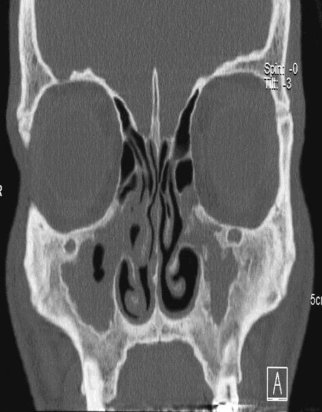
2 Conventional Radiology NNH-Status: okzipito-frontal: frontal sinus, anterior ethmoid okzipito-nasal : maxillary and frontal sinus axial: sphenoid and ethmoidal sinus Radiation exposure: 2 mgy per exposure (dose for cataract 2 Gy)

3 Conventional Radiology inadequate sensivity and specifity superseded by CT


4 Single detector row CT Multi detector row CT Multislice Spiral CT Zeilen Zeilen Zeilen Zeilen Zeilen x 64 Zeilen x 128 Zeilen 2009
5 Paranasal Sinus CT Protocol high resolution axial scan from alveolar process to frontal sinus collimation 0.6mm overlapping slice reconstruction 0.6/0.4mm, mas <200 reconstruction: axial, coronal, sagittal iv contrast*: tumor, inflammation, vascular lesion not required for preop assessment of anatomy
6 Review: Radiation Dose radiation dose paranasal sinus CT conventional x-ray paranasal sinus average diagnostic dose /per capita transatlantic flight (round trip) 0.5-1mGy 0.2 mgy/expos. CH: 1mGy, GB 0.33mGy 0.1mGy cataract of eye lens: > 2 Gy
7 CT preoperative anatomy, residual mucosal disease pattern of bone destruction, skull base defects navigation- CT, CT guided biopsies MRI soft tissue differentiation: retention, tumor, cellularity, vascularity meningeal, neural, vascular involvement
8 Complementary Role CT MR

9 Meningo-Encephalocele
10

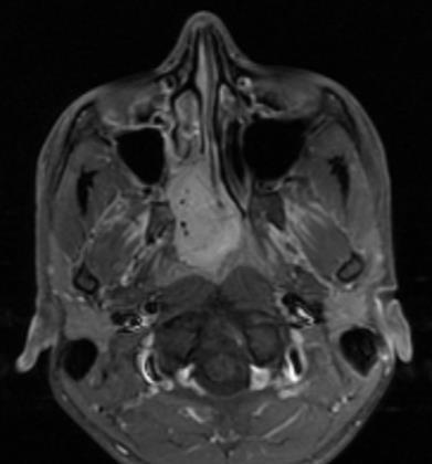
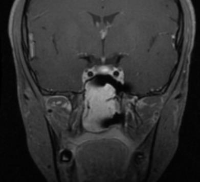
11 Clival Chordoma
12 Angiography diagnostic use for selected cases superselective embolisation: intractable epistaxis; M. Osler embolisation of tumors
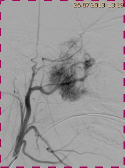
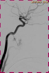
13 Juvenile Nasopharyngeal Angiofibroma
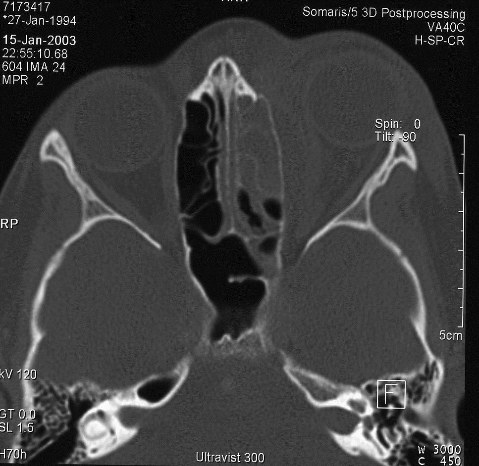
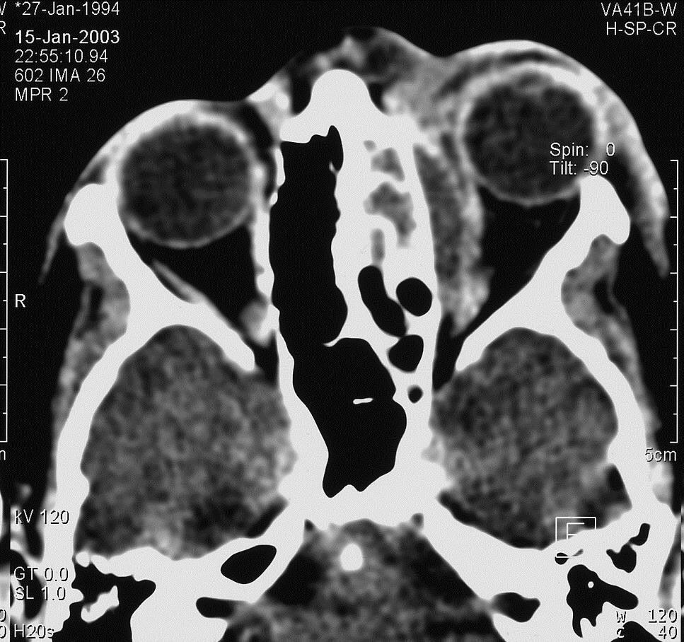
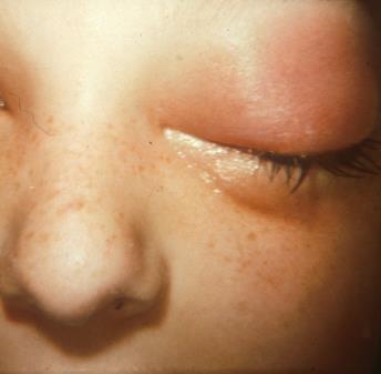
14 Orbital Complication: Subperiostal Abscess


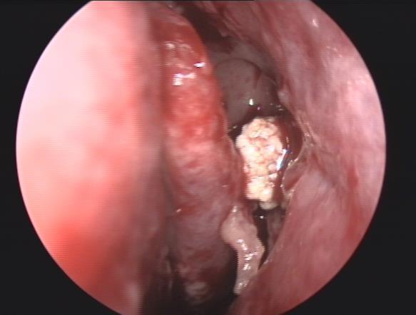
15 Endocranial Complikation: Epidural and Soft Tissue Abscess in Frontal Sinusitis
 N.B.: after medical treatment")
16 Chronic Rhinosinusitis (without Nasal Polyps) N.B.: after medical treatment
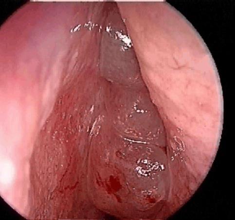

17 Chronic Rhinosinusitis (with Nasal Polyps) N.B.: after medical treatment
 severe frontal headache, frontal swelling signs of meningitis neurological signs urgent investigation and")
18 Red Flags Consider other diagnosis: unilateral symptoms bleeding, crusting cacosmia orbital symptoms( oedema, displaced globe, double vision) severe frontal headache, frontal swelling signs of meningitis neurological signs urgent investigation and intervention
19 Granulomatose mit Polyangiitis (M. Wegener)
20 Aspergilloma (fungus ball)
21 Odontogenic Sinusitis

22 Inverted Papilloma

23 Inverted Papilloma


24 Mucocele of Maxillary Sinus


25 Mucocele of Sphenoid Sinus Dehiscent Internal Carotid Artery

26 Complicated Nose Fracture


27 Zygomatic Fracture

28 CSF-Leak High resolution CT MRI (T2) β2-transferrin or β-trace In selected cases: Intrathecal fluorescein injection
29 CSF-Leak

30 Dacryo CT Scan

31 Dacryo CT Scan
32 FESS: Preoperative Evaluation (CT Checklist) uncinate process ethmoidal roof, skull base ethmoidal arteries orbit, optic nerve internal carotid artery anatomical variations of ethmoidal cells

33 Uncinate Process A B1 B2 Simmen D, Jones N: Manual of Endoscopic Sinus Surgery. Thieme 2014
34 Preoperative Checklist: Uncinate Process Typ A (~ 75%): Insertion at Lamina papyracea «Recessus terminalis» Frontal sinus drainage pathway directly in middle meatus

35 Preoperative Checklist: Uncinate Process Typ B1: Insertion at skull base Frontal sinus drainage pathway in ethmoidal infundibulum

36 Preoperative Checklist: Uncinate Process Typ B2: Insertion at middle turbinate Frontal sinus drainage pathway in ethmoidal infundibulum

37 Frontal Recess Wormald PJ: The agger nasi cell: the key to understand the anatomy of the frontal recess Otolaryngol Head Neck Surg 2003; 129:

38 Anatomy of the Frontal Recess "frontal drainage pathway" "fronto-ethmoidal cell" "agger nasi cell"

39 Frontal Recess: Sagittal Reconstruction Suprabullar cells Frontal bullar cells Interfrontal sinus septal cell Fronto-ethmoidal cells: Agger nasi cell K1 to K4 cells
40
41 anterior (medial) posterior
42

43 Preoperative Checklist: Ethmoid Roof Cribriform Plate


44 Preoperative Checklist: Anterior Ethmoidal Artery

45 Preoperative Checklist: Optic nerve
46 Preoperative Checklist: Internal Carotid Artery

47 Concha bullosa media

48 Haller Cell


49 Haller Cell

50 Onodi Cell

51 Pneumatized Inferior Turbinate

52 3 Nervs??? Nervus opticus Nervus maxillaris Nervus vidianus

53 Cone Beam CT (Digitale Volumentomographie)
54 Cone Beam CT Corresponds to a C-arm with 3D images Digital volume tomography Cone revolves around patient for 360 Since 2000 Office use, no need for a radiologist
55 Low dose multislice CT Cone beam CT Acquisition time ( motion artefacts) Radiation exposure MSCT DVT 2 sec sec msv ( %) Image homogenity + + Bone window Soft tissue window Cost - + Image reconstruction msv + - De Cock et al. A comparative study for image quality and radiation dose of a cone beam computed tomography scanner and a multislice computed tomography scanner for paranasal sinus imaging. Eur Radiol (2015) 25: Al Abduwani et al. Cone beam CT paranasal ainuses versus low dose meltidetector CT studies. Am J Otolaryngol (2016)37:59-64


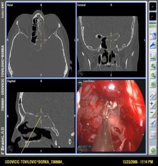
56 Navigation: Localization Systems Optical tracking systems Electromagnetic tracking systems + cordeless + free movement of surgeon - line of sight - magnetic interference
57 Accuracy Depends on: CT scan and its reconstruction Registration! Patient motion, motion of reference points! Generally accepted range: ± 1 mm (=2 mm) Shift in accuracy from anterior to posterior up to 9 mm (z axis)

58 Intraoperative Manual Registration Refinement New feature to adjust disaccuracy i.e. in the depth Before After

:")

59 Navigation starts the day before surgery by: Analysis of the CT-scan (coronar,axial,sagittal): where is the frontal drainage pathway? Step by step surgical plan
60 Suggested Reading Simmen D, Schuknecht B: Computed tomography of paranasal sinuses. A preoperative checklist. Laryngo-Rhino-Otology 1997; 76:8-13 Simmen D, Jones N: Manual of endoscopic Sinus and Skull Base surgery. Thieme 2014, second edition Wormald PJ: Endoscopic Sinus Surgery: Anatomy, Three-Dimensional Reconstruction, and Surgical Technique. Thieme 2018, fourth edition
Radiological anatomy of frontal sinus By drtbalu
 2009 Radiological anatomy of frontal sinus By drtbalu Anatomy of frontal sinus is highly variable. Precise understanding of these variables will help a surgeon to avoid unnecessary complications during
Boundaries Septum Turbinates & Meati Lamellae Drainage Pathways Variants
 The Fastest 20 Minutes in Michelle A. Michel, MD Professor of Radiology and Otolaryngology Medical College of Wisconsin, Milwaukee Overview Nasal cavity Anterior skull base Ostiomeatal complex Frontal
Communication issue - What should the radiologist report before functional endoscopic sinus surgery
 Communication issue - What should the radiologist report before functional endoscopic sinus surgery Poster No.: C-0509 Congress: ECR 2015 Type: Educational Exhibit Authors: A. M. Dobra 1, C. A. Badiu 1,
Skull Base Danger Zones in FESS
 Skull Base Danger Zones in FESS Poster No.: C-2278 Congress: ECR 2014 Type: Educational Exhibit Authors: L. Renza Lozada, R. Carreño Gonzalez, G. Quintana Sanchez, 1 2 1 1 1 2 R. E. Figueroa ; Malaga/ES,
JMSCR Vol 05 Issue 09 Page September 2017
 www.jmscr.igmpublication.org Impact Factor 5.84 Index Copernicus Value: 71.58 ISSN (e)-2347-176x ISSN (p) 2455-0450 DOI: https://dx.doi.org/10.18535/jmscr/v5i9.52 Relationship of Agger Nasi Cell and Uncinate
Chapter Five. 1 of 8 11/3/2008 2:52 PM.
 1 of 8 11/3/2008 2:52 PM Email : myousefmian@hotmail.com Chapter Five FRONT COVER Introduction Acknowledgement CHAPTERS Chapter One Chapter Two Chapter Three Chapter Four Chapter Five Chapter Six Chapter
Computed tomography road map of the paranasal sinuses for treatment planning
 Computed tomography road map of the paranasal sinuses for treatment planning Poster No.: C-2607 Congress: ECR 2013 Type: Educational Exhibit Authors: N. Schembri, A. S. Gatt, D. Ellul, J. Brunton; Dundee/UK
Three-Dimensional Volumetric Display of the Nasal Ostiomeatal Channels and Paranasal Sinuses
 Downloaded from www.ajronline.org by 37.44.202.192 on 12/22/17 from IP address 37.44.202.192. Copyright RRS. For personal use only; all rights reserved Three-Dimensional Volumetric Display of the Nasal
FESS imaging - the role of MDCT
 FESS imaging - the role of MDCT Poster No.: C-0179 Congress: ECR 2013 Type: Educational Exhibit Authors: J. Plascak, K. Makaruha, B. Klasic, L. Kavur, V. Vidjak; Zagreb/HR Keywords: Image verification,
FRONTAL SINUPLASTY P R E P A R E D A N D P R E S E N T E D B Y : D R. Y A H Y A F A G E E H R 4 16/ 12/ 2013
 FRONTAL SINUPLASTY P R E P A R E D A N D P R E S E N T E D B Y : D R. Y A H Y A F A G E E H R 4 16/ 12/ 2013 ANATOMY: FRONTAL SINUS Not present at birth Starts developing at 4 years Radiographically visualized
PROBLEM RECOMMENDATION
 PREVENTION (MINIMIZING) IN ENDOSCOPIC Steven D. Schaefer, MD Professor and Chair Department of Otolaryngology PREVENTION AND Intraoperative Hemorrhage Loss of Orientation Inability to Identify/Preserve
Professor Dr.Muhammad Ajmal Dr.Tehmina Nazir. HOLY FAMILY HOSPITAL Rawalpindi
 Professor Dr.Muhammad Ajmal Dr.Tehmina Nazir HOLY FAMILY HOSPITAL Rawalpindi SCHEME OF PRESENTATION PLAIN X-RAYS CT SCAN MRI CONCLUSION IMAGING MODALITIES PLAIN X-RAYS CT SCAN MRI OCCIPITOMENTAL/WATER
CT OF THE PARANASAL SINUSES : NORMAL ANATOMY, VARIANTS AND PATHOLOGY
 Journal of Optoelectronics and Biomedical Materials Vol.2 Issue 4, October-December 2010, p. 281 289 CT OF THE PARANASAL SINUSES : NORMAL ANATOMY, VARIANTS AND PATHOLOGY AMIT N D DWIVEDI *, KAPIL KUMAR
Anatomical Variations in Osteomeatal Complex among Patients undergoing Functional Endoscopic Sinus Surgery
 V Narendrakumar, V Subramanian Original article 10.5005/jp-journals-10013-1259 Anatomical Variations in Osteomeatal Complex among Patients undergoing Functional Endoscopic Sinus Surgery 1 V Narendrakumar,
Reasons for Failure and Surgical Revisions. Stil Kountakis, MD, PhD Professor and Chief, Division of Rhinology
 Reasons for Failure and Surgical Revisions Stil Kountakis, MD, PhD Professor and Chief, Division of Rhinology Medical College of Georgia of Georgia Regents University Department of Otolaryngology / Head
SINUS ANATOMY AND FUNCTION
 EMBRYOLOGY AND DEVELOPMENT SINUS ANATOMY AND FUNCTION -4 th week gestation: -frontonasal process identified, arises over developing forebrain -ectodermal -contributes to nasal capsule -9 th and 10 th week
International Journal of Biological & Medical Research
 Int J Biol Med Res.2015;6(1):4775-4781 Contents lists available at BioMedSciDirect Publications International Journal of Biological & Medical Research Journal homepage: www.biomedscidirect.com BioMedSciDirect
Anatomical Analysis of the Frontal Recess Cells in Endoscopic Sinus Surgery An Indian Perspective
 ORIGINAL ARTICLE Anatomical Analysis of the Frontal Recess Cells in Endoscopic Sinus Surgery An Indian Perspective 1 Dhingra Shruti, 2 Agarwal AK, 3 Passey JC, 4 Kaul JM 1 Resident, Department of Otolaryngology
Katya A. Shpilberg 1 Simon C. Daniel 1 Amish H. Doshi 1 William Lawson 2 Peter M. Som 1. Neuroradiology/Head and Neck Imaging Original Research
 Neuroradiology/Head and Neck Imaging Original Research Shpilberg et al. CT of Paranasal Sinuses and Nasal Cavity Neuroradiology/Head and Neck Imaging Original Research Katya A. Shpilberg 1 Simon C. Daniel
COMPLICATIONS IN ENDOSCOPIC SINUS SURGERY
 COMPLICATIONS IN ENDOSCOPIC SINUS SURGERY John M. DelGaudio, MD Professor and Vice Chair Chief of Rhinology and Sinus Surgery Department of Otolaryngology-Head and Neck Surgery Emory University School
A radiological study of anatomical variations in ostiomeatal complex in patients with chronic rhinosinusitis
 International Journal of Otorhinolaryngology and Head and Neck Surgery Rajneesh et al. Int J Otorhinolaryngol Head Neck Surg. 2017 Jul;3(3):528-533 http://www.ijorl.com pissn 2454-5929 eissn 2454-5937
Anatomical Variants in Frontal Recess Region and their Impact on Frontal Sinus Surgery in Chronic Sinusitis
 American Journal of Health Research 2015; 3(3): 140-145 Published online April 23, 2015 (http://www.sciencepublishinggroup.com/j/ajhr) doi: 10.11648/j.ajhr.20150303.15 ISSN: 2330-8788 (Print); ISSN: 2330-8796
National Imaging Associates, Inc. Clinical guidelines/considerations SINUS & MAXILLOFACIAL AREA CT 70486, 70487, 70488
 National Imaging Associates, Inc. Clinical guidelines/considerations SINUS & MAXILLOFACIAL AREA CT 70486, 70487, 70488 Date: September 1997 Page 1 of 5 LIMITED OR LOCALIZED FOLLOW UP - SINUS CT 76380 Guideline
The Relation between Anatomical Variations of Osteomeatal Complex & Nasal Structures and Chronic Sinusitis by Computed Tomography
 International Journal of Medical Imaging 2015; 3(2): 16-20 Published online March 6, 2015 (http://www.sciencepublishinggroup.com/j/ijmi) doi: 10.11648/j.ijmi.20150302.12 ISSN: 2330-8303 (Print); ISSN:
Tomographical Findings in Adult Patients Undergoing Endoscopic Sinus Surgery Revision
 THIEME Original Research 73 Tomographical Findings in Adult Patients Undergoing Endoscopic Sinus Surgery Revision Jan Alessandro Socher 1 Jonas Mello 2 Barbara Batista Baltha 2 1 Department of Otorhinolaryngology,
ADVANCED ENDOSCOPIC SURGERY OF THE PARANASAL SINUSES & SKULL BASE
 26 th International Course on ADVANCED ENDOSCOPIC SURGERY OF THE PARANASAL SINUSES & SKULL BASE Ghent (Belgium) 27-30 August 2014 INTERNATIONAL FACULTY Bachert C. (Ghent) Bernal-Sprekelsen M. (Barcelona)
A Study of Anatomical Variations in Patients with Chronic Rhinosinusitis.
 DOI: 10.2127/aimdr.201..2.EN1 Original Article ISSN (O):239-222; ISSN (P):239-21 A Study of Anatomical Variations in Patients with Chronic Rhinosinusitis. Smruti Swain 1 1 Associate Professor, Department
Nasal region. cartilages: septal cartilage (l); lateral nasal cartilage (2); greater alar cartilages (2); lesser alar cartilages (?
; lateral nasal cartilage (2); greater alar cartilages (2); lesser alar cartilages (?") Nasal region skull bones: nasal and frontal processes of maxilla cartilages: septal cartilage (l); lateral nasal cartilage (2); greater alar cartilages (2); lesser alar cartilages (?) 1 Nasal cavity Roof
Nasal region skull bones: nasal and frontal processes of maxilla cartilages: septal cartilage (l); lateral nasal cartilage (2); greater alar cartilages (2); lesser alar cartilages (?) 1 Nasal cavity Roof
Relationship of the Optic Nerve to the Posterior Paranasal Sinuses: A CT Anatomic Study
 Relationship of the Optic Nerve to the Posterior Paranasal Sinuses: A CT Anatomic Study Mark C. DeLano, F. Y. Fun, and S. James Zinreich PURPOSE: To delineate the relationship between the optic nerves
Morphological Changes of the Ethmoid and Maxillary Cavities after Endoscopic Sinus Surgery, A Quantitative Digital Analysis.
 Morphological Changes of the Ethmoid and Maxillary Cavities after Endoscopic Sinus Surgery, A Quantitative Digital Analysis Thesis Submitted for fulfillment of M.D. degree in Otorhinolaryngology By: Hisham
Computed tomographic evaluation of anatomical variations of paranasal sinus region
 International Journal of Research in Medical Sciences Gupta S et al. Int J Res Med Sci. 2016 Jul;4(7):2909-2913 www.msjonline.org pissn 2320-6071 eissn 2320-6012 Research Article DOI: http://dx.doi.org/10.18203/2320-6012.ijrms20161975
Juvenile Angiofibroma
 Juvenile Angiofibroma Disclaimer The pictures used in this presentation have been obtained from a number of sources. Their use is purely for academic and teaching purposes. The contents of this presentation
We are IntechOpen, the world s leading publisher of Open Access books Built by scientists, for scientists. International authors and editors
 We are IntechOpen, the world s leading publisher of Open Access books Built by scientists, for scientists 3,350 108,000 1.7 M Open access books available International authors and editors Downloads Our
Original Article Effect of lamina papyracea ingression on orbito-ocular complications after functional endoscopic sinus surgery
 Int J Clin Exp Med 2016;9(6):10317-10321 www.ijcem.com /ISSN:1940-5901/IJCEM0025371 Original Article Effect of lamina papyracea ingression on orbito-ocular complications after functional endoscopic sinus
ROLE OF ANATOMICAL OBSTRUCTION IN THE PATHOGENESIS OF CHRONIC SINUSITIS
 From the SelectedWorks of Balasubramanian Thiagarajan July 1, 2012 ROLE OF ANATOMICAL OBSTRUCTION IN THE PATHOGENESIS OF CHRONIC SINUSITIS Balasubramanian Thiagarajan Available at: https://works.bepress.com/drtbalu/51/
Fracture frontal bone and its management
 From the SelectedWorks of Balasubramanian Thiagarajan March 1, 2013 Fracture frontal bone and its management Balasubramanian Thiagarajan Available at: https://works.bepress.com/drtbalu/14/ ISSN: 2250-0359
Chronic Frontal Rhinosinusitis: Diagnosis and Management
 Chapter Chronic Frontal Rhinosinusitis: Diagnosis and Management Core Messages Despite significant advances in surgical techniques, technology, and knowledge of pathophysiology, management of chronic frontal
CT anatomy of paranasal sinuses.
 CT anatomy of paranasal sinuses. Poster No.: C-2117 Congress: ECR 2017 Type: Educational Exhibit Authors: O. Dib, H. Chahinez, B. Asma, C. abdelouahab, M. Ourrad El, 1 2 1 1 1 2 1 3 3 B. Nacereddine ;
Commen Nose Diseases
 Commen Nose Diseases Symptoms List: Nasal obstruction. Nasal discharge: Anterior (Rhinorrhea). Posterior (Postnasal discharge). Epistaxis. Hyposmia and Anosmia. Headache. Snoring. Nasal Obstruction Definition:
The Egyptian Journal of Hospital Medicine (July 2017) Vol.68 (3), Page
 Vol.68 (3), Page") The Egyptian Journal of Hospital Medicine (July 2017) Vol.68 (3), Page 1390-1394 Anatomical Variations of Nasal Structures in Chronic Rhinosinusitis as Detected by Computed Tomography Scan Omar Adnan Hasan,
The Egyptian Journal of Hospital Medicine (July 2017) Vol.68 (3), Page 1390-1394 Anatomical Variations of Nasal Structures in Chronic Rhinosinusitis as Detected by Computed Tomography Scan Omar Adnan Hasan,
HEAD AND NECK IMAGING. James Chen (MS IV)
") HEAD AND NECK IMAGING James Chen (MS IV) Anatomy Course Johns Hopkins School of Medicine Sept. 27, 2011 OBJECTIVES Introduce cross sectional imaging of head and neck Computed tomography (CT) Review head
HEAD AND NECK IMAGING James Chen (MS IV) Anatomy Course Johns Hopkins School of Medicine Sept. 27, 2011 OBJECTIVES Introduce cross sectional imaging of head and neck Computed tomography (CT) Review head
Pathological consequences of anatomical variations in the sino-nasal region: how can radiologists help clinicians?
 Pathological consequences of anatomical variations in the sino-nasal region: how can radiologists help clinicians? Poster No.: C-0735 Congress: ECR 2016 Type: Educational Exhibit Authors: M. E. Laino,
Diagnostic Performance of Multidetector Computed Tomography (MDCT) in Diagnosis of Sinus Variations
 in Diagnosis of Sinus Variations") Signature: Pol J Radiol, 2017; 82: 713-725 DOI: 10.12659/PJR.903684 ORIGINL RTICLE Received: 2017.02.08 ccepted: 2017.02.23 Published: 2017.11.17 uthors Contribution: Study Design Data Collection C Statistical
Signature: Pol J Radiol, 2017; 82: 713-725 DOI: 10.12659/PJR.903684 ORIGINL RTICLE Received: 2017.02.08 ccepted: 2017.02.23 Published: 2017.11.17 uthors Contribution: Study Design Data Collection C Statistical
ENDOSCOPIC SURGERY OF THE PARANASAL SINUSES & SKULL BASE
 30 th International Course on ENDOSCOPIC SURGERY OF THE PARANASAL SINUSES & SKULL BASE Ghent (Belgium) 29 August-1 September 2018 INTERNATIONAL FACULTY Claus Bachert (Ghent, Belgium) Philippe Gevaert (Ghent,
Sinonasal Imaging. Mamdouh Mahfouz MD Professor of Radiology Cairo University. ssregypt.com
 Sinonasal Imaging Mamdouh Mahfouz MD Professor of Radiology Cairo University ssregypt.com Scanning Techniques Routine Study CORONAL Coronal 3-5mm sections from the posterior wall of the sphenoid sinus
Provider Led Entity. CDI Quality Institute PLE Rhinosinusitis AUC 12/04/2018
 Provider Led Entity CDI Quality Institute PLE Rhinosinusitis AUC 12/04/2018 Appropriateness of advanced imaging procedures* in patients with rhinosinusitis and the following clinical presentations or diagnoses:
SINONASAL IMAGING. Kim O. Learned, MD. Assistant Professor Department of Radiology/Division of Neuroradiology University of Pennsylvania Health System
 SINONASAL IMAGING Kim O. Learned, MD Assistant Professor Department of Radiology/Division of Neuroradiology University of Pennsylvania Health System REVIEWS Key Anatomy: Sinus Drainage Pathways Practical
A COMPUTERIZED TOMOGRAPHIC STUDY OF UNCINATE PROCESS OF ETHMOID BONE
 Original Article A COMPUTERIZED TOMOGRAPHIC STUDY OF UNCINATE PROCESS OF ETHMOID BONE N. Vinay Kumar * 1, E. Kamala 2, T. S. Guga Priya 3, S. D. NalinaKumari 4. *1,2 Assistant professor, Department of
Variation in frontal cells in relation to chronic frontal sinusitis
 International Journal of Current Research in Medical Sciences ISSN: 244-71 P-ISJN: A472-04, E -ISJN: A472-01 www.ijcrims.com Original Research Article Volume, Issue 1-2019 DOI: http://dx.doi.org/10.22192/ijcrms.2019.0.01.00
A Computer-Assisted Anatomical Study of the Nasofrontal Region
 The Laryngoscope Lippincott Williams & Wilkins, Inc., Philadelphia 2001 The American Laryngological, Rhinological and Otological Society, Inc. A Computer-Assisted Anatomical Study of the Nasofrontal Region
 www.oralradiologists.com CONE BEAM CT REPORT CASE ---- Case Information Referring Doctor: - Patient Name: - Scan Date: December 1, 2015 Patient DOB: - Reason for Exam: - Study Details: icat Flex, 160x160x112
Bones of the skull & face
 Bones of the skull & face Cranium= brain case or helmet Copyright The McGraw-Hill Companies, Inc. Permission required for reproduction or display. The cranium is composed of eight bones : frontal Occipital
Incidence of sinonasal anatomical variations associated with chronic sinusitis by CT scan in Karaikal, South India
 International Journal of Otorhinolaryngology and Head and Neck Surgery Gouripur K et al. Int J Otorhinolaryngol Head Neck Surg. 2017 Jul;3(3):576-580 http://www.ijorl.com pissn 2454-5929 eissn 2454-5937
Image quality and dose reduction in sinus computed tomography using iterative reconstruction: a cadaver study*
 ORIGINAL CONTRIBUTION Image quality and dose reduction in sinus computed tomography using iterative reconstruction: a cadaver study* Adam J. Kimple 1, Stanley W. McClurg 1, Benjamin Y. Huang 2, Satyan
JMSCR Vol 04 Issue 05 Page May 2016
 www.jmscr.igmpublication.org Impact Factor 5.244 Index Copernicus Value: 5.88 ISSN (e)-2347-176x ISSN (p) 2455-0450 DOI: http://dx.doi.org/10.18535/jmscr/v4i5.25 Radiologic Variations of Nose and Paranasal
Anatomical variants of the uncinate process CT scan imaging study
 Romanian Journal of Rhinology, Vol. 2, No. 7, July - September 2012 original Study Anatomical variants of the uncinate process CT scan imaging study Vasilica Baldea 1, Mihail Dan Cobzeanu 2, Florina Mihalcea
The View through the Nose: ENT considerations for Pituitary/Skull Base Surgery
 The View through the Nose: ENT considerations for Pituitary/Skull Base Surgery Edsel Kim, M.D. Otolaryngology-Head and Neck Surgery The Oregon Clinic Providence Brain and Spine Institute Pituitary, Thyroid
3D Accuitomo Clinical Case Evidence
 3D Accuitomo Clinical Case Evidence The Advantages of DVT for Ear-, Nose- & Throat-Diagnostic Thinking ahead. Focused on life. Editorial Dear Colleagues, Index of Contents I am very happy to present you
A Targeted Endoscopic Approach to Chronic Isolated Frontal Sinusitis
 Otolaryngology Head and Neck Surgery (2006) 134, 28-32 ORIGINAL RESEARCH A Targeted Endoscopic Approach to Chronic Isolated Frontal Sinusitis Roee Landsberg, MD, Yoram Segev, MD, Michael Friedman, MD,
Original Research THE USE OF REFORMATTED CONE BEAM CT IMAGES IN ASSESSING MID-FACE TRAUMA, WITH A FOCUS ON THE ORBITAL FLOOR FRACTURES
 DOI: 10.15386/cjmed-601 Original Research THE USE OF REFORMATTED CONE BEAM CT IMAGES IN ASSESSING MID-FACE TRAUMA, WITH A FOCUS ON THE ORBITAL FLOOR FRACTURES RALUCA ROMAN 1, MIHAELA HEDEȘIU 1, FLOAREA
ORIGINAL ARTICLE RELATIONSHIP OF CONCHA BULLOSA WITH OSTEOMEATAL UNIT BLOCKAGE. TOMOGRAPHIC STUDY IN 200 PATIENTS.
 RELATIONSHIP OF CONCHA BULLOSA WITH OSTEOMEATAL UNIT BLOCKAGE. TOMOGRAPHIC STUDY IN 200 PATIENTS. Shrikrishna B H 1, Jyothi A C 2, Sanjay G 3, Sandeep Samson G 4. 1. Associate Professor, Department of
Spheno-Ethmoidectomy
 Diagnostic and Therapeutic Endoscopy, Vol. 5, pp. 1-8 Reprints available directly from the publisher Photocopying permitted by license only (C) 1998 OPA (Overseas Publishers Association) N.V. Published
Functional Endoscopic Sinus Surgery
 WHAT IS FUNCTIONAL ENDOSCOPIC SINUS SURGERY (FESS)? The nasal telescope has greatly changes the evaluation and treatment of rhino-sinusitis. This instrument, which provides a view of the structures in
Endoscopic Management Of A Giant Ethmoid Mucocele
 ISPUB.COM The Internet Journal of Otorhinolaryngology Volume 6 Number 1 S Ceylan, F Bora Citation S Ceylan, F Bora.. The Internet Journal of Otorhinolaryngology. 2006 Volume 6 Number 1. Abstract We present
The Nose and Sinuses. Ophir Ilan, MD, PhD Department of Otolaryngology/Head&Neck surgery Hadassah University Hospital
 The Nose and Sinuses Ophir Ilan, MD, PhD Department of Otolaryngology/Head&Neck surgery Hadassah University Hospital Nasal Mucociliary System Function of the Nasal Mucosa warming and humidifying the
Conventional Sinus Surgery Vs Fess
 IOSR Journal of Dental and Medical Sciences (IOSR-JDMS) e-issn: 2279-0853, p-issn: 2279-0861.Volume 16, Issue 7 Ver. III (July. 2017), PP 44-51 www.iosrjournals.org Conventional Sinus Surgery Vs Fess *
A comprehensive study on complications of endoscopic sinus surgery
 International Journal of Otorhinolaryngology and Head and Neck Surgery Shyras JAD et al. Int J Otorhinolaryngol Head Neck Surg. 2017 Jul;3(3):472-477 http://www.ijorl.com pissn 2454-5929 eissn 2454-5937
ENT NAVIGATION SIMPLE AND INTUITIVE
 ENT NAVIGATION SIMPLE AND INTUITIVE REVOLUTIONARY NAVIGATION TECHNOLOGY FOR ROUTINE AND COMPLEX ENT PROCEDURES We at Fiagon believe patient care should be centered around the patient. For us, patient-centered-care
Head & Neck Clinical Sub Group. Network Agreed Imaging Guidelines for UAT and Thyroid Cancer. Measure Nos: 11-1C-105i & 11-1C-106i
 Greater Manchester, Lancashire & South Cumbria Strategic Clinical Network & Senate Head & Neck Clinical Sub Group Network Agreed Imaging Guidelines for UAT and Thyroid Cancer Measure Nos: 11-1C-105i &
DIGITAL PANORAMIC RADIOGRAPHY IN DETERMINING THE PREVALENCE OF HALLER S CELLS: A RETROSPECTIVE RADIOGRAPHIC STUDY
 Original Article International Journal of Dental and Health Sciences Volume 02,Issue 04 DIGITAL PANORAMIC RADIOGRAPHY IN DETERMINING THE PREVALENCE OF HALLER S CELLS: A RETROSPECTIVE RADIOGRAPHIC STUDY
The Frontal Sinus Drainage Pathway and Related Structures
 Pictorial Essay The Frontal Sinus Drainage Pathway and Related Structures David L. Daniels, Mahmood F. Mafee, Michelle M. Smith, Timothy L. Smith, Thomas P. Naidich, W. Douglas Brown, William E. Bolger,
Transnasal Endoscopic Sinonasal Surgery
 Reda kamel, Cadaveric dissection 1 Transnasal Endoscopic Sinonasal Surgery Cadaver Dissection Guide For Endoscopic Sinus Surgery Cairo University Egypt Reda Kamel Professor of Rhinology Cairo University
Prevalence of Anatomical Variations of the Sinonasal Region and their Relationship with Chronic Rhinosinusitis
 Prevalence of Anatomical Variations of the Sinonasal Region and their Relationship with Chronic Rhinosinusitis Karki S, 1 Pokharel M, 2 Suwal S, 1 Poudel R 1 ABSTRACT Background 1 Department of Radiology
International Journal of Health Sciences and Research ISSN:
 International Journal of Health Sciences and Research www.ijhsr.org ISSN: 2249-9571 Original Research Article Anatomical Study of the Middle Meatus with Emphasis to the Maxillary Ostium and Their Clinical
FOR CMS (MEDICARE) MEMBERS ONLY NATIONAL COVERAGE DETERMINATION (NCD) FOR COMPUTED TOMOGRAPHY:
 MEMBERS ONLY NATIONAL COVERAGE DETERMINATION (NCD) FOR COMPUTED TOMOGRAPHY:") National Imaging Associates, Inc. Clinical guidelines SINUS & MAXILLOFACIAL AREA CT LIMITED OR LOCALIZED FOLLOW UP SINUS CT Original Date: September 1997 Page 1 of 5 CPT Codes: 70486, 70487, 70488, 76380
National Imaging Associates, Inc. Clinical guidelines SINUS & MAXILLOFACIAL AREA CT LIMITED OR LOCALIZED FOLLOW UP SINUS CT Original Date: September 1997 Page 1 of 5 CPT Codes: 70486, 70487, 70488, 76380
Mucocele of paranasal sinuses
 From the SelectedWorks of Balasubramanian Thiagarajan March 7, 2012 Mucocele of paranasal sinuses Balasubramanian Thiagarajan Available at: https://works.bepress.com/drtbalu/57/ Mucoceles of paranasal
Crista galli sinusitis a radiological impression or a real clinical entity
 Romanian Journal of Rhinology, Vol. 6, No. 23, July - September 2016 ORIGINAL STUDY Crista galli sinusitis a radiological impression or a real clinical entity Claudiu Manea 1,2, Ranko Mladina 3 1 CESITO
Skull and Axial Skeleton
 Published on Second Faculty of Medicine, Charles University (http://www.lf2.cuni.cz ) Skull and Axial Skeleton Description of the test The examination of the skull skeleton is in oral format. It consists
Nasal Polyposis. DEPARTMENT OF ENT K.S.Hegde Medical Academy Deralakatte, Mangalore
 Nasal Polyposis DEPARTMENT OF ENT K.S.Hegde Medical Academy Deralakatte, Mangalore Def: INTRODUCTION Chronic inflammatory disease of the mucous membrane in the nose & PNS, presenting as pedunculated smooth
CT of Maxillofacial Fracture Patterns. CT of Maxillofacial Fracture Patterns
 CT of Maxillofacial Fracture Patterns CT of Maxillofacial Fracture Patterns Stuart E. Mirvis, M.D., FACR Department of Radiology University of Maryland School of Medicine Viking 1 1976 MGS 2001 Technology
Dr. Sami Zaqout Faculty of Medicine IUG
 The Nose External Nose Nasal Cavity External Nose Blood and Nerve Supplies of the External Nose Blood Supply of the External Nose The skin of the external nose Branches of the ophthalmic and the maxillary
The Incidence of Concha Bullosa and Its Association with Chronic Rhinosinusitis Deviated Nasal Septum and Osteomeatal Complex Obstruction
 1 Bahrain Medical Bulletin, Vol. 33, No. 4, December 2011 The Incidence of Concha Bullosa and Its Association with Chronic Rhinosinusitis Deviated Nasal Septum and Osteomeatal Complex Obstruction Fatma
The International Frontal Sinus Anatomy Classification (IFAC) and Classification of the Extent of Endoscopic Frontal...
 and Classification of the Extent of Endoscopic Frontal...") See discussions, stats, and author profiles for this publication at: https://www.researchgate.net/publication/298902054 The International Frontal Sinus Anatomy Classification (IFAC) and Classification
See discussions, stats, and author profiles for this publication at: https://www.researchgate.net/publication/298902054 The International Frontal Sinus Anatomy Classification (IFAC) and Classification
CT - Brain Examination
 CT - Brain Examination Submitted by: Felemban 1 CT - Brain Examination The clinical indication of CT brain are: a) Chronic cases (e.g. headache - tumor - abscess) b) ER cases (e.g. trauma - RTA - child
Imaging Anatomy in Revision Sinus Surgery
 Chapter 1 Imaging Anatomy in Revision Sinus Surgery Ramon E. Figueroa 1 Core Messages An intimate knowledge of sinus anatomy and a clear understanding of the baseline postsurgical anatomy are required
Extranodal Natural Killer/T-Cell Lymphoma Nasal Type: Detection by Computed Tomography Features
 The Laryngoscope VC 2014 The American Laryngological, Rhinological and Otological Society, Inc. Extranodal Natural Killer/T-Cell Lymphoma Nasal Type: Detection by Computed Tomography Features Yin-Ping
Cranium Facial bones. Sternum Rib
 Figure 7.1 The human skeleton. Skull Thoracic cage (ribs and sternum) Cranium Facial bones Sternum Rib Bones of pectoral girdle Vertebral column Sacrum Vertebra Bones of pelvic girdle (a) Anterior view
MANAGEMENT OF CSF. Steven D. Schaefer, MD, FACS. Department of Otolaryngology New York Eye and Ear Infirmary
 MANAGEMENT OF CSF RHINORRHEA, MENIGIOCELES, Steven D. Schaefer, MD, FACS Professor and Chair Department of Otolaryngology New York Eye and Ear Infirmary New York Medical College Anatomy and Physiology
Clinical and imagistic correlations in the inflammatory pathology of nasosinusal cavities
 Romanian Journal of Rhinology, Volume 8, No. 29, January-March 2018 ORIGINAL STUDY DOI: 10.2478/rjr-2018-0003 Clinical and imagistic correlations in the inflammatory pathology of nasosinusal cavities Emilia
ISSN: Volume 4 Issue FACTORS CONTRIBUTING FOR THE BEST OUTCOME OF FUNCTIONAL ENDOSCOPIC SINUS SURGERY
 1 ISSN: 2250-0359 Volume 4 Issue 4 2014 FACTORS CONTRIBUTING FOR THE BEST OUTCOME OF FUNCTIONAL ENDOSCOPIC SINUS SURGERY SANGEETHA Madha Medical College and Research Institute, Kovoor, Chennai Abstract
The frequency of nasal septal deviation and concha bullosa and their relationship with maxillary sinusitis based on CBCT finding
 Available online at www.ijmrhs.com ISSN No: 2319-5886 International Journal of Medical Research & Health Sciences, 2016, 5, 11:152-156 The frequency of nasal septal deviation and concha bullosa and their
A comparative analysis of CT scan versus diagnostic nasal endoscopy in chronic rhino sinusitis
 International Journal of Otorhinolaryngology and Head and Neck Surgery Pullarat AN et al. Int J Otorhinolaryngol Head Neck Surg. 2018 Jul;4(4):930-934 http://www.ijorl.com pissn 2454-5929 eissn 2454-5937
Evaluation of Anatomical Variations in Ostiomeatal Unit by Computed Tomography
 Original Article Print ISSN: 2321-6379 Online ISSN: 2321-595X DOI: 10.17354/ijss/2018/83 Evaluation of Anatomical Variations in Ostiomeatal Unit by Computed Tomography Sushilkumar Kale 1, K Preetha 2 1
The Role of Computed Tomography in the Evaluation of Paranasal Sinuses Lesions
 ORIGINAL ARTICLE The Role of Computed Tomography in the Evaluation of Paranasal Sinuses Lesions Bhumikaben P. Suthar 1 *, Divya Vaidya 2, Pukhraj P. Suthar 3. 1 Assistant Professor, 2 Third Year Resident,
The surgical approach to the sphenoid sinus continues to
 A comparison of two sphenoidotomy approaches using a novel computerized tomography grading system Heitham Gheriani, F.R.C.S.C., F.R.C.S.I., David Flamer, B.Sc., Trent Orton, M.D., Brad Mechor, F.R.C.S.C.,
The Role of Computed Tomography in the Evaluation of Paranasal Sinuses Lesions
 ORIGINAL ARTICLE The Role of Computed Tomography in the Evaluation of Paranasal Sinuses Lesions Bhumikaben P. Suthar 1 *, Divya Vaid 2, Pukhraj P. Suthar 3. 1 Assistant Professor, 2 Third Year Resident,
Bones of the Skull Lateral View
 Bones of the Skull Lateral View Frontal Bone Parietal Bone Occipital Bone Temporal Bone Sphenoid Bone Pterion Sutures of the Skull Lateral View Coronal Suture Lambdoid Suture Squamous Suture Sutures of
Frontal sinus disease continues to be one of the great
 Unilateral transnasal endoscopic approach to frontal sinuses: Draf IIc Mohammed K. Al Komser, M.D., M.A.S. and Andrew N. Goldberg, M.D., M.S.C.E. ABSTRACT For chronic sinusitis surgery, the Draf III approach
Pneumatization of Mastoid Air Cells, Temporal Bone, Ethmoid and Sphenoid Sinuses. Any Correlation?
 DOI 10.1007/s12070-014-0745-z ORIGINAL ARTICLE Pneumatization of Mastoid Air Cells, Temporal Bone, Ethmoid and Sphenoid Sinuses. Any Correlation? Khalid Hindi Sarmad Alazzawi Rajagopalan Raman Narayanan
A CORRELATION STUDY OF PARANASAL SINUSES BETWEEN OPERATIVE ENDOSCOPIC FINDINGS IN FESS AND PREOPERATIVE CT SCAN
 A CORRELATION STUDY OF PARANASAL SINUSES BETWEEN OPERATIVE ENDOSCOPIC FINDINGS IN FESS AND PREOPERATIVE CT SCAN Polisetti Ravi Babu 1, Bhennur Durga Prasad 2, Lanke Sowmya 3, K.S.B.S. Krishna Sasanka 4