Penetration of the Optic Nerve or Chiasm by Anterior Communicating Artery Aneurysms. - Three Case Reports-
|
|
|
- Elfrieda Golden
- 5 years ago
- Views:
Transcription
1 Penetration of the Optic Nerve or Chiasm by Anterior Communicating Artery Aneurysms. - Three Case Reports- Tetsuyoshi Horiuchi 1, Toshiya Uchiyama 1, Yoshikazu Kusano 1, Maki Okada 1, Kazuhiro Hongo 1, and Shigeaki Kobayashi 2 1 Department of Neurosurgery, Shinshu University School of Medicine, Matsumoto, Nagano, Japan 2 Department of Neurosurgery, Aizawa Hospital, Matsumoto, Nagano, Japan Key words: aneurysm, anterior communicating artery, optic chiasm, optic nerve, penetration Running head: Penetration of optic nerve or chiasm Correspondence to: Tetsuyoshi Horiuchi, M.D. Department of Neurosurgery, Shinshu University School of Medicine, Asahi, Matsumoto, (postal code), Japan Telephone: Fax: tetuyosi@shinshu-u.ac.jp 1
2 Abstract Although large and giant aneurysms can induce visual disturbance by compression of the anterior visual pathway, splitting and penetration of the optic apparatus are extremely rare. The authors describe 3 patients who underwent clipping surgery for the anterior communicating artery aneurysm infiltrating into the optic nerve or chiasm. These findings were suspected on the preoperative magnetic resonance imaging and confirmed at surgery. Two aneurysms were ruptured and one unruptured. We review the literature and discuss the mechanism of cranial nerve penetration by an aneurysm. 2
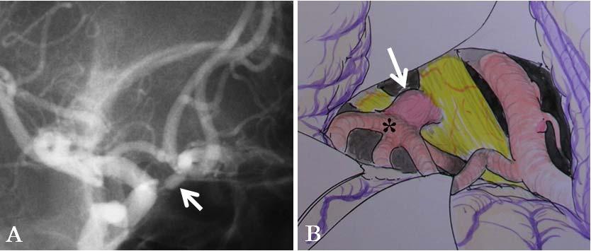
3 Introduction Since cerebral aneurysms of the internal carotid artery (ICA) and the anterior communicating artery (ACoA) are located closely to the optic nerve and chiasm, some of 3, 10) them can compress the anterior visual pathway causing visual disturbance. However, its penetration and splitting by a cerebral aneurysm is rare. Hongo et al. 5) first reported in 1981, a case of the right ICA aneurysm that penetrated and split the optic chiasm. To our knowledge, there have been only 8 cases reporting penetration and splitting of the optic nerve or chiasm by ICA 1, 4, 5, 7, 8, 11) 2, 9) or ACoA aneurysms. We recently encountered a case (Case 3) with the penetration of optic chiasm by the unruptured ACoA aneurysm. Therefore, similar cases were retrospectively searched using the operation records written by senior authors (K.H. and S.K.). Additional 2 cases were found. Here, we describe total 3 cases of ACoA aneurysm penetrating the anterior visual pathway and review the literature. Case report Case 1 History and Examination. This 62-year-old man presented with sudden onset of headache. His neurological examination revealed no abnormalities. He did not complain visual disturbances. Computed tomography (CT) demonstrated subarachnoid hemorrhage (SAH) and cerebral angiography detected an ACoA aneurysm (Figure 1). Since the emergency surgery was needed to prevent the re-rupture, no detailed ophthalmological evaluations were performed. Operation and Postoperative Course. A right pterional approach was used and the aneurysm was found impinging upon the right optic nerve (Figure 1). It was not fully dissected from 3
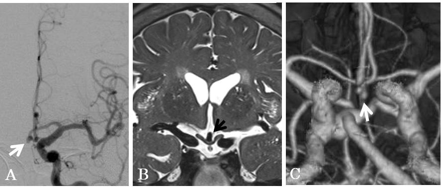
4 the optic nerve not to damage the nerve. This impingement was verified by the senior author s operative reports. A straight clip was applied for obliteration. The patient tolerated the procedure well and was discharged without neurological deficits. Case 2 History and Examination. This 53-year-old man presented to a near-by hospital with one-month history of difficulty seeing with the right eye and intermittent severe headaches. His visual acuity was 20/20 in both eyes. The nasal hemianopia of the right eye was detected on Goldmann visual field examination (Figure 2-1). CT and magnetic resonance imaging (MRI) revealed a mass in the right prechiasmal region (Figure 2-2). The patient was referred to us. Angiogram showed an ACoA aneurysm (Figure 2-2). Operation and Postoperative Course. A fronto-temporal craniotomy was performed and transsylvian approach was used. There was an evidence of recent SAH. A red mark was seen on the optic nerve just distal to the optic canal; this was suspectedly caused by the hemorrhage. The aneurysmal dome was located inside the right optic nerve (Figure 2-2). The aneurysm was successfully clipped with a bayonet clip and punctured for decompression. Postoperative course was uneventful. His visual acuity was unchanged. The nasal hemianopia of the right eye improved after surgery (Figure 2-3). Follow-up angiogram showed complete obliteration of the aneurysm. Case 3 History and Examination. This 65-year-old woman was diagnosed as having multiple cerebral aneurysms on brain check-up. Cerebral angiogram demonstrated aneurysms in the bilateral middle cerebral arteries, right superior cerebellar artery, and ACoA. The ACoA aneurysm was irregular in shape with bleb and infiltrated the optic chiasm on MRI (Figure 3). 4
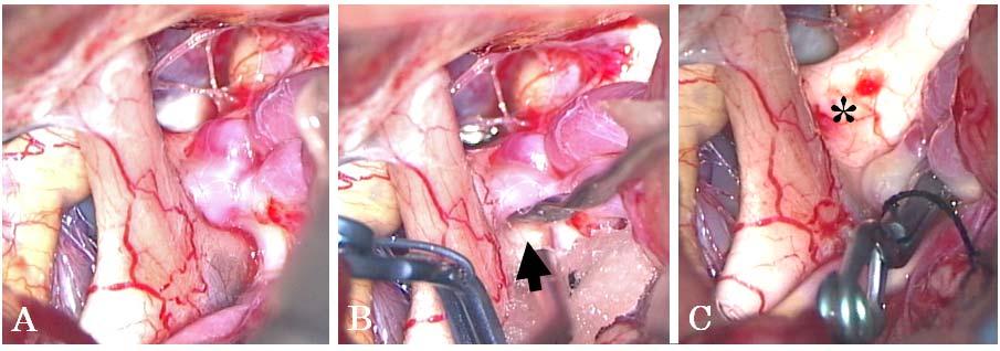
5 Preoperatively, her visual acuity was 20/20 in both eyes and no visual field defect was detected. Operation and Postoperative Course. Under general anesthesia, the ACoA and left middle cerebral artery aneurysms were exposed through an ordinary left pterional route. The ACoA aneurysm was tightly embedded in the optic chiasm (Figure 4). The aneurysm dome was carefully dissected away from the chiasm. Neither tiny artery nor hemosiderin deposit suggesting the previous hemorrhage were seen in the penetrated chiasm. The 2 unruptured aneurysms were clipped successfully. The ACoA aneurysm was punctured to confirm complete occlusion and for decompression. No visual disturbance occurred after surgery and she was discharged in good condition. Discussion In the present study, we reported 3 cases of ACoA aneurysms penetrating the optic nerve and chiasm. Cerebral aneurysms arising from the circle of Willis are located closely 3, 10) to cranial nerves such as optic and oculomotor nerves. Therefore, aneurysms can produce compression of these nerves causing their dysfunction. However, penetration, splitting, or fenestration of cranial nerves by aneurysms is extremely rare. 6) Table 1 summarizes clinical characteristics of 8 reported aneurysms penetrating optic nerve or chiasm and the 3 present cases. There are 6 ICA and 5 ACoA aneurysms. In 7 of these cases, the patients presented with an aneurysmal rupture. The mean age of ACoA aneurysm patients is 55years with male preponderance. By contrast, sex distribution is opposite in ICA aneurysms. The ACoA aneurysm size (mean: 6.5 mm) is smaller than the ICA (18.1 mm). In 3 of the 5 ACoA cases, the patients presented with aneurysmal rupture and preoperative visual symptoms in 2 patients were suspected to be caused by bleeding in the anterior visual pathway and not by compression. 5
6 In the previously reported case by Date et al., 2 the visual disturbance including filed defects and decreased acuity did not improve after clipping surgery, while unilateral hemianopia recovered well in the present Case 2. There are 3 possibilities to explain this difference. First, visual field is more likely to improve than visual acuity. 3) Second, this difference might depend on the initial damage to the visual pathway by aneurysmal rupture. Third, the aneurysm of Case 2 was well decompressed by the puncture because the aneurysm was a saccular type. By contrast, the aneurysm in Date s case 2) was partially thrombosed and the thrombosed part of the aneurysm remained in the right optic nerve after clipping. No deterioration of visual function occurred after the surgery in the reported cases. There are several mechanisms to explain penetration or splitting of the optic nerve and chiasm by cerebral aneurysm. 1) Previously, we reported a case in which the oculomotor nerve was split by ICA-posterior communicating artery aneurysm. 6) We speculated that a sentinel hemorrhage caused a rent in the nerve and subsequent growth of the aneurysm resulted in mechanical fenestration. This speculation was applicable to the present Case 2 according to the history and operative findings. By contrast, in the Case 1 and 3, there were no visual disturbances although the aneurysms tightly infiltrated the nerve and the chiasm, respectively. Other two possibilities causing the fenestration were growth of the aneurysm along the congenital splitting of the nerve, enlarging through the perforating artery of the nerve. 6) Based upon intraoperative findings of the Case 3, the aneurysm actually penetrated without congenital splitting and perforating artery. In Case 1, no dissection was performed between the nerve and the aneurysm not to injure the nerve. Therefore, neither congenital splitting nor perforating artery could be confirmed. Fujita and colleagues 4) proposed that the rapid ICA aneurysm growth without rupture induced visual symptoms and fenestration of the optic chiasm by allowing the aneurysm to infiltrate the tightly bound fibers of the optic nerve. By contrast, in the absence of visual disturbance, Jea et al. 7) speculated 6
7 that the aneurysm grew slowly and parted the nerve because the unruptured aneurysm did not usually have the potentially aggressive behavior as proposed by Fujita and colleagues. 7) Therefore, it is possible that the local aneurysmal arterial pulsation can create the gap between fibers of the chiasm without causing rupture and penetrate the nerve as observed in the present Case 3. Additionally, anatomical relationship between the anterior visual pathway and surrounding structures might support its hypothesis because the optic nerve is anchored with the chiasm and the optic canal, and likewise the chiasm with the nerve and the optic tract. Therefore, the small unruptured pulssatile aneurysm can chronically exert local compression to the anterior visual pathway resulting in penetration along the nerve fibers. We propose that the unruptured ACoA aneurysm protruding inferiorly should be evaluated carefully to exclude a penetration of the optic nerve and chiasm by preoperative MR imagings. We reported new 3 cases of ruptured and unruptured ACoA aneurysms infiltrating the anterior visual pathway. The mechanism of fenestration would be different between ruptured and unruptured aneurysms. Further accumulation of similar cases will be necessary to prove the exact mechanism of the optic nerve and chiasm fenestration. 7
8 Reference List 1) Beatty RA: Splitting of the optic nerve by a carotid-ophthalmic artery aneurysm. Case report. J Neurosurg 65: , ) Date I, Akioka T, Ohmoto T: Penetration of the optic chiasm by a ruptured anterior communicating artery aneurysm. Case report. J Neurosurg 87: , ) Date I, Asari S, Ohmoto T: Cerebral aneurysms causing visual symptoms: their features and surgical outcome. Clin Neurol Neurosurg 100: , ) Fujita A, Tamaki N, Yasuo K, Nagashima T, Ehara K: Complete penetration of the optic chiasm by an unruptured aneurysm of the ophthalmic segment: case report. Surg Neurol 57: , ) Hongo K, Kobayashi S, Inoue T, Hara H, Ohigashi Y, Sugita K: Two cases of the posterior chiasmal lesion with interesting intraoperative findings. Gannka Rinsyo Iho 75: , ) Horiuchi T, Kyoshima K, Oya F, Kobayashi S: Fenestrated oculomotor nerve caused by internal carotid-posterior communicating artery aneurysm: case report. Neurosurgery 40: , ) Jea A, Baskaya MK, Morcos JJ: Penetration of the optic nerve by an internal carotid artery-ophthalmic artery aneurysm: case report and literature review. Neurosurgery 53: , ) Kanamaru K, Ishida F, Taki W: Splitting and penetration of the optic nerve by an 8
9 aneurysm arising from the anterior wall of internal carotid artery: case report. J Neurol Neurosurg Psychiatry 71: , ) Kuzu Y, Ogasawara K, Kikuchi Y, Ogawa A: Splitting and penetration of the optic nerve by an unruptured aneurysm arising from the anterior communicating artery. Jpn J Neurosurg 13: , ) Nonaka T, Haraguchi K, Baba T, Koyanagi I, Houkin K: Clinical manifestations and surgical results for paraclinoid cerebral aneurysms presenting with visual symptoms. Surg Neurol 67: , ) Sato T, Sasaki T, Sakuma J, Suzuki K, Matsumoto M, Sato M, Itakura T, Kodama N: [Case of ruptured carotid-ophthalmic aneurysm splitting the optic nerve]. No Shinkei Geka 37: ,
10 Figure legends Figure 1: Case 1. A: Oblique projection of the right carotid artery injection revealing a 5-mm saccular aneurysm (arrow) projecting inferiorly at the anterior communicating artery (ACoA). B: Intraoperative drawing showing the aneurysm (arrow) infiltrating the right optic nerve. Asterisk indicating the ACoA. Figure 2-1: Case 2. Preoperative Goldmann perimetry showing the nasal hemianopia of the right eye. Figure 2-2: Coronal (A) and sagittal (B) MR images showing a signal flow void representing the anterior communicating artery aneurysm (arrow) within the right optic nerve. Oblique view of right carotid angiogram (C) showing a 9-mm ACoA aneurysm (arrow) projecting infero-laterally. Intraoperative drawing through the right pterional approach (D) demonstrating the aneurysm (arrow) infiltrating the right optic nerve. Figure 2-3: Postopertative Goldmann perimetry showing the improvement of the nasal hemianopia of the right eye. Figure 3: Case 3. Anteroposterior projection of left carotid artery injection (A) showing a 5-mm ACoA aneurysm (arrow). Coronal section of MR images (B) demonstrating the chiasm penetrated by the aneurysm (arrow). Three-dimensional computed tomography angiography (C) revealing the aneurysm (arrow) with bleb projecting inferiorly. Figure 4: Case 3. Intraoperative photographs (A: before clipping, B: during dissection, C: after clipping) showing the aneurysm protruding inferiorly. Note that the aneurysm 10
11 infiltrates the chiasm tightly (arrow and asterisk) and there are no perforating artery and no evidence of rupture. 11
12 Figure 1

13 Figure 2-1

14 Figure 2-2

15 Figure 2-3
16 Figure 3
17 Figure 4
According to the related branching artery or anatomical
 clinical article J Neurosurg 123:460 466, 2015 Role of superior hypophyseal artery in visual function impairment after paraclinoid carotid artery aneurysm surgery Tetsuyoshi Horiuchi, MD, 1 Tetsuya Goto,
Title Review of the Literature. Honda, Masaru; Ando, Takeo. Issue Date Right
 NAOSITE: Nagasaki University's Ac Title Author(s) Proximal Anterior Cerebral Artery A Review of the Literature Honda, Masaru; Ando, Takeo Citation Acta medica Nagasakiensia, 57(3), p Issue Date 2013-02
Carotid cave aneurysms of the internal carotid artery
 J Neurosurg 70:216-221, 1989 Carotid cave aneurysms of the internal carotid artery SHIGEAKI KOBAYASHI, M.D., KAZUHIKO KYOSHIMA, M.D., HIROHIKO GIBO, M.D., SATHYARANJANDAS A. HEGDE, M.D., TOSHIKI TAKEMAE,
Treatment of Unruptured Vertebral Artery Dissecting Aneurysms
 33 Treatment of Unruptured Vertebral Artery Dissecting Aneurysms Isao NAITO, M.D., Shin TAKATAMA, M.D., Naoko MIYAMOTO, M.D., Hidetoshi SHIMAGUCHI, M.D., and Tomoyuki IWAI, M.D. Department of Neurosurgery,
POSTOPERATIVE CHRONIC SUBDURAL HEMATOMA FOLLOWING CLIP- PING SURGERY
 Nagoya postoperative Med. J., chronic subdural hematoma after aneurysmal clipping 13 POSTOPERATIVE CHRONIC SUBDURAL HEMATOMA FOLLOWING CLIP- PING SURGERY TAKAYUKI OHNO, M.D., YUSUKE NISHIKAWA, M.D., KIMINORI
Saharsh Patel, 1 Kyle M Fargen, 2 Keith Peters, 3 Peter Krall, 1 Hazem Samy, 1 Brian L Hoh 2 CASE REPORT. Hemorrhagic stroke
 1 Department of Ophthalmology, 2 Department of Neurosurgery, 3 Department of Radiology, Correspondence to Dr Kyle Michael Fargen, Department of Neurosurgery, Box 100265, Gainesville, Florida 32610, USA;
Moyamoya Syndrome with contra lateral DACA aneurysm: First Case report with review of literature
 Romanian Neurosurgery Volume XXXI Number 3 2017 July-September Article Moyamoya Syndrome with contra lateral DACA aneurysm: First Case report with review of literature Ashish Kumar Dwivedi, Pradeep Kumar,
Surgical treatment of a dissecting aneurysm of the superior cerebellar artery: case report
 Romanian Neurosurgery (2014) XXI 3: 269-273 269 Surgical treatment of a dissecting aneurysm of the superior cerebellar artery: case report Florin Stefanescu 1, Stefanita Dima 2, Mugurel Petrinel Radoi
T HE direct surgical approach to an aneurysm on
 J Neurosurg 66:500-505, 1987 Aneurysms of the basilar artery trunk KENICHIRO SUGITA, M.D., SHIGEAKI KOBAYASHI, M.D., TOSHIKI TAKEMAE, M.D., TSUYOSHI TADA, M.D., AND YUICHIRO TANAKA, M.D. Department of
Endosaccular aneurysm occlusion with Guglielmi detachable coils for obstructive hydrocephalus caused by a large basilar tip aneurysm Case report
 Neurosurg Focus 7 (4):Article 5, 1999 Endosaccular aneurysm occlusion with Guglielmi detachable coils for obstructive hydrocephalus caused by a large basilar tip aneurysm Case report Akira Watanabe, M.D.,
Surgical anatomy of the juxtadural ring area
 Surgical anatomy of the juxtadural ring area Susumu Oikawa, M.D., Kazuhiko Kyoshima, M.D., and Shigeaki Kobayashi, M.D. Department of Neurosurgery, Shinshu University School of Medicine, Matsumoto, Japan
Surgical anatomy of the juxta dural ring area
 J Neurosurg 89:250 254, 1998 Surgical anatomy of the juxta dural ring area SUSUMU OIKAWA, M.D., KAZUHIKO KYOSHIMA, M.D., AND SHIGEAKI KOBAYASHI, M.D. Department of Neurosurgery, Shinshu University School
Emergency EC-IC bypass for symptomatic atherosclerotic ischemic stroke
 Emergency EC-IC bypass for symptomatic atherosclerotic ischemic stroke Tetsuyoshi Horiuchi, Junpei Nitta, Shigetoshi Ishizaka, Kohei Kanaya, Takao Yanagawa, and Kazuhiro Hongo. Department of Neurosurgery,
NIH Public Access Author Manuscript J Am Coll Radiol. Author manuscript; available in PMC 2013 June 24.
 NIH Public Access Author Manuscript Published in final edited form as: J Am Coll Radiol. 2010 January ; 7(1): 73 76. doi:10.1016/j.jacr.2009.06.015. Cerebral Aneurysms Janet C. Miller, DPhil, Joshua A.
Posterior Cerebral Artery Aneurysms with Common Carotid Artery Occlusion: A Report of Two Cases
 Journal of Neuroendovascular Therapy 2017; 11: 371 375 Online March 3, 2017 DOI: 10.5797/jnet.cr.2016-0114 Posterior Cerebral Artery Aneurysms with Common Carotid Artery Occlusion: A Report of Two Cases
Effect of early operation for ruptured aneurysms on prevention of delayed ischemic symptoms
 J Neurosurg 57:622-628, 1982 Effect of early operation for ruptured aneurysms on prevention of delayed ischemic symptoms MAMORU TANEDA, M.D. Department of Neurosurgery, Hanwa Memorial Hospital, Osaka,
T HE visual field changes that accompany
 J. Neurosurg. / Volume 31 / September, 1969 The Arterial Supply of the Human Optic Chiasm RICHARD BERGLAND, M.D.,* AND BRONSON S. RAY, M.D. Department of Surgery (Neurosurgery), New York Hospital-Cornell
Effect of clot removal on cerebral vasospasm TETSUJI INAGAWA, M.D., MITSUO YAMAMOTO, M.D., AND KAZUKO KAMIYA, M.D.
 J Neurosurg 72:224-230, 1990 Effect of clot removal on cerebral vasospasm TETSUJI INAGAWA, M.D., MITSUO YAMAMOTO, M.D., AND KAZUKO KAMIYA, M.D. Department of Neurosurgery, Shimane Prefectural Central Hospital,
Application of three-dimensional angiography in elderly patients with meningioma
 Application of three-dimensional angiography in elderly patients with meningioma Poster No.: C-0123 Congress: ECR 2012 Type: Scientific Paper Authors: X. Han, J. Chen, K. Shi; Haikou/CN Keywords: Neuroradiology
Intentional body clipping of wide-necked basilar artery bifurcation aneurysms*
 J Neurosurg 93:169 174, 2000 Intentional body clipping of wide-necked basilar artery bifurcation aneurysms* YUICHIRO TANAKA, M.D., SHIGEAKI KOBAYASHI, M.D., KAZUHIRO HONGO, M.D., TSUYOSHI TADA, M.D., HISASHI
Computer-generated microsurgical anatomy of the basilar artery bifurcation
 J Neurosurg 91:145 152, 1999 Computer-generated microsurgical anatomy of the basilar artery bifurcation Technical note TORU KOYAMA, M.D., HIROSHI OKUDERA, M.D., HIROHIKO GIBO, M.D., AND SHIGEAKI KOBAYASHI,
In some patients with pituitary macroadenoma, visual acuity
 ORIGINAL RESEARCH A.M. Tokumaru I. Sakata H. Terada S. Kosuda H. Nawashiro M. Yoshii Optic Nerve Hyperintensity on T2-Weighted Images among Patients with Pituitary Macroadenoma: Correlation with Visual
Brain AVM with Accompanying Venous Aneurysm with Intracerebral and Intraventricular Hemorrhage
 Cronicon OPEN ACCESS EC PAEDIATRICS Case Report Brain AVM with Accompanying Venous Aneurysm with Intracerebral and Intraventricular Hemorrhage Dimitrios Panagopoulos* Neurosurgical Department, University
Case Report Ocular Symptomatology, Management, and Clinical Outcome of a Giant Intracranial Aneurysm
 Volume 2012, Article ID 643965, 4 pages doi:10.1155/2012/643965 Case Report Ocular Symptomatology, Management, and Clinical Outcome of a Giant Intracranial Aneurysm Chryssa Terzidou, 1 Georgios Dalianis,
Azygos anterior cerebral artery aneurysm with subarachnoid hemorrhage
 Chowdhury et al. Neuroimmunol Neuroinflammation 2018;5:39 DOI: 10.20517/2347-8659.2018.37 Neuroimmunology and Neuroinflammation Letter to Editor Open ccess zygos anterior cerebral artery aneurysm with
DIRECT SURGERY FOR INTRA-AXIAL
 Kitakanto Med. J. (S1) : 23 `28, 1998 23 DIRECT SURGERY FOR INTRA-AXIAL BRAINSTEM LESIONS Kazuhiko Kyoshima, Susumu Oikawa, Shigeaki Kobayashi Department of Neurosurgery, Shinshu University School of Medicine,
Enlargement of internal carotid artery aneurysm presenting with severe visual sequela: A case report and anatomy review
 Optometry (2009) 80, 76-82 Enlargement of internal carotid artery aneurysm presenting with severe visual sequela: A case report and anatomy review Amanda Mendez Roberts, O.D., and Amy L. Grimes, O.D. Southern
Carotid Cavernous Fistula
 Chief Complaint: Double vision. Carotid Cavernous Fistula Alex W. Cohen, MD, PhD; Richard Allen, MD, PhD May 14, 2010 History of Present Illness: A 46 year old female patient presented to the Oculoplastics
Cryptogenic Enlargement Of Bilateral Superior Ophthalmic Veins
 ISPUB.COM The Internet Journal of Radiology Volume 18 Number 1 Cryptogenic Enlargement Of Bilateral Superior Ophthalmic Veins K Kragha Citation K Kragha. Cryptogenic Enlargement Of Bilateral Superior Ophthalmic
Ruptured Cerebral Aneurysm of the Anterior Circulation
 Original Articles * Division of Neurosurgery Department of Surgery Ruptured Cerebral Aneurysm of the Anterior Circulation Management and Microsurgical Treatment Ossama Al-Mefty, MD* ABSTRACT Based on the
Literature Review: Neurosurgery
 NANOS 2018 Kona, Hawaii Literature Review: Neurosurgery Neil R. Miller, MD FACS Frank B. Walsh Professor of Neuro-Ophthalmology Professor of Ophthalmology, Neurology & Neurosurgery Johns Hopkins University
Unruptured cerebral aneurysms are identified more
 J Neurosurg 117:20 25, 2012 Annual rupture risk of growing unruptured cerebral aneurysms detected by magnetic resonance angiography Clinical article Takashi Inoue, M.D., Ph.D., 1 Hiroaki Shimizu, M.D.,
Longitudinal anterior-to-posterior shift of collateral channels in patients with moyamoya disease: an implication for its hemorrhagic onset
 CLINICAL ARTICLE Longitudinal anterior-to-posterior shift of collateral channels in patients with moyamoya disease: an implication for its hemorrhagic onset Shusuke Yamamoto, MD, Satoshi Hori, MD, PhD,
Studying Aneurysm Devices in the Intracranial Neurovasculature
 Studying Aneurysm Devices in the Intracranial Neurovasculature The benefits and risks of treating unruptured aneurysms depend on the anatomical location. One approach to studying devices to treat unruptured
An Unruptured Anterior Communicating Artery Aneurysm with Bilateral Infraoptic Anterior Cerebral Arteries. Case Report and Review of the Literature
 An Unruptured Anterior Communicating Artery Aneurysm with Bilateral Infraoptic Anterior Cerebral Arteries. Case Report and Review of the Literature The Harvard community has made this article openly available.
TABLES. Table 1 Terminal vessel aneurysms. Table. Aneurysm location. Bypass flow** Symptoms Strategy Bypass recipient. Age/ Sex.
 Table TABLES Table 1 Terminal vessel aneurysms Age/ Sex Aneurysm location Symptoms Strategy Bypass recipient Recipient territory Recipient territory flow* Cut flow Bypass flow** Graft Patent postop F/U
Clinical Review of 20 Cases of Terson s Syndrome
 34 Clinical Review of 20 Cases of Terson s Syndrome Takashi SUGAWARA, M.D., Yoshio TAKASATO, M.D., Hiroyuki MASAOKA, M.D., Yoshihisa OHTA, M.D., Takanori HAYAKAWA, M.D., Hiroshi YATSUSHIGE, M.D., Shogo
Criteria for early CLINICAL STUDY. N Fujimoto 1, N Saeki 2, O Miyauchi 1
 (2002) 16, 731 738 2002 Nature Publishing Group All rights reserved 0950-222X/02 $25.00 www.nature.com/eye N Fujimoto 1, N Saeki 2, O Miyauchi 1 Criteria for early and E Adachi-Usami 1 detection of temporal
Letter to the Editor: test occlusion under monitoring of motor-evoked potentials for giant distal
 Letter to the Editor: test occlusion under monitoring of motor-evoked potentials for giant distal anterior cerebral artery aneurysm Acta Neurochirurgica Kampei Shimizu, Shoichi Tani, Hirotoshi Imamura,
Distal anterior cerebral artery (DACA) aneurysms are. Case Report
 aneurysms are. Case Report") 248 Formos J Surg 2010;43:248-252 Distal Anterior Cerebral Artery Aneurysm: an Infrequent Cause of Transient Ischemic Attack Followed by Diffuse Subarachnoid Hemorrhage: Report of a Case Che-Chuan Wang
248 Formos J Surg 2010;43:248-252 Distal Anterior Cerebral Artery Aneurysm: an Infrequent Cause of Transient Ischemic Attack Followed by Diffuse Subarachnoid Hemorrhage: Report of a Case Che-Chuan Wang
Bilateral Carotid and Vertebral Rete Mirabile Presenting with Subarachnoid Hemorrhage Caused by the Rupture of Spinal Artery Aneurysm
 Tohoku J. Exp. Med., 2013, 230, 205-209 Carotid and Vertebral Rete Mirabile Presenting with SAH 205 Bilateral Carotid and Vertebral Rete Mirabile Presenting with Subarachnoid Hemorrhage Caused by the Rupture
Department of Neurosurgery, Showa University School of Medicine; and 2 Tokyo Midtown Medical Center, Tokyo, Japan
 CLINICAL ARTICLE Detection rates and sites of unruptured intracranial aneurysms according to sex and age: an analysis of MR angiography based brain examinations of 4070 healthy Japanese adults Yohichi
Pterional-subolfactory Approach for Treatment of High Positioned Anterior Communicating Artery Aneurysms
 Journal of Cerebrovascular and Endovascular Neurosurgery ISSN 2234-8565, EISSN 2287-3139, http://dx.doi.org/10.7461/jcen.2013.15.3.177 Clinical Article Pterional-subolfactory Approach for Treatment of
A Case of Carotid-Cavernous Fistula
 A Case of Carotid-Cavernous Fistula By : Mohamed Elkhawaga 2 nd Year Resident of Ophthalmology Alexandria University A 19 year old male patient came to our outpatient clinic, complaining of : -Severe conjunctival
Daniel A Capen MD Downey Orthopedic Group COMPLICATIONS IN CERVICAL AND LUMBAR SPINAL SURGERY
 Daniel A Capen MD Downey Orthopedic Group COMPLICATIONS IN CERVICAL AND LUMBAR SPINAL SURGERY Complications in Spinal Surgery Positioning Complications Approach Complications Procedure Complications Post-surgical
Transient Bilateral Oculomotor Nerve. Palsy (TOP) Associated with Ruptured. Anterior Communicating Artery Aneurysm: A Case Report
 Associated with Ruptured. Anterior Communicating Artery Aneurysm: A Case Report") Case Report imedpub Journals http://www.imedpub.com Insights in Neurosurgery ISSN 2471-9633 DOI: 10.21767/2471-9633.100012 Abstract Transient Bilateral Oculomotor Nerve Palsy (TOP) Associated with Ruptured
Case Report imedpub Journals http://www.imedpub.com Insights in Neurosurgery ISSN 2471-9633 DOI: 10.21767/2471-9633.100012 Abstract Transient Bilateral Oculomotor Nerve Palsy (TOP) Associated with Ruptured
Original Article Remote cerebellar hemorrhage after microsurgical clipping of intracranial aneurysms: diagnosis and treatment a review of 13 cases
 Int J Clin Exp Med 2016;9(2):3681-3686 www.ijcem.com /ISSN:1940-5901/IJCEM0012155 Original Article Remote cerebellar hemorrhage after microsurgical clipping of intracranial aneurysms: diagnosis and treatment
Carotid Endarterectomy for Symptomatic Complete Occlusion of the Internal Carotid Artery
 2011 65 4 239 245 Carotid Endarterectomy for Symptomatic Complete Occlusion of the Internal Carotid Artery a* a b a a a b 240 65 4 2011 241 9 1 60 10 2 62 17 3 67 2 4 64 7 5 69 5 6 71 1 7 55 13 8 73 1
The dura is sensitive to stretching, which produces the sensation of headache.
 Dural Nerve Supply Branches of the trigeminal, vagus, and first three cervical nerves and branches from the sympathetic system pass to the dura. Numerous sensory endings are in the dura. The dura is sensitive
Principles Arteries & Veins of the CNS LO14
 Principles Arteries & Veins of the CNS LO14 14. Identify (on cadaver specimens, models and diagrams) and name the principal arteries and veins of the CNS: Why is it important to understand blood supply
Case Report 1. CTA head. (c) Tele3D Advantage, LLC
 Tele3D Advantage, LLC") Case Report 1 CTA head 1 History 82 YEAR OLD woman with signs and symptoms of increased intra cranial pressure in setting of SAH. CT Brain was performed followed by CT Angiography of head. 2 CT brain Extensive
Case Report 1 CTA head 1 History 82 YEAR OLD woman with signs and symptoms of increased intra cranial pressure in setting of SAH. CT Brain was performed followed by CT Angiography of head. 2 CT brain Extensive
Index. Note: Page numbers of article titles are in boldface type.
 Index Note: Page numbers of article titles are in boldface type. A Acetazolamide, in idiopathic intracranial hypertension, 49 52, 60 Angiography, computed tomography, in cranial nerve palsy, 103 107 digital
5. COMMON APPROACHES. Each of the described approaches is also demonstrated on supplementary videos, please see Appendix 2.
 5. COMMON APPROACHES Each of the described approaches is also demonstrated on supplementary videos, please see Appendix 2. 5.1. LATERAL SUPRAORBITAL APPROACH The most common craniotomy approach used in
No Financial Interest
 Pituitary Apoplexy Michael Vaphiades, D.O. Professor Department of Ophthalmology, Neurology, Neurosurgery University of Alabama at Birmingham, Birmingham, AL No Financial Interest N E U R O L O G I C
Depicting Cerebral Veins by Three-Dimensional CT Angiography before Surgical Clipping of Aneurysms
 AJNR Am J Neuroradiol 23:85 91, January 2001 Depicting Cerebral Veins by Three-Dimensional CT Angiography before Surgical Clipping of Aneurysms Makio Kaminogo, Hideyuki Hayashi, Hideki Ishimaru, Minoru
CEREBRAL ANEURYSMS PRESENTING WITH VISUAL FIELD DEFECTS*
 Brit. J. Ophthal. (1966) 50, 251 CEREBRAL ANEURYSMS PRESENTING WITH VISUAL FIELD DEFECTS* BY University Department of Ophthalmology and Royal Infirmary, Edinburgh ANEURYSMS occur more frequently within
Cerebral aneurysms A case study
 August 2001 Cerebral aneurysms A case study Heather L. Hinds, Harvard Medical School Year III Our Patient 57yr old woman History of migraines Presents with persistent headache several months duration different
Techniques in cerebral aneurysm surgery. Anatomical variation of middle meningeal artery origin ophthalmic artery
 SYⅤ-1 Anatomical variation of middle meningeal artery origin ophthalmic artery Yu Kinoshita, Rokuya Tanikawa, Kosumo Noda, Nakao Ota, Tomomasa Kondo, Takanori Miyazaki, Kiyotaka Toyoda, Syuichi Tanada,
I T IS well known that aneurysms occur at
 The Lateral Perforating Branches of the Anterior and Middle Cerebral Arteries* HARRY A. KAPLAN, M.D. Division of Neurosurgery, Seton Hall College of Medicine, and Jersey City Medical Center, Jersey City,
Cavernous Malformations at Optic Apparatus: Three Cases
 Journal of Cerebrovascular and Endovascular Neurosurgery pissn 2234-8565, eissn 2287-3139, https//doi.org/10.7461/jcen.2018.20.3.176 Case Report Cavernous Malformations at Optic Apparatus: Three Cases
Two Cases of Carotid Artery Stenting Combined Balloon- and Self-expanding Stent for the Spontaneous Internal Carotid Artery Dissections
 Journal of Neuroendovascular Therapy 2017; 11: 437 442 Online June 13, 2017 DOI: 10.5797/jnet.tn.2016-0059 Two Cases of Carotid Artery Stenting Combined Balloon- and Self-expanding Stent for the Spontaneous
CASE REPORT TREATMENT OF A CEREBRAL DISSECTING ANEURYSM IN ANTERIOR CIRCULATION: REPORT OF 11 SUBARACHNOID HEMORRHAGE CASES
 Nagoya J. Med. Sci. 74. 325 ~ 338, 2012 CASE REPORT TREATMENT OF A CEREBRAL DISSECTING ANEURYSM IN ANTERIOR CIRCULATION: REPORT OF 11 SUBARACHNOID HEMORRHAGE CASES HIROFUMI OYAMA, AKIRA KITO, HIDEKI MAKI,
CASE REPORT AIR VENT OF VEIN GRAFT IN EXTRACRANIAL-INTRACRANIAL BYPASS SURGERY
 Nagoya J. Med. Sci. 74. 339 ~ 345, 2012 CASE REPORT AIR VENT OF VEIN GRAFT IN EXTRACRANIAL-INTRACRANIAL BYPASS SURGERY HIROFUMI OYAMA, AKIRA KITO, HIDEKI MAKI, KENICHI HATTORI, TOMOYUKI NODA and KENTARO
Perforating branches from ovending arteries in hemifacial spasm: anatomical correlation with vertebrobasilar configuration
 J Neurol Neurosurg Psychiatry 1999;67:73 77 73 Department of Neurosurgery, Nagoya University School of Medicine, Nagoya, Japan T Nagatani S Inao Y Suzuki J Yoshida Correspondence to: Dr T Nagatani, Department
Occipital lobe infarction: a rare presentation of bilateral giant cavernous carotid aneurysms: a case report
 Vanikieti et al. BMC Ophthalmology (2018) 18:25 DOI 10.1186/s12886-018-0687-4 CASE REPORT Open Access Occipital lobe infarction: a rare presentation of bilateral giant cavernous carotid aneurysms: a case
What Is the Significance of a Large Number of Ruptured Aneurysms Smaller than 7 mm in Diameter?
 online ML Comm www.jkns.or.kr 10.3340/jkns.2009.45.2.85 J Korean Neurosurg Soc 45 : 85-89, 2009 Print ISSN 2005-3711 On-line ISSN 1598-7876 Copyright 2009 The Korean Neurosurgical Society Clinical Article
Chapter Five. Anosmia after aneurysmal subarachnoid hemorrhage. M.J.H. Wermer, M. Donswijk, P. Greebe, B. Verweij and G.J.E.
 Chapter Anosmia after aneurysmal subarachnoid hemorrhage M.J.H. Wermer, M. Donswijk, P. Greebe, B. Verweij and G.J.E. Rinkel Abstract Background and purpose Anosmia has an important impact on well-being,
T HE prognostic significance of postoperative aneurysm
 J Neurosurg 66:30-34, 1987 Natural history of postoperative aneurysm rests ISAAC FEUERBERG, M.D., CHRISTER LINDQUIST, M.D., PH.D., MELKER LINDQVIST, M.D., PH.D., AND LADISLAU STEINER, M.D., PH.D. Departments
Techniques in cerebral aneurysm surgery
 SYⅤ-1 Surgery for large and giant cerebral aneurysm Hidetoshi Murata, Ryohei Miyazaki, Mitsuru Sato, Nobuyuki Shimizu, Takahiro Tanaka, Taishi Nakamura, Shigeta Miyake, Jun Suenaga, Tetsuya Yamamoto Department
Pituitary apoplexy with minor cerebral infarction
 Case Report Brunei Int Med J. 2016; 12 (2): 80-84 Pituitary apoplexy with minor cerebral infarction Adi SYAZNI MUHAMMED 1, Azizi ABU BAKAR 1, Kamalanathan PALANIANDY 1, Redzuan ISMAIL 2, Ramesh KUMAR 1,
Ruptured Aneurysm of the Accessory Middle Cerebral Artery Associated with Moyamoya Disease A Case Report
 Case Report 541 Ruptured Aneurysm of the Accessory Middle Cerebral Artery Associated with Moyamoya Disease A Case Report Cheng-Chi Lee, MD; Zhuo-Hao Liu, MD; Shih-Ming Jung 1, MD; Tao-Chieh Yang, MD The
Neurosurgical decision making in structural lesions causing stroke. Dr Rakesh Ranjan MS, MCh, Dip NB (Neurosurgery)
") Neurosurgical decision making in structural lesions causing stroke Dr Rakesh Ranjan MS, MCh, Dip NB (Neurosurgery) Subarachnoid Hemorrhage Every year, an estimated 30,000 people in the United States experience
Neurosurgical decision making in structural lesions causing stroke Dr Rakesh Ranjan MS, MCh, Dip NB (Neurosurgery) Subarachnoid Hemorrhage Every year, an estimated 30,000 people in the United States experience
Transorbital blood flow sound recordings have the
 397 Noninvasive Detection of Intracranial Vascular Lesions by Recording Blood Flow Sounds Yasushi Kurokawa, MD; Seisho Abiko, MD; Kohsaku Watanabe, MD Background and Purpose Transorbital blood flow sound
N EOPLASMS of the optic nerves occur
 Tumors of the optic nerve and optic chiasm COLLINS. MAcCARTY~ M.D., ALLEN S. BOYD, JR., M.D., AND DONALD S. CHILDS, JR,, M.D. Departments of Neurologic Surgery and Therapeutic Radiology, Mayo Clinic and
Anton-Babinski syndrome as a rare complication of chronic bilateral subdural hematomas
 DOI: 10.2478/romneu-2018-0050 Article Anton-Babinski syndrome as a rare complication of chronic bilateral subdural hematomas D. Adam, D. Iftimie, Cristiana Moisescu, Gina Burduşa ROMANIA Romanian Neurosurgery
Unruptured Aneurysms with Cranial Nerve Symptoms: Efficacy of Endosaccular Guglielmi Detachable Coil Treatment
 nruptured Aneurysms with Cranial Nerve Symptoms: Efficacy of Endosaccular Guglielmi Detachable Coil Treatment Dong Joon Kim, MD Dong Ik Kim, MD Seung-Koo Lee, MD Si Yeon Kim, MD Objective: To evaluate
Coil Embolization of Cerebral Tiny Aneurysms
 Journal of Neuroendovascular Therapy 2016; 10: 243 248 Online November 9, 2016 DOI: 10.5797/jnet.oa.2016-0035 Coil Embolization of Cerebral Tiny Aneurysms Terumasa Kuroiwa, 1 Fuminori Shimizu, 2 Taro Yamashita,
A Novel Technique of Microcatheter Shaping with Cerebral Aneurysmal Coil Embolization: In Vivo Printing Method
 Journal of Neuroendovascular Therapy 2017; 11: 48 52 Online November 28, 2016 DOI: 10.5797/jnet.tn.2016-0051 A Novel Technique of Microcatheter Shaping with Cerebral Aneurysmal Coil Embolization: In Vivo
Rupture of Very Small Intracranial Aneurysms: Incidence and Clinical Characteristics
 Journal of Cerebrovascular and Endovascular Neurosurgery pissn 2234-8565, eissn 2287-3139, http://dx.doi.org/10.7461/jcen.2015.17.3.217 Original Article Rupture of Very Small Intracranial Aneurysms: Incidence
Variant of Persistent Primitive Trigeminal Artery Associated with Giant Internal Carotid Artery Pseudoaneurysm
 Chin J Radiol 2001; 26: 173-178 173 CASE REPORT Variant of Persistent Primitive Trigeminal Artery Associated with Giant Internal Carotid Artery Pseudoaneurysm WEI-CHEN LIN 1 KUO-LUON KUNG 2 WEN-YUH HSIEH
Ruptured fusiform aneurysm of the proximal anterior cerebral artery in young patient - case report
 286 Ion et al Ruptured fusiform aneurysm of the proximal anterior cerebral artery Ruptured fusiform aneurysm of the proximal anterior cerebral artery in young patient - case report Georgiana Ion*, A. Chiriac,
Carotid Artery Dissection Causing an Isolated Hypoglossal. Nerve Palsy
 Archives of Clinical and Medical Case Reports doi: 10.26502/acmcr.96550035 Volume 2, Issue 5 Case Report Carotid Artery Dissection Causing an Isolated Hypoglossal Muzzammil Ali*, Yatin Sardana Nerve Palsy
Epidemiology And Treatment Of Cerebral Aneurysms At An Australian Tertiary Level Hospital
 ISPUB.COM The Internet Journal of Neurosurgery Volume 9 Number 2 Epidemiology And Treatment Of Cerebral Aneurysms At An Australian Tertiary Level Hospital A Granger, R Laherty Citation A Granger, R Laherty.
Large suprasellar aneurysms imitating pituitary
 Journal ofneurology, Neurosurgery, and Psychiatry, 1978, 41, 83-87 Large suprasellar aneurysms imitating pituitary tumour L. A. RAYMOND AND J. TEW From the Department of Ophthalmology and Division of Neurosurgery,
Surgical techniques and procedures for cerebrovascular surgery. Surgery for the AVF at the cranio-cervical junction and high cervical spine
 VS-1 Surgery for the AVF at the cranio-cervical junction and high cervical spine Hiroyuki Kinouchi University of Yamanashi, Department of Neurosurgery Dural AVFs have been recognized as common type of
Ruptured aberrant internal carotid artery pseudoaneurysm presenting with spontaneous massive ear bleeding following a single sneeze: a case report
 Case eport JNET 7:312-316, 2013 uptured aberrant internal carotid artery pseudoaneurysm presenting with spontaneous massive ear bleeding following a single sneeze: a case report Seiichiro HIONO 1) Eiichi
Update in Diagnosis and Management of Intracranial Aneurysms for Primary Health Care Providers November 15, 2012 Boston, Massachusetts
 Update in Diagnosis and Management of Intracranial Aneurysms for Primary Health Care Providers November 15, 2012 Boston, Massachusetts Educational Partner: Session 1: Update in Diagnosis and Management
Small UIAs, <7 mm in diameter, uncommonly cause aneurysmal symptoms and are the most frequently detected incidentally.
 Research grant from Stryker Neurovascular Research grant from Covidien/ Medtronic Consultant and proctor for Stryker Neurovascular Consultant and proctor for Covidien/ Medtronic Consultant for Codman Neurovascular
Department of Neurosurgery, Faculty of Medicine, Universitas Padjdajaran-Dr. Hasan Sadikin General Hospital 2
 Case Rare Distal Anterior Choroidal Artery Aneurysm Muhammad Zafrullah Arifin, 1 Julius July, 2 Bilzardy Ferry, 1 Ahmad Faried 1 1 Department of Neurosurgery, Faculty of Medicine, Universitas Padjdajaran-Dr.
Internal Carotid Artery Dissection
 May 2011 Internal Carotid Artery Dissection Carolyn April, HMS IV Agenda Presentation of a clinical case Discussion of the clinical features of ICA dissection Discussion of the imaging modalities used
Neuroscience. Journal. Moyamoya disease a review and case illustration. P A L M E T T O H E A L T H Vol. 2 Issue 3 Summer 2016
 Neuroscience P A L M E T T O H E A L T H Vol. 2 Issue 3 Summer 2016 Journal Moyamoya disease a review and case illustration pg. 5 Choroid Plexus Papilloma in adults pg. 8 As physician co-leaders of Palmetto
Kiyoshi Ito, MD, Tatsuro Aoyama, MD, Takuya Nakamura, MD, Yoshiki Hanaoka, MD, Tetsuyoshi Horiuchi, MD, and Kazuhiro Hongo, MD
 technical note J Neurosurg Spine 25:620 625, 2016 Novel dural incision and closure procedure for preventing postoperative cerebrospinal fluid leakage during the surgical removal of dumbbell-shaped spinal
Contralateral clipping of bilateral middle cerebral artery aneurysms. Case report
 Romanian Neurosurgery Volume XXXI Number 1 2017 January - March Article Contralateral clipping of bilateral middle cerebral artery aneurysms. Case report Georgiana Ion, Alexandru Chiriac, Ziyad Faiyad,
lek Magdalena Puławska-Stalmach
 lek Magdalena Puławska-Stalmach tytuł pracy: Kliniczne i radiologiczne aspekty tętniaków wewnątrzczaszkowych a wybór metody leczenia Summary An aneurysm is a localized, abnormal distended lumen of the
Intracranial-to-intracranial vascular anastomosis created using a microanastomotic device for the treatment of distal middle cerebral artery aneurysms
 J Neurosurg 97:486 491, 2002 Intracranial-to-intracranial vascular anastomosis created using a microanastomotic device for the treatment of distal middle cerebral artery aneurysms Technical note DAVID
Clinical Analysis of Risk Factors Affecting Rebleeding in Patients with an Aneurysm. Gab Teug Kim, M.D.
 / 119 = Abstract = Clinical Analysis of Risk Factors Affecting Rebleeding in Patients with an Aneurysm Gab Teug Kim, M.D. Department of Emergency Medicine, College of Medicine, Dankook University, Choenan,
Spontaneous cervicocephalic arterial dissection with headache and neck pain as the only symptom
 J Headache Pain (2012) 13:247 253 DOI 10.1007/s10194-012-0420-2 BRIEF REPORT Spontaneous cervicocephalic arterial dissection with headache and neck pain as the only symptom Hajime Maruyama Harumitsu Nagoya
What You Should Know About Cerebral Aneurysms
 American Society of Neuroradiology American Society of Interventional & Therapeutic Neuroradiology What You Should Know About Cerebral Aneurysms From the Cerebrovascular Imaging and Intervention Committee
Surgery of Middle Cerebral Artery (MCA) Aneurysm
 Aneurysm") Surgery of Middle Cerebral Artery (MCA) Aneurysm 15 Fusao Ikawa 15.1 Sign and Symptoms Middle cerebral artery (MCA) aneurysm is one of the most popular cerebral aneurysm. The incidence of ruptured MCA
Surgery of Middle Cerebral Artery (MCA) Aneurysm 15 Fusao Ikawa 15.1 Sign and Symptoms Middle cerebral artery (MCA) aneurysm is one of the most popular cerebral aneurysm. The incidence of ruptured MCA
Microsurgery for ruptured cerebellar arteriovenous malformations
 European Review for Medical and Pharmacological Sciences Microsurgery for ruptured cerebellar arteriovenous malformations S.-F. GONG 1,2, X.-B. WANG 1,3, Y.-Q. LIAO 1,2, T.-P. JIANG 1,2, J.-B. HE 1,2,