SINONASAL IMAGING. Kim O. Learned, MD. Assistant Professor Department of Radiology/Division of Neuroradiology University of Pennsylvania Health System
|
|
|
- Phebe Bridges
- 6 years ago
- Views:
Transcription
1 SINONASAL IMAGING Kim O. Learned, MD Assistant Professor Department of Radiology/Division of Neuroradiology University of Pennsylvania Health System
2 REVIEWS Key Anatomy: Sinus Drainage Pathways Practical approach to CT and MR Pathologies

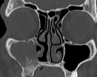
3 DRAINAGE PATHWAYS Ostiomeatal Units Anterior Middle Meatus Frontal sinus Maxillary sinus Anterior Ethmoid air cells Posterior Superior Meatus Sphenoid sinus Posterior ethmoid air cells


4 Ostio-Meatal Unit Anterior OMU Middle Meatus Frontal sinus Ostium & Recess Maxillary sinus ostium Infundibulum Anterior Ethmoid cells


5 Sphenoethmoidal Recess Posterior OMU Sphenoid sinus Ostium Sphenoethmoidal Recess Posterior Ethmoid cells Superior Meatus

 Planum")
6 Ventral Skull Base Nasal vault: Cribriform plate Ethmoid Fovealis (frontal bone) Planum Sphenoidale

7 Skull Base Pterygo-Maxillary Fissure Pterygo-Palatine Fossa (PPF) Foramen Rotundum Cavernous sinus Inferior orbital fissure Orbital Apex

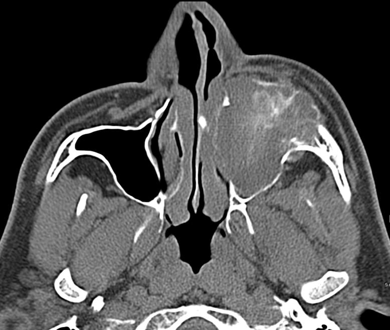
8 PRACTICAL APPROACH Sinonasal Imaging Pathology Location Pattern CT MR


9 Approach to CT Calcification Fungus ball center, punctate Concretion of CRS: periphery, egg-shell, marginated

10 Approach to CT Calcification Chondroid/Osteoid Matrix Bone Destruction or Dehiscence
11 Approach to CT Attenuation Low density: Mucoid, Fluid, Polyps Hyperdense: Fungus, Concretion, Blood Soft tissue: Neoplasm, Scar, Thick Secretion
12 Approach to CT Bone changes Deficiency/Dehiscence Long standing Mucocele, Polyps, IP Slow growing neoplasm Schwannoma?Cephalocele Destruction/Erosion Aggressive Tumor SCCA, SNUC, SNEC, esthesioneuroblastoma Lymphoma, RCC met Osteomyelitis sinusitis Invasive fungal sinusitis Granulomatous disease


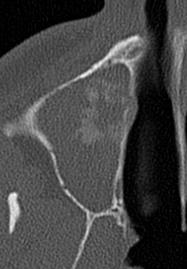
13 Fibrous Dysplasia Begin and End with CT
14 Why MR? Pathology Location Pattern CT MR


15 SINONASAL IMAGING Begin and end with CT: Bone change Bone Matrix MR: Tissue characteristic Extent Intracranial
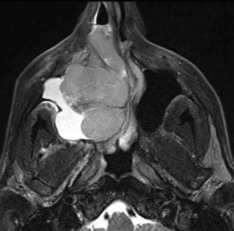
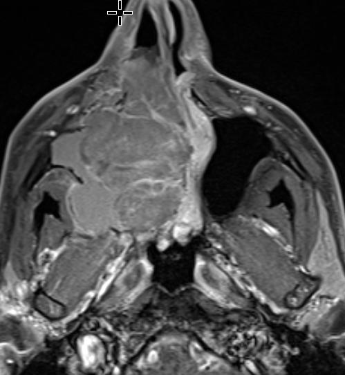
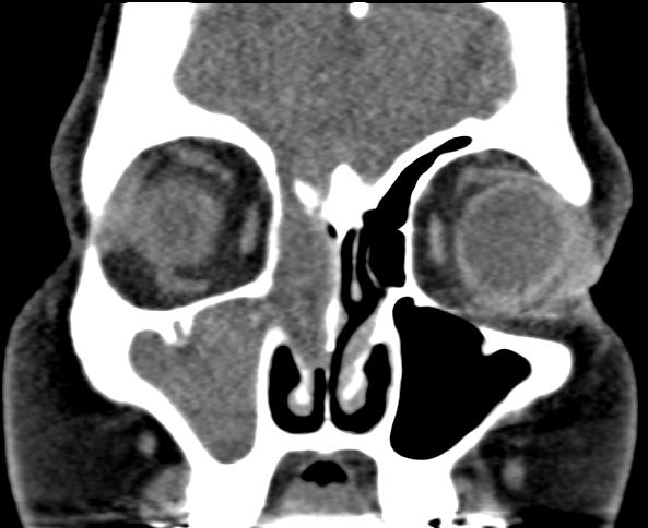
16 Approach to MR T2, Peripheral Gd + T2, Solid Gd + Retention cyst Submucosal Mucinous/Serous gland collection Partially aerated sinus Polyp Fluid deep to lamina propria Mass effect Mucocele Trapped secretion in obstructed sinus Airless Expanded sinus Neoplasm


17 Sinonasal Polyposis Frontal Mucocele Polypoid T2 Hyperintensity Polyps MR Pitfall: concretion & fungus signal void Severe Deficiency at skull base mimics Destruction Mucocele, Sinonasal Polyposis, Inverted Papilloma


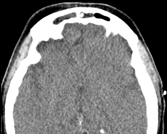
18 Acute Sinusitis Complications T2 hyperintensity NOT tumor Osteomyelitis Sinus Epidural empyema Meningitis, Cerebritis

19 Tumor Mapping

20 PATTERN Approach Diffuse/Pan-sinus Rhinosinusitis Focal Obstructive pattern Rhinosinusitis Neoplasm
21 RhinoSinusitis Poor Correlation of Symptoms with CT/Endoscopy Acute RS: 1-4 weeks Bacterial infection Fluid level Chronic RS: 12 weeks Multiple factors, Idiopathic, Allergy, Impaired Cilliary function, Granulomatous disease Hypertrophic Mucosa, Polyp, Scar, Atrophy Osteitis Neo-osteogenesis


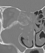
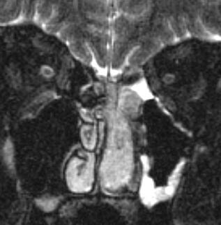
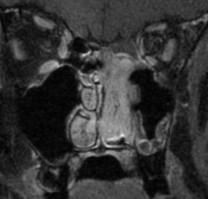
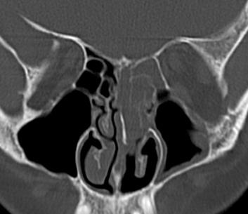
22 Chronic Rhino-Sinusitis Neo-osteogenesis Cystic Fibrosis o o Bronchiectasis Hypoplastic sinuses Wegener, Sarcoid, Churg Strauss o Chronic inflammatory/ granulomatous destruction o Systemic disease


23 Pan-sinusitis Polypoid Opacification Allergic Rhinosinusitis Sinonasal Polyposis Allergic Fungal Sinusitis Jack Jill
24 Fungal Sinusitis Immuno-competent Non-invasive Mycetoma Allergic Fungal Sinusitis Immuno-compromised Invasive Acute Immunocompromised, DM Chronic DM
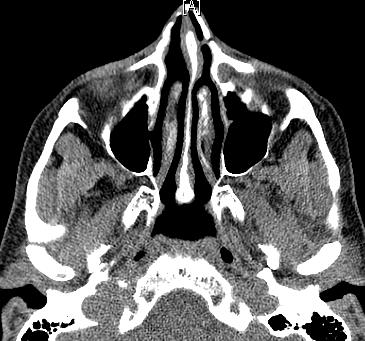
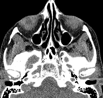
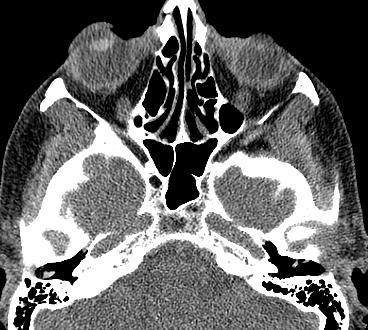
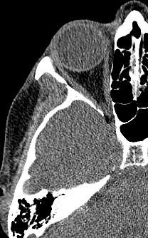
25 Allergic Fungal Sinusitis High density NOT tumor Inspissated secretion or fungal Focal or Diffuse Allergy, Fungus-specific IgE, Allergic Mucin Debridement, Path: no invasion of mucosa Rx steroid


26 Acute Invasive Fungal Sinusitis Mucormycosis 25 yo ALL Path shows Fungal invasion: Mucosa dark ulcer Vessel vasculitis, thrombosis, hemorrhage, tissue infaction Invasion of Orbit & CNS


27 Angioinvasive Fungal Sinusistis Mycotic Aneurysm 68 yo NHL on Chemotherapy, ESRD on HD. Acute right eye ptosis blurry vision


28 Chronic Invasive Fungal Sinusitis 3 WEEKS Epidural abscess, Meningitis, Cerebritis, Abscess Slowly progressive, low-grade invasive fungal infection Path: Necrosis of the mucosa, submucosa, and blood vessels, with low-grade inflammation

29 PATTERN Diffuse Pansinus Rhinosinusitis Focal Obstructive pattern Rhinosinusitis Neoplasm


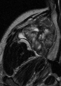
30 OMU obstructive lesion Inverted Papilloma Cerebriform pattern can be seen with other neoplasm? Antrochoanal polyp
31 PRACTICAL APPROACH Sinonasal Imaging Pathology Location Pattern CT MR
32 Approach to Sinonasal Neoplasm Location Pathology Imaging feature Clinical presentation
33 Sinonasal Neoplasm Most common locations for Primary CA: Maxillary sinus > Nasal cavity > Ethmoid cells Frontal/Sphenoid < 2 % Most common tumors in Adults SCCA >> Esthesioneuroblastoma, Melanoma, Adenoid cystic carcinoma Odontogenic (Odontoma, Ameloblastoma) Osteoid/Chondroid Fibrous dysplasia Osteo/Chondro-Sarcoma
34 Obstructive Lesions OMU: Infundibulum-Maxillary ostium Frontal recess Nasal cavity Naso-ethmoidal Sphenoid sinus


35 Nasal Cavity lesion Nasal septum Lateral nasal wall Inferior turbinate T1 of Melanin Melanoma
36 Esthesioneuroblastoma Widening of Nasal vault Intermediate T2 Enhancement (Similar to mucosa)


37 Sphenoid sinus Rarely sinonasal tumor Adjacent process Fungal Sinusitis Pituitary adenoma Clival/skull base lesion AFS

38 Osteosarcoma Focal disease Maxillary sinus Sunburst periostitis

: Without or")
39 Ameloblastoma Soap-Bubble lesion Hard, painless yo. 2 nd most common odontogenic lesion 20 % Maxilla. 20% associated with Dentigerous cyst & unerupted teeth Locally aggressive, high recurrence Simple or luminal (mural): Without or with nodule(s) in the wall of the cyst


40 Nasal obstruction Refractory seizure x 13 years


41 Juvenile Nasopharyngeal Angiofibroma Internal maxillary artery feeder Tumor starts in the nose, spreads to NP Benign, locally invasive Adolescent male Tx: preop embolization resection adjuvant radiation for unresectable intracranial disease
42 References Daniels DL, et al. The Frontal Sinus Drainage Pathway and Related Structures. AJNR 2003 Sep;24(8): Huang BY, et at. Failed Endoscopic Sinus Surgery: Spectrum of CT Findings in the Frontal Recess. Radiographics 2009 Younis R et at. The role of computed tomography and magnetic resonance imaging in patients with sinusitis with complications. Laryngoscope 2002;112(2): Stewart MG, et al. Chronic sinusitis: symptoms versus CT scan findings. Curr Opin Otolaryngol Head Neck Surg 2004 Feb;12(1):27-9. Som PM, et al. Sinonasal tumors and inflammatory tissues: differentiation with MR imaging. Radiology 1988;167(3): Yousem DM. Imaging of sinonasal inflammatory disease. Radiology 1993;188(2): Yoon JH, et al. Calcification in Chronic maxillary sinusitis: comparison of CT findings with histopathologic results. AJNR 1999;20: Ilica AT, et al. Clinical and Radiologic features of fungal diseases of the paranasal sinuses. Comput Assist Tomogr Sep;36(5):570-6 Loevner LA, Sonners AI. Imaging of neoplasms of the paranasal sinuses. Neuroimaging Clin N Am 2004 Nov;14(4): Jeon T.Y, et al. Sinonasal Inverted Papilloma: Value of Convoluted Cerebriform Pattern on MR Imaging. AJNR 29:
Sinonasal Imaging. Mamdouh Mahfouz MD Professor of Radiology Cairo University. ssregypt.com
 Sinonasal Imaging Mamdouh Mahfouz MD Professor of Radiology Cairo University ssregypt.com Scanning Techniques Routine Study CORONAL Coronal 3-5mm sections from the posterior wall of the sphenoid sinus
Clinical and imagistic correlations in the inflammatory pathology of nasosinusal cavities
 Romanian Journal of Rhinology, Volume 8, No. 29, January-March 2018 ORIGINAL STUDY DOI: 10.2478/rjr-2018-0003 Clinical and imagistic correlations in the inflammatory pathology of nasosinusal cavities Emilia
Juvenile Angiofibroma
 Juvenile Angiofibroma Disclaimer The pictures used in this presentation have been obtained from a number of sources. Their use is purely for academic and teaching purposes. The contents of this presentation
Sinonasal Tumors. Objectives. Objectives. Incidence of Paranasal Sinus Tumors. Demographics of Paranasal Sinus Tumors. Paranasal Sinus Tumors
 Sinonasal Tumors Objectives Incidence and demographics of sinonasal tumors Separating tumors from inflammatory changes Common and notable histologic types of sinonasal tumors Staging of sinonasal tumors
Imaging of the Paranasal Sinuses
 14. Sommerschule Imaging of the Paranasal Sinuses Bettlach 24.08.2018 Christoph Schlegel Conventional Radiology NNH-Status: okzipito-frontal: frontal sinus, anterior ethmoid okzipito-nasal : maxillary
Boundaries Septum Turbinates & Meati Lamellae Drainage Pathways Variants
 The Fastest 20 Minutes in Michelle A. Michel, MD Professor of Radiology and Otolaryngology Medical College of Wisconsin, Milwaukee Overview Nasal cavity Anterior skull base Ostiomeatal complex Frontal
Neuroradiology Case of the Day
 Neuroradiology Case of the Day 76 th CAR Annual Meeting, Montreal, Quebec April 27, 2013 Eugene Yu, MD Assistant Professor of Radiology and Otolaryngology-Head and Neck Surgery Head and Neck Imaging Princess
Reasons for Failure and Surgical Revisions. Stil Kountakis, MD, PhD Professor and Chief, Division of Rhinology
 Reasons for Failure and Surgical Revisions Stil Kountakis, MD, PhD Professor and Chief, Division of Rhinology Medical College of Georgia of Georgia Regents University Department of Otolaryngology / Head
Nasal Polyposis. DEPARTMENT OF ENT K.S.Hegde Medical Academy Deralakatte, Mangalore
 Nasal Polyposis DEPARTMENT OF ENT K.S.Hegde Medical Academy Deralakatte, Mangalore Def: INTRODUCTION Chronic inflammatory disease of the mucous membrane in the nose & PNS, presenting as pedunculated smooth
Case Studies in the Skull Base
 Case Studies in the Skull Base Amy C Tsai, MD Neuroradiology Fellow Department of Radiology and Imaging Sciences University of Utah Health Sciences Center Salt Lake City, Utah, USA No disclosures related
Paranasal Sinuses: Neoplastic Lesions
 Pravin Mundada Department of Radiology, Geneva University Hospital, Switzerland Paranasal Sinuses: Neoplastic Lesions ESHNR 2017 Lisbon, Portugal Layout of the presentation Clinical & imaging features
RADIOLOGY TEACHING CONFERENCE
 RADIOLOGY TEACHING CONFERENCE John Athas, MD Monica Tadros, MD Columbia University, College of Physicians & Surgeons Department of Otolaryngology- Head & Neck Surgery September 27, 2007 CT SCAN IMAGING
Histopathology of Nasal Masses
 ORIGINAL ARTICLE Histopathology of Nasal Masses 1 Hemant Chopra, 2 Kapil Dua, 3 Neha Chopra, 4 Vikrant Mittal AIJCR Histopathology of Nasal Masses 1 Professor and Head, Dayanand Medical College, Ludhiana,
Commen Nose Diseases
 Commen Nose Diseases Symptoms List: Nasal obstruction. Nasal discharge: Anterior (Rhinorrhea). Posterior (Postnasal discharge). Epistaxis. Hyposmia and Anosmia. Headache. Snoring. Nasal Obstruction Definition:
OSTEITIS IN CRS. Rhinology Chair Meeting presented by Amal Binhazza a
 OSTEITIS IN CRS Rhinology Chair Meeting presented by Amal Binhazza a ROAD MAP Definition. pathophysiology. Diagnosis. Grading systems. Clinical implications. Management. OSTEITIS Presence of new bone formation,
Destructive Giant Maxillary Sinus Mucocele: A Case Report
 Destructive Giant Maxillary Sinus Mucocele: A Vahit Mutlu 1, Ozgur Yoruk 1, Ozalkan Ozkan 2 1 Atatürk University Faculty of Medicine, Department of Ears, Nose and Throat, Erzurum, Turkey 2 Erzincan University
FESS imaging - the role of MDCT
 FESS imaging - the role of MDCT Poster No.: C-0179 Congress: ECR 2013 Type: Educational Exhibit Authors: J. Plascak, K. Makaruha, B. Klasic, L. Kavur, V. Vidjak; Zagreb/HR Keywords: Image verification,
The Nose and Sinuses. Ophir Ilan, MD, PhD Department of Otolaryngology/Head&Neck surgery Hadassah University Hospital
 The Nose and Sinuses Ophir Ilan, MD, PhD Department of Otolaryngology/Head&Neck surgery Hadassah University Hospital Nasal Mucociliary System Function of the Nasal Mucosa warming and humidifying the
Conventional Sinus Surgery Vs Fess
 IOSR Journal of Dental and Medical Sciences (IOSR-JDMS) e-issn: 2279-0853, p-issn: 2279-0861.Volume 16, Issue 7 Ver. III (July. 2017), PP 44-51 www.iosrjournals.org Conventional Sinus Surgery Vs Fess *
Radiological anatomy of frontal sinus By drtbalu
 2009 Radiological anatomy of frontal sinus By drtbalu Anatomy of frontal sinus is highly variable. Precise understanding of these variables will help a surgeon to avoid unnecessary complications during
Rhinosinusitis. John Ramey, MD Joseph Russell, MD
 Rhinosinusitis John Ramey, MD Joseph Russell, MD Disclosure Statement RSFH as a continuing medical education provider, accredited by the South Carolina Medical Association, it is the policy of RSFH to
Chapter Five. 1 of 8 11/3/2008 2:52 PM.
 1 of 8 11/3/2008 2:52 PM Email : myousefmian@hotmail.com Chapter Five FRONT COVER Introduction Acknowledgement CHAPTERS Chapter One Chapter Two Chapter Three Chapter Four Chapter Five Chapter Six Chapter
A CONTRIBUTION TO THE ETIOPATHOGENESIS, DIAGNOSIS AND MANAGEMENT OF SINONASAL INVERTED PAPILLOMAS
 UNIVERSITY OF MEDICINE AND PHARMACY OF TÂRGU MUREȘ ROMANIA A CONTRIBUTION TO THE ETIOPATHOGENESIS, DIAGNOSIS AND MANAGEMENT OF SINONASAL INVERTED PAPILLOMAS PhD THESIS ABSTRACT PhD Student Viorel Emanoil
Aggressive Inflammatory and Neoplastic Processes of the Paranasal Sinuses
 Aggressive Inflammatory and Neoplastic Processes of the Paranasal Sinuses Michael J. Hartman, MD*, Lindell R. Gentry, MD KEYWORDS Sinus Nasal Neoplasms Infection Aggressive KEY POINTS A thorough knowledge
Nasal region. cartilages: septal cartilage (l); lateral nasal cartilage (2); greater alar cartilages (2); lesser alar cartilages (?
; lateral nasal cartilage (2); greater alar cartilages (2); lesser alar cartilages (?") Nasal region skull bones: nasal and frontal processes of maxilla cartilages: septal cartilage (l); lateral nasal cartilage (2); greater alar cartilages (2); lesser alar cartilages (?) 1 Nasal cavity Roof
Nasal region skull bones: nasal and frontal processes of maxilla cartilages: septal cartilage (l); lateral nasal cartilage (2); greater alar cartilages (2); lesser alar cartilages (?) 1 Nasal cavity Roof
DISCLOSURES LEARNING OBJECTIVES WE WILL NOT DISCUSS. CSB: Birdseye View MESSAGE NAVIGATING THE SELLA AND CENTRAL SKULL BASE
 NAVIGATING THE SELLA AND CENTRAL SKULL BASE Christopher P. Hess, M.D., Ph.D. DISCLOSURES Research Support, General Electric SLIDES: http://www.radiology.ucsf.edu/research/meetings/rsna LEARNING OBJECTIVES
Tumors of the Paranasal Sinuses:
 Tumors of the Paranasal Sinuses: Approaches to Diagnostic Imaging Nir J. Harish September 2007 Head and Neck Cancers Oral cavity Pharynx Larynx Nasal cavity Paranasal sinuses Salivary glands Incidence
FRONTAL SINUPLASTY P R E P A R E D A N D P R E S E N T E D B Y : D R. Y A H Y A F A G E E H R 4 16/ 12/ 2013
 FRONTAL SINUPLASTY P R E P A R E D A N D P R E S E N T E D B Y : D R. Y A H Y A F A G E E H R 4 16/ 12/ 2013 ANATOMY: FRONTAL SINUS Not present at birth Starts developing at 4 years Radiographically visualized
Omran Saeed. Luma Taweel. Mohammad Almohtaseb. 1 P a g e
 2 Omran Saeed Luma Taweel Mohammad Almohtaseb 1 P a g e I didn t include all the photos in this sheet in order to keep it as small as possible so if you need more clarification please refer to slides In
Rhinosinusitis: A Prospective Study
 االله الرحمن الرحيم بسم Pathological l & Radiological i l Evidences in the Pathogenesis es s of Chronic Rhinosinusitis: A Prospective Study Prof. Dr. H. ABDEL FATAH, MD A. Prof. Dr. K. NOWEAM, MD Dr. Z.
Allergic fungal rhinosinusitis: detection of fungal DNA in sinus aspirate using polymerase chain reaction
 1- Allergic fungal rhinosinusitis: detection of fungal DNA in sinus aspirate using polymerase chain reaction Abstract Objective: This study investigated allergic fungal rhinosinusitis cases, and aimed
Extranodal Natural Killer/T-Cell Lymphoma Nasal Type: Detection by Computed Tomography Features
 The Laryngoscope VC 2014 The American Laryngological, Rhinological and Otological Society, Inc. Extranodal Natural Killer/T-Cell Lymphoma Nasal Type: Detection by Computed Tomography Features Yin-Ping
Dr.Adel A. Al Ibraheem
 Rhinology Chair Weekly Activity Dr.Adel A. Al Ibraheem www.rhinologychair.org conference@rhinologychair.org Rhinology Chair Introduction: It is important to classify and stage nasal polyposis. ( decide
Mucocele of paranasal sinuses
 From the SelectedWorks of Balasubramanian Thiagarajan March 7, 2012 Mucocele of paranasal sinuses Balasubramanian Thiagarajan Available at: https://works.bepress.com/drtbalu/57/ Mucoceles of paranasal
Provider Led Entity. CDI Quality Institute PLE Rhinosinusitis AUC 12/04/2018
 Provider Led Entity CDI Quality Institute PLE Rhinosinusitis AUC 12/04/2018 Appropriateness of advanced imaging procedures* in patients with rhinosinusitis and the following clinical presentations or diagnoses:
Tomographical Findings in Adult Patients Undergoing Endoscopic Sinus Surgery Revision
 THIEME Original Research 73 Tomographical Findings in Adult Patients Undergoing Endoscopic Sinus Surgery Revision Jan Alessandro Socher 1 Jonas Mello 2 Barbara Batista Baltha 2 1 Department of Otorhinolaryngology,
White Paper: Balloon Sinuplasty for Chronic Sinusitis, The Latest Recommendations
 White Paper: Balloon Sinuplasty for Chronic Sinusitis, The Latest Recommendations For Health Plans, Medical Management Organizations and TPAs Executive Summary Despite recent advances in instrumentation
The Role of Computed Tomography in the Evaluation of Paranasal Sinuses Lesions
 ORIGINAL ARTICLE The Role of Computed Tomography in the Evaluation of Paranasal Sinuses Lesions Bhumikaben P. Suthar 1 *, Divya Vaidya 2, Pukhraj P. Suthar 3. 1 Assistant Professor, 2 Third Year Resident,
The Role of Computed Tomography in the Evaluation of Paranasal Sinuses Lesions
 ORIGINAL ARTICLE The Role of Computed Tomography in the Evaluation of Paranasal Sinuses Lesions Bhumikaben P. Suthar 1 *, Divya Vaid 2, Pukhraj P. Suthar 3. 1 Assistant Professor, 2 Third Year Resident,
Corporate Medical Policy
 Corporate Medical Policy Surgical Treatment of Sinus Disease Description of Procedure or Service Sinusitis refers to infection or inflammation of the sinuses, which are small openings in the bones of the
Laurie A. Loevner, MD
 Laurie A. Loevner, MD Chief, Division of Neuroradiology UPHS Professor of Radiology, Otorhinolaryngology: Head & Neck Surgery, Neurosurgery, and Ophthalmology University of Pennsylvania Health System Disclosures
Fungal ball.. Clinical and radiological features DR. AHMED ALTUWAIJRI 1/5/2017
 Fungal ball.. Clinical and radiological features DR. AHMED ALTUWAIJRI 1/5/2017 Fungal Rhinosinusitis (FRS) Rhinosinusitis, is a common disorder affecting approximately 20% of the population at some time
Surgical Management of Sinusitis (What About Balloons?) Relative Indications for Sinus Surgery in Children
 Relative Indications for Sinus Surgery in Children") Surgical Management of Sinusitis (What About Balloons?) Andrew N. Goldberg M.D. Andrew H. Murr M.D. Michael J. Cunningham, M.D. Department of Otolaryngology and Communication Enhancement Children s Hospital
Surgical Management of Sinusitis (What About Balloons?) Andrew N. Goldberg M.D. Andrew H. Murr M.D. Michael J. Cunningham, M.D. Department of Otolaryngology and Communication Enhancement Children s Hospital
Nasal Cavity and Paranasal Sinuses
 Chapter 2 Nasal Cavity and Paranasal Sinuses Introduction Included in this chapter are nasal cavities, frontal sinus, ethmoid complex, sphenoid sinus, and maxillary sinuses. These cavities and sinuses
ISSN: Volume 4 Issue Role of Modified Endoscopic Medial Maxillectomy in persistent chronic maxillary. sinusitis
 ISSN: 2250-0359 Volume 4 Issue 3 2014 Role of Modified Endoscopic Medial Maxillectomy in persistent chronic maxillary sinusitis Thulasidas P ¹ Venkatraman V ² ¹ Sinus and Nose Hospital, Chennai, India,
Vascular. Extravasated blood. Melanocytic. Tattoo. Epidermolysis bullosa. Lichen planus. Pemphigoid Pemphigus Lupus. Candidosis. Surface Epithelial
 Oral Soft Tissue Pathology Epithelial Thickening (white) Combination Erythema migrans Epithelial atrophy (red) Surface Lesions Clinical Impression Enlargements Surface Debris Pigmented Vesicular Ulcerated
PITUITARY PARASELLAR LESIONS. Kim Learned, MD
 PITUITARY PARASELLAR LESIONS Kim Learned, MD DIFFERENTIALS Pituitary Sella Clivus, Sphenoid Sinus Suprasellar Optic chiasm, Hypothalamus, Circle of Willis Parasellar Cavernous Sinus Case 1 17 YEAR-OLD
Role of Modified Endoscopic Medial Maxillectomy in Persistent Chronic Maxillary Sinusitis
 THIEME Original Research 159 Role of Modified Endoscopic Medial Maxillectomy in Persistent Chronic Maxillary Sinusitis Ponnaiah Thulasidas 1 Venkatraman Vaidyanathan 2 1 Department of Otolaryngology Head
A CASE OF A Huge Submandibular Pleomorphic Adenoma
 ISPUB.COM The Internet Journal of Head and Neck Surgery Volume 4 Number 2 S VERMA Citation S VERMA.. The Internet Journal of Head and Neck Surgery. 2009 Volume 4 Number 2. Abstract Pleomorphic adenoma
*in general the blood supply of the nose comes from branches of the internal and external carotid arteries.
 In the previous lecture we talked about the anatomy of the nasal cavity, today we will talk about its blood supply, venous drainage, innervations, and finally about the paranasal sinuses. When we describe
Skull Base Danger Zones in FESS
 Skull Base Danger Zones in FESS Poster No.: C-2278 Congress: ECR 2014 Type: Educational Exhibit Authors: L. Renza Lozada, R. Carreño Gonzalez, G. Quintana Sanchez, 1 2 1 1 1 2 R. E. Figueroa ; Malaga/ES,
Isolated sphenoid inflammatory diseases
 International Journal of Otorhinolaryngology and Head and Neck Surgery http://www.ijorl.com pissn 2454-5929 eissn 2454-5937 Original Research Article DOI: http://dx.doi.org/10.18203/issn.2454-5929.ijohns20183688
National Imaging Associates, Inc. Clinical guidelines/considerations SINUS & MAXILLOFACIAL AREA CT 70486, 70487, 70488
 National Imaging Associates, Inc. Clinical guidelines/considerations SINUS & MAXILLOFACIAL AREA CT 70486, 70487, 70488 Date: September 1997 Page 1 of 5 LIMITED OR LOCALIZED FOLLOW UP - SINUS CT 76380 Guideline
Kathleen R. Fink, MD Virginia Mason Medical Center. 6 th Nordic Emergency Radiology Course 2017
 Kathleen R. Fink, MD Virginia Mason Medical Center 6 th Nordic Emergency Radiology Course 2017 Disclosure My spouse has a financial relationship with a commercial organization that may have a direct or
PROBLEM RECOMMENDATION
 PREVENTION (MINIMIZING) IN ENDOSCOPIC Steven D. Schaefer, MD Professor and Chair Department of Otolaryngology PREVENTION AND Intraoperative Hemorrhage Loss of Orientation Inability to Identify/Preserve
CT OF THE PARANASAL SINUSES : NORMAL ANATOMY, VARIANTS AND PATHOLOGY
 Journal of Optoelectronics and Biomedical Materials Vol.2 Issue 4, October-December 2010, p. 281 289 CT OF THE PARANASAL SINUSES : NORMAL ANATOMY, VARIANTS AND PATHOLOGY AMIT N D DWIVEDI *, KAPIL KUMAR
Bisection of Head & Nasal Cavity 頭部對切以及鼻腔. 解剖學科馮琮涵副教授 分機
 Bisection of Head & Nasal Cavity 頭部對切以及鼻腔 解剖學科馮琮涵副教授 分機 3250 E-mail: thfong@tmu.edu.tw Outline: The structure of nose The concha and meatus in nasal cavity The openings of paranasal sinuses Canals, foramens
Chapter 7: Head & Neck
 Chapter 7: Head & Neck Osteology I. Overview A. Skull The cranium is composed of irregularly shaped bones that are fused together at unique joints called sutures The skull provides durable protection from
Functional Endoscopic Sinus Surgery
 WHAT IS FUNCTIONAL ENDOSCOPIC SINUS SURGERY (FESS)? The nasal telescope has greatly changes the evaluation and treatment of rhino-sinusitis. This instrument, which provides a view of the structures in
NASAL SEPTUM ADENOID CYSTIC CARCINOMA: A CASE REPORT
 NASAL SEPTUM ADENOID CYSTIC CARCINOMA: A CASE REPORT Shu-Yu Tai, 1 Chen-Yu Chien, 2 Chih-Feng Tai, 2,4 Wen-Rei Kuo, 2,4 Wan-Ting Huang, 3 and Ling-Feng Wang 2,4 Departments of 1 Family Medicine, 2 Otolaryngology
objectives Pitfalls and Pearls in PET/CT imaging Kevin Robinson, DO Assistant Professor Department of Radiology Michigan State University
 objectives Pitfalls and Pearls in PET/CT imaging Kevin Robinson, DO Assistant Professor Department of Radiology Michigan State University To determine the regions of physiologic activity To understand
The Pterygopalatine Fossa: Postoperative MR Imaging Appearance
 AJNR Am J Neuroradiol 21:1315 1319, August 2000 The Pterygopalatine Fossa: Postoperative MR Imaging Appearance Ling-Ling Chan, June Chong, Ann M. Gillenwater, and Lawrence E. Ginsberg BACKGROUND AND PURPOSE:
FUNCTIONAL ENDOSCOPIC SINUS SURGERY (FESS)
") FUNCTIONAL ENDOSCOPIC SINUS SURGERY (FESS) Protocol: SUR055 Effective Date: June 1, 2018 Table of Contents Page COMMERCIAL, MEDICAID AND MEDICARE COVERAGE RATIONALE... 1 DEFINITIONS... 2 DESCRIPTION OF
FUNCTIONAL ENDOSCOPIC SINUS SURGERY (FESS) Protocol: SUR055 Effective Date: June 1, 2018 Table of Contents Page COMMERCIAL, MEDICAID AND MEDICARE COVERAGE RATIONALE... 1 DEFINITIONS... 2 DESCRIPTION OF
Anatomic Relations Summary. Done by: Sohayyla Yasin Dababseh
 Anatomic Relations Summary Done by: Sohayyla Yasin Dababseh Anatomic Relations Lecture 1 Part-1 - The medial wall of the nose is the septum. - The vestibule lies directly inside the nostrils (Nares). -
Three-Dimensional Volumetric Display of the Nasal Ostiomeatal Channels and Paranasal Sinuses
 Downloaded from www.ajronline.org by 37.44.202.192 on 12/22/17 from IP address 37.44.202.192. Copyright RRS. For personal use only; all rights reserved Three-Dimensional Volumetric Display of the Nasal
SINUS ANATOMY AND FUNCTION
 EMBRYOLOGY AND DEVELOPMENT SINUS ANATOMY AND FUNCTION -4 th week gestation: -frontonasal process identified, arises over developing forebrain -ectodermal -contributes to nasal capsule -9 th and 10 th week
Skull-2. Norma Basalis Interna Norma Basalis Externa. Dr. Heba Kalbouneh Associate Professor of Anatomy and Histology
 Skull-2 Norma Basalis Interna Norma Basalis Externa Dr. Heba Kalbouneh Associate Professor of Anatomy and Histology Norma basalis interna Base of the skull- superior view The interior of the base of the
PTERYGOPALATINE FOSSA
 PTERYGOPALATINE FOSSA Outline Anatomical Structure and Boundaries Foramina and Communications with other spaces and cavities Contents Pterygopalatine Ganglion Especial emphasis on certain arteries and
Allergic Fungal Rhinosinusitis Involving Frontal Sinus: A Prospective Study comparing Surgical Modalities
 10.5005/jp-journals-10013-1141 H Verma et al ORIGINAL ARTICLE Allergic Fungal Rhinosinusitis Involving Frontal Sinus: A Prospective Study comparing Surgical Modalities H Verma, Rijuneeta, AK Gupta, A Chakrabarti
NEXT STOP : Central Station "Pterygopalatine fossa"
 NEXT STOP : Central Station "Pterygopalatine fossa" Poster No.: C-1359 Congress: ECR 2015 Type: Educational Exhibit Authors: I. Alba de Caceres, A. Paniagua, L. Ibañez, J. A. Blanco ; 1 1 1 1 2 2 Madrid/ES,
Nose & Mouth OUTLINE. Nose. - Nasal Cavity & Its Walls. - Paranasal Sinuses. - Neurovascular Structures. Mouth. - Oral Cavity & Its Contents
 Dept. of Human Anatomy, Si Chuan University Zhou hongying eaglezhyxzy@163.com Nose & Mouth OUTLINE Nose - Nasal Cavity & Its Walls - Paranasal Sinuses - Neurovascular Structures Mouth - Oral Cavity & Its
Imaging: When to get MRI, CT or PET-CT?
 Imaging: When to get MRI, CT or PET-CT? Alina Uzelac, D.O. Assistant Clinical Professor Neuroradiology UCSF Department of Radiology and Biomedical Imaging San Francisco General Hospital Overview CT MRI
Vascular and Parameningeal Infections of the Head and Neck
 Vascular and Parameningeal Infections of the Head and Neck Kevin B. Laupland, MD, MSc, FRCPC Associate Professor Departments of Medicine, Critical Care Medicine, Pathology and Laboratory Medicine, and
The orbit-1. Dr. Heba Kalbouneh Assistant Professor of Anatomy and Histology
 The orbit-1 Dr. Heba Kalbouneh Assistant Professor of Anatomy and Histology Orbital plate of frontal bone Orbital plate of ethmoid bone Lesser wing of sphenoid Greater wing of sphenoid Lacrimal bone Orbital
2017 OPTIONS FOR INDIVIDUAL MEASURES: REGISTRY ONLY. MEASURE TYPE: Efficiency
 Measure #334: Adult Sinusitis: More than One Computerized Tomography (CT) Scan Within 90 Days for Chronic Sinusitis (Overuse) National Quality Strategy Domain: Efficiency and Cost Reduction 2017 OPTIONS
2016 PQRS OPTIONS FOR INDIVIDUAL MEASURES: REGISTRY ONLY
 Measure #334: Adult Sinusitis: More than One Computerized Tomography (CT) Scan Within 90 Days for Chronic Sinusitis (Overuse) National Quality Strategy Domain: Efficiency and Cost Reduction 2016 PQRS OPTIONS
 www.oralradiologists.com CONE BEAM CT REPORT CASE ---- Case Information Referring Doctor: - Patient Name: - Scan Date: December 1, 2015 Patient DOB: - Reason for Exam: - Study Details: icat Flex, 160x160x112
A Study of Anatomical Variations in Patients with Chronic Rhinosinusitis.
 DOI: 10.2127/aimdr.201..2.EN1 Original Article ISSN (O):239-222; ISSN (P):239-21 A Study of Anatomical Variations in Patients with Chronic Rhinosinusitis. Smruti Swain 1 1 Associate Professor, Department
Endoscopic Management Of A Giant Ethmoid Mucocele
 ISPUB.COM The Internet Journal of Otorhinolaryngology Volume 6 Number 1 S Ceylan, F Bora Citation S Ceylan, F Bora.. The Internet Journal of Otorhinolaryngology. 2006 Volume 6 Number 1. Abstract We present
FOR CMS (MEDICARE) MEMBERS ONLY NATIONAL COVERAGE DETERMINATION (NCD) FOR COMPUTED TOMOGRAPHY:
 MEMBERS ONLY NATIONAL COVERAGE DETERMINATION (NCD) FOR COMPUTED TOMOGRAPHY:") National Imaging Associates, Inc. Clinical guidelines SINUS & MAXILLOFACIAL AREA CT LIMITED OR LOCALIZED FOLLOW UP SINUS CT Original Date: September 1997 Page 1 of 5 CPT Codes: 70486, 70487, 70488, 76380
National Imaging Associates, Inc. Clinical guidelines SINUS & MAXILLOFACIAL AREA CT LIMITED OR LOCALIZED FOLLOW UP SINUS CT Original Date: September 1997 Page 1 of 5 CPT Codes: 70486, 70487, 70488, 76380
Richard J. Harvey 26 Wall of the Maxillary Sinus Christos C. Georgalas and Wytske J. Fokkens 27 Pterygopalatine Space...
 Contents............................................................ viii Preface......................................................................... xi Acknowledgments.............................................................
ORIGINAL ARTICLE RELATIONSHIP OF CONCHA BULLOSA WITH OSTEOMEATAL UNIT BLOCKAGE. TOMOGRAPHIC STUDY IN 200 PATIENTS.
 RELATIONSHIP OF CONCHA BULLOSA WITH OSTEOMEATAL UNIT BLOCKAGE. TOMOGRAPHIC STUDY IN 200 PATIENTS. Shrikrishna B H 1, Jyothi A C 2, Sanjay G 3, Sandeep Samson G 4. 1. Associate Professor, Department of
 www.oralradiologists.com CONE BEAM CT REPORT CASE XXXX Patient information Patient Name: - Referring Doctor: - Patient DOB: - Scan Date: [Start date] Reason for Exam: Maxillary facial pain Doctor Notes:
Chapter 7 Part A The Skeleton
 Chapter 7 Part A The Skeleton Why This Matters Understanding the anatomy of the skeleton enables you to anticipate problems such as pelvic dimensions that may affect labor and delivery The Skeleton The
FUNCTIONAL ENDOSCOPIC SINUS SURGERY (FESS)
") UnitedHealthcare Community Plan Medical Policy FUNCTIONAL ENDOSCOPIC SINUS SURGERY (FESS) Policy Number: CS144.D Effective Date: January 1, 2018 Table of Contents Page INSTRUCTIONS FOR USE... 1 BENEFIT
UnitedHealthcare Community Plan Medical Policy FUNCTIONAL ENDOSCOPIC SINUS SURGERY (FESS) Policy Number: CS144.D Effective Date: January 1, 2018 Table of Contents Page INSTRUCTIONS FOR USE... 1 BENEFIT
CT evaluation : odontogenic origin causing obstructive maxillary sinusitis
 CT evaluation : odontogenic origin causing obstructive maxillary sinusitis Poster No.: C-0362 Congress: ECR 2016 Type: Scientific Exhibit Authors: G. Kim; jejusi, jejudo/kr Keywords: Emergency, Interventional
Anatomy #1; Respiratory Nose and the Nasal Cavity December 1st, 2013
 Note #1: the doctor skipped some slides in the lecture. Those slides are not included in this sheet and so you will have to review the slides to study them. The reason they were not included is because
Benign Neoplasms of the Nose
 Department of Otolaryngology Head and Neck Surgery Pursuing Wellness Through Teaching, Learning and Healing Benign Neoplasms of the Nose Ivan El Sayed, MD Disclosure Principal Investigator: Grant Support
Computed tomography road map of the paranasal sinuses for treatment planning
 Computed tomography road map of the paranasal sinuses for treatment planning Poster No.: C-2607 Congress: ECR 2013 Type: Educational Exhibit Authors: N. Schembri, A. S. Gatt, D. Ellul, J. Brunton; Dundee/UK
The View through the Nose: ENT considerations for Pituitary/Skull Base Surgery
 The View through the Nose: ENT considerations for Pituitary/Skull Base Surgery Edsel Kim, M.D. Otolaryngology-Head and Neck Surgery The Oregon Clinic Providence Brain and Spine Institute Pituitary, Thyroid
ORIGINAL ARTICLE. Group Chandler Maloney. First Inflammatory oedema Preseptal cellulitis. Second Orbital Cellulitis Subperiosteal abscess
 ORBITAL MANIFESTATIONS OF SINUS DISEASE T. Jyothirmayi 1, V. Meenakshi 2, M. Deepika 3 HOW TO CITE THIS ARTICLE: T. Jyothirmayi, V. Meenakshi, M. Deepika. Orbital Manifestations of Sinus Disease. Journal
Incidence of accessory ostia in patients with chronic maxillary sinusitis
 International Journal of Otorhinolaryngology and Head and Neck Surgery Ghosh P et al. Int J Otorhinolaryngol Head Neck Surg. 2018 Mar;4(2):443-447 http://www.ijorl.com pissn 2454-5929 eissn 2454-5937 Original
PLEOMORPHIC ADENOMA OF LATERAL WALL OF NOSE A RARE PRESENTATION
 ISSN: 2250-0359 Volume 4 Issue 1 2014 PLEOMORPHIC ADENOMA OF LATERAL WALL OF NOSE A RARE PRESENTATION *USHA KUMAR MAHESH *RATNAKAR MADHAVARAO POTEKAR * B.L.D.E UNIVERSITY ABSTRACT: The aim of the article
FUNCTIONAL ENDOSCOPIC SINUS SURGERY (FESS)
") UnitedHealthcare Oxford Clinical Policy FUNCTIONAL ENDOSCOPIC SINUS SURGERY (FESS) Policy Number: ENT 022.5 T2 Effective Date: April 1, 2018 Table of Contents Page INSTRUCTIONS FOR USE... 1 CONDITIONS
UnitedHealthcare Oxford Clinical Policy FUNCTIONAL ENDOSCOPIC SINUS SURGERY (FESS) Policy Number: ENT 022.5 T2 Effective Date: April 1, 2018 Table of Contents Page INSTRUCTIONS FOR USE... 1 CONDITIONS
Bones of the skull & face
 Bones of the skull & face Cranium= brain case or helmet Copyright The McGraw-Hill Companies, Inc. Permission required for reproduction or display. The cranium is composed of eight bones : frontal Occipital
JMSCR Vol 05 Issue 09 Page September 2017
 www.jmscr.igmpublication.org Impact Factor 5.84 Index Copernicus Value: 71.58 ISSN (e)-2347-176x ISSN (p) 2455-0450 DOI: https://dx.doi.org/10.18535/jmscr/v5i9.52 Relationship of Agger Nasi Cell and Uncinate
Dr.Ban I.S. head & neck anatomy 2 nd y جامعة تكريت كلية طب االسنان مادة التشريح املرحلة الثانية أ.م.د. بان امساعيل صديق 6102/6102
 جامعة تكريت كلية طب االسنان مادة التشريح املرحلة الثانية أ.م.د. بان امساعيل صديق 6102/6102 Pterygopalatine fossa: The pterygopalatine fossa is a cone-shaped depression, It is located between the maxilla,
Cranium Facial bones. Sternum Rib
 Figure 7.1 The human skeleton. Skull Thoracic cage (ribs and sternum) Cranium Facial bones Sternum Rib Bones of pectoral girdle Vertebral column Sacrum Vertebra Bones of pelvic girdle (a) Anterior view
Transnasal Endoscopic Sinonasal Surgery
 Reda kamel, Cadaveric dissection 1 Transnasal Endoscopic Sinonasal Surgery Cadaver Dissection Guide For Endoscopic Sinus Surgery Cairo University Egypt Reda Kamel Professor of Rhinology Cairo University