Juvenile Angiofibroma
|
|
|
- Winfred Gilbert
- 6 years ago
- Views:
Transcription
1 Juvenile Angiofibroma
2 Disclaimer The pictures used in this presentation have been obtained from a number of sources. Their use is purely for academic and teaching purposes. The contents of this presentation do not have any intended commercial use. In case the owner of any of the pictures has any objection and seeks their removal please contact at dineshkumarsharma@gmail.com. These pictures will be removed immediately.
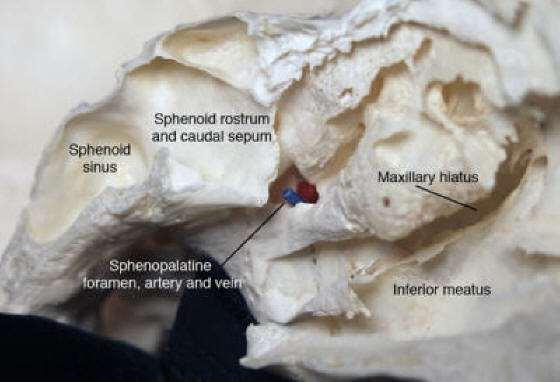
3 Sphenopalatine Foramen
4

5
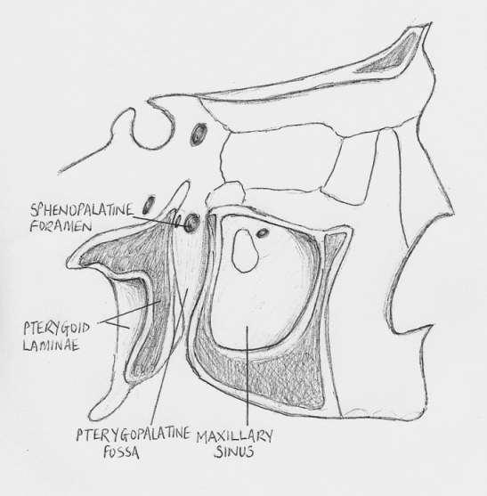
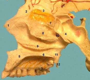
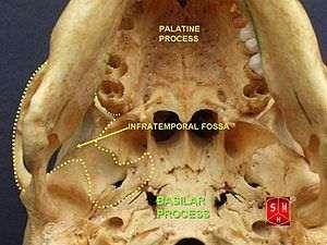
6 Pterygopalatine Fossa
7 Laterally with the infratemporal fossa through the pterygomaxillary fissure Anteriorly to the orbit via the infraorbital fissure Posteriorly to the middle cranial fossa through the foramen rotundum and the pterygoid canal Medially to the inferior portion of the sphenoethmoid recess through the sphenopalatine foramen To the oral cavity via the greater and lesser palatine foramina.
8 Infratemporal Fossa
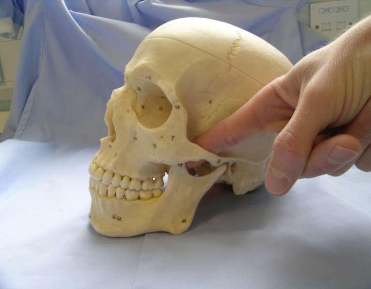
9 Infratemporal Fossa
10 Infratemporal Fossa

11 Infratemporal Fossa
12 Juvenile nasopharyngeal angiofibroma (JNA) It is a rare, benign, vascular neoplasm that accounts for less than 0.5% of all head and neck tumours. JNAs occur almost exclusively in the nasopharynx of adolescent males. The site of origin of JNA remains controversial.
13 Age: Onset most commonly is in the second decade; range is 7-21 years. Mean age at diagnosis is 14 years May regress in late teens but may persist into adulthood Rare in patients older than 25 years.
14 Etiopathogenesis Many theories have been propounded but none is entirely convincing Hormonal theory has been suggested due to the lesion's occurrence in adolescent males Other theories include: Desmoplastic response of the nasopharyngeal periosteum or Embryonic fibrocartilage between the basiocciput and the basisphenoid Harmatomas Nest cells [undiff. epitheloid] Vestiges of atrophied stapedial artery
15 Site of Origin Superior lip of the sphenopalatine foramen at the junction of the pterygoid process of the sphenoid bone and the sphenoid process of the palatine bone. Bone of the vidian canal

16 Gross Examination Usually sessile, lobulated, rubbery, and red-pink to tan-gray in appearance. In rare cases, the tumor is polypoid or pedunculated Usually is encapsulated and composed of vascular tissue and fibrous stroma with coarse or fine collagen fibers.
17 Spread JNAs are slow growing and initially expand intranasally into the nasopharynx and nasal cavity and then into the pterygomaxillary space. Over time, JNAs will eventually erode bone and invade the infratemporal fossa, orbit, and middle cranial fossa.

18 The blood supply to these benign tumours is most commonly from the internal maxillary artery. May also be supplied by the: External carotid artery Internal carotid artery Common carotid artery Ascending pharyngeal artery Blood Supply
19 Structure Histologically, JNAs originate from myofibroblasts. The tumour spreads submucosally. It is composed of a fibrous abundance of single endothelial cell lined vascular spaces or channels. These channels are surrounded by a collagenous tissue network and lack a complete muscular layer.

20 Histopathology The tumour is composed of a variable admixture of blood vessels and fibrous tissue
21 Presentation Unilateral nasal obstruction. Epistaxis Nasopharyngeal mass in adolescent males with an average age of onset of 15 years of age
22 Not so rare presentations Conductive hearing loss Dacrocystits Rhinolalia Hard and soft palate deformity Hyposmia or anosmia

23 Advanced lesions may cause Facial swelling Proptosis Cranial neuropathy Massive hemorrhage

24 Investigations Plain x-ray: View of the sinuses may demonstrate nasopharyngeal polyp. Bowing of the posterior wall of the maxillary sinus and maxillary sinus opacification is very suggestive of JNA. Newer radiographic modalities have surpassed plain films in usefulness
25 Holman-Miller sign CT Scan is excellent for evaluation of bone detail and will enhance with contrast. The characteristic anterior bowing of the posterior maxillary wall due to the presence of a mass in the pterygomaxillary space known as the Holman-Miller sign is a finding noted on CT Scan.
26
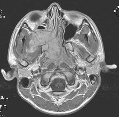
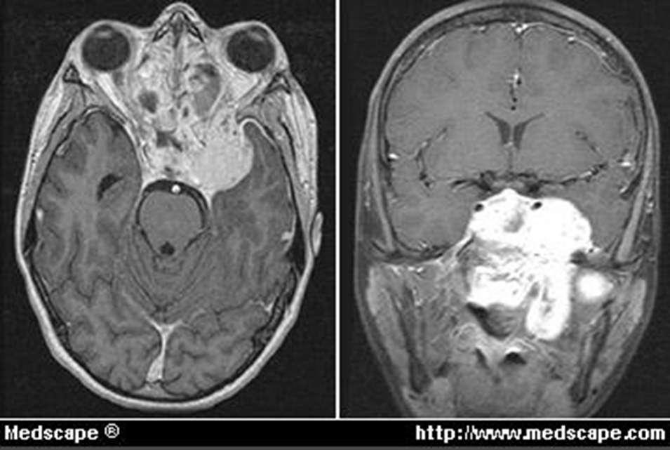
27 MRI
28
29
30
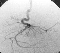
31 Angiography Angiography confirms the hyper vascularity of the lesion, which is supplied by a hypertrophic maxillary artery (arrow).
32 A patient presenting with the above described signs and symptoms should not undergo biopsy due to the risk of bleeding.
33 Staging: Classification according to Fisch Stage I - Tumors limited to nasal cavity, nasopharynx with no bony destruction Stage II - Tumors invading pterygomaxillary fossa, paranasal sinuses with bony destruction Stage III - Tumors invading infratemporal fossa, orbit and/or parasellar region remaining lateral to cavernous sinus Stage IV - Tumors invading cavernous sinus, optic chiasmal region, and/or pituitary fossa
34 Differentials Other causes of nasal obstruction: Nasal polyps Antrochoanal polyp Teratoma Encephalocele Dermoids Inverting papilloma Rhabdomyosarcoma Squamous cell carcinoma
35 Treatment options for JNAs Surgery Radiation therapy Chemotherapy Hormone therapy Surgery is the gold standard of treatment
36 External beam radiation Generally reserved for larger and/or unressectable tumors and tumors that are life threatening do to their location. Reason for limited use of radiation: Carcinogenic side effects Growth retardation Temporal lobe radionecrosis Panhypopituitarism Cataracts Radiation induced keratopathy
37 Chemotherapy is used when previous surgery and radiation have failed. Hormone therapy has been proposed due to the androgen receptors associated with JNAs in an attempt to decrease tumour size and vascularity. Estrogen has been shown to decrease size and vascularity of the tumour, but has feminizing side effects.
38 The treatment of choice in the vast majority of patients is surgical resection. Preoperative selective arterial embolization of feeding vessels from the external carotid artery has significantly decreased intraoperative blood loss and facilitated resection of larger tumors. Embolization is typically performed hours prior to resection. Materials often used include gelfoam and polyvinyl alcohol foam. Gelfoam lasts approximately two weeks, while polyvinyl alcohol foam is more permanent.

39 Pre and Post Embolization


40 Lateral rhinotomy, transpalatal, transmaxillary, or sphenoethmoidal route is used for small tumors (Fisch stage I or II).
41 Endoscopic Excision It has been suggested that tumors involving the ethmoid, maxillary, or sphenoid sinus, the sphenopalatine foramen, nasopharynx, or pterygomaxillary fossa and have limited extension into the infratemporal fossa are amenable to endoscopic resection. JNAs that involve the orbit or middle cranial fossa are not ideal for endoscopic excision
42 Spontaneous Regression JNAs have the potential to regress which usually occurs when the patient is years old. Complete regression does not occur in all patients. Spontaneous regression is valuable for residual tumor following treatment. Recurrence rates have been reported between 30 and 50%.
PTERYGOPALATINE FOSSA
 PTERYGOPALATINE FOSSA Outline Anatomical Structure and Boundaries Foramina and Communications with other spaces and cavities Contents Pterygopalatine Ganglion Especial emphasis on certain arteries and
Dr.Ban I.S. head & neck anatomy 2 nd y جامعة تكريت كلية طب االسنان مادة التشريح املرحلة الثانية أ.م.د. بان امساعيل صديق 6102/6102
 جامعة تكريت كلية طب االسنان مادة التشريح املرحلة الثانية أ.م.د. بان امساعيل صديق 6102/6102 Pterygopalatine fossa: The pterygopalatine fossa is a cone-shaped depression, It is located between the maxilla,
Omran Saeed. Luma Taweel. Mohammad Almohtaseb. 1 P a g e
 2 Omran Saeed Luma Taweel Mohammad Almohtaseb 1 P a g e I didn t include all the photos in this sheet in order to keep it as small as possible so if you need more clarification please refer to slides In
Mohammad Hisham Al-Mohtaseb. Lina Mansour. Reyad Jabiri. 0 P a g e
 2 Mohammad Hisham Al-Mohtaseb Lina Mansour Reyad Jabiri 0 P a g e This is only correction for the last year sheet according to our record. If you already studied this sheet just read the yellow notes which
Anatomic Relations Summary. Done by: Sohayyla Yasin Dababseh
 Anatomic Relations Summary Done by: Sohayyla Yasin Dababseh Anatomic Relations Lecture 1 Part-1 - The medial wall of the nose is the septum. - The vestibule lies directly inside the nostrils (Nares). -
Dr. Sami Zaqout, IUG Medical School
 The skull The skull is composed of several separate bones united at immobile joints called sutures. Exceptions? Frontal bone Occipital bone Vault Cranium Sphenoid bone Zygomatic bones Base Ethmoid bone
Benign Neoplasms of the Nose
 Department of Otolaryngology Head and Neck Surgery Pursuing Wellness Through Teaching, Learning and Healing Benign Neoplasms of the Nose Ivan El Sayed, MD Disclosure Principal Investigator: Grant Support
Infratemporal fossa: Tikrit University college of Dentistry Dr.Ban I.S. head & neck Anatomy 2 nd y.
 Infratemporal fossa: This is a space lying beneath the base of the skull between the lateral wall of the pharynx and the ramus of the mandible. It is also referred to as the parapharyngeal or lateral pharyngeal
Temporal region. temporal & infratemporal fossae. Zhou Hong Ying Dept. of Anatomy
 Temporal region temporal & infratemporal fossae Zhou Hong Ying Dept. of Anatomy Temporal region is divided by zygomatic arch into temporal & infratemporal fossae. Temporal Fossa Infratemporal fossa Temporal
Bisection of Head & Nasal Cavity 頭部對切以及鼻腔. 解剖學科馮琮涵副教授 分機
 Bisection of Head & Nasal Cavity 頭部對切以及鼻腔 解剖學科馮琮涵副教授 分機 3250 E-mail: thfong@tmu.edu.tw Outline: The structure of nose The concha and meatus in nasal cavity The openings of paranasal sinuses Canals, foramens
Sinonasal Tumors. Objectives. Objectives. Incidence of Paranasal Sinus Tumors. Demographics of Paranasal Sinus Tumors. Paranasal Sinus Tumors
 Sinonasal Tumors Objectives Incidence and demographics of sinonasal tumors Separating tumors from inflammatory changes Common and notable histologic types of sinonasal tumors Staging of sinonasal tumors
Skull-2. Norma Basalis Interna Norma Basalis Externa. Dr. Heba Kalbouneh Associate Professor of Anatomy and Histology
 Skull-2 Norma Basalis Interna Norma Basalis Externa Dr. Heba Kalbouneh Associate Professor of Anatomy and Histology Norma basalis interna Base of the skull- superior view The interior of the base of the
ISSN: Volume 6 Issue Angiofibroma. Abdelrahman Ezzat, Hussein Roshdy. Al Azhar University- Cairo-Egypt
 ISSN: 2250-0359 Volume 6 Issue 1 2016 Atypical clinical and radiological presentation of Juvenile Nasopharyngeal Angiofibroma Abdelrahman Ezzat, Hussein Roshdy Al Azhar University- Cairo-Egypt ABSTRACT
*in general the blood supply of the nose comes from branches of the internal and external carotid arteries.
 In the previous lecture we talked about the anatomy of the nasal cavity, today we will talk about its blood supply, venous drainage, innervations, and finally about the paranasal sinuses. When we describe
Temporal fossa Infratemporal fossa Pterygopalatine fossa Terminal branches of external carotid artery Pterygoid venous plexus
 Outline of content Temporal fossa Infratemporal fossa Pterygopalatine fossa Terminal branches of external carotid artery Pterygoid venous plexus Boundary Content Communication Mandibular division of trigeminal
Maxilla, ORBIT and infratemporal fossa. Neophytos C Demetriades MD, DDS, MSc Associate professor European University of Cyprus School of Medicine
 Maxilla, ORBIT and infratemporal fossa Neophytos C Demetriades MD, DDS, MSc Associate professor European University of Cyprus School of Medicine MAXILLA Superior, middle, and inferior meatus Frontal sinus
The orbit-1. Dr. Heba Kalbouneh Assistant Professor of Anatomy and Histology
 The orbit-1 Dr. Heba Kalbouneh Assistant Professor of Anatomy and Histology Orbital plate of frontal bone Orbital plate of ethmoid bone Lesser wing of sphenoid Greater wing of sphenoid Lacrimal bone Orbital
MAXILLA, ORBIT & PTERYGOPALATINE FOSSA. Neophytos C Demetriades MD, DDS, MSc Associate professor European University of Cyprus School of Medicine
 MAXILLA, ORBIT & PTERYGOPALATINE FOSSA Neophytos C Demetriades MD, DDS, MSc Associate professor European University of Cyprus School of Medicine Maxilla MAXILLA Superior, middle, and inferior meatus Frontal
Chapter 7: Head & Neck
 Chapter 7: Head & Neck Osteology I. Overview A. Skull The cranium is composed of irregularly shaped bones that are fused together at unique joints called sutures The skull provides durable protection from
Neuroradiology Case of the Day
 Neuroradiology Case of the Day 76 th CAR Annual Meeting, Montreal, Quebec April 27, 2013 Eugene Yu, MD Assistant Professor of Radiology and Otolaryngology-Head and Neck Surgery Head and Neck Imaging Princess
Major Anatomic Components of the Orbit
 Major Anatomic Components of the Orbit 1. Osseous Framework 2. Globe 3. Optic nerve and sheath 4. Extraocular muscles Bony Orbit Seven Bones Frontal bone Zygomatic bone Maxillary bone Ethmoid bone Sphenoid
EXTRACRANIAL MENINGIOMA PRESENTING AS INFRATEMPORAL FOSSA MASS: A CASE SERIES
 Case Series EXTRACRANIAL MENINGIOMA PRESENTING AS INFRATEMPORAL FOSSA MASS: A CASE SERIES Sunil Mathew * 1, Reddy Ravikanth 2, Vijaykishan B 3. ABSTRACT Extradural meningioma occurs as extracranial extension
Trigeminal Nerve Worksheets, Distributions Page 1
 Trigeminal Nerve Worksheet #1 Distribution by Nerve Dr. Darren Hoffmann Dental Gross Anatomy, Spring 2013 We have drawn out each of the branches of CN V in lecture and you have an idea now for their basic
Paranasal Sinuses: Neoplastic Lesions
 Pravin Mundada Department of Radiology, Geneva University Hospital, Switzerland Paranasal Sinuses: Neoplastic Lesions ESHNR 2017 Lisbon, Portugal Layout of the presentation Clinical & imaging features
No IN THE SUPREME COURT OF ALABAMA
 E-Filed 09/22/2017 @ 03:05:41 PM Honorable Julia Jordan Weller Clerk Of The Court No. 1881555 IN THE SUPREME COURT OF ALABAMA Ex parte Doyle Lee Hamm, * * In re. State of Alabama * Petitioner, * Fourth
Anatomy and Physiology. Bones, Sutures, Teeth, Processes and Foramina of the Human Skull
 Anatomy and Physiology Chapter 6 DRO Bones, Sutures, Teeth, Processes and Foramina of the Human Skull Name: Period: Bones of the Human Skull Bones of the Cranium: Frontal bone: forms the forehead and the
Bones of the skull & face
 Bones of the skull & face Cranium= brain case or helmet Copyright The McGraw-Hill Companies, Inc. Permission required for reproduction or display. The cranium is composed of eight bones : frontal Occipital
Skull-2. Norma Basalis Interna. Dr. Heba Kalbouneh Assistant Professor of Anatomy and Histology
 Skull-2 Norma Basalis Interna Dr. Heba Kalbouneh Assistant Professor of Anatomy and Histology Norma basalis interna Base of the skull- superior view The interior of the base of the skull is divided into
Anatomy #1; Respiratory Nose and the Nasal Cavity December 1st, 2013
 Note #1: the doctor skipped some slides in the lecture. Those slides are not included in this sheet and so you will have to review the slides to study them. The reason they were not included is because
NEXT STOP : Central Station "Pterygopalatine fossa"
 NEXT STOP : Central Station "Pterygopalatine fossa" Poster No.: C-1359 Congress: ECR 2015 Type: Educational Exhibit Authors: I. Alba de Caceres, A. Paniagua, L. Ibañez, J. A. Blanco ; 1 1 1 1 2 2 Madrid/ES,
Cranium Facial bones. Sternum Rib
 Figure 7.1 The human skeleton. Skull Thoracic cage (ribs and sternum) Cranium Facial bones Sternum Rib Bones of pectoral girdle Vertebral column Sacrum Vertebra Bones of pelvic girdle (a) Anterior view
OPEN ACCESS ATLAS OF OTOLARYNGOLOGY, HEAD & NECK OPERATIVE SURGERY
 OPEN ACCESS ATLAS OF OTOLARYNGOLOGY, HEAD & NECK OPERATIVE SURGERY INFERIOR MAXILLECTOMY Tumours of the hard palate and superior alveolus may be resected by inferior maxillectomy (Figure 1). A Le Fort
Introduction to Local Anesthesia and Review of Anatomy
 5-Sep Introduction and Anatomy Review 12-Sep Neurophysiology and Pain 19-Sep Physiology and Pharmacology part 1 26-Sep Physiology and Pharmacology part 2 Introduction to Local Anesthesia and Review of
Four cases of Pleomorphic Adenoma of the nasal cavity: An unusual entity
 J. Acad. Indus. Res. Vol. 1(4) September 2012 203 RESEARCH ARTICLE ISSN: 2278-5213 Four cases of Pleomorphic Adenoma of the nasal cavity: An unusual entity Kiran Naik Dept. of ENT, Adichunchanagiri Inst.
Commen Nose Diseases
 Commen Nose Diseases Symptoms List: Nasal obstruction. Nasal discharge: Anterior (Rhinorrhea). Posterior (Postnasal discharge). Epistaxis. Hyposmia and Anosmia. Headache. Snoring. Nasal Obstruction Definition:
Trigeminal Nerve (V)
") Trigeminal Nerve (V) Lecture Objectives Discuss briefly how the face is developed. Follow up the course of trigeminal nerve from its point of central connections, exit and down to its target areas. Describe
Trigeminal Nerve (V) Lecture Objectives Discuss briefly how the face is developed. Follow up the course of trigeminal nerve from its point of central connections, exit and down to its target areas. Describe
Trigeminal Nerve Anatomy. Dr. Mohamed Rahil Ali
 Trigeminal Nerve Anatomy Dr. Mohamed Rahil Ali Trigeminal nerve Largest cranial nerve Mixed nerve Small motor root and large sensory root Motor root Nucleus of motor root present in the pons and medulla
Bones of the Skull Lateral View
 Bones of the Skull Lateral View Frontal Bone Parietal Bone Occipital Bone Temporal Bone Sphenoid Bone Pterion Sutures of the Skull Lateral View Coronal Suture Lambdoid Suture Squamous Suture Sutures of
Nose & Mouth OUTLINE. Nose. - Nasal Cavity & Its Walls. - Paranasal Sinuses. - Neurovascular Structures. Mouth. - Oral Cavity & Its Contents
 Dept. of Human Anatomy, Si Chuan University Zhou hongying eaglezhyxzy@163.com Nose & Mouth OUTLINE Nose - Nasal Cavity & Its Walls - Paranasal Sinuses - Neurovascular Structures Mouth - Oral Cavity & Its
Exclusively endoscopic surgery for juvenile nasopharyngeal angiofibroma
 Otolaryngology Head and Neck Surgery (2007) 137, 492-496 ORIGINAL RESEARCH Exclusively endoscopic surgery for juvenile nasopharyngeal angiofibroma Nilvano A. Andrade, PhD, José Antonio Pinto, MD, Mônica
Bony orbit Roof The orbital plate of the frontal bone Lateral wall: the zygomatic bone and the greater wing of the sphenoid
 Bony orbit Roof: Formed by: The orbital plate of the frontal bone, which separates the orbital cavity from the anterior cranial fossa and the frontal lobe of the cerebral hemisphere Lateral wall: Formed
Skeletal System: Skull.
 Skeletal System: Skull www.fisiokinesiterapia.biz Bones of the Skull SPLANCHNOCRANIUM Nasal (2) Maxilla (2) Lacrimal (2) Zygomatic (2) Palatine (2) Inferior concha (2) Vomer Mandible NEUROCRANIUM Frontal
The sebaceous glands (glands of Zeis) open directly into the eyelash follicles, ciliary glands (glands of Moll) are modified sweat glands that open
 open directly into the eyelash follicles, ciliary glands (glands of Moll) are modified sweat glands that open") The Orbital Region The orbits are a pair of bony cavities that contain the eyeballs; their associated muscles, nerves, vessels, and fat; and most of the lacrimal apparatus upper eyelid is larger and more
The Orbital Region The orbits are a pair of bony cavities that contain the eyeballs; their associated muscles, nerves, vessels, and fat; and most of the lacrimal apparatus upper eyelid is larger and more
Nasal region. cartilages: septal cartilage (l); lateral nasal cartilage (2); greater alar cartilages (2); lesser alar cartilages (?
; lateral nasal cartilage (2); greater alar cartilages (2); lesser alar cartilages (?") Nasal region skull bones: nasal and frontal processes of maxilla cartilages: septal cartilage (l); lateral nasal cartilage (2); greater alar cartilages (2); lesser alar cartilages (?) 1 Nasal cavity Roof
Nasal region skull bones: nasal and frontal processes of maxilla cartilages: septal cartilage (l); lateral nasal cartilage (2); greater alar cartilages (2); lesser alar cartilages (?) 1 Nasal cavity Roof
The Role of Computed Tomography in the Evaluation of Paranasal Sinuses Lesions
 ORIGINAL ARTICLE The Role of Computed Tomography in the Evaluation of Paranasal Sinuses Lesions Bhumikaben P. Suthar 1 *, Divya Vaidya 2, Pukhraj P. Suthar 3. 1 Assistant Professor, 2 Third Year Resident,
Head & Neck Clinical Sub Group. Network Agreed Imaging Guidelines for UAT and Thyroid Cancer. Measure Nos: 11-1C-105i & 11-1C-106i
 Greater Manchester, Lancashire & South Cumbria Strategic Clinical Network & Senate Head & Neck Clinical Sub Group Network Agreed Imaging Guidelines for UAT and Thyroid Cancer Measure Nos: 11-1C-105i &
The Role of Computed Tomography in the Evaluation of Paranasal Sinuses Lesions
 ORIGINAL ARTICLE The Role of Computed Tomography in the Evaluation of Paranasal Sinuses Lesions Bhumikaben P. Suthar 1 *, Divya Vaid 2, Pukhraj P. Suthar 3. 1 Assistant Professor, 2 Third Year Resident,
Abstract. Samuel Hahn, M.D. 1 James N. Palmer, M.D. 1 Nithin D. Adappa, M.D. 1
 19 A Catecholamine-Secreting Skull Base Sinonasal Paraganglioma Presenting with Labile Hypertension in a Patient with Previously Undiagnosed Genetic Mutation Samuel Hahn, M.D. 1 James N. Palmer, M.D. 1
Parotid Gland. Parotid Gland. Largest of 3 paired salivary glands (submandibular; sublingual) Ramus of Mandible. Medial pterygoid.
 Ramus of Mandible. Medial pterygoid.") Parotid region Parotid Gland Largest of 3 paired salivary glands (submandibular; sublingual) Ramus of Mandible Medial pterygoid Cross section of mandible Masseter D S SCM Parotid Gland Mastoid Process
Parotid region Parotid Gland Largest of 3 paired salivary glands (submandibular; sublingual) Ramus of Mandible Medial pterygoid Cross section of mandible Masseter D S SCM Parotid Gland Mastoid Process
Chapter 7 Part A The Skeleton
 Chapter 7 Part A The Skeleton Why This Matters Understanding the anatomy of the skeleton enables you to anticipate problems such as pelvic dimensions that may affect labor and delivery The Skeleton The
Sinonasal Imaging. Mamdouh Mahfouz MD Professor of Radiology Cairo University. ssregypt.com
 Sinonasal Imaging Mamdouh Mahfouz MD Professor of Radiology Cairo University ssregypt.com Scanning Techniques Routine Study CORONAL Coronal 3-5mm sections from the posterior wall of the sphenoid sinus
Vascular supply with angio-ct for superselective intra-arterial chemotherapy in advanced maxillary sinus cancer
 The British Journal of Radiology, 83 (2010), 171 178 PICTORIAL REVIEW Vascular supply with angio-ct for superselective intra-arterial chemotherapy in advanced maxillary sinus cancer 1 N KASHIWAGI, MD,
Nasopharyngeal Carcinoma. Rusty Stevens, MD Christopher Rassekh, MD
 Nasopharyngeal Carcinoma Rusty Stevens, MD Christopher Rassekh, MD Introduction Rare in the US, more common in Asia High index of suspicion required for early diagnosis Nasopharyngeal malignancies SCCA
Imaging of the Paranasal Sinuses
 14. Sommerschule Imaging of the Paranasal Sinuses Bettlach 24.08.2018 Christoph Schlegel Conventional Radiology NNH-Status: okzipito-frontal: frontal sinus, anterior ethmoid okzipito-nasal : maxillary
PLEOMORPHIC ADENOMA OF LATERAL WALL OF NOSE A RARE PRESENTATION
 ISSN: 2250-0359 Volume 4 Issue 1 2014 PLEOMORPHIC ADENOMA OF LATERAL WALL OF NOSE A RARE PRESENTATION *USHA KUMAR MAHESH *RATNAKAR MADHAVARAO POTEKAR * B.L.D.E UNIVERSITY ABSTRACT: The aim of the article
Extranasopharyngeal Angiofibroma Of The Cartilaginous Nasal Septum: Possible Origin From Ectopic Tissue Trapped During Septal Development
 ISPUB.COM The Internet Journal of Otorhinolaryngology Volume 14 Number 1 Extranasopharyngeal Angiofibroma Of The Cartilaginous Nasal Septum: Possible Origin From Ectopic Tissue Trapped During Septal Development
Dr.Noor Hashem Mohammad Lecture (5)
") Dr.Noor Hashem Mohammad Lecture (5) 2016-2017 If the mandible is discarded, the anterior part of this aspect of the skull is seen to be formed by the hard palate. The palatal processes of the maxillae
Dr.Noor Hashem Mohammad Lecture (5) 2016-2017 If the mandible is discarded, the anterior part of this aspect of the skull is seen to be formed by the hard palate. The palatal processes of the maxillae
Structure Location Function
 Frontal Bone Cranium forms the forehead and roof of the orbits Occipital Bone Cranium forms posterior and inferior portions of the cranium Temporal Bone Cranium inferior to the parietal bone forms the
Professor Dr.Muhammad Ajmal Dr.Tehmina Nazir. HOLY FAMILY HOSPITAL Rawalpindi
 Professor Dr.Muhammad Ajmal Dr.Tehmina Nazir HOLY FAMILY HOSPITAL Rawalpindi SCHEME OF PRESENTATION PLAIN X-RAYS CT SCAN MRI CONCLUSION IMAGING MODALITIES PLAIN X-RAYS CT SCAN MRI OCCIPITOMENTAL/WATER
Boundaries Septum Turbinates & Meati Lamellae Drainage Pathways Variants
 The Fastest 20 Minutes in Michelle A. Michel, MD Professor of Radiology and Otolaryngology Medical College of Wisconsin, Milwaukee Overview Nasal cavity Anterior skull base Ostiomeatal complex Frontal
Skull basic structures. Neurocranium
 Assoc. Prof. Květuše Lovásová, M.V.D., PhD. Skull basic structures Skull consists of two groups of bones: neurocranium (bones forming the brain box) splanchnocranium (bones forming the facial skeleton)
SINONASAL IMAGING. Kim O. Learned, MD. Assistant Professor Department of Radiology/Division of Neuroradiology University of Pennsylvania Health System
 SINONASAL IMAGING Kim O. Learned, MD Assistant Professor Department of Radiology/Division of Neuroradiology University of Pennsylvania Health System REVIEWS Key Anatomy: Sinus Drainage Pathways Practical
Research Article Expanded Endoscopic Endonasal Treatment of Primary Intracranial Tumors within the Paranasal Sinuses
 ISRN Minimally Invasive Surgery Volume 2013, Article ID 129780, 5 pages http://dx.doi.org/10.1155/2013/129780 Research Article Expanded Endoscopic Endonasal Treatment of Primary Intracranial Tumors within
Research Article Length and Geometric Patterns of the Greater Palatine Canal Observed in Cone Beam Computed Tomography
 International Dentistry Volume 2010, Article ID 292753, 6 pages doi:10.1155/2010/292753 Research Article Length and Geometric Patterns of the Greater Palatine Canal Observed in Cone Beam Computed Tomography
Histopathology of Nasal Masses
 ORIGINAL ARTICLE Histopathology of Nasal Masses 1 Hemant Chopra, 2 Kapil Dua, 3 Neha Chopra, 4 Vikrant Mittal AIJCR Histopathology of Nasal Masses 1 Professor and Head, Dayanand Medical College, Ludhiana,
The Pterygopalatine Fossa: Postoperative MR Imaging Appearance
 AJNR Am J Neuroradiol 21:1315 1319, August 2000 The Pterygopalatine Fossa: Postoperative MR Imaging Appearance Ling-Ling Chan, June Chong, Ann M. Gillenwater, and Lawrence E. Ginsberg BACKGROUND AND PURPOSE:
Tracing the Cranial Nerves Osteologically
 CN I II III IV V 1 Supra-orbital ethmoidal nn. Ext. nasal V 2 Tracing the Cranial Nerves Osteologically Nucleus of Origin Olfactory tracts of frontal lobe of cerebrum Optic tracts from optic chiasma and
Trigeminal nerve. Slide in bold and please go back to see the pictures, if I skipped any part of record that because it wasn t clear to me
 Trigeminal nerve Slide in bold and please go back to see the pictures, if I skipped any part of record that because it wasn t clear to me Hala nsour 2/26/2018 P a g e 1 this lecture contain two topics
Perineural Tumor Spread (PNS) Perineural Tumor Spread (PNS) PNS Anatomic Considerations. Perineural Tumor Spread-Imaging
 Perineural Tumor Spread (PNS) PNS Anatomic Considerations. Perineural Tumor Spread-Imaging") Imaging of Perineural Tumor Spread in Head and Neck Cancer Lawrence E. Ginsberg, MD Departments of Diagnostic Radiology and Head and Neck Surgery University of Texas M.D. Anderson Cancer Center Houston,
Imaging of Perineural Tumor Spread in Head and Neck Cancer Lawrence E. Ginsberg, MD Departments of Diagnostic Radiology and Head and Neck Surgery University of Texas M.D. Anderson Cancer Center Houston,
University of Palestine. Midterm Exam 2013/2014 Total Grade:
 Course No: DNTS2208 Course Title: Head and Neck Anatomy Date: 09/11/2013 No. of Questions: (50) Time: 1hour Using Calculator (No) University of Palestine Midterm Exam 2013/2014 Total Grade: Instructor
APPENDICULAR SKELETON 126 AXIAL SKELETON SKELETAL SYSTEM. Cranium. Skull. Face. Skull and associated bones. Auditory ossicles. Associated bones.
 SKELETAL SYSTEM 206 AXIAL SKELETON 80 APPENDICULAR SKELETON 26 Skull Skull and associated s 29 Cranium Face Auditory ossicles 8 4 6 Associated s Hyoid Thoracic cage 25 Sternum Ribs 24 Vertebrae 24 column
Endoscopic Assisted resection for congenital Midline Nasal Mass
 Endoscopic Assisted resection for congenital Midline Nasal Mass Ahmed Aly Ibrahim A.prof ORL Department Alexandria University Emad. A Magdy prof ORL Department Alexandria University Haytham Morsi,MD Mohammad
Occurrence of benign tumors of the paranasal sinuses by computed tomography study in Karnataka
 Journal of Drug Discovery and Therapeutics Available Online at www.jddt.in CODEN: - JDDTBP (Source: - American Chemical Society) Volume 3, Issue 27, 2015, 25-31 Research Article ISSN: 2320-4230 Occurrence
ENDOSCOPIC SURGERY has. Endoscopic Transnasal Approach to the Pterygopalatine Fossa ORIGINAL ARTICLE. John M. DelGaudio, MD
 Endoscopic Transnasal Approach to the Pterygopalatine Fossa John. DelGaudio, D ORIGINAL ARTICLE Objective: To describe an endoscopic transnasal approach to the pterygopalatine fossa (PPF). Design: Case
Nasal Polyposis. DEPARTMENT OF ENT K.S.Hegde Medical Academy Deralakatte, Mangalore
 Nasal Polyposis DEPARTMENT OF ENT K.S.Hegde Medical Academy Deralakatte, Mangalore Def: INTRODUCTION Chronic inflammatory disease of the mucous membrane in the nose & PNS, presenting as pedunculated smooth
Schwannoma of the nasal septum-a case report
 Volume 2 Issue 3 2012 ISSN: 2250-0359 Schwannoma of the nasal septum-a case report *SUNIL JANARDHANAN *KULOTHUNGAN *VINOD FELIX * KERF ENT HOSPITAL TRIVANDRUM KERALA Abstract: Schwannomas of the nasal
Multimodality approach for advanced-stage juvenile nasopharyngeal angiofibromas
 ORIGINAL ARTICLE Multimodality approach for advanced-stage juvenile nasopharyngeal angiofibromas Fernando Lopez Alvarez, MD, PhD,* Vanessa Suarez, MD, PhD, Carlos Suarez, MD, PhD, Jose L. Llorente, MD,
Exposure techniques in endoscopic skull base surgery: Posterior septectomy, medial maxillectomy, transmaxillary and transpterygoid approach
 European Annals of Otorhinolaryngology, Head and Neck diseases (2012) 129, 284 288 Available online at www.sciencedirect.com TECHNICAL NOTE Exposure techniques in endoscopic skull base surgery: Posterior
Malignant growth Maxilla management an analysis
 ISSN: 2250-0359 Volume 3 Issue 2 2013 Malignant growth Maxilla management an analysis *Balasubramanian Thiagarajan *Geetha Ramamoorthy *Stanley Medical College Abstract: Malignant tumors involving maxilla
AJCC Staging of Head & Neck Cancer (7 th edition, 2010) -LIP & ORAL CAVITY-
 -LIP & ORAL CAVITY-") TX: primary tumor cannot be assessed T0: no evidence of primary tumor Tis: carcinoma in situ. T1: tumor is 2 cm or smaller AJCC Staging of Head & Neck Cancer (7 th edition, 2010) -LIP & ORAL CAVITY- T2:
TX: primary tumor cannot be assessed T0: no evidence of primary tumor Tis: carcinoma in situ. T1: tumor is 2 cm or smaller AJCC Staging of Head & Neck Cancer (7 th edition, 2010) -LIP & ORAL CAVITY- T2:
Infection of the Pharyngeal Spaces
 Lecture (4) pharynx د.سنمار Infection of the Pharyngeal Spaces Parapharyngeal Abscess Definition: Collection of pus in the parapharyngeal space which is a connective tissue space lies on the lateral side
SCHOOL OF ANATOMICAL SCIENCES Mock Run Questions. 4 May 2012
 SCHOOL OF ANATOMICAL SCIENCES Mock Run Questions 4 May 2012 1. With regard to the muscles of the neck: a. the platysma muscle is supplied by the accessory nerve. b. the stylohyoid muscle is supplied by
SKULL AS A WHOLE + ANTERIOR CRANIAL FOSSA
 SKULL AS A WHOLE + ANTERIOR CRANIAL FOSSA LEARNING OBJECTIVES At the end of this lecture, the student should be able to know: Parts of skeleton (axial and appendicular) Parts of skull Sutures of skull
University of Palestine. Midterm Exam 2013/2014 Total Grade:
 [ Course No: DNTS2208 Course Title: Head and Neck Anatomy Date: 17/11/1024 No. of Questions: (52) Time: 2hours Using Calculator (No) University of Palestine Midterm Exam 2013/2014 Total Grade: Instructor
Perineural Tumor Spread. In Head & Neck Cancer
 Head and Neck Imaging Conference University of Perineural Tumor Spread In Head & Neck Cancer Philip Chapman MD University of Alabama, Birmingham OBJECTIVES: 1. Define (PNTS) 2. Distinguish from pathologic
Radiotherapy in feline and canine head and neck cancer
 Bettina Kandel Like surgery radiotherapy is usually a localized type of treatment. Today it is more readily available for the treatment of cancer in companion animals and many clients are well informed
OBSTRUCTIVE NASAL AND NASOPHARYNGEAL AIRWAY MASSES IN CHILDREN AND ADOLESCENTS
 OBSTRUCTIVE NASAL AND NASOPHARYNGEAL AIRWAY MASSES IN CHILDREN AND ADOLESCENTS Michael J. Cunningham MD Department of Otolaryngology and Communication Enhancement Children s Hospital Boston Nasal Airway
ORIGINAL ARTICLE. Le Fort I Osteotomy and Skull Base Tumors. Tyler M. Lewark, MD; Gregory C. Allen, MD; Khalid Chowdhury, MD, FRCSC; Kenny H.
 Le Fort I Osteotomy and Skull Base Tumors A Pediatric Experience ORIGINAL ARTICLE Tyler M. Lewark, MD; Gregory C. Allen, MD; Khalid Chowdhury, MD, FRCSC; Kenny H. Chan, MD Background: The Le Fort I maxillary
Parotid Gland, Temporomandibular Joint and Infratemporal Fossa
 M1 - Anatomy Parotid Gland, Temporomandibular Joint and Infratemporal Fossa Jeff Dupree Sanger 9-057 jldupree@vcu.edu Parotid gland: wraps around the mandible positioned between the mandible and the sphenoid
M. PIEMONTE SOC O.R.L. Az. Ospedaliero-Universitaria S.M.M., Udine
 M. PIEMONTE SOC O.R.L. Az. Ospedaliero-Universitaria S.M.M., Udine LIMITS OF ENDOSCOPIC RESECTIONS IN ANTERIOR SKULL BASE TUMORS Limiti delle resezioni endoscopiche nei tumori della rinobase anteriore
Experience with malignant tumours of the maxillary sinus in the Department of Otolaryngology Universiti Kebangsaan Malaysia, Kuala Lumpur
 Med. J. Malaysia Vol. 44 No. 1 March 1989 Experience with malignant tumours of the maxillary sinus in the Department of Otolaryngology Universiti Kebangsaan Malaysia, Kuala Lumpur S. Lokman, MD (UKMalaysia)
Bones Ethmoid bone Inferior nasal concha Lacrimal bone Maxilla Nasal bone Palatine bone Vomer Zygomatic bone Mandible
 splanchnocranium - Consists of part of skull that is derived from branchial arches - The facial bones are the bones of the anterior and lower human skull Bones Ethmoid bone Inferior nasal concha Lacrimal
 www.oralradiologists.com CONE BEAM CT REPORT CASE XXXX Patient information Patient Name: - Referring Doctor: - Patient DOB: - Scan Date: [Start date] Reason for Exam: Maxillary facial pain Doctor Notes:
Diagnostic Role of CT in the Evaluation of Proptosis
 IOSR Journal of Dental and Medical Sciences (IOSR-JDMS) e-issn: 2279-0853, p-issn: 2279-0861.Volume 14, Issue 4 Ver. IX (Apr. 2015), PP 25-31 www.iosrjournals.org Diagnostic Role of CT in the Evaluation
MR and CT anatomy and the pathology of skull base focusing on pterygopalatine fossa
 MR and CT anatomy and the pathology of skull base focusing on pterygopalatine fossa Poster No.: C-1688 Congress: ECR 2010 Type: Educational Exhibit Topic: Head and Neck Authors: S. Kandatsu, R. Kishimoto,
THE PALATOVAGINAL CANAL. Done by: Sultan alanazy. 30/3/2016
 THE PALATOVAGINAL CANAL Done by: Sultan alanazy. 30/3/2016 Why? It is rarely mentioned in the medical literature and is even omitted in articles and textbook chapters describing the anatomy of the pterygopalatine
Brain ميهاربا لض اف دمح ا د The Meninges 1- Dura Mater of the Brain endosteal layer does not extend meningeal layer falx cerebri tentorium cerebelli
 .احمد د فاضل ابراهيم Lecture 15 Brain The Meninges Three protective membranes or meninges surround the brain in the skull: the dura mater, the arachnoid mater, and the pia mater 1- Dura Mater of the Brain
Allergic fungal rhinosinusitis: detection of fungal DNA in sinus aspirate using polymerase chain reaction
 1- Allergic fungal rhinosinusitis: detection of fungal DNA in sinus aspirate using polymerase chain reaction Abstract Objective: This study investigated allergic fungal rhinosinusitis cases, and aimed