NUCLEAR CARDIOLOGY AND ADVANCED VASCULAR IMAGING. Joel Kahn, MD, FACC
|
|
|
- Cody Hancock
- 5 years ago
- Views:
Transcription
1 NUCLEAR CARDIOLOGY AND ADVANCED VASCULAR IMAGING Joel Kahn, MD, FACC
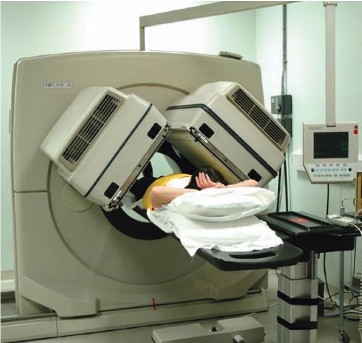
2 Short History Nuclear Cardiology Hermann blumgart-1927-injected radon to measure circulation time Hal Anger-1952-gamma camera-beginning of clinical nuclear cardiology 1976-thallium201-two dimensional planar imaging 1980s-SPECT using rotating anger camera 1990-technetium99m based agents and gated SPECT 90+% of SPECT in U.S use technetium and 90% are gated SPECT
3
4
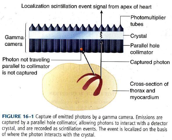
5 SPECT
6 SPECT perfusion tracers Thallium 201 Technetium 99m Sestamibi (Cardiolyte) Tetrafosmin (Myoview) Teboroxime Dual Isotope Thallium injected for resting images Tech -99m injected at peak stress
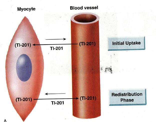
7 Thallium-201 Monovalent cation, similar to potassium Half life 73 hours, emits 80keV photons, 85% first pass extraction Peak myocardial concentration in 5 min, rapid clearance from intravascular compartment Redistribution of thallium-begins in min
8
9 Thallium protocols- Stress protocols-injected at peak stress and images taken at peak stress and at 4 hrs,24hrs Reversal of a thallium defect marker of reversible ischemia Rest protocols-thallium defect reversibility from initial rest images to delayed redistribution images reflect viable myocardium with resting hypoperfusion Initial defect persists-irreversible defect
10 Technetium-99m labelled tracers Half life 6 hrs, 140keV photons, 60% extraction Uptake by passive distribution by gradient and trapped in myocardial cell Minimal redistribution-require two separate injections-one at peak stress and one at rest Single day study-first injected dose is low
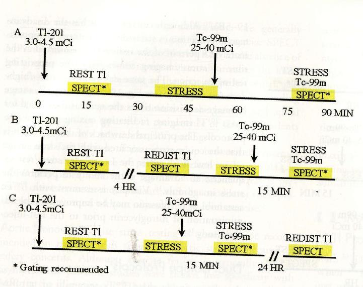
11 Dual isotope protocol Anger camera can collect image in different energy windows Thallium at rest followed by Tc 99m tracer at peak stress If there is rest perfusion defect,redistribution imaging taken either 4 hrs prior or 24hrs after Tc99m injection
12
13 Radionuclide Properties Property Thallous Chloride Tc-Sestamibi Chemistry +1 cation, hydrophilic +1 cation, lipophilic half life 73 hrs 6 hours Photon energy kev 140 kev Uptake Active: Na-K ATPase pump Extraction fraction 85% 66% Heart uptake 4% 1.2% Redistribution Redistributes Fixed Passive diffusion (if intact membrane potentials)
14 Pharmacologic Stress Dipyridamole infusion for 4 min-isotope injection 3 min after infusion Adenosine infusion for 6 min-isotope given 3 min into infusion Regadenoson (Lexiscan) infusion for 6 mins Dobutamine progressive infusion to increase HR
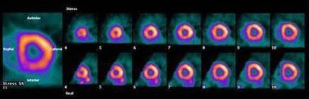
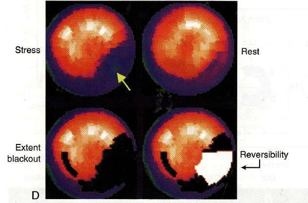
15 Interpretation of the Findings-SPECT Stress Rest Interpretation No defects No defects Normal Defect No defect Ischemia (Stress-induced ischemia) Defect Defect Scar/ hibernating Defect location (anterior, posterior, lateral, or septal wall), size (small, medium, or big), severity (mild, moderate, absent), degree of reversibility at rest (completely reversible, partially reversible, irreversible) Regional wall motion, EDV, ESV, EF

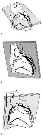
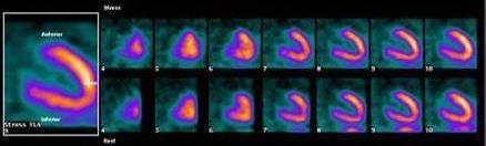
16 Ant Stress Apex Rest Inf Septum Lateral Stress Apex Rest Sep Lat Inferior Anterior Ant Stress Reversible Ischeamia, defect appears at stress and disappears during rest Rest Sep Lat Inf Apex Base

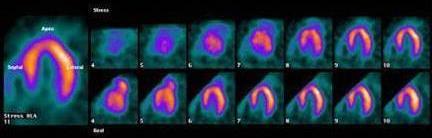
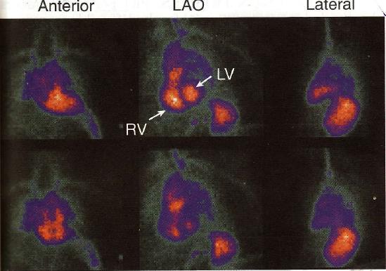
17 Ant Stress Apex Rest Inf Septum Lateral Stress Apex Rest Sep Lat Inferior Anterior Ant Stress Rest Sep Inf Lat Fixed Scar, defect is seen in both stress and rest Apex Base

18
19
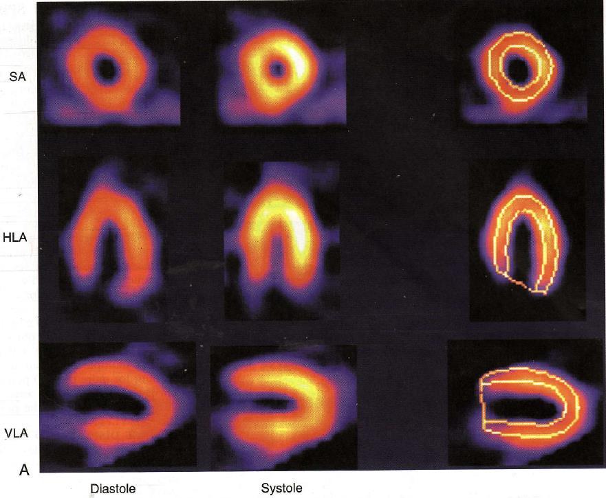
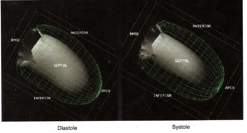
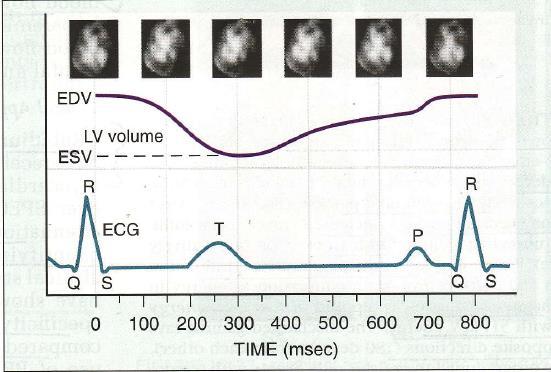
20 Gated SPECT (Like a MUGA) Simultaneous assessment of LV function and perfusion Each R-R interval is devided into prespecified number of frames Frame one represent end diastole,middle frames end systole An average of several hundred beats of a particular cycle length acquired over 8-15 min.
21
22
23
24 Radionuclide ventriculography MUGA scanning-multiple gated acquisition Tc 99m labelled r.b.c or albumin Image constructed over an average cardiac cycle by e.c.g gating,16-32 frames /cycle Image acquired in antr.,lao, left lateral projections Size of chambers, RWMA, LV function
25
26

27 PET imaging of the heart
28 PET Radiotracers labelled with positron emitting isotopes Perfusion tracers-rb82 and n13 ammonia Metabolic tracer-f18 FDG Beta decay-positron emission PET scanner detects opposing photons in coincidence-spatial and temporal resolution
29 Advantage of PET Higher spatial resolution Improved attenuation correction Quantification regional blood flow SPECT may fail to detect balanced ischemia in multivessel CAD blood flow reserve by PET early identification of CAD Higher sensitivity and specificity (95%) for detection of CAD
30 Limitations High cost Requirement of cyclotron Short half life-pharmacological stress only
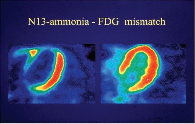
31 Metabolic tracers C-11 palmitate I-123 BMIPP-Ischemic memory-fatty acid metabolism suppressed for longer time after an ischemic event F18 FDG-imaging myocardial glucose utilisation with PET Phosphorylated and trapped in myocardium Uptake may be increased in hibernating but viable myocardium
32 *
33 PET Viability Scan Patterns Contractility Perfusion Metabolism Normal N N N Stunning - N N - Hibernation Scar

34 Cardiac PET/CT: The Ultimate

35 PET/CT Scanners: The Ultimate
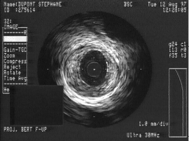
36 ADVANCED IMAGING OF THE VULNERABLE PLAQUE

37 The Old Friend: Angiography
38 Angiography: the good and the bad Good Bad Extensively used > 60 years Entire coronary anatomy, including small and distal vessels Excellent PPV Validated QCA Helpful in clinical decision making Relative % stenosis Reference segment assessment Eccentricity Post PTCA/dissections Limited correlation with physiology

39 Pitfall: lesion eccentricity
40 Vascular Remodelling (Glagov s phenomenon)

41 Plaque Pathogenesis Morphologic traits associated with rupture prone plaques are found in thin-cap fibroatheromas.
42 Intravascular Assessment of Plaque Vulnerability IVUS IVUS-RF (Virtual Histology) Palpography Optical coherence tomography Near-infrared spectroscopy Intravascular MR Angioscopy Thermography
43 Intravascular Ultrasound Real-time cross-sectional tomographic images in the short axis. Backscatter signal is processed into gray scale with spatial resolution of 150 µm and frame rate of 10 to 30 frames/sec. Plaque vulnerability features include: Eccentric pattern Echolucent core Positive remodeling Presence of thrombi Plaque length Lumen narrowing Spotty calcifications.
44 IVUS: the good and the bad Good Bad Tomographic views Vessel wall + lumen visualization Excellent NPV+PPV Validated quantitative software Plaque characterization Need to instrument vessels Limited to proximal segments Cost Not as well validated for clinical decision making Limited correlation with physiology Not always perpendicular to vessel axis

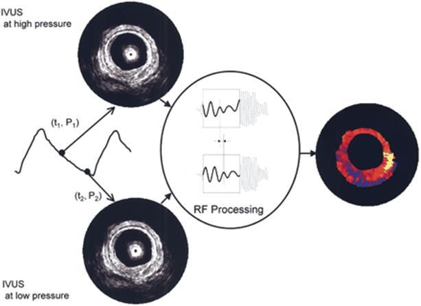
45 IVUS Imaging 2D Cross-Sectional Imaging

46 Distal LMT


47 Soft Fibrous Superficial Ca Deep calcification

48 IVUS: Potentially unstable coronary lesion Echolucent
49 Intravascular Coronary Ultrasound Angio remains the most widely and conveniently used coronary imaging modality IVUS has helped better use/understand angiography Not IVUS vs Angio, more Angio ± IVUS Need to understand the pitfalls of each technique and use them appropriately
50 IVUS-RF (Virtual Histology) Mathmatical autoregression modeling of each line in the radiofrequency signal is performed on a region of interest and averaged over that region. Results are displayed as a color-coded map superimposed on gray scale IVUS images. Sensitivity, specificity, and PPV to detect necrotic cores in initial ex-vivo and in vivo studies were 67%, 93%, and 88% respectively.
, precludes fibrous cap thickness Conflicting data regarding assessment of necrotic")
51 IVUS-RF Limitations: Unable to distinguish thrombi from other plaque components Limited spatial resolution (equivalent to IVUS), precludes fibrous cap thickness Conflicting data regarding assessment of necrotic cores
52 IVUS - Palpography Measures the mechanical properties of tissue through RF-ultrasound signals recorded at different pressures i.e. measures the local rate of plaque deformation (strain) in response to the pulsating force of blood pressure. Fibrous plaques are less elastic than lipid rich plaques. Validated ex vivo, in pig models, and in small in vitro studies.
53
54 IVUS - Palpography Limitations: Has worse spatial and temporal resolution than conventional IVUS or IVUS-RF (~200 µm). Cardiac motion and pullback of catheter can create artifacts. J Am Coll Cardiol, 2006; 47:86-91
55 Optical coherence tomography Uses optical scattering to generate an ultra-high resolution (4 to 20 µm) 2-dimensional image. Limited tissue penetration due to use of light to create the image. Attenuated by blood. Compares favorably with IVUS for plaque characterization.
 29 (16):")
56 Eur Heart J (2008) 29 (16): 2023
57 Near-infrared spectroscopy Based on absorbance of light of organic molecules. NIRS allows for the chemical characterization of biological tissues can be used to assess lipid and protein content in plaques. Resultant image termed a chemogram. Validated against histologic hallmarks of plaque vulnerability lipid pool, thin cap, and inflammatory cells.

58 NIRS Limitations Limited tissue penetration on par, or slightly worse than, OCT Cardiac motion artifact
59 Detection by Near-Infrared Spectroscopy of Large Lipid Core Plaques at Culprit Sites in Patients With Acute ST- Segment Elevation Myocardial Infarction We performed NIRS within the culprit vessels of 20 patients with acute STEMI and compared the STEMI culprit findings to findings in nonculprit segments of the artery and to findings in autopsy control segments. Culprit and control segments were analyzed for the maximum lipid core burden index in a 4-mm length of artery (maxlcbi 4mm ). MaxLCBI 4mm was 5.8-fold higher in STEMI culprit segments than in 87 nonculprit segments of the STEMI culprit vessel and 87-fold higher than in 279 coronary autopsy segments free of large LCP by histology. Within the STEMI culprit artery, NIRS accurately distinguished culprit from nonculprit segments. Conclusions The present study has demonstrated in vivo that a maxlcbi 4mm >400, as detected by NIRS, is a signature of plaques causing STEMI. Madder R et al. JACC Intevent July 17, 2013

60 Detection by Near-Infrared Spectroscopy of Large Lipid Core Plaques at Culprit Sites in Patients With Acute ST- Segment Elevation Myocardial Infarction
61 Intravascular Magnetic Resonance Pulsed field MRI has been used to calculate the water diffusion coefficient in atherosclerotic plaques. Water diffusion is less in lipid-rich than fibrous plaques. In ex vivo studies, correlation between MRI and histology was good, with a sensitivity of 100% and specificity of 89% for lipid cores.

62 IV MRI Limitations: Requires hybrid lab or transport to MRI suite Catheter-coil needs to be stabilized with occlusive balloon Gadolinium contrast utilization Atherosclerosis; 196; 2;
63 Angioscopy Direct visualization of the surface of plaque. Number of yellow plaques is a strong predictor of ACS Subjective, needs blood displacment, factual accuracy per patient is poor

64 Angioscopy Cover of JACC intervention 2/1/2008
65 Thermography Based on assumption that plaque inflammation and neoangiogenesis produce heat that can be measured by dedicated catheter. Temperature difference of up to 1.5 C between plaque and healthy vessel in ACS has been shown in human subjects. Major limitation is blood flows cooling effect, requiring interruption of flow. JACC; 57, 20,
66 Noninvasive Assessment of Plaque Vulnerability Multidetector Computed Tomography Magnetic Resonance Imaging Nuclear Imaging Contrast-enhanced Ultrasonography
67 Multidetector CT MDCT can detect features associated with plaque vulnerability including positive remodeling, spotty calcification, lower plaque density, intra-plaque dye penetration, and ulceration. Currently considered a first-line method in detecting vulnerable plaque.

68 Circulation: Cardiovascular Imaging. 2010;3:
69 Magnetic Resonance Imaging Due to cardiac motion, MRI best suited for study of large, static arteries. Lipid and fibrotic plaque components have been accurately quantified on T2 weighted imaging. T2 weighted imaging has also been utilized to measure fibrous cap thickness, ruptures, and intraplaque hemorrhages.

70 MRI Limitations: Clinical utility remains to be determined Technical improvements still needed prior to better visualization of coronary arteries.
71 Conclusions Detection of plaque vulnerability is becoming a reality. More evidence needed for many of the imaging modalities correlating to clinical events. Further research into the morphologic, molecular, biologic, and mechanical features of vulnerable plaques is needed.
NUCLEAR CARDIOLOGY UPDATE
 Nuclear Cardiology David K. Shelton, Jr., MD NUCLEAR CARDIOLOGY UPDATE No Conflicts. No Disclosures. No Smoking. David K. Shelton UCDMC Nuclear Cardiology Nuclear Cardiology Radionuclide Ventriculography
Invasive Coronary Imaging Modalities for Vulnerable Plaque Detection
 Invasive Coronary Imaging Modalities for Vulnerable Plaque Detection Gary S. Mintz, MD Cardiovascular Research Foundation New York, NY Greyscale IVUS studies have shown Plaque ruptures do not occur randomly
OTHER NON-CARDIAC USES OF Tc-99m CARDIAC AGENTS Tc-99m Sestamibi for parathyroid imaging, breast tumor imaging, and imaging of other malignant tumors.
 DEFINITION OF CARDIAC RADIOPHARMACEUTICAL: A radioactive drug which, when administered for purpose of diagnosis of heart disease, typically elicits no physiological response from the patient. Even though
Cardiac Imaging Tests
 Cardiac Imaging Tests http://www.medpagetoday.com/upload/2010/11/15/23347.jpg Standard imaging tests include echocardiography, chest x-ray, CT, MRI, and various radionuclide techniques. Standard CT and
Pearls & Pitfalls in nuclear cardiology
 Pearls & Pitfalls in nuclear cardiology Maythinee Chantadisai, MD., NM physician Division of Nuclear Medicine, Department of radiology, KCMH Principle of myocardial perfusion imaging (MPI) Radiotracer
Myocardial viability testing. What we knew and what is new
 Myocardial viability testing. What we knew and what is new Dr B K S Sastry, MD, DM. CARE Hospitals, Hyderabad What is Viability Viability Dysfunctional myocardium subtended by diseased coronary arteries
Radiologic Assessment of Myocardial Viability
 November 2001 Radiologic Assessment of Myocardial Viability Joshua Moss, Harvard Medical School Year III Patient EF 66yo female with a 3-year history of intermittent chest pain previously relieved by sublingual
Added Value of Invasive Coronary Imaging for Plaque Rupture and Erosion
 Assessment of Coronary Plaque Rupture and Erosion Added Value of Invasive Coronary Imaging for Plaque Rupture and Erosion Yukio Ozaki, MD, PhD, FACC, FESC Cardiology Dept., Fujita Health Univ. Toyoake,
Prior Authorization for Non-emergency Cardiac Imaging Procedures
 Attention: All Providers Prior Authorization for Non-emergency Cardiac Imaging Procedures The N.C. Medicaid Program is considering implementation of a prior authorization (PA) program for non-emergency
Imaging Atheroma The quest for the Vulnerable Plaque
 Imaging Atheroma The quest for the Vulnerable Plaque P.J. de Feijter 1. Department of Cardiology 2. Department of Radiology Coronary Heart Disease Remains the Leading Cause of Death in the U.S, Causing
Fundamentals of Nuclear Cardiology. Terrence Ruddy, MD, FRCPC, FACC
 Fundamentals of Nuclear Cardiology Terrence Ruddy, MD, FRCPC, FACC Objectives To understand the Principles of Nuclear Cardiac Imaging Radiotracers Image acquisition and processing Stress protocols To appreciate
Gated blood pool ventriculography: Is there still a role in myocardial viability?
 Gated blood pool ventriculography: Is there still a role in myocardial viability? Oliver C. Alix, MD Adult Clinical and Nuclear Cardiology St. Luke s Medical Centre - Global City Case Presentation A 62-year-old
Cardiac Imaging. Kimberly Delcour, DO, FACC. Mahi Ashwath, MD, FACC, FASE. Director, Cardiac CT. Director, Cardiac MRI
 Cardiac Imaging Kimberly Delcour, DO, FACC Director, Cardiac CT Mahi Ashwath, MD, FACC, FASE Director, Cardiac MRI Cardiac Imaging Discuss the clinical applications of and indications for: Cardiac CT Nuclear
I have no financial disclosures
 Manpreet Singh MD I have no financial disclosures Exercise Treadmill Bicycle Functional capacity assessment Well validated prognostic value Ischemic assessment ECG changes ST segments Arrhythmias Hemodynamic
CT Imaging of Atherosclerotic Plaque. William Stanford MD Professor-Emeritus Radiology University of Iowa College of Medicine Iowa City, IA
 CT Imaging of Atherosclerotic Plaque William Stanford MD Professor-Emeritus Radiology University of Iowa College of Medicine Iowa City, IA PREVALENCE OF CARDIOVASCULAR DISEASE In 2006 there were 80 million
SPECT-CT: Τι πρέπει να γνωρίζει ο Καρδιολόγος
 SPECT-CT: Τι πρέπει να γνωρίζει ο Καρδιολόγος Δρ Αναστασία Κίτσιου Διευθύντρια, Καρδιολογική Κλινική, Σισμανόγλειο ΓΝΑ Chair, Education Committee, Section on Nuclear Cardiology & Cardiac CT, EACVI, ESC
Rational use of imaging for viability evaluation
 EUROECHO and other imaging modalities 2011 Rational use of imaging for viability evaluation Luc A. Pierard, MD, PhD, FESC, FACC Professor of Medicine Head, Department of Cardiology, CHU Liège, Belgium
State of the Art. Advances in Cardiovascular Imaging. ESC Congres Stockholm September 1, 2010 Frank E. Rademakers, MD, PhD, FESC
 State of the Art Advances in Cardiovascular Imaging ESC Congres Stockholm September 1, 2010 Frank E. Rademakers, MD, PhD, FESC Coronary Artery Disease Content Patho Physiology Imaging requirements Economical
EMPHISIS ON PHYSIOLOGY PHYSIOLOGY REQUIRES TIME QUALITATIVE vs. QUANTITATIVE ISOTOPES TO RADIOPHARMACEUTICALS
 1926 Herman Blumgard used solutions of radon gas to measure what he called velocity of the circulation. 1927 Blumberg and Soma Weiss wrote article in (Journal of Clinical Investigation) 1929 Werner Forssmann
Click here for Link to References: CMS Website HOPPS CY 2018 Final Rule. CMS Website HOPPS CY2018 Final Rule Updated November 2017.
 Final Compared to 3Q 2017 Rates Medicare Hospital Outpatient Prospective Payment System HOPPS () Nuclear Cardiology Procedures, Radiopharmaceuticals, and Drugs Click here for Link to References: CMS Website
Cardiovascular nuclear imaging employs non-invasive techniques to assess alterations in coronary artery flow, and ventricular function.
 National Imaging Associates, Inc. Clinical guidelines CARDIOVASCULAR NUCLEAR MEDICINE -MYOCARDIAL PERFUSION IMAGING -MUGA CPT4 Codes: Refer to pages 6-9 LCD ID Number: L33960 J 15 = KY, OH Responsible
Evaluation of myocardial ischaemia
 l2 TOPIC Evaluation of myocardial ischaemia Topic Contents Markers of myocardial injury and infarction 6 Myocardial territories supplied by coronary arteries 8 The 17 segment model 9 Regional assessment
CLINICAL APPLICATIONS OF OPTICAL COHERENCE TOMOGRAPHY. Konstantina P. Bouki, FESC 2 nd Department of Cardiology General Hospital Of Nikea, Pireaus
 CLINICAL APPLICATIONS OF OPTICAL COHERENCE TOMOGRAPHY Konstantina P. Bouki, FESC 2 nd Department of Cardiology General Hospital Of Nikea, Pireaus OPTICAL COHERENCE TOMOGRAPHY (OCT) IVUS and OCT IVUS OCT
Imaging Overview for Vulnerable Plaque: Data from IVUS Trial and An Introduction to VH-IVUS Imgaging
 Imaging Overview for Vulnerable Plaque: Data from IVUS Trial and An Introduction to VH-IVUS Imgaging Gary S. Mintz,, MD Cardiovascular Research Foundation New York, NY Today, in reality, almost everything
SPECT TRACERS Tl-201, Tc-99m Sestamibi, Tc-99m Tetrofosmin
 SPECT TRACERS Tl-201, Tc-99m Sestamibi, Tc-99m Tetrofosmin Elmer Jasper B. Llanes, M.D. Nuclear Cardiology St. Luke s Medical Center Outline Ideal Physiologic Characteristics of MPI radioactive tracers
Cardiovascular nuclear imaging employs non-invasive techniques to assess alterations in coronary artery flow, and ventricular function.
 National Imaging Associates, Inc. Clinical guidelines CARDIOVASCULAR NUCLEAR MEDICINE -MYOCARDIAL PERFUSION IMAGING -MUGA Original Date: October 2015 Page 1 of 9 FOR CMS (MEDICARE) MEMBERS ONLY CPT4 Codes:
ASSOCIATION BETWEEN LEFT VENTRICULAR EJECTION FRACTION (LVEF) IN PATIENTS WITH REGIONAL ISCHEMIA AND INFARCTION ON MYOCARDIAL PERFUSION IMAGES
 IN PATIENTS WITH REGIONAL ISCHEMIA AND INFARCTION ON MYOCARDIAL PERFUSION IMAGES") University of New Mexico UNM Digital Repository Biomedical Sciences ETDs Electronic Theses and Dissertations 7-1-2015 ASSOCIATION BETWEEN LEFT VENTRICULAR EJECTION FRACTION (LVEF) IN PATIENTS WITH REGIONAL
University of New Mexico UNM Digital Repository Biomedical Sciences ETDs Electronic Theses and Dissertations 7-1-2015 ASSOCIATION BETWEEN LEFT VENTRICULAR EJECTION FRACTION (LVEF) IN PATIENTS WITH REGIONAL
Cardiovascular Imaging
 Cardiovascular Imaging Cardiovascular Imaging Cardio and Vascular Imaging Vascularization / Angiogenesis Cardiovascular Imaging metabolic imaging of the heart myocardial perfusion imaging Cardiac CT Vascularization
CARDIAC PET PERFUSION IMAGING with RUBIDIUM-82
 CARDIAC PET PERFUSION IMAGING with RUBIDIUM-82 Pr Denis AGOSTINI Président du Groupe de Cardiologie Nucléaire et IRM CHU Caen Bordeaux 2006 Cardiac Perfusion-Metabolism Mismatch with PET Cumulative Survival
Tc-99m Sestamibi/Tetrofosmin Stress-Rest Myocardial Perfusion Scintigraphy
 APPROVED BY: Director of Radiology Page 1 of 6 Tc-99m Sestamibi/Tetrofosmin Stress-Rest Myocardial Primary Indications: Evaluation of myocardial perfusion and viability in patients with known or suspected
Noninvasive Coronary Imaging: Plaque Imaging by MDCT
 Coronary Physiology & Imaging Summit 2007 Noninvasive Coronary Imaging: Plaque Imaging by MDCT Byoung Wook Choi Department of Radiology Yonsei University, Seoul, Korea Stary, H. C. et al. Circulation
RADIOTRACERS FOR MYOCARDIAL PERFUSION IMAGING
 RADIOTRACERS FOR MYOCARDIAL PERFUSION IMAGING RAYMOND TAILLEFER, M.D. FRCP(c), ABNM DIRECTOR, DEPARTMENT OF NUCLEAR MEDICINE HOPITAL ST-JEAN-SUR-RICHELIEU Disclosures to Report: Grant Research Support:
The Value of Stress MRI in Evaluation of Myocardial Ischemia
 The Value of Stress MRI in Evaluation of Myocardial Ischemia Dr. Saeed Al Sayari, MBBS, EBCR, MBA Department of Radiology and Nuclear Medicine Mafraq Hospital, Abu Dhabi United Arab Emirates Introduction
Η Πυρηνική Καρδιολογία Το 2017 ΟΜΑΔΑ ΕΡΓΑΣΙΑΣ ΑΠΕΙΚΟΝΙΣΤΙΚΩΝ ΤΕΧΝΙΚΩΝ
 Η Πυρηνική Καρδιολογία Το 2017 ΟΜΑΔΑ ΕΡΓΑΣΙΑΣ ΑΠΕΙΚΟΝΙΣΤΙΚΩΝ ΤΕΧΝΙΚΩΝ huma human n Setting diagnosis of the early stages of chronic diseases (i.e cancer, neuropsychiatric, cardiovascular disorders), in
Cardiac CT Angiography
 Cardiac CT Angiography Dr James Chafey, Radiologist Why do we need a better test for C.A.D? 1. CAD is the leading cause of death in the US CAD 31% Cancer 23% Stroke 7% 2. The prevalence of atherosclerosis
Cardial MRI; Approaching the Level of Gold Standard for Viability Assessment
 Cardial MRI; Approaching the Level of Gold Standard for Viability Assessment 용환석 고려대학교구로병원영상의학과 Viability Hibernating myocardium a state of myocardial hypocontractility during chronic hypoperfusion, in
High-risk vulnerable plaques. Kostis Raisakis G.Gennimatas General Hospital of Athens
 High-risk vulnerable plaques. Kostis Raisakis G.Gennimatas General Hospital of Athens Overview: 1 Definition-Pathology 2 3 Diagnostic Strategies Invasive Non Invasive Prognostic Value of Detection 4 Treatment
Nuclear Cardiology Pierre-Yves MARIE Department of Nuclear Medicine CHU-Nancy, FRANCE.
 Nuclear Cardiology Pierre-Yves MARIE Department of Nuclear Medicine CHU-Nancy, FRANCE. Nuclear Cardiology I - A remaining need of a functional information on myocardial perfusion II - The future: - combining
Photon Attenuation Correction in Misregistered Cardiac PET/CT
 Photon Attenuation Correction in Misregistered Cardiac PET/CT A. Martinez-Möller 1,2, N. Navab 2, M. Schwaiger 1, S. G. Nekolla 1 1 Nuklearmedizinische Klinik der TU München 2 Computer Assisted Medical
Gary S. Mintz,, MD. IVUS Observations in Acute (vs Chronic) Coronary Artery Disease: Structure vs Function
 Coronary Artery Disease: Structure vs Function") Gary S. Mintz,, MD IVUS Observations in Acute (vs Chronic) Coronary Artery Disease: Structure vs Function Important IVUS Observations: Remodeling Originally used (first by Glagov) ) to explain atherosclerosis
Gary S. Mintz,, MD IVUS Observations in Acute (vs Chronic) Coronary Artery Disease: Structure vs Function Important IVUS Observations: Remodeling Originally used (first by Glagov) ) to explain atherosclerosis
Anthem Blue Cross and Blue Shield Virginia Advanced Imaging Procedures Requiring Precertification Revised 02/13/2013
 Anthem Blue Cross and Blue Shield Virginia Advanced Imaging Procedures Requiring Precertification Revised 02/13/2013 Modality and CT Head CTA Head: Cerebrovascular MRI Head MRA Head: Cerebrovascular Functional
Multiple Gated Acquisition (MUGA) Scanning
 Scanning") Multiple Gated Acquisition (MUGA) Scanning Dmitry Beyder MPA, CNMT Nuclear Medicine, Radiology Barnes-Jewish Hospital / Washington University St. Louis, MO Disclaimers/Relationships Standard of care research
Multiple Gated Acquisition (MUGA) Scanning Dmitry Beyder MPA, CNMT Nuclear Medicine, Radiology Barnes-Jewish Hospital / Washington University St. Louis, MO Disclaimers/Relationships Standard of care research
Assessment of plaque morphology by OCT in patients with ACS
 Assessment of plaque morphology by OCT in patients with ACS Takashi Akasaka, M.D. Department of Cardiovascular Medicine Wakayama, Japan Unstable plaque Intima Lipid core Plaque rupture and coronary events
1. LV function and remodeling. 2. Contribution of myocardial ischemia due to CAD, and
 1 The clinical syndrome of heart failure in adults is commonly associated with the etiologies of ischemic and non-ischemic dilated cardiomyopathy, hypertrophic cardiomyopathy, hypertensive heart disease,
Cardiology for the Practitioner Advanced Cardiac Imaging: Worth the pretty pictures?
 Keenan Research Centre Li Ka Shing Knowledge Institute Cardiology for the Practitioner Advanced Cardiac Imaging: Worth the pretty pictures? Howard Leong-Poi, MD, FRCPC Associate Professor of Medicine St.
Value of Assessment of Viable and Ischemic Myocardium and Techniques Such as MRI, Radionuclide Imaging
 Chapter 2 Imaging for Viable and Ischemic Myocardium Value of Assessment of Viable and Ischemic Myocardium and Techniques Such as MRI, Radionuclide Imaging Catalin Loghin and K. Lance Gould Introduction
Abnormal, Autoquant Adenosine Myocardial Perfusion Heart Imaging. ID: GOLD Date: Age: 46 Sex: M John Doe Phone (310)
") Background: Reason: preoperative assessment of CAD, Shortness of Breath Symptom: atypical chest pain Risk factors: hypertension Under influence: a beta blocker Medications: digoxin Height: 66 in. Weight:
Background: Reason: preoperative assessment of CAD, Shortness of Breath Symptom: atypical chest pain Risk factors: hypertension Under influence: a beta blocker Medications: digoxin Height: 66 in. Weight:
Intervention: How and to which extent is technology helping us?
 Cardiological Society of India Congress 12th February 2016 Chennai, India Intervention: How and to which extent is technology helping us? SIMONE BISCAGLIA MD CARDIOVASCULAR INSTITUTE, FERRARA, ITALY Introduction
Optical Coherence Tomography (OCT): A New Imaging Tool During Carotid Artery Stenting
: A New Imaging Tool During Carotid Artery Stenting") Chapter 6 Optical Coherence Tomography (OCT): A New Imaging Tool During Carotid Artery Stenting Shinichi Yoshimura, Masanori Kawasaki, Kiyofumi Yamada, Arihiro Hattori, Kazuhiko Nishigaki, Shinya Minatoguchi
Chapter 6 Optical Coherence Tomography (OCT): A New Imaging Tool During Carotid Artery Stenting Shinichi Yoshimura, Masanori Kawasaki, Kiyofumi Yamada, Arihiro Hattori, Kazuhiko Nishigaki, Shinya Minatoguchi
Use of Nuclear Cardiology in Myocardial Viability Assessment and Introduction to PET and PET/CT for Advanced Users
 Use of Nuclear Cardiology in Myocardial Viability Assessment and Introduction to PET and PET/CT for Advanced Users February 1 5, 2011 University of Santo Tomas Hospital Angelo King A-V Auditorium Manila,
Yukio Ozaki, M Okumura, TF Ismail 2, S Motoyama, H. Naruse, K. Hattori, H. Kawai, M. Sarai, J. Ishii, Jagat Narula 3
 Culprit Lesion Characteristics in Acute Coronary Syndrome and Stable Angina Assessed by Optical Coherence Tomography (OCT), Angioscopy, IVUS and Multidetector Computed Tomography (MDCT) Yukio Ozaki, M
CT or PET/CT for coronary artery disease
 CT or PET/CT for coronary artery disease Rotterdam 2012 Juhani Knuuti, MD, PhD, FESC Turku PET Centre University of Turku Turku, Finland Juhani.knuuti@utu.fi Turku PET Centre University of Turku Åbo Akademi
Evaluation of Intermediate Coronary lesions: Can You Handle the Pressure? Jeffrey A Southard, MD May 4, 2013
 Evaluation of Intermediate Coronary lesions: Can You Handle the Pressure? Jeffrey A Southard, MD May 4, 2013 Disclosures Consultant- St Jude Medical Boston Scientific Speaker- Volcano Corporation Heart
Medical imaging X-ray, CT, MRI, scintigraphy, SPECT, PET Györgyi Műzes
 Medical imaging X-ray, CT, MRI, scintigraphy, SPECT, PET Györgyi Műzes Semmelweis University, 2nd Dept. of Medicine Medical imaging: definition technical process of creating visual representations about
CHRONIC CAD DIAGNOSIS
 CHRONIC CAD DIAGNOSIS Chest Pain Evaluation 1. Approach to diagnosis of CAD 2. Classification of chest pain 3. Pre-test likelihood CAD 4. Algorithm for chest pain evaluation in women 5. Indications for
1st Department of Cardiology, University of Athens, Hippokration Hospital, Athens, Greece
 Konstantinos Toutouzas, Maria Riga, Antonios Karanasos, Eleftherios Tsiamis, Andreas Synetos, Maria Drakopoulou, Chrysoula Patsa, Georgia Triantafyllou, Aris Androulakis, Christodoulos Stefanadis 1st Department
OCT. molecular imaging J Jpn Coll Angiol, 2008, 48: molecular imaging MRI positron-emission tomography PET IMT
 48 6 CT MRI PET OCT molecular imaging J Jpn Coll Angiol, 2008, 48: 456 461 atherosclerosis, imaging gold standard computed tomography CT magnetic resonance imaging MRI CT B intima media thickness IMT B
General Cardiovascular Magnetic Resonance Imaging
 2 General Cardiovascular Magnetic Resonance Imaging 19 Peter G. Danias, Cardiovascular MRI: 150 Multiple-Choice Questions and Answers Humana Press 2008 20 Cardiovascular MRI: 150 Multiple-Choice Questions
Κλινική Χρήση IVUS και OCT PERIKLIS A. DAVLOUROS ASSOCIATE PROFESSOR OF CARDIOLOGY INVASIVE CARDIOLOGY & CONGENITAL HEART DISEASE
 Κλινική Χρήση IVUS και OCT PERIKLIS A. DAVLOUROS ASSOCIATE PROFESSOR OF CARDIOLOGY INVASIVE CARDIOLOGY & CONGENITAL HEART DISEASE Conflict of interest None to declare While IVUS is the most used intravascular
How to evaluate heart disease - Do we need new tools Focus on myocardial circulation
 How to evaluate heart disease - Do we need new tools Focus on myocardial circulation Alf Inge Larsen Professor department of clinical science University of Bergen Head of Cardiovascular Research Group
PET myocard perfusion & viability Riemer Slart
 PET myocard perfusion & viability Riemer Slart Nuclear Medicine Physician Dept. of Nuclear Medicine and Molecular Imaging University Medical Center Groningen, the Netherlands Professor in Molecular Imaging,
Chapter 43 Noninvasive Coronary Plaque Imaging
 hapter 43 Noninvasive oronary Plaque Imaging NIRUDH KOHLI The goal of coronary imaging is to define the extent of luminal narrowing as well as composition of an atherosclerotic plaque to facilitate appropriate
Non Invasive Diagnostic Modalities for Coronary Artery Disease. Dr. Amitesh Aggarwal
 Non Invasive Diagnostic Modalities for Coronary Artery Disease Dr. Amitesh Aggarwal Ebers papyrus, ca. 1555 BCE If thou examine a man for illness in his cardia, and he has pains in his arms, in his breasts
Functional aspects of anatomical imaging techniques
 Functional aspects of anatomical imaging techniques Nilendu Purandare Associate Professor & Consultant Radiologist Tata Memorial Centre Functional/metabolic/molecular imaging (radioisotope scanning) PET
Reversible defect of 123 I-15-(p-iodophenyl)-9-(R,S)-methylpentadecanoic acid indicates residual viability within infarct-related area
-9-(R,S)-methylpentadecanoic acid indicates residual viability within infarct-related area") ORIGINAL ARTICLE Annals of Nuclear Medicine Vol. 16, No. 3, 183 187, 2002 Reversible defect of 123 I-15-(p-iodophenyl)-9-(R,S)-methylpentadecanoic acid indicates residual viability within infarct-related
ORIGINAL ARTICLE Annals of Nuclear Medicine Vol. 16, No. 3, 183 187, 2002 Reversible defect of 123 I-15-(p-iodophenyl)-9-(R,S)-methylpentadecanoic acid indicates residual viability within infarct-related
A Snapshot on Nuclear Cardiac Imaging
 Editorial A Snapshot on Nuclear Cardiac Imaging Khalil, M. Department of Physics, Faculty of Science, Helwan University. There is no doubt that nuclear medicine scanning devices are essential tool in the
Nuclear Cardiology Cardiac Myocardial Perfusion with 82 Rb. Dominique Delbeke, MD, PhD Vanderbilt University Medical Center Nashville, TN
 Nuclear Cardiology Cardiac Myocardial Perfusion with 82 Rb Dominique Delbeke, MD, PhD Vanderbilt University Medical Center Nashville, TN VUMC PET/CT conference 2009 82 Rb Cardiac Perfusion PET 82 Rb is
Pathology of Coronary Artery Disease
 Pathology of Coronary Artery Disease Seth J. Kligerman, MD Pathology of Coronary Artery Disease Seth Kligerman, MD Assistant Professor Medical Director of MRI University of Maryland Department of Radiology
Sung A Chang Department of Internal Medicine, Division of Cardiology, Sungkyunkwan University School of Medicine, Samsung Medical Center
 CMR Perfusion and Viability A STICH Out of Time? Sung A Chang Department of Internal Medicine, Division of Cardiology, Sungkyunkwan University School of Medicine, Samsung Medical Center Can Imaging Improve
Perspectives of new imaging techniques for patients with known or suspected coronary artery disease
 Perspectives of new imaging techniques for patients with known or suspected coronary artery disease Department of Cardiology, Leiden University Medical Center, Leiden, The Netherlands Correspondence: Jeroen
Emerging role of PET in nuclear cardiology. Dr. Erick Alexánderson. Rosas
 Emerging role of PET in nuclear cardiology Dr. Erick Alexánderson Rosas PET principles e + e - 180 PET/CT Cyclotron Unit UNAM Cyclotron Radiopharmacy PET Camera CT PET PET CT Perfusion Anatomic evaluation
Rotation: Imaging 2. Nuclear Cardiology (in Imaging 1 and 2)
") Rotation: Imaging 2 Imaging 2 provides addition nuclear cardiology experience and COCATS Level 1 cardiac MRI experience. Fellows administer, process, and read VHVI cardiac nuclear studies with cardiology
Rotation: Imaging 2 Imaging 2 provides addition nuclear cardiology experience and COCATS Level 1 cardiac MRI experience. Fellows administer, process, and read VHVI cardiac nuclear studies with cardiology
Radiopharmaceuticals For Nuclear Cardiology Studies. Mark Soffing, PharmD, MBA, MS, RPh, BCNP
 Radiopharmaceuticals For Nuclear Cardiology Studies Mark Soffing, PharmD, MBA, MS, RPh, BCNP marksoffing@gmail.com Objectives Understand basics and advantages of myocardial imaging Describe isotopes and
INTRAVENOUS ADENOSINE MYOCARDIAL PERFUSION STUDY (rest/pharmacologic stress SPECT with gated SPECT wall motion studies at rest and post-stress)
") nucware.com, LLC Product Demo Anytown Cardiac Specialists, Inc. Janet Jones, MD, FACC Ed Wilson, MD, FACC Tom Smith, MD, FACC Jim Wilson, MD, FACC John Womack, MD, FACC JOHNSON, VICTOR DOB: 09/06/1938
nucware.com, LLC Product Demo Anytown Cardiac Specialists, Inc. Janet Jones, MD, FACC Ed Wilson, MD, FACC Tom Smith, MD, FACC Jim Wilson, MD, FACC John Womack, MD, FACC JOHNSON, VICTOR DOB: 09/06/1938
Hybrid cardiac imaging Advantages, limitations, clinical scenarios and perspectives for the future
 Hybrid cardiac imaging Advantages, limitations, clinical scenarios and perspectives for the future Prof. Juhani Knuuti, MD, FESC Turku, Finland Disclosure: Juhani Knuuti, M.D. Juhani Knuuti, M.D. has financial
Nuclear Cardiology Reimbursement. Todd Lamb, BS, AS, CNMT Clinical Operations Mgr Regions Hospital St. Paul, MN
 Nuclear Cardiology Reimbursement Todd Lamb, BS, AS, CNMT Clinical Operations Mgr Regions Hospital St. Paul, MN Slides are not to be reproduced without the permission of the author Slides are not to be
Cardiac PET. John Buscombe
 Cardiac PET John Buscombe Why PET? Improved resolution-not really required in cardiology Improved sensitivity this may be important-financially as reduced acquisition time Improved attenuation correction-good
Je bénéficie régulièrement de fonds privés, dans le cadre de projets de recherche ou d activités de formation.
 Je bénéficie régulièrement de fonds privés, dans le cadre de projets de recherche ou d activités de formation. Ces fonds proviennent essentiellement d industriels travaillant dans les domaines de l imagerie
SPECT CT Current Status and Future Direction Nuclear Cardiology
 SPECT CT Current Status and Future Direction Nuclear Cardiology BIR London 25 th February 2013 Nik Sabharwal Consultant Cardiologist Oxford Heart Centre nikant.sabharwal@ouh.nhs.uk No conflict of interest
Ischemic heart disease
 Ischemic heart disease Introduction In > 90% of cases: the cause is: reduced coronary blood flow secondary to: obstructive atherosclerotic vascular disease so most of the time it is called: coronary artery
Coronary Artery Thermography
 Coronary Artery Thermography The 10th Anniversary, Interventional Vascular Therapeutics Angioplasty Summit 2005 TCT Asia Pacific Christodoulos Stefanadis Professor of Cardiology Athens Medical School In
Microvascular Disease: How to Diagnose and What s its Treatment
 Microvascular Disease: How to Diagnose and What s its Treatment Laxmi S. Mehta, MD, FACC The Ohio State University Medical Center Assistant Professor of Clinical Internal Medicine Clinical Director of
Cardiac Viability Testing A Clinical Perspective Annual Cardiac Imaging Symposium. Lisa M Mielniczuk MD FRCPC University of Ottawa Heart Institute
 Cardiac Viability Testing A Clinical Perspective Annual Cardiac Imaging Symposium Lisa M Mielniczuk MD FRCPC University of Ottawa Heart Institute 62 year old male Anterior STEMI late presentation, occluded
Cardiovascular Research Foundation and Columbia University Medical Center, New York.
 Virtual Histology Intravascular Ultrasound Analysis of Non-culprit Attenuated Plaques Detected by Grayscale Intravascular Ultrasound in Patients with Acute Coronary Syndromes Xiaofan Wu, Akiko Maehara,
Optical Coherence Tomography
 Optical Coherence Tomography Disclosure Information Demetrius Lopes MD The following relationships exist related to this presentation: University Grant/Research Support: Rush University Industry Grant
The role of Magnetic Resonance Imaging in the diagnosis of viability & Coronary Artery Disease
 The role of Magnetic Resonance Imaging in the diagnosis of viability & Coronary Artery Disease G.P. Spanos, MSc, Phd Head of CardioVascular Imaging Tomographia Diagnostic Center Cardiovascular magnetic
Magnetic Resonance Imaging (MRI) for the Assessment of Myocardial Viability
 for the Assessment of Myocardial Viability") Ontario Health Technology Assessment Series 2010; Vol. 10, No. 15 Magnetic Resonance Imaging (MRI) for the Assessment of Myocardial Viability An Evidence-Based Analysis Presented to the Ontario Health
Ontario Health Technology Assessment Series 2010; Vol. 10, No. 15 Magnetic Resonance Imaging (MRI) for the Assessment of Myocardial Viability An Evidence-Based Analysis Presented to the Ontario Health
Advantages of PET Myocardial Imaging
 Advantages of PET Myocardial Imaging Legal Disclaimers These materials were prepared in good faith by MITA as a service to the profession and are believed to be reliable based on current scientific literature.
Diagnostic and Prognostic Value of Coronary Ca Score
 Diagnostic and Prognostic Value of Coronary Ca Score Dr. Ghormallah Alzahrani Cardiac imaging division, Adult Cardiology department Prince Sultan Cardiac Center ( PSCC) Madina, June 2 Coronary Calcium
Cigna - Prior Authorization Procedure List: Radiology & Cardiology
 Cigna - Prior Authorization Procedure List: Radiology & Cardiology Product Category CPT Code CPT Code Description Radiology MR 70336 MRI Temporomandibular Joint(s), (TMJ) Radiology CT 70450 CT Head or
Case-Based Pitfalls of SPECT and PET: Recognizing and Working with Artifacts
 2:20 PM Friday WORKSHOP V Case-Based Pitfalls of SPECT and PET: Recognizing and Working with Artifacts Sean W. Hayes, MD Associate Clinical Professor of Medicine UCLA School of Medicine Cedars-Sinai Heart
Conflict of Interest Disclosure
 Comparative Advantages of PET Over SPECT: Is PET Really Better? Timothy M. Bateman M.D. Co-Director, Cardiovascular Radiologic Imaging Mid America Heart Institute Professor of Medicine University of Missouri-Kansas
EAE Teaching Course. Magnetic Resonance Imaging. Competitive or Complementary? Sofia, Bulgaria, 5-7 April F.E. Rademakers
 EAE Teaching Course Magnetic Resonance Imaging Competitive or Complementary? Sofia, Bulgaria, 5-7 April 2012 F.E. Rademakers Complementary? Of Course N Engl J Med 2012;366:54-63 Clinical relevance Treatment
Medical Policy An independent licensee of the Blue Cross Blue Shield Association
 PET Scanning: Cardiac Applications Page 1 of 31 Medical Policy An independent licensee of the Blue Cross Blue Shield Association Title: Positron Emission Tomography (PET) Scanning: Cardiac Applications
Nuclear Perfusion Imaging of Angina
 January 2002 Nuclear Perfusion Imaging of Angina Davin Quinn HMS III Beth Israel Deaconess Medical Center Radiology Department Goals of Presentation Understand what a perfusion scan is Understand what
Nuclear medicine in general practice. Dr Reza Garzan MD, FRACP, FAANMS
 Nuclear medicine in general practice Dr Reza Garzan MD, FRACP, FAANMS Myocardial perfusion study Bone scans in general practice Thyroid scans in general practice Gamma camera Detection of gamma rays Myocardial
Evidence for myocardial CT perfusion imaging in the diagnosis of hemodynamically significant coronary artery disease
 Editorial Evidence for myocardial CT perfusion imaging in the diagnosis of hemodynamically significant coronary artery disease Zhonghua Sun Discipline of Medical Imaging, Department of Imaging and Applied
Myocardial Stress Perfusion Imaging using CMR
 Clinical Cardiovascular MRI Myocardial Stress Perfusion Imaging using CMR Andrew E. Arai, M.D. National Heart, Lung and Blood Institute, National Institutes of Health, Bethesda, M.D., USA Stress testing
Qualitative and Quantitative Assessment of Perfusion
 APCDE 2011 Qualitative and Quantitative Assessment of Perfusion Hyun Ju Yoon Chonnam National University Hospital Gwangju, Korea ISCHEMIC CASCADE Blood flow mismatch Perfusion defects on nuclear imaging,
Myocardial Perfusion: Positron Emission Tomography
 Myocardial Perfusion: Positron Emission Tomography TH. Schindler, MD University Hospitals of Geneva, Cardiovascular Center, Geneva, Switzerland ESC 2010 Stockholm Personal Disclosure Research Grant support