Functional aspects of anatomical imaging techniques
|
|
|
- Vanessa Doyle
- 5 years ago
- Views:
Transcription

1 Functional aspects of anatomical imaging techniques Nilendu Purandare Associate Professor & Consultant Radiologist Tata Memorial Centre
2 Functional/metabolic/molecular imaging (radioisotope scanning) PET SPECT Perfusion,flow Metabolsim (glucose, AA) Receptor expression Hypoxia,Apoptosis Anatomical imaging techniques Ultrasonography CT scan MRI Location Size/dimensions Density Morphology Spatial relations
3 Oncology / tumor imaging
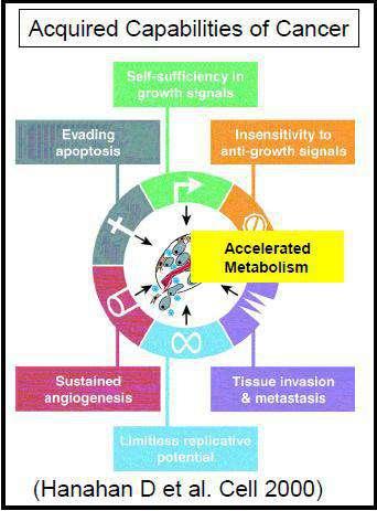
4
5 Functional imaging Tumors Metabolism (glucose, amino acid) Proliferation Hypoxia PET Angiogenesis Apoptosis Image receptors
6 Functional imaging Tumors Metabolism (glucose, amino acid) Angiogenesis Proliferation Perfusion Hypoxia Diffusion characteristics Angiogenesis Metabolite quantification Apoptosis Hypoxia Image receptors CT/MR
7 Tumor imaging Angiogenesis Perfusion Diffusion characteristics CT/MR Metabolite quantification Hypoxia
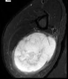
8 Angiogenesis (DCE MRI and CT Perfusion) Physiological imaging (ultrafast imaging) Follows the early enhancement kinetics of contrast within first few minutes of IV bolus injection (TIC). Depicts tissue vascularisation, capillary permeability, perfusion, volume of interstitial space, thus indirectly tissue cellularity.
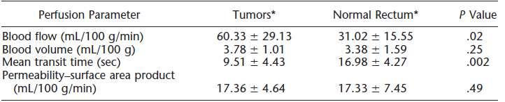
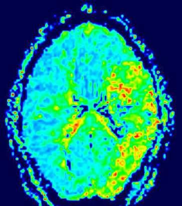
9 Angiogenesis (CT Perfusion) Assesses physiological parameters Blood flow (BF) Blood volume (BV) Mean transit time (MTT) Capillary permeability (CP/PS) In vivo markers of micro vessel density and angiogenesis
10 Angiogenesis (CT Perfusion) De-convolution based analysis technique Operative derived ROI are placed on artery and representative portion of the tumor for input functions Functional maps of perfusion parameters obtained
11 Angiogenesis (CT Perfusion)

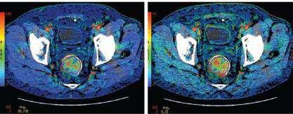
12 Angiogenesis (CT Perfusion) Rectal Cancer : response to neo-adjuvant therapy
 SI Time")
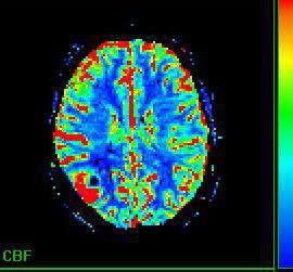
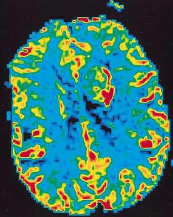
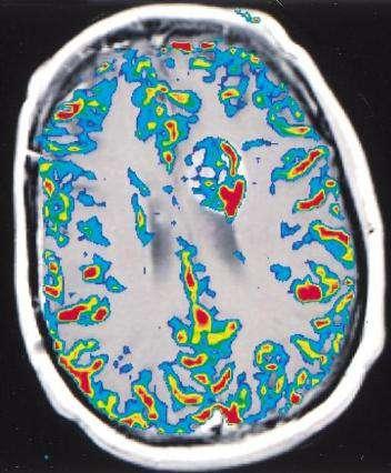
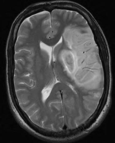
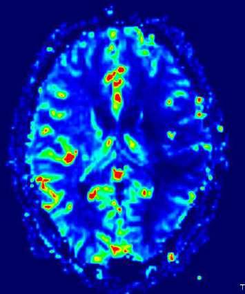
13 MR Perfusion / DSC MRI (Brain tumors) SI Time
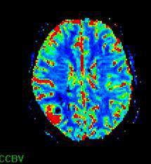
14 MR Perfusion / DSC MRI (Brain tumors) Cerebral blood volume Cerebral blood flow Mean Transit Time Time to Peak
15 MR Perfusion / DSC MRI (Brain tumors) Establish the diagnosis of tumor Pre- operative assessment of tumor histology Guide tumor biopsy Assessment of true lesion extent Monitoring response to therapy (surrogate marker)
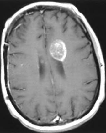
16 Hyperperfusion Possible glioma
17 Delineation of true tumor extent

18 Radiation Necrosis Vs tumor recurrence
19 Angiogenesis (bone & soft tissue tumors) Dynamic contrast enhanced (DCE) MRI Tissue characterization (vascularity & perfusion) Identifying viable areas to biopsy Staging of local extent Monitoring response to therapy (surrogate marker) (K-trans, tissue permeability)
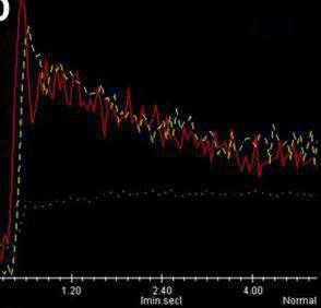
20 Advanced techniques : DCE MR- Curve types

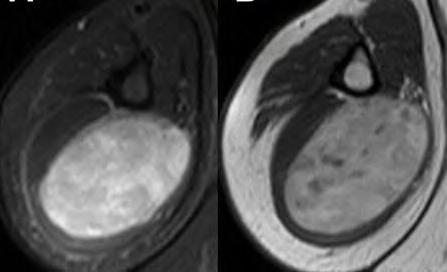
21 Advanced techniques : MRI Dynamic contrast enhanced (DCE) MRI Tissue characterization Highly perfused and vascularised tumor with small volume interstitial space Type IV curve-malignant


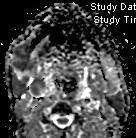
 Avoid edema, necrosis,")
22 Advanced techniques : MRI DCE MRI : Ideal site for biopsy Highly vascularised, highly perfused viable portion of tumor (Synovial Sarc) Avoid edema, necrosis, normal tissue
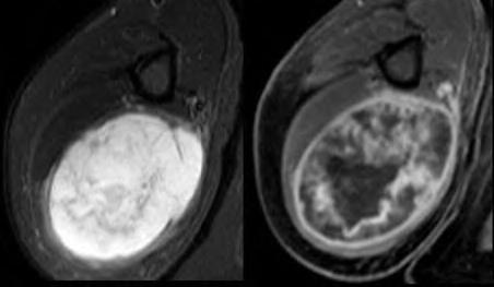
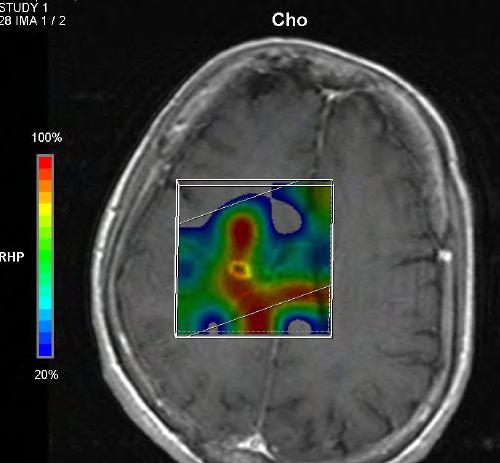
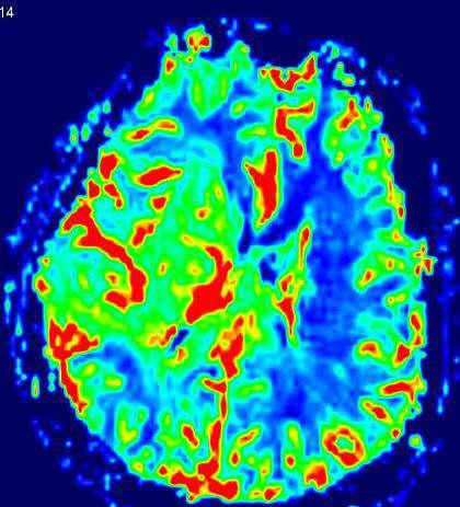
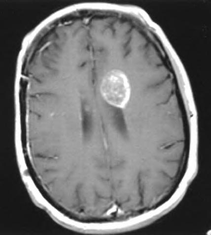
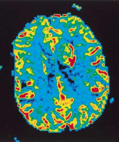
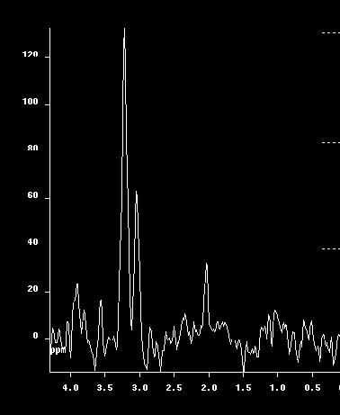
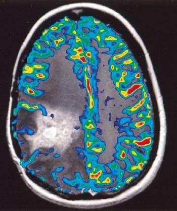
23 Advanced techniques : MRI DCE MRI : Surrogate marker of response to chemo Rx MFH


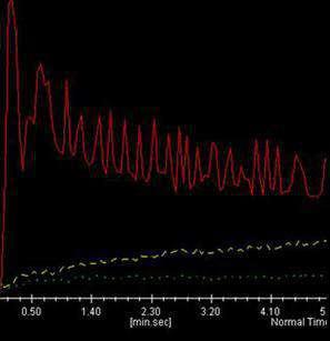
24 Advanced techniques : MRI DCE MRI : Residual/recurrent tumor from post Rx change Gradual slope pseudonodule Bx- negative for malignancy
25 Tumor imaging Angiogenesis Perfusion Diffusion characteristics Metabolite quantification Hypoxia
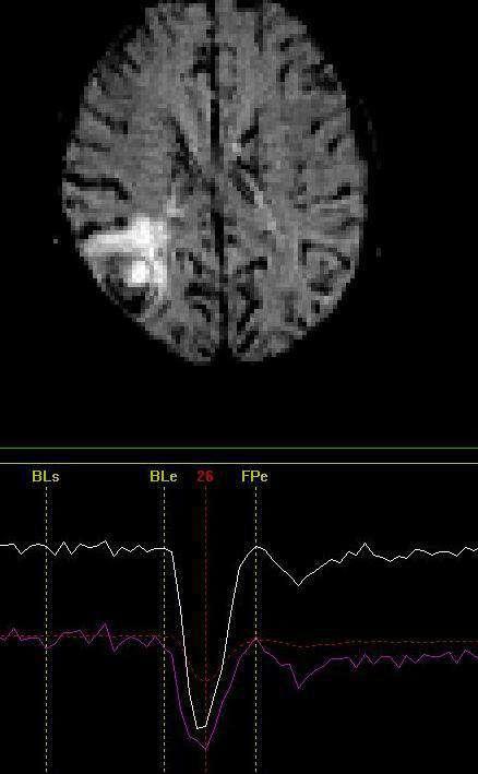
26 Diffusion weighted imaging (DWI) Measures the motion of water molecules in the intra & extravascular spaces Motion of water molecules is more restricted in tissues with high cellularity, intact cell membranes and reduced intracellular space Malignant tumors in general have high cellularity and thus more restricted diffusion. Quantitatively ADC values are used to study diffusion restriction

 A B")
27 Diffusion weighted imaging (DWI) A B C D E F
28 Diffusion weighted imaging (DWI)

29 Diffusion weighted imaging (DWI)
 Synovial")


30 Diffusion weighted imaging (DWI) Synovial sarcoma post surgery Enhancing nodule at surgery site ADC map- restricted diffusion Local recurrence
31 Tumor imaging Angiogenesis Perfusion Diffusion characteristics Metabolite quantification Hypoxia
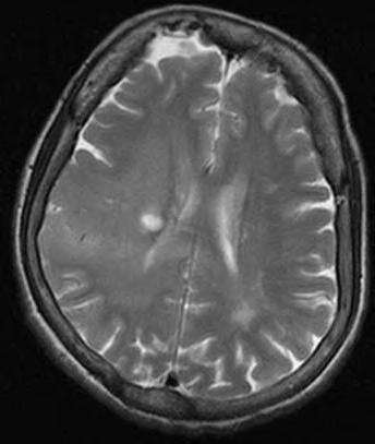
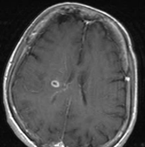
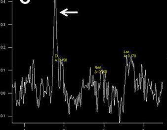
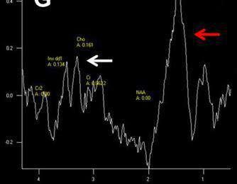
32 Metabolite quantification MR spectroscopy Maps metabolite signal intensity from tissues (choline, creatinine, lipids, NAA, lactate, citrate) Detects increased levels of choline Marker of cell membrane turnover- feature of malignancy Proton (H1) MR spectroscopy is used.

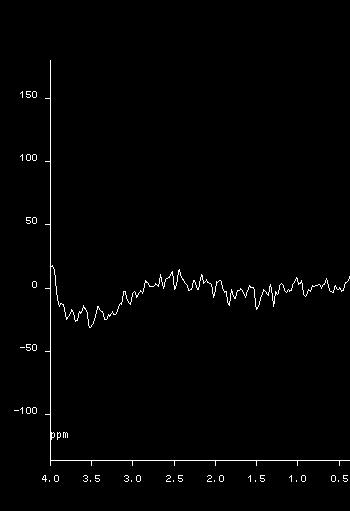
33 Normal Brain spectrum

34 Glioma spectrum
35 Metabolite quantification-mr spectroscopy Radiation Necrosis Vs tumor recurrence

36 Metabolite quantification MR spectroscopy : Response evaluation
37 Tumor imaging Angiogenesis Perfusion Diffusion characteristics Metabolite quantification Hypoxia
38 BOLD (Blood oxygen level dependant imaging) BOLD MR detects hypoxic subfraction and patients suitable for hypoxia modifying agents. Provide a means of evaluating changes in tumor oxygenation in response to chemotherapy Used for Brain, breast, pancreatic and prostate cancers BOLD-MRI images are more likely to reflect on acute (perfusion-related) tissue hypoxia.

39 BOLD
40 Functional techniques Tumor imaging Metabolism (glucose, amino acid) Proliferation Hypoxia Angiogenesis Apoptosis Image receptors PET CT MRI Size & Volume Angiogenesis Perfusion Diffusion characteristics Metabolite quantification Hypoxia
41 Neurology
42 Diffusion weighted imaging (DWI) Measures the motion of water molecules in the intra & extravascular spaces Motion of water molecules is more restricted in tissues with high cellularity, intact cell membranes and reduced intracellular space Infarcts show restricted diffusion in a few hours Quantitatively ADC values are used to study diffusion restriction

43 Central irreversible infarcted tissue core Neurology Significance of penumbra Peripheral region of ischaemic but salvagable tissue called penumbra
44 Comparison of Diffusion & Perfusion Abnormalities Mismatch State of the art imaging of acute stroke

45 Comparison of Diffusion & Perfusion Abnormalities Match State of the art imaging of acute stroke


46 2 hours after deficit CBF MTT 7 days later

47 3 hours


48 3 hours after deficit 7 days later Penumbra is reversed after endovascular clot retrieval & revascularisation
")
49 Functional MRI ( F MRI) BOLD
50 Functional MRI ( F MRI) Defined cognitive task Increased neuronal activity Localized vasodilatation and increase in blood flow Increase in dimagnetic oxyhemoglobin Signal response

51 Epilepsy: cortical dysplasia

52 Epilepsy: cortical dysplasia, fmri
53 Cardiology
54 Cardiology MRI (Myocardial perfusion) use the first pass of an intravenously injected Gd contrast agent at rest and during administration of a vasodilator (i.e. adenosine ) to depict hemodynamically significant coronary artery stenosis MRI (Cardiac Viability) Contrast enhanced CMR is a newly established technique for infarct assessment. Regions of myocardial infarction exhibit high signal intensity (contrast enhancement) on T1 weighted images after administration of contrast such as gadolinium based agents.

55 MR Perfusion Top panel: Subendocardial infarct, prominent perfusion defects larger than infarct on stress MR Bottom panel: Matched stress and rest defects, no infarction, CA is normal- artifactual


56 MRI: Viability 58 yrs old man (EF-20%) Dilated cardiomyopathy Thinning of inferior wall on still images Late ce- MRI shows transmural enhancement suggesting old MI In chronic CAD patients, myocardial enhancement in areas of dysfunctional myocardium corresponds closely to fixed defects on thallium SPECT, and areas of flow-metabolism matched defects on FDG-PET scans, histologically representing scarred or fibrotic tissue


57 Color Doppler Ultrasound Estimates blood flow and velocity Locate and grade vascular stenosis 2D echo Pumping function (ejection fraction)
58 Summary CT/MRI (DCE MRI, DWI, spectroscopy, BOLD) Oncology Angiogenesis (perfusion), diffusion, metabolites, hypoxia Neurology Functional maps, CBF,CBV, MTT (epilepsy, stroke) Cardiology Myocardial perfusion, viability
59 Thank you
Rational use of imaging for viability evaluation
 EUROECHO and other imaging modalities 2011 Rational use of imaging for viability evaluation Luc A. Pierard, MD, PhD, FESC, FACC Professor of Medicine Head, Department of Cardiology, CHU Liège, Belgium
occlusions. Cerebral perfusion is driven fundamentally by regional cerebral
 Appendix Figures Figure A1. Hemodynamic changes that may occur in major anterior circulation occlusions. Cerebral perfusion is driven fundamentally by regional cerebral perfusion pressure (CPP). In response
NUCLEAR CARDIOLOGY UPDATE
 Nuclear Cardiology David K. Shelton, Jr., MD NUCLEAR CARDIOLOGY UPDATE No Conflicts. No Disclosures. No Smoking. David K. Shelton UCDMC Nuclear Cardiology Nuclear Cardiology Radionuclide Ventriculography
Perfusion Physics. ICMRI2018 March 29-31, 2018 Grand Hilton Hotel, Seoul, Korea. Asian Forum Ⅱ: Perfusion MRI SY24-1.
 SY24-1 Perfusion Physics Hiroyuki Kabasawa MR Collaborations and Development, GE Healthcare, Tokyo, Japan Perfusion is referred as the blood supply to micro capillary in tissue. Perfusion parameter such
Use of Nuclear Cardiology in Myocardial Viability Assessment and Introduction to PET and PET/CT for Advanced Users
 Use of Nuclear Cardiology in Myocardial Viability Assessment and Introduction to PET and PET/CT for Advanced Users February 1 5, 2011 University of Santo Tomas Hospital Angelo King A-V Auditorium Manila,
On Call Guide to CT Perfusion. Updated: March 2011
 On Call Guide to CT Perfusion Updated: March 2011 CT Stroke Protocol 1. Non contrast CT brain 2. CT perfusion: contrast 40cc bolus dynamic imaging at 8 slice levels ~ 60 sec creates perfusion color maps
Cardiac Imaging Tests
 Cardiac Imaging Tests http://www.medpagetoday.com/upload/2010/11/15/23347.jpg Standard imaging tests include echocardiography, chest x-ray, CT, MRI, and various radionuclide techniques. Standard CT and
The Value of Stress MRI in Evaluation of Myocardial Ischemia
 The Value of Stress MRI in Evaluation of Myocardial Ischemia Dr. Saeed Al Sayari, MBBS, EBCR, MBA Department of Radiology and Nuclear Medicine Mafraq Hospital, Abu Dhabi United Arab Emirates Introduction
A Light in the Dark: Cardiac MRI and Risk Mitigation. J. Ronald Mikolich MD Professor of Internal Medicine Northeast Ohio Medical University (NEOMED)
") A Light in the Dark: Cardiac MRI and Risk Mitigation J. Ronald Mikolich MD Professor of Internal Medicine Northeast Ohio Medical University (NEOMED) Dr. Mikolich has NO financial disclosures relative to
A Light in the Dark: Cardiac MRI and Risk Mitigation J. Ronald Mikolich MD Professor of Internal Medicine Northeast Ohio Medical University (NEOMED) Dr. Mikolich has NO financial disclosures relative to
Multiparametric imaging in oncology
 Multiparametric imaging in oncology p1 T p2 p2 T T p3 p1 p3 T Marco Ravanelli Roberto Maroldi The goal of traditional imaging is high spatial and contrast resolution diagnosis, tumor extent treatment planning,
Cardiovascular Imaging
 Cardiovascular Imaging Cardiovascular Imaging Cardio and Vascular Imaging Vascularization / Angiogenesis Cardiovascular Imaging metabolic imaging of the heart myocardial perfusion imaging Cardiac CT Vascularization
Imaging of Coronary Artery Disease: II
 Acta Radiológica Portuguesa, Vol.XIX, nº 74, pág. 45-51, Abr.-Jun., 2007 Imaging of Coronary Artery Disease: II Jean Jeudy University of Maryland School of Medicine Department of Diagnostic Radiology Armed
Rotation: Imaging 2. Nuclear Cardiology (in Imaging 1 and 2)
") Rotation: Imaging 2 Imaging 2 provides addition nuclear cardiology experience and COCATS Level 1 cardiac MRI experience. Fellows administer, process, and read VHVI cardiac nuclear studies with cardiology
Rotation: Imaging 2 Imaging 2 provides addition nuclear cardiology experience and COCATS Level 1 cardiac MRI experience. Fellows administer, process, and read VHVI cardiac nuclear studies with cardiology
I have no financial disclosures
 Manpreet Singh MD I have no financial disclosures Exercise Treadmill Bicycle Functional capacity assessment Well validated prognostic value Ischemic assessment ECG changes ST segments Arrhythmias Hemodynamic
Sung A Chang Department of Internal Medicine, Division of Cardiology, Sungkyunkwan University School of Medicine, Samsung Medical Center
 CMR Perfusion and Viability A STICH Out of Time? Sung A Chang Department of Internal Medicine, Division of Cardiology, Sungkyunkwan University School of Medicine, Samsung Medical Center Can Imaging Improve
Advanced Neuroimaging for Acute Stroke
 Advanced Neuroimaging for Acute Stroke E. Bradshaw Bunney, MD, FACEP Professor Department Of Emergency Medicine University of Illinois at Chicago Swedish American Belvidere Hospital Disclosures FERNE Board
PET IMAGING (POSITRON EMISSION TOMOGRAPY) FACT SHEET
 FACT SHEET") Positron Emission Tomography (PET) When calling Anthem (1-800-533-1120) or using the Point of Care authorization system for a Health Service Review, the following clinical information may be needed to
Positron Emission Tomography (PET) When calling Anthem (1-800-533-1120) or using the Point of Care authorization system for a Health Service Review, the following clinical information may be needed to
Cardiac Imaging. Kimberly Delcour, DO, FACC. Mahi Ashwath, MD, FACC, FASE. Director, Cardiac CT. Director, Cardiac MRI
 Cardiac Imaging Kimberly Delcour, DO, FACC Director, Cardiac CT Mahi Ashwath, MD, FACC, FASE Director, Cardiac MRI Cardiac Imaging Discuss the clinical applications of and indications for: Cardiac CT Nuclear
Qualitative and Quantitative Assessment of Perfusion
 APCDE 2011 Qualitative and Quantitative Assessment of Perfusion Hyun Ju Yoon Chonnam National University Hospital Gwangju, Korea ISCHEMIC CASCADE Blood flow mismatch Perfusion defects on nuclear imaging,
Imaging Acute Stroke and Cerebral Ischemia
 Department of Radiology University of California San Diego Imaging Acute Stroke and Cerebral Ischemia John R. Hesselink, M.D. Causes of Stroke Arterial stenosis Thrombosis Embolism Dissection Hypotension
Myocardial viability testing. What we knew and what is new
 Myocardial viability testing. What we knew and what is new Dr B K S Sastry, MD, DM. CARE Hospitals, Hyderabad What is Viability Viability Dysfunctional myocardium subtended by diseased coronary arteries
Clinical indications for positron emission tomography
 Clinical indications for positron emission tomography Oncology applications Brain and spinal cord Parotid Suspected tumour recurrence when anatomical imaging is difficult or equivocal and management will
Multimodality Imaging in Cardiac Stem Cell Research
 Multimodality Imaging in Cardiac Stem Cell Research IL SUK SOHN, MD, PhD Department of Cardiology Kyung Hee University Hospital at Gangdong Kyung Hee University School of Medicine, Seoul, Korea Stem Cell
Perfusion, Viability, Edema and Hemorrhage: How it Can (and Should) Change Clinical Practice. Rohan Dharmakumar, Ph.D.
 Change Clinical Practice. Rohan Dharmakumar, Ph.D.") Perfusion, Viability, Edema and Hemorrhage: How it Can (and Should) Change Clinical Practice Rohan Dharmakumar, Ph.D. Director, Translational Cardiac Imaging Research Associate Director, Biomedical Imaging
Perfusion, Viability, Edema and Hemorrhage: How it Can (and Should) Change Clinical Practice Rohan Dharmakumar, Ph.D. Director, Translational Cardiac Imaging Research Associate Director, Biomedical Imaging
Perfusion MRI. Youngkyoo Jung, PhD Associate Professor Radiology, Biomedical Engineering, and Clinical & Translational Science Institute
 Perfusion MRI Youngkyoo Jung, PhD Associate Professor Radiology, Biomedical Engineering, and Clinical & Translational Science Institute Perfusion The delivery of blood to a capillary bed in tissue Perfusion
Gated blood pool ventriculography: Is there still a role in myocardial viability?
 Gated blood pool ventriculography: Is there still a role in myocardial viability? Oliver C. Alix, MD Adult Clinical and Nuclear Cardiology St. Luke s Medical Centre - Global City Case Presentation A 62-year-old
Disclosures. GETTING TO THE HEART OF THE MATTER WITH MULTIMODALITY CARDIAC IMAGING Organ Review Meeting 25 September. Overview
 GETTING TO THE HEART OF THE MATTER WITH MULTIMODALITY CARDIAC IMAGING Organ Review Meeting 25 September Disclosures None relevant to this presentation Mini Pakkal Assistant Professor of Radiology University
Disclosures. Diffusion and Perfusion Imaging in the Head and Neck. Learning objectives ???
 Disclosures No relevant financial disclosures Diffusion and Perfusion Imaging in the Head and Neck Ashok Srinivasan, MD Associate Professor Director of Neuroradiology University of Michigan Health System
Goals for this Lecture. Case 1. Key Points MRI TECHNIQUES FOR DIFFERENTIAL DIAGNOSIS OF RECURRENT BRAIN LESIONS
 MRI TECHNIQUES FOR DIFFERENTIAL DIAGNOSIS OF RECURRENT BRAIN LESIONS Goals for this Lecture 1. Review common appearances for recurrent tumor and treatment effects on conventional MRI 2. Discuss current
Detection and Assessment of MI: Use of Imaging Methods. Robert O. Bonow, M.D.
 Detection and Assessment of MI: Use of Imaging Methods Robert O. Bonow, M.D. Detection and Assessment of MI: Use of Imaging Methods Robert O. Bonow, M.D. No Relationships to Disclose Expert Consensus Document
PERFUSION MRI CONTRAST BASED TECHNIQUES
 PERFUSION MRI CONTRAST BASED TECHNIQUES by Kenny K Israni Mar 28, 2006 PERFUSION - MRI Dynamic Susceptibility contrast Dynamic Relaxivity contrast STEADY-STATE STATE TECHNIQUES Steady-state Susceptibility
Place for Interventional Radiology in Acute Stroke
 Place for Interventional Radiology in Acute Stroke Dr Lakmalie Paranahewa MBBS, MD(Radiology), FRCR Consultant Interventional Radiologist Asiri Group of Hospitals Objectives Imaging in Stroke Neurovascular
Laura Tormoehlen, M.D. Neurology and EM-Toxicology Indiana University
 Laura Tormoehlen, M.D. Neurology and EM-Toxicology Indiana University Disclosures! No conflicts of interest to disclose Neuroimaging 101! Plain films! Computed tomography " Angiography " Perfusion! Magnetic
Radiology Codes Requiring Authorization*
 70336 Magnetic resonance (eg, proton) imaging, temporomandibular joint(s) 70450 Computed tomography, head or brain; without contrast material 70460 Computed tomography, head or brain; with contrast material(s)
CHAP 11 Contrast Enhanced MRI - Perfusion. CHAP 11 Contrast Enhanced MRI - Perfusion
 CHAP 11 Contrast Enhanced MRI - Perfusion Contrast agents Paramagnetic / superparamagnetic Relaxivity Water exchange Susceptibility effect Brain perfusion pathologies Perfusion measurement T2* (DSC-MRI)
A Light in the Dark: Cardiac MRI and Risk Mitigation. J. Ronald Mikolich MD Professor of Internal Medicine Northeast Ohio Medical University (NEOMED)
") A Light in the Dark: Cardiac MRI and Risk Mitigation J. Ronald Mikolich MD Professor of Internal Medicine Northeast Ohio Medical University (NEOMED) Dr. Mikolich has NO financial disclosures relative to
A Light in the Dark: Cardiac MRI and Risk Mitigation J. Ronald Mikolich MD Professor of Internal Medicine Northeast Ohio Medical University (NEOMED) Dr. Mikolich has NO financial disclosures relative to
Outline. Why Image Animals?
 Small Animal Magnetic Resonance Imaging: Current Trends, Challenges and Perspectives for Pathological Imaging C. Chad Quarles Vanderbilt University Institute of Imaging Science Outline Why image animals?
CHRONIC CAD DIAGNOSIS
 CHRONIC CAD DIAGNOSIS Chest Pain Evaluation 1. Approach to diagnosis of CAD 2. Classification of chest pain 3. Pre-test likelihood CAD 4. Algorithm for chest pain evaluation in women 5. Indications for
Related Symposia in AAPM 2007
 Related Symposia in AAPM 7 Functional and Physiological MR Imaging for Therapy Assessment Yue Cao,, Ph.D. Departments of Radiation Oncology and Radiology, University of Michigan President s s symposium:
What Radiologists do?
 Multimodality Imaging in Oncology 2018 March 5 th 9th Diagnostic Imaging in Oncology What Radiologists do? Chikako Suzuki, MD, PhD Department of Diagnostic Radiology, KS Solna Department of Molecular Medicine
Cigna - Prior Authorization Procedure List: Radiology & Cardiology
 Cigna - Prior Authorization Procedure List: Radiology & Cardiology Product Category CPT Code CPT Code Description Radiology MR 70336 MRI Temporomandibular Joint(s), (TMJ) Radiology CT 70450 CT Head or
Anthem Blue Cross and Blue Shield Virginia Advanced Imaging Procedures Requiring Precertification Revised 02/13/2013
 Anthem Blue Cross and Blue Shield Virginia Advanced Imaging Procedures Requiring Precertification Revised 02/13/2013 Modality and CT Head CTA Head: Cerebrovascular MRI Head MRA Head: Cerebrovascular Functional
1) Diffusion weighted imaging DWI is a term used to describe moving molecules due to random thermal motion. This motion is restricted by boundaries
 Diffusion weighted imaging DWI is a term used to describe moving molecules due to random thermal motion. This motion is restricted by boundaries") 1) Diffusion weighted imaging DWI is a term used to describe moving molecules due to random thermal motion. This motion is restricted by boundaries such as ligaments, membranes and macro molecules. Diffusion
1) Diffusion weighted imaging DWI is a term used to describe moving molecules due to random thermal motion. This motion is restricted by boundaries such as ligaments, membranes and macro molecules. Diffusion
AMERICAN IMAGING MANAGEMENT
 2012 CPT Codes Computerized Tomography (CT) CPT Description Abdomen 74150 CT abdomen; w/o 74160 CT abdomen; with 74170 CT abdomen; w/o followed by Chest 71250 CT thorax; w/o 71260 CT thorax; with 71270
AMERICAN IMAGING MANAGEMENT
 2010 BCBS of Georgia CPT Codes With Grouper Numbers Computerized Tomography (CT) CPT Description Abdomen 74150 CT abdomen; w/o contrast 6 74160 CT abdomen; with contrast 74170 CT abdomen; w/o contrast
J. Schwitter, MD, FESC Section of Cardiology
 J. Schwitter, MD, FESC Section of Cardiology CMR Center of the CHUV University Hospital Lausanne - CHUV Switzerland Centre de RM Cardiaque J. Schwitter, MD, FESC Section of Cardiology CMR Center of the
MRS and Perfusion of Brain Tumors
 Department of Radiology University of California San Diego MRS and Perfusion of Brain Tumors John R. Hesselink, M.D. MRS & Perfusion of Brain Tumors Tumor histology Degree of malignancy Delineate tumor
Imaging of Pediatric MSK Tumors
 Imaging of Pediatric MSK Tumors Kirsten Ecklund, M.D. Boston Children s Hospital Harvard Medical School kirsten.ecklund@childrens.harvard.edu Tumor Imaging Goals Diagnosis Lesion characterization Benign
Cigna - Prior Authorization Procedure List: Radiology & Cardiology
 Cigna - Prior Authorization Procedure List: Radiology & Cardiology Category CPT Code CPT Code Description 93451 Right heart catheterization 93452 Left heart catheterization 93453 Combined right and left
The role of Magnetic Resonance Imaging in the diagnosis of viability & Coronary Artery Disease
 The role of Magnetic Resonance Imaging in the diagnosis of viability & Coronary Artery Disease G.P. Spanos, MSc, Phd Head of CardioVascular Imaging Tomographia Diagnostic Center Cardiovascular magnetic
Imaging Decisions Start Here SM
 Owing to its high resolution and wide anatomic coverage, dynamic first-pass perfusion 320-detector-row CT outperforms PET/CT for distinguishing benign from malignant lung nodules, researchers from Japan
MRI/MRS Biomarkers. Robert E. Lenkinski, Ph.D.
 MRI/MRS Biomarkers Robert E. Lenkinski, Ph.D. Disclosure GE Healthcare-Research Grant Aspect MR-Scientific Advisor Aposense-Scientific Advisor Brainwatch-Scientific Advisor I will be discussing off-label
Medical imaging X-ray, CT, MRI, scintigraphy, SPECT, PET Györgyi Műzes
 Medical imaging X-ray, CT, MRI, scintigraphy, SPECT, PET Györgyi Műzes Semmelweis University, 2nd Dept. of Medicine Medical imaging: definition technical process of creating visual representations about
T2, T2*, ute. Yeo Ju Kim. Radiology, Inha University Hospital, Incheon, Korea
 SY28-1 T2, T2*, ute Yeo Ju Kim Radiology, Inha University Hospital, Incheon, Korea T2 relaxation times relate to the rate of transverse magnetization decay, caused by the loss of phase coherence induced
High Tech Imaging Quick Reference Guide
 High Tech Imaging Quick Reference Guide 1 High Tech Imaging Authorizations may now be requested through our secure provider portal, BlueAccess. Getting Started Step 1: Log into BlueAccess from www.bcbst.com
Combined Anatomical and Functional Imaging with Revolution * CT
 GE Healthcare Case studies Combined Anatomical and Functional Imaging with Revolution * CT Jean-Louis Sablayrolles, M.D. Centre Cardiologique du Nord, Saint-Denis, France Case 1 Whole Brain Perfusion and
Prior Authorization for Non-emergency Cardiac Imaging Procedures
 Attention: All Providers Prior Authorization for Non-emergency Cardiac Imaging Procedures The N.C. Medicaid Program is considering implementation of a prior authorization (PA) program for non-emergency
Pearls & Pitfalls in nuclear cardiology
 Pearls & Pitfalls in nuclear cardiology Maythinee Chantadisai, MD., NM physician Division of Nuclear Medicine, Department of radiology, KCMH Principle of myocardial perfusion imaging (MPI) Radiotracer
Radiologic Assessment of Myocardial Viability
 November 2001 Radiologic Assessment of Myocardial Viability Joshua Moss, Harvard Medical School Year III Patient EF 66yo female with a 3-year history of intermittent chest pain previously relieved by sublingual
Bone PET/MRI : Diagnostic yield in bone metastases and malignant primitive bone tumors
 Bone PET/MRI : Diagnostic yield in bone metastases and malignant primitive bone tumors Lars Stegger, Benjamin Noto Department of Nuclear Medicine University Hospital Münster, Germany Content From PET to
Role of DE-CT in Oncology
 Role of DE-CT in Oncology Dushyant Sahani, M.D Director of CT Associate Professor of Radiology Massachusetts General Hospital Harvard Medical School Email-dsahani@partners.org Disclosure Research Grant
2014 CPT Radiology Codes Requiring Review
 CT Head 1 70480 CT orbit, sella or posterior fossa; w/o contrast 1 CT Head 1 70481 CT orbit, sella or posterior fossa; with CT orbit, sella or posterior fossa; w/o contrast CT Head 1 70482 followed by
Diagnostic Imaging Utilization Management and Consultation Management Programs Imaging Code Listing for Connecticut, Maine and New Hampshire
 Diagnostic Imaging Utilization Management and Consultation Management Programs Imaging Code Listing for Connecticut, Maine and New Hampshire The grid below contains the CPT * codes that are subject to
High Risk PCI for Heart Failure
 High Risk PCI for Heart Failure Ray Matthews MD Professor of Clinical Medicine Chief, Division of Cardiovascular Medicine University of Southern California Los Angeles, California Disclosures Abiomed Research
Click here for Link to References: CMS Website HOPPS CY 2018 Final Rule. CMS Website HOPPS CY2018 Final Rule Updated November 2017.
 Final Compared to 3Q 2017 Rates Medicare Hospital Outpatient Prospective Payment System HOPPS () Nuclear Cardiology Procedures, Radiopharmaceuticals, and Drugs Click here for Link to References: CMS Website
Cardiology for the Practitioner Advanced Cardiac Imaging: Worth the pretty pictures?
 Keenan Research Centre Li Ka Shing Knowledge Institute Cardiology for the Practitioner Advanced Cardiac Imaging: Worth the pretty pictures? Howard Leong-Poi, MD, FRCPC Associate Professor of Medicine St.
BlueAdvantage SM. & BlueChoice SM Radiology Prior Authorization Program Code List CPT /HCPS
 BlueAdvantage SM & BlueChoice SM Radiology Prior Authorization Program Code List CPT /HCPS 70336 MRI TMJ 70450 CT Head Without Contrast 70460 CT Head With Contrast 70470 CT Head Without & With Contrast
Current Guidelines for Diagnosis of AMI Chest pain ST change on EKG Cardiac Enzymes
 Noninvasive Cardiac Imaging in Myocardial Infarction Sangchol Lee Sungkyunkwan University Samsung Medical Center Current Guidelines for Diagnosis of AMI Chest pain ST change on EKG Cardiac Enzymes Do We
Cardiac PET. John Buscombe
 Cardiac PET John Buscombe Why PET? Improved resolution-not really required in cardiology Improved sensitivity this may be important-financially as reduced acquisition time Improved attenuation correction-good
Biomarkers and the Future of. John R. Votaw CBIS 5 th Year Anniversary Celebration/Look to the future February 8, 2013
 Biomarkers and the Future of Radiology John R. Votaw CBIS 5 th Year Anniversary Celebration/Look to the future February 8, 2013 Statistics/Radiology Collaboration The utility of Radiologic procedures
MR Functional Imaging to Guide Radiotherapy: Challenges and Opportunities
 Abstract No. 1234 MR Functional Imaging to Guide Radiotherapy: Challenges and Opportunities Michael Milosevic, MD Department of Radiation Oncology, University of Toronto Radiation Medicine Program, Princess
PET myocard perfusion & viability Riemer Slart
 PET myocard perfusion & viability Riemer Slart Nuclear Medicine Physician Dept. of Nuclear Medicine and Molecular Imaging University Medical Center Groningen, the Netherlands Professor in Molecular Imaging,
Diagnostic Imaging Utilization Management and Consultation Management Programs Imaging Code Listing for Connecticut, Maine and New Hampshire
 Diagnostic Imaging Utilization Management and Consultation Management Programs Imaging Code Listing for Connecticut, Maine and New Hampshire The grid below contains the CPT * codes that are subject to
CT for Myocardial Characterization of Cardiomyopathy. Byoung Wook Choi, Yonsei University Severance Hospital, Seoul, Korea
 CT for Myocardial Characterization of Cardiomyopathy Byoung Wook Choi, Yonsei University Severance Hospital, Seoul, Korea Cardiomyopathy Elliott P et al. Eur Heart J 2008;29:270-276 The European Society
The use of Cardiac CT and MRI in Clinical Practice
 The use of Cardiac CT and MRI in Clinical Practice Matthew W. Martinez, MD Assistant Professor of Medicine LVPG - Lehigh Valley Heart Specialists Lehigh Valley Health Network Oct. 3, 2009 DISCLOSURE Relevant
screening; including image post processing CT, heart; without contrast material; with Requires authorization
 0042T Cerebral perfusion analysis using CT; with ; including of parametric maps with determination of cerebral blood flow, cerebral blood volume, and mean transit time 74263 Computed tomographic (CT) colonography,
MR Assessment of Myocardial Viability
 MR Assessment of Myocardial Viability Definition of Viability Clinical Metabolism: Presence of glucose uptake Perfusion / Perfusion reserve Morphology: Wall thickness, wall thickening Contractility: Recovery
Assessment Of Myocardial Viability
 Assessment Of Myocardial Viability James K. Min, MD FACC President, Society of Cardiovascular Computed Tomography Associate Professor of Medicine, UCLA School of Medicine Associate Professor of Medicine
Speed, Comfort and Quality with NeuroDrive
 Speed, Comfort and Quality with NeuroDrive Echelon Oval provides a broad range of capabilities supporting fast, accurate diagnosis of brain conditions and injuries. From anatomical depiction to vascular
Codes Requiring Authorization from MedSolutions (MSI): Updated 3/2014
: Updated 3/2014") s Requiring Authorization from MedSolutions (): Updated 3/2014 0042T Cerebral Perfusion Analysis using CT with contrast 0159T CAD, including computer algorithm analysis, BREAST MRI 0195T prepare interspace,
s Requiring Authorization from MedSolutions (): Updated 3/2014 0042T Cerebral Perfusion Analysis using CT with contrast 0159T CAD, including computer algorithm analysis, BREAST MRI 0195T prepare interspace,
Cardiac magnetic resonance imaging in rheumatoid arthritis: promising or misleading? Sophie Mavrogeni MD FESC
 Cardiac magnetic resonance imaging in rheumatoid arthritis: promising or misleading? Sophie Mavrogeni MD FESC Onassis Cardiac Surgery Center Athens Greece Nothing to disclose Financial disclosure Cardiac
Imaging in Ischemic Heart Disease: Role of Cardiac MRI
 Imaging in Ischemic Heart Disease: Role of Cardiac MRI Chiara Bucciarelli Ducci MD, PhD, FESC, FRCP Consultant Senior Lecturer Cardiologist Bristol Heart Institute, University of Bristol, UK Chair elect,
Cardiac MRI in ACHD What We. ACHD Patients
 Cardiac MRI in ACHD What We Have Learned to Apply to ACHD Patients Faris Al Mousily, MBChB, FAAC, FACC Consultant, Pediatric Cardiology, KFSH&RC/Jeddah Adjunct Faculty, Division of Pediatric Cardiology
05/02/ CPT Preauthorization Groupings Effective May 2, Computerized Tomography (CT) Abdomen 6. CPT Description SEGR CT01
 Abdomen 6. CPT Description SEGR CT01") Computerized Tomography (CT) 6 & 101 5 Upper Extremity 11 Lower Extremity 12 Head 3 Orbit 1 Sinus 2 Neck 4 7 Cervical Spine 8 Thoracic Spine 9 Lumbar Spine 10 Colon 13 CPT Preauthorization Groupings CPT
Computerized Tomography (CT) 6 & 101 5 Upper Extremity 11 Lower Extremity 12 Head 3 Orbit 1 Sinus 2 Neck 4 7 Cervical Spine 8 Thoracic Spine 9 Lumbar Spine 10 Colon 13 CPT Preauthorization Groupings CPT
Table 1. Summary of PET and fmri Methods. What is imaged PET fmri BOLD (T2*) Regional brain activation. Blood flow ( 15 O) Arterial spin tagging (AST)
 Regional brain activation. Blood flow ( 15 O) Arterial spin tagging (AST)") Table 1 Summary of PET and fmri Methods What is imaged PET fmri Brain structure Regional brain activation Anatomical connectivity Receptor binding and regional chemical distribution Blood flow ( 15 O)
Table 1 Summary of PET and fmri Methods What is imaged PET fmri Brain structure Regional brain activation Anatomical connectivity Receptor binding and regional chemical distribution Blood flow ( 15 O)
Why Cardiac MRI? Presented by:
 Why Cardiac MRI? Presented by: Lisa G. Carkner, MD, FACC 1 Disclosures I have no financial disclosures Objectives Review basic principles of Cardiac MRI. What patient characteristics do I need to consider
Prostate MRI Hamidreza Abdi, MD,FEBU Post Doctoral Fellow Vancouver Prostate Centre UBC Department of Urologic Sciences May-20144
 Prostate MRI Hamidreza Abdi, MD,FEBU Post Doctoral Fellow Vancouver Prostate Centre UBC Department of Urologic Sciences May-20144 Objectives: Detection of prostate cancer the need for better imaging What
PET-MRI in malignant bone tumours. Lars Stegger Department of Nuclear Medicine University Hospital Münster, Germany
 PET-MRI in malignant bone tumours Lars Stegger Department of Nuclear Medicine University Hospital Münster, Germany Content From PET to PET/MRI General considerations Bone metastases Primary bone tumours
CARDIAC PET PERFUSION IMAGING with RUBIDIUM-82
 CARDIAC PET PERFUSION IMAGING with RUBIDIUM-82 Pr Denis AGOSTINI Président du Groupe de Cardiologie Nucléaire et IRM CHU Caen Bordeaux 2006 Cardiac Perfusion-Metabolism Mismatch with PET Cumulative Survival
Conflict Disclosures. Vermont Cardiac Network. Outline. Series Learning Objectives 4/27/2016. Scott E. Friedman April 28, 2016
 Conflict Disclosures Vermont Cardiac Network The Speaker has reported no significant financial relationship with any companies whose product may be germane to the content of their presentations or who
Cardiac MRI: Clinical Application to Disease
 Cardiac MRI: Clinical Application to Disease Jessi Smith, MD Cardiothoracic imaging, Indiana University Slides courtesy of Stacy Rissing, MD Outline Imaging planes Disease findings Pulse sequences used
Current Indications for Cardiac MRI: What You See is What You Get?
 Current Indications for Cardiac MRI: What You See is What You Get? Javier Ganame, MD, PhD, FASE No disclosures Cardiology Update, Niagara, Sept 24th, 2016 The Ideal Diagnostic Technique Easy to apply Accurate
Diagnostic Imaging Prior Review Code List 2 nd Quarter 2018
 Computerized Tomography (CT) Abdomen 6 Abdomen/Pelvis Combination 101 Service 74150 CT abdomen; w/o 74160 CT abdomen; with 74170 CT abdomen; w/o followed by 74176 Computed tomography, abdomen and pelvis;
Usefulness of Delayed Enhancement by Magnetic Resonance Imaging in Hypertrophic Cardiomyopathy as a Marker of Disease and Its Severity
 Usefulness of Delayed Enhancement by Magnetic Resonance Imaging in Hypertrophic Cardiomyopathy as a Marker of Disease and Its Severity G.D.Aquaro, MD Fondazione G.Monasterio Regione Toscana/CNR Pisa, Italy
Introduction to Brain Imaging
 Introduction to Brain Imaging Human Brain Imaging NEUR 570 & BIC lecture series September 9, 2013 Petra Schweinhardt, MD PhD Montreal Neurological Institute McGill University Montreal, Canada Various techniques
Proc. Intl. Soc. Mag. Reson. Med. 22 (2014)
") Tumor Physiology Natalie J. Serkova, PhD Department of Anesthesiology and Radiology, University of Colorado at Denver, Anschutz Medical Center, Aurora, CO This course will describe the distinct characteristics
Tumor Physiology Natalie J. Serkova, PhD Department of Anesthesiology and Radiology, University of Colorado at Denver, Anschutz Medical Center, Aurora, CO This course will describe the distinct characteristics
Cardiac MRI: Cardiomyopathy
 Cardiac MRI: Cardiomyopathy Laura E. Heyneman, MD I do not have any relevant financial relationships with any commercial interests Cardiac MRI: Cardiomyopathy Laura E. Heyneman, MD Duke University Medical
Ventricular tachycardia and ischemia. Martin Jan Schalij Department of Cardiology Leiden University Medical Center
 Ventricular tachycardia and ischemia Martin Jan Schalij Department of Cardiology Leiden University Medical Center Disclosure: Research grants from: Boston Scientific Medtronic Biotronik Sudden Cardiac
MOLINA HEALTHCARE OF MICHIGAN PRIOR AUTHORIZATION / PRE-SERVICE REVIEW GUIDE IMAGING CODES REQUIRING PRIOR AUTHORIZATION EFFECTIVE 1/1/2014
 70336 MRI MRI, temporomandibular joint(s) 70450 CT/CTA CT, head or brain; without contrast material 70460 CT/CTA CT, head or brain; with contrast material(s) 70470 CT/CTA CT, head or brain; without contrast
The Egyptian Journal of Hospital Medicine (July 2018) Vol. 72 (10), Page
 Vol. 72 (10), Page") The Egyptian Journal of Hospital Medicine (July 2018) Vol. 72 (10), Page 5398-5402 The Role of Susceptibility Weighted Imaging (SWI) in Evaluation of Acute Stroke Maha Abdelhamed El Nouby*, Eman Ahmed
The Egyptian Journal of Hospital Medicine (July 2018) Vol. 72 (10), Page 5398-5402 The Role of Susceptibility Weighted Imaging (SWI) in Evaluation of Acute Stroke Maha Abdelhamed El Nouby*, Eman Ahmed