High-risk vulnerable plaques. Kostis Raisakis G.Gennimatas General Hospital of Athens
|
|
|
- Dylan Lucas
- 5 years ago
- Views:
Transcription
1 High-risk vulnerable plaques. Kostis Raisakis G.Gennimatas General Hospital of Athens
2 Overview: 1 Definition-Pathology 2 3 Diagnostic Strategies Invasive Non Invasive Prognostic Value of Detection 4 Treatment General Focused
3 Overview: 1 Definition-Pathology 2 3 Diagnostic Strategies Invasive Non Invasive Prognostic Value of Detection 4 Treatment General Focused
4 Necrotomic Examination Culprit Lesions Histological Similarities with Non-culprit Lesions CV Death Vulnerable Plaques Treatment Efficiency Prognostic Value Algorithms Ex-vivo verification of Modalities Ex-vivo verification In-vivo comparison New Modalities Morphological Molecular Biochemical
5 1 Vulnerable Plaque Clinical Definition Any thrombosis-prone plaque or plaque at a risk of rapid progression, with potential of becoming a culprit lesion and triggering an ACS, independent of its specific morphology Naghavi et al, Circulation 2003

6 1 Vulnerable Plaque Definition Pathological Substrate of Thrombosis Virmani R, et al. Arterioscler Thromb Vasc Biol 2000
 Increased Macrophage Density, ~26% of cap Reduced Smooth Muscle Cells Increased Angiogenesis New Microvessels Intraplaque hemorrhage")
7 1 Vulnerable Plaque Definition Thin-cap Fibroatheroma (TCFA) Increased Plaque size Positive remodeling Increased Necrotic core ~34% of plaque area ~3.8 mm 2 & ~9 mm long Fibrous cap Reduced Thickness (<65 μm) Increased Macrophage Density, ~26% of cap Reduced Smooth Muscle Cells Increased Angiogenesis New Microvessels Intraplaque hemorrhage Perivascular inflammation Reduced Calcification & Spotty Calcification Virmani R, et al. JACC 2006
8 1 Vulnerable Plaque Definition Challenges to the VP concept Multiple Vulnerable plaques co-exist in the coronaries How many of them did rupture? All ruptures all fatal? All fatal events are caused by plaque rupture? The vast majority of so called Vulnerable plaques do not exhibit clinical instability and seldom provokes ACS Virmani R, et al. Arterioscler Thromb Vasc Biol 2000


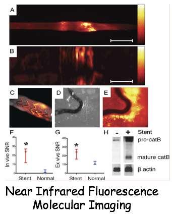
9 1 From Vulnerable Plaque to Vulnerable Patient Toutouzas et al, EHJ 2015
10 Overview: 1 Definition-Pathology 2 3 Diagnostic Strategies Invasive Non Invasive Prognostic Value of Detection 4 Treatment General Focused

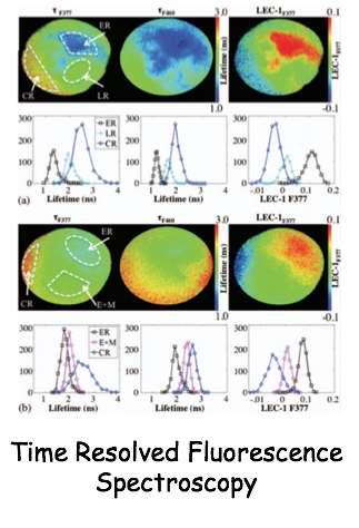
11 2 a Invasive Imaging Available Techniques for VP assessment

12 2 a Invasive Imaging/IVUS Grey Scale Stenosis Severity Plaque Burden Positive Remodeling Calcification

13 2 a Invasive Imaging/IVUS Virtual Histology Tissue components of the plaque are correlated to specific colors
 thin-cap fibroatheroma Fibro-calcified")
14 2 a Invasive Imaging/IVUS Plaque Characterization with IVUS-VH Intima thickness thick-cap fibroatheroma) thin-cap fibroatheroma Fibro-calcified Plaque


15 2 a Invasive Imaging/OCT Plaque Characteristics High resolution Cap thickness Lipid core Macrophages Microvessels Spotty Calcification


16 2 a Invasive Imaging/OCT Plaque Characterization with μoct Liu L et al, Nat Med. 2011


17 2 a Invasive Imaging/NIRS Chemical Characterization of the Arterial Wall Garg et al, Eurointervention 2013 Madder et al, JACC Cardiovasc Interv. 2013

18 2 a Invasive Imaging/NIRS Chemical Characterization of the Arterial Wall Toutouzas et al, JACC 2007
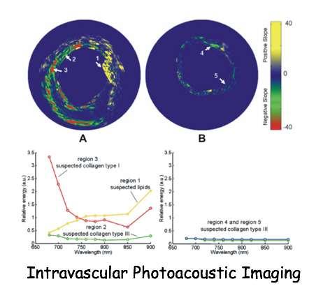
19 2 a Novel Intravascular Imaging Bourantas et al, EHJ. 2016


20 2 a Hybrid Intravascular Imaging IVUS-OCT Bourantas et al, EHJ Li et al, J Biomed Opt 2013
 does not permit the direct visualization of thin fibrous cap Dimensions of the large necrotic core reach the detection threshold of CTA")
21 2 b Non Invasive Imaging/MDCT Detection of Calcium/Calcium score positive remodeling NCP plaque (areas of 30 HU) spotty calcification Spatial resolution ( μm) does not permit the direct visualization of thin fibrous cap Dimensions of the large necrotic core reach the detection threshold of CTA Napkin-Ring Sign

22 2 b Non Invasive Imaging/CTA Plaque Characterization Motoyama et al, JACC 2007
 spotty calcification Stable Angina Pectoris Motoyama et al,")
23 2b Non Invasive Imaging/MDCT Plaque Characterization ACS positive remodeling NCP plaque (areas of 30 HU) spotty calcification Stable Angina Pectoris Motoyama et al, JACC 2007

24 2 b Non Invasive Imaging/CTA Napkin-Ring Sign plaque core with low CT attenuation surrounded by a rim-like area of higher CT attenuation as napkin ring like large central lipid core surrounded by fibrous plaque tissue. deep micro-calcifications intramural thrombus neovascularization, Maurovich-Horvat et al, JACC Cardiov. Imag. 2010
 spotty calcification Ferencik et al, J Cardiovasc")
25 2b Non Invasive Imaging/MDCT Plaque Characterization/ Semi-automated software tool. positive remodeling NCP plaque (areas of 30 HU) spotty calcification Ferencik et al, J Cardiovasc Comput Tomogr. 2015
 Naghavi et")
26 2 b Non Invasive Imaging/PET-CT fluorodeoxyglucose F 18 ( 18 F- FDG) Naghavi et al, Circulation 2003
 Joshi et al, Lancet")
27 2 b Non Invasive Imaging/PET-CT 18 F- FDG versus ⁸F-sodium fluoride( 1 ⁸F-NaF) Joshi et al, Lancet 2014
28 Overview: 1 Definition-Pathology 2 3 Diagnostic Strategies Invasive Non Invasive Prognostic Value of Detection 4 Treatment General Focused 5 Future Perspectives

29 3 Prognostic Value of Imaging VP IVUS-VH/PROSPECT Study Stone et al. N Engl J Med. 2011


30 3 Prognostic Value of Imaging VP IVUS-ESS/ PREDICTION Study Stone P et al. Circulation 2012

31 3 Prognostic Value of Imaging VP ATHEROREMO-NIRS Study LCBI=Lipid Core Burden Index Oemrawsingh et al, JACC 2014
32 3 Prognostic Value of Imaging VP CTA-Napkin ring sign PR=Positive Remodeling LAP=Low Attenuation Plaque Otsuka et al. JACC Caridiovasc Imaging 2013
33 3 Prognostic Value of Imaging VP CTA HRP=High Risk Plaque (positive remodeling and low attenuation plaques) SS=Significant stenosis Motoyama et al. JACC 2015
34 3 Prognostic Value of Imaging VP PET/CT 18 F- FDG TBR=Target to Background Ratio
35 3 Prognostic Value of Imaging VP PET/18F-NaF
36 Overview: 1 Definition-Pathology 2 3 Diagnostic Strategies Invasive Non Invasive Prognostic Value of Detection 4 Treatment General Focused

37 4 a Medical Treatment for VP/Statins Plaque regression Tsujita et al. J Am Coll Cardiol. 2015
38 4 a Medical Treatment for VP/Statins Atheroma Calcification promotion PAV=percent atheroma volume Puri et al. J Am Coll Cardiol. 2015
39 4 a Medical Treatment for VP/Statins NIRS/Plaque lipid depletion LRNC=Lipid-Rich Necrotic Core Zhao et al. JACC Cardiovasc Imaging. 2011


40 4 a Medical Treatment for VP/Statins PET/CT, Vessel Inflammation TBR=Target to Background Ratio Tawakol et al. J Am Coll Cardiol. 2013
41 4 a Medical Treatment/Statins Metanalysis for major events Baigent et al. Lancet. 2010
42 4 a Medical Treatment for VP/Olmesartan Plaque burden regression/ivus Hirohata et al. J Am Coll Cardiol. 2010

43 4 a Medical Treatment for VP/Evolocumab GLAGOV study/ivus Puri et al. Am. Heart J. 2016

44 4 a Medical Treatment for VP Phase III Trials De Caterina et al. Thromb Haemost 2016

45 4 b Invasive Sealing of VP

46 4 b Invasive Sealing of VP
47 Summary Accurate detection of VP is a battle and not war winning. Novel imaging techniques provided new morphological information regarding VP. Available Imaging is unable to completely assess plaque s pathology and function. Prognostic implications of the presence of VPs is questionable. More accurate multi-modality techniques needed for stratification and identification of patients at risk for future events. Novel treatment targeting both the vulnerable plaque and vulnerable patient needed

48 Thank you for your attention
Added Value of Invasive Coronary Imaging for Plaque Rupture and Erosion
 Assessment of Coronary Plaque Rupture and Erosion Added Value of Invasive Coronary Imaging for Plaque Rupture and Erosion Yukio Ozaki, MD, PhD, FACC, FESC Cardiology Dept., Fujita Health Univ. Toyoake,
Imaging Atheroma The quest for the Vulnerable Plaque
 Imaging Atheroma The quest for the Vulnerable Plaque P.J. de Feijter 1. Department of Cardiology 2. Department of Radiology Coronary Heart Disease Remains the Leading Cause of Death in the U.S, Causing
Vulnerable Plaque Pathophysiology, Detection, and Intervention. VP: A Local Problem or Systemic Disease. Erling Falk, Denmark
 Vulnerable Plaque Pathophysiology, Detection, and Intervention VP: A Local Problem or Systemic Disease Erling Falk, Denmark Vulnerable Plaque Pathophysiology, Detection, and Intervention VP: A Local Problem
Cottrell Memorial Lecture. Has Reversing Atherosclerosis Become the New Gold Standard in the Treatment of Cardiovascular Disease?
 Cottrell Memorial Lecture Has Reversing Atherosclerosis Become the New Gold Standard in the Treatment of Cardiovascular Disease? Stephen Nicholls MBBS PhD @SAHMRI_Heart Disclosures Research support: AstraZeneca,
Imaging Overview for Vulnerable Plaque: Data from IVUS Trial and An Introduction to VH-IVUS Imgaging
 Imaging Overview for Vulnerable Plaque: Data from IVUS Trial and An Introduction to VH-IVUS Imgaging Gary S. Mintz,, MD Cardiovascular Research Foundation New York, NY Today, in reality, almost everything
State of the Art. Advances in Cardiovascular Imaging. ESC Congres Stockholm September 1, 2010 Frank E. Rademakers, MD, PhD, FESC
 State of the Art Advances in Cardiovascular Imaging ESC Congres Stockholm September 1, 2010 Frank E. Rademakers, MD, PhD, FESC Coronary Artery Disease Content Patho Physiology Imaging requirements Economical
Invasive Coronary Imaging Modalities for Vulnerable Plaque Detection
 Invasive Coronary Imaging Modalities for Vulnerable Plaque Detection Gary S. Mintz, MD Cardiovascular Research Foundation New York, NY Greyscale IVUS studies have shown Plaque ruptures do not occur randomly
Plaque Characteristics in Coronary Artery Disease. Chourmouzios Arampatzis MD, PhD, FESC
 Plaque Characteristics in Coronary Artery Disease Chourmouzios Arampatzis MD, PhD, FESC Disclosure Statement of Financial Interest Regarding this Presentation NONE Atherosclerosis Model proposed by Stary
Chapter 43 Noninvasive Coronary Plaque Imaging
 hapter 43 Noninvasive oronary Plaque Imaging NIRUDH KOHLI The goal of coronary imaging is to define the extent of luminal narrowing as well as composition of an atherosclerotic plaque to facilitate appropriate
1st Department of Cardiology, University of Athens, Hippokration Hospital, Athens, Greece
 Konstantinos Toutouzas, Maria Riga, Antonios Karanasos, Eleftherios Tsiamis, Andreas Synetos, Maria Drakopoulou, Chrysoula Patsa, Georgia Triantafyllou, Aris Androulakis, Christodoulos Stefanadis 1st Department
Can We Identify Vulnerable Patients & Vulnerable Plaque?
 Can We Identify Vulnerable Patients & Vulnerable Plaque? We Know Enough to Treat High-Risk Lesions? Takashi Akasaka, MD, PhD Department of Cardiovascular Medicine, Japan Disclosure Statement of Financial
2yrs 2-6yrs >6yrs BMS 0% 22% 42% DES 29% 41% Nakazawa et al. J Am Coll Cardiol 2011;57:
 Pathology of In-stent Neoatherosclerosis in BMS and DES 197 BMS, 103 SES, and 106 PES with implant duration >30 days The incidence of neoatherosclerosis was significantly greater in DES (31%) than BMS
Noninvasive Coronary Imaging: Plaque Imaging by MDCT
 Coronary Physiology & Imaging Summit 2007 Noninvasive Coronary Imaging: Plaque Imaging by MDCT Byoung Wook Choi Department of Radiology Yonsei University, Seoul, Korea Stary, H. C. et al. Circulation
CT Imaging of Atherosclerotic Plaque. William Stanford MD Professor-Emeritus Radiology University of Iowa College of Medicine Iowa City, IA
 CT Imaging of Atherosclerotic Plaque William Stanford MD Professor-Emeritus Radiology University of Iowa College of Medicine Iowa City, IA PREVALENCE OF CARDIOVASCULAR DISEASE In 2006 there were 80 million
Coronary Artery Thermography
 Coronary Artery Thermography The 10th Anniversary, Interventional Vascular Therapeutics Angioplasty Summit 2005 TCT Asia Pacific Christodoulos Stefanadis Professor of Cardiology Athens Medical School In
Can IVUS Define Plaque Features that Impact Patient Care?
 Can IVUS Define Plaque Features that Impact Patient Care? A Pichard L Satler, K Kent, R Waksman, W Suddath, N Bernardo, N Weissman, M Angelo, D Harrington, J Lindsay, J Panza. Washington Hospital Center
OCT. molecular imaging J Jpn Coll Angiol, 2008, 48: molecular imaging MRI positron-emission tomography PET IMT
 48 6 CT MRI PET OCT molecular imaging J Jpn Coll Angiol, 2008, 48: 456 461 atherosclerosis, imaging gold standard computed tomography CT magnetic resonance imaging MRI CT B intima media thickness IMT B
Assessment of plaque morphology by OCT in patients with ACS
 Assessment of plaque morphology by OCT in patients with ACS Takashi Akasaka, M.D. Department of Cardiovascular Medicine Wakayama, Japan Unstable plaque Intima Lipid core Plaque rupture and coronary events
Assessment of Vulnerable Plaque by IVUS and VH-IVUS
 Assessment of Vulnerable Plaque by IVUS and VH-IVUS Akiko Maehara, MD Director of Intravascular Imaging & Physiology Core Laboratories Associate Director of MRI/MDCT Core Laboratory Cardiovascular Research
Pathology of Coronary Artery Disease
 Pathology of Coronary Artery Disease Seth J. Kligerman, MD Pathology of Coronary Artery Disease Seth Kligerman, MD Assistant Professor Medical Director of MRI University of Maryland Department of Radiology
Gary S. Mintz,, MD. IVUS Observations in Acute (vs Chronic) Coronary Artery Disease: Structure vs Function
 Coronary Artery Disease: Structure vs Function") Gary S. Mintz,, MD IVUS Observations in Acute (vs Chronic) Coronary Artery Disease: Structure vs Function Important IVUS Observations: Remodeling Originally used (first by Glagov) ) to explain atherosclerosis
Gary S. Mintz,, MD IVUS Observations in Acute (vs Chronic) Coronary Artery Disease: Structure vs Function Important IVUS Observations: Remodeling Originally used (first by Glagov) ) to explain atherosclerosis
Failure of positive. Recanalization and CTO formation. TCFA rupture with (fatal) thrombotic occlusion. TCFA Lipid pool
 thrombotic occlusion. TCFA Lipid pool") Vulnerable Plaque features on coronary CT Jin Ho Choi, MD, PhD Department of Internal Medicine, Emergency Medicine Samsung Medical Center, Sungkyunkwan University School of Medicine, Seoul, Korea IPS /
Vulnerable Plaque features on coronary CT Jin Ho Choi, MD, PhD Department of Internal Medicine, Emergency Medicine Samsung Medical Center, Sungkyunkwan University School of Medicine, Seoul, Korea IPS /
OCT Findings: Lesson from Stable vs Unstable Plaques
 ANGIOPLASTY SUMMIT TCTAP 2010 Imaging Workshop OCT Findings: Lesson from Stable vs Unstable Plaques Giulio Guagliumi MD Ospedali Riuniti di Bergamo, Italy DISCLOSURE OF FINANCIAL INTERESTS Consultant Boston
Multimodality Imaging Atlas of Coronary Atherosclerosis
 JCC: CRDIOVSCUR IMGING VO. 3, NO. 8, 2010 2010 BY THE MERICN COEGE OF CRDIOOGY FOUNDTION ISSN 0735-1097/$36.00 PUBISHED BY ESEVIER INC. DOI:10.1016/j.jcmg.2010.06.006 IMGING VIGNETTE Multimodality Imaging
Κλινική Χρήση IVUS και OCT PERIKLIS A. DAVLOUROS ASSOCIATE PROFESSOR OF CARDIOLOGY INVASIVE CARDIOLOGY & CONGENITAL HEART DISEASE
 Κλινική Χρήση IVUS και OCT PERIKLIS A. DAVLOUROS ASSOCIATE PROFESSOR OF CARDIOLOGY INVASIVE CARDIOLOGY & CONGENITAL HEART DISEASE Conflict of interest None to declare While IVUS is the most used intravascular
Left main coronary artery (LMCA): The proximal segment
: The proximal segment") Anatomy and Pathology of Left main coronary artery G Nakazawa Tokai Univ. Kanagawa, Japan 1 Anatomy Difinition Left main coronary artery (LMCA): The proximal segment RCA AV LAD LM LCX of the left coronary
Anatomy and Pathology of Left main coronary artery G Nakazawa Tokai Univ. Kanagawa, Japan 1 Anatomy Difinition Left main coronary artery (LMCA): The proximal segment RCA AV LAD LM LCX of the left coronary
Assessment of vulnerable plaque by OCT
 Assessment of vulnerable plaque by OCT Comparison with histology and possible clinical applications Takashi Akasaka, M.D. Department of Cardiovascular Medicine Wakayama, Japan Identification of vulnerable
Pathology of Vulnerable Plaque Angioplasty Summit 2005 TCT Asia Pacific, Seoul, April 28-30, 2005
 Pathology of Vulnerable Plaque Angioplasty Summit 25 TCT Asia Pacific, Seoul, April 28-3, 25 Renu Virmani, MD CVPath, A Research Service of the International Registry of Pathology Gaithersburg, MD Plaque
CLINICAL APPLICATIONS OF OPTICAL COHERENCE TOMOGRAPHY. Konstantina P. Bouki, FESC 2 nd Department of Cardiology General Hospital Of Nikea, Pireaus
 CLINICAL APPLICATIONS OF OPTICAL COHERENCE TOMOGRAPHY Konstantina P. Bouki, FESC 2 nd Department of Cardiology General Hospital Of Nikea, Pireaus OPTICAL COHERENCE TOMOGRAPHY (OCT) IVUS and OCT IVUS OCT
FESC, FACC, MAHA, MSCAI, MEAPSI, ESH
 «Απεικόνιση και φυσιολογία στο αιμοδυναμικό εργαστήριο». Eυάλωτη αθηρωματική πλάκα. Πού βρισκόμαστε? Ηλίας Α. Σανίδας MD, PhD, FESC, FACC, MAHA, MSCAI, MEAPSI, ESH Specialist Επεμβατικός Kαρδιολόγος Επιμελητής,
OCT; Comparative Imaging Results with IVUS, VH and Angioscopy
 OCT; Comparative Imaging Results with IVUS, VH and Angioscopy Takashi Akasaka, M.D. Department of Cardiovascular Medicine Wakayama, Japan Comparison among coronary imaging techniques OCT IVUS MRI CAG Angioscopy
Molecular Imaging of Coronary Plaques
 Molecular Imaging of Coronary Plaques Daniel S. Berman, MD Director, Cardiac Imaging Cedars-Sinai Heart Institute CSMC 20113 Professor of Medicine David Geffen School of Medicine at UCLA Zahi A. Fayad,
Malaysian Healthy Ageing Society
 Organised by: Co-Sponsored: Malaysian Healthy Ageing Society CAD INVESTIGATIONS PHYSIOLOGICAL / FUNCTIONAL VS ANATOMICAL / STRUCTURAL Disclosure iheal medical centre is a one stop cardiac centre with
Atherosclerosis xxx (2012) 1e7. Contents lists available at SciVerse ScienceDirect. Atherosclerosis
 1e7. Contents lists available at SciVerse ScienceDirect. Atherosclerosis") Atherosclerosis xxx (2012) 1e7 Contents lists available at SciVerse ScienceDirect Atherosclerosis journal homepage: www.elsevier.com/locate/atherosclerosis Histopathological correlates of the napkin-ring
Atherosclerosis xxx (2012) 1e7 Contents lists available at SciVerse ScienceDirect Atherosclerosis journal homepage: www.elsevier.com/locate/atherosclerosis Histopathological correlates of the napkin-ring
Review Article Optical Coherence Tomography Imaging in Acute Coronary Syndromes
 SAGE-Hindawi Access to Research Cardiology Research and Practice Volume 2011, Article ID 312978, 7 pages doi:10.4061/2011/312978 Review Article Optical Coherence Tomography Imaging in Acute Coronary Syndromes
Invasive Evaluation of High Risk or Vulnerable Plaque A Powerful Tool to Address Potential Pharmacological Agents? Jagat Narula, MD PhD MACC
 Invasive Evaluation of High Risk or Vulnerable Plaque A Powerful Tool to Address Potential Pharmacological Agents? Jagat Narula, MD PhD MACC No Conflicts of Interest to Declare Histological Signatures
Journal of Cardiovascular Emergencies 2016;2(4): DOI: /jce
: DOI: /jce") Journal of Cardiovascular Emergencies 2016;2(4):173-184 DOI: 10.1515/jce-2016-0028 JOURNAL OF CARDIOVASCULAR EMERGENCIES ORIGINAL RESEARCH Assessment of Coronary Plaque Vulnerability in Acute Coronary
Journal of Cardiovascular Emergencies 2016;2(4):173-184 DOI: 10.1515/jce-2016-0028 JOURNAL OF CARDIOVASCULAR EMERGENCIES ORIGINAL RESEARCH Assessment of Coronary Plaque Vulnerability in Acute Coronary
Usefulness of OCT during coronary intervention
 Usefulness of OCT during coronary intervention Takashi Akasaka, M.D. Department of Cardiovascular Medicine Wakayama, Japan Predictors at 12 Months of Stent Thrombosis and Target Lesion Revascularization
Comprehensive plaque assessment by coronary CT angiography. Pál Maurovich-Horvat, Maros Ferencik, Szilard Voros, Béla Merkely and Udo Hoffmann
 Comprehensive plaque assessment by coronary CT angiography Pál Maurovich-Horvat, Maros Ferencik, Szilard Voros, Béla Merkely and Udo Hoffmann Abstract Most acute coronary syndromes are caused by sudden
Inflammation, plaque progression and vulnerability: evidence from intravascular ultrasound imaging
 Review Article Inflammation, plaque progression and vulnerability: evidence from intravascular ultrasound imaging Yu Kataoka, Rishi Puri, Stephen J. Nicholls South Australian Health & Medical Research
Le espressioni della placca che preoccupano il clinico: progressione o vulnerabilità? CardioLUCCA Marzo 2017
 Le espressioni della placca che preoccupano il clinico: progressione o vulnerabilità? CardioLUCCA Marzo 2017 F Prati San Giovanni H. and CLI Foundation, Rome Euro Image Research Che cosa preoccupa il cardiologo
We are IntechOpen, the first native scientific publisher of Open Access books. International authors and editors. Our authors are among the TOP 1%
 We are IntechOpen, the first native scientific publisher of Open Access books 3,350 108,000 1.7 M Open access books available International authors and editors Downloads Our authors are among the 151 Countries
Optical Coherence Tomography for Intracoronary Imaging
 Optical Coherence Tomography for Intracoronary Imaging Lorenz Räber Stephan Windecker Department of Cardiology Swiss Cardiovascular Center and Clinical Trials Unit Bern Bern University Hospital, Switzerland
From the Vulnerable Atherosclerotic Plaque to CAD Management
 33 rd Panhellenic Congress of Cardiology Athens, November 1-3, 2012 From the Vulnerable Atherosclerotic Plaque to CAD Management Filippos Triposkiadis, MD, FESC, FACC Professor of Cardiology Director,
Pathology of the Vulnerable Plaque
 Journal of the American College of Cardiology Vol. 47, No. 8 Suppl C 2006 by the American College of Cardiology Foundation ISSN 0735-1097/06/$32.00 Published by Elsevier Inc. doi:10.1016/j.jacc.2005.10.065
The PROSPECT Trial. A Natural History Study of Atherosclerosis Using Multimodality Intracoronary Imaging to Prospectively Identify Vulnerable Plaque
 The PROSPECT Trial Providing Regional Observations to Study Predictors of Events in the Coronary Tree A Natural History Study of Atherosclerosis Using Multimodality Intracoronary Imaging to Prospectively
Ambiguity in Detection of Necrosis in IVUS Plaque Characterization Algorithms and SDH as Alternative Solution
 Ambiguity in Detection of Necrosis in IVUS Plaque Characterization Algorithms and SDH as Alternative Solution Amin Katouzian, Ph.D., Debdoot Sheet, M.S., Abouzar Eslami, Ph.D., Athanasios Karamalis, M.Sc.,
as a Mechanism of Stent Failure
 In-Stent t Neoatherosclerosis e osc e os s as a Mechanism of Stent Failure Soo-Jin Kang MD., PhD. University of Ulsan College of Medicine, Heart Institute Asan Medical Center, Seoul, Korea Disclosure I
Non-invasive Plaque Imaging
 Characterization of Coronary Atherosclerotic Plaques Using CTA Koen Nieman, MD Erasmus University Medical Center Thoraxcenter, dept. of Cardiology Dept. of Radiology Rotterdam, The Netherlands Massachusetts
Cardiovascular Research Foundation and Columbia University Medical Center, New York.
 Virtual Histology Intravascular Ultrasound Analysis of Non-culprit Attenuated Plaques Detected by Grayscale Intravascular Ultrasound in Patients with Acute Coronary Syndromes Xiaofan Wu, Akiko Maehara,
FFR and outcome: The mechanistic link
 FFR and outcome: The mechanistic link Bernard De Bruyne Cardiovascular Center Aalst Belgium Mechanisms of Plaque Destabilization Stenosis Hemodynamics Thrombotic Occlusion Blood/ Platelets Histopathology
actually rupture! Challenges to the vulnerable plaque concept
 An Update on the Pathogenesis of the Acute Coronary Syndromes Peter Libby Brigham & Women s Hospital Harvard Medical School ADVANCES IN HEART DISEASE University of California San Francisco December 20,
Optical Coherence Tomography
 Optical Coherence Tomography Disclosure Information Demetrius Lopes MD The following relationships exist related to this presentation: University Grant/Research Support: Rush University Industry Grant
04RC2. The biology of vulnerable plaques. Jozef L. Van Herck 1, Christiaan J. Vrints 1, Arnold G. Herman 2
 04RC2 The biology of vulnerable plaques Jozef L. Van Herck 1, Christiaan J. Vrints 1, Arnold G. Herman 2 1 Department of Cardiology, Antwerp University Hospital, Edegem, Belgium 2 Department of Pharmacology,
Culprit Lesion Remodeling and Long-term (> 5years) Prognosis in Patients with Acute Coronary Syndrome
 Prognosis in Patients with Acute Coronary Syndrome") Culprit Lesion Remodeling and Long-term (> 5years) Prognosis in Patients with Acute Coronary Syndrome Hiroyuki Okura*, MD; Nobuya Matsushita**,MD Kenji Shimeno**, MD; Hiroyuki Yamaghishi**, MD Iku Toda**,
Culprit Lesion Remodeling and Long-term (> 5years) Prognosis in Patients with Acute Coronary Syndrome Hiroyuki Okura*, MD; Nobuya Matsushita**,MD Kenji Shimeno**, MD; Hiroyuki Yamaghishi**, MD Iku Toda**,
F-18 Fluoride Positron Emission Tomography-Computed Tomography for Detecting Atherosclerotic Plaques
 Review Article Nuclear Medicine http://dx.doi.org/10.3348/kjr.2015.16.6.1257 pissn 1229-6929 eissn 2005-8330 Korean J Radiol 2015;16(6):1257-1261 F-18 Fluoride Positron Emission Tomography-Computed Tomography
Intervention: How and to which extent is technology helping us?
 Cardiological Society of India Congress 12th February 2016 Chennai, India Intervention: How and to which extent is technology helping us? SIMONE BISCAGLIA MD CARDIOVASCULAR INSTITUTE, FERRARA, ITALY Introduction
Cardiac CT Angiography
 Cardiac CT Angiography Dr James Chafey, Radiologist Why do we need a better test for C.A.D? 1. CAD is the leading cause of death in the US CAD 31% Cancer 23% Stroke 7% 2. The prevalence of atherosclerosis
Optical Coherence Tomography (OCT): A New Imaging Tool During Carotid Artery Stenting
: A New Imaging Tool During Carotid Artery Stenting") Chapter 6 Optical Coherence Tomography (OCT): A New Imaging Tool During Carotid Artery Stenting Shinichi Yoshimura, Masanori Kawasaki, Kiyofumi Yamada, Arihiro Hattori, Kazuhiko Nishigaki, Shinya Minatoguchi
Chapter 6 Optical Coherence Tomography (OCT): A New Imaging Tool During Carotid Artery Stenting Shinichi Yoshimura, Masanori Kawasaki, Kiyofumi Yamada, Arihiro Hattori, Kazuhiko Nishigaki, Shinya Minatoguchi
The PROSPECT Trial. A Natural History Study of Atherosclerosis Using Multimodality Intracoronary Imaging to Prospectively Identify Vulnerable Plaque
 The PROSPECT Trial Providing Regional Observations to Study Predictors of Events in the Coronary Tree A Natural History Study of Atherosclerosis Using Multimodality Intracoronary Imaging to Prospectively
Assessment of Coronary Plaque Vulnerability with Optical Coherence Tomography
 Review Article Acta Cardiol Sin 2014;30:1 9 Assessment of Coronary Plaque Vulnerability with Optical Coherence Tomography Shiro Uemura, Tsunenari Soeda, Yu Sugawara, Tomoya Ueda, Makoto Watanabe and Yoshihiko
Defining Plaque Composition by CTA: The Latest Tool to Monitor Therapy?
 Defining Plaque Composition by CTA: The Latest Tool to Monitor Therapy? John McB. Hodgson, M.D., FSCAI Chairman, Department of Cardiology Geisinger Health System Wilkes Barre,, Pa Disclosure Information
Subclinical atherosclerosis in CVD: Risk stratification & management Raul Santos, MD
 Subclinical atherosclerosis in CVD: Risk stratification & management Raul Santos, MD Sao Paulo Medical School Sao Paolo, Brazil Subclinical atherosclerosis in CVD risk: Stratification & management Prof.
Plaque Imaging: What It Can Tell Us. Kenneth Snyder, MD, PhD L Nelson Hopkins MD FACS Elad Levy MD MBA FAHA FACS Adnan Siddiqui MD PhD
 Plaque Imaging: What It Can Tell Us Kenneth Snyder, MD, PhD L Nelson Hopkins MD FACS Elad Levy MD MBA FAHA FACS Adnan Siddiqui MD PhD Buffalo Disclosure Information FINANCIAL DISCLOSURE: Research and consultant
대한심장학회춘계학술대회 Satellite Symposium
 대한심장학회춘계학술대회 Satellite Symposium Coronary Plaque Regression and Compositional Changes by Lipid-Lowering Therapy: IVUS Substudy in Livalo (Pitavastatin) in Acute Myocardial Infarction Study (LAMIS) Livalo
CPIS So-Yeon Choi, MD., PhD. Department of Cardiology Ajou University School of MedicineSuwon, Korea
 So-Yeon Choi, MD., PhD. Department of Cardiology Ajou University School of MedicineSuwon, Korea Coronary Artery Imaging The ideal coronary imaging technology would be capable of identifying not only vessel
Yukio Ozaki, M Okumura, TF Ismail 2, S Motoyama, H. Naruse, K. Hattori, H. Kawai, M. Sarai, J. Ishii, Jagat Narula 3
 Culprit Lesion Characteristics in Acute Coronary Syndrome and Stable Angina Assessed by Optical Coherence Tomography (OCT), Angioscopy, IVUS and Multidetector Computed Tomography (MDCT) Yukio Ozaki, M
Measuring Natriuretic Peptides in Acute Coronary Syndromes
 Measuring Natriuretic Peptides in Acute Coronary Syndromes Peter A. McCullough, MD, MPH, FACC, FACP, FAHA, FCCP Consultant Cardiologist Chief Academic and Scientific Officer St. John Providence Health
Head-to-Head Comparison of Coronary Plaque Evaluation Between Multislice Computed Tomography and Intravascular Ultrasound Radiofrequency Data Analysis
 JACC: CARDIOVASCULAR INTERVENTIONS VOL. 1, NO. 2, 2008 2008 BY THE AMERICAN COLLEGE OF CARDIOLOGY FOUNDATION ISSN 1936-8798/08/$34.00 PUBLISHED BY ELSEVIER INC. DOI: 10.1016/j.jcin.2008.01.007 Head-to-Head
Coronary Artery Calcification
 Coronary Artery Calcification Julianna M. Czum, MD OBJECTIVES CORONARY ARTERY CALCIFICATION Julianna M. Czum, MD Dartmouth-Hitchcock Medical Center 1. To review the clinical significance of coronary heart
TVA_C02.qxd 8/8/06 10:27 AM Page 19 PART 2. Pathology
 TVA_C2.qxd 8/8/6 :27 AM Page 19 2 PART 2 Pathology TVA_C2.qxd 8/8/6 :27 AM Page TVA_C2.qxd 8/8/6 :27 AM Page 21 2 CHAPTER 2 The pathology of vulnerable plaque Renu Virmani, Allen P Burke, James T Willerson,
Detection of carotid plaque neovascularization with Superb Micro-Vascular Imaging
 Detection of carotid plaque neovascularization with Superb Micro-Vascular Imaging Yong Qiang Professor Ultrasonic Department, Beijing An-Zhen Hospital, Capital Medical University, China 1. Background Conventional
The Dynamic Nature of Coronary Artery Lesion Morphology Assessed by Serial Virtual Histology Intravascular Ultrasound Tissue Characterization
 Journal of the American College of Cardiology Vol. 55, No. 15, 2010 2010 by the American College of Cardiology Foundation ISSN 0735-1097/10/$36.00 Published by Elsevier Inc. doi:10.1016/j.jacc.2009.07.078
Biological Markers & Diagnosis of Unstable Plaques
 Identification of vulnerable atherosclerotic plaques and impact on treatment decision Chairmen : A. K. Banerjee, C. Erol Stocholm, August 30 th, 2010 Biological Markers & Diagnosis of Unstable Plaques
The vulnerable coronary plaque: update on imaging technologies
 Review Article 1 The vulnerable coronary plaque: update on imaging technologies Gian Marco Rosa 1 ; Matteo Bauckneht 1 ; Giovanni Masoero 1 ; François Mach 2 ; Alessandra Quercioli 2 ; Sara Seitun 3 ;
Non-invasive Imaging of Carotid Artery Atherosclerosis
 Non-invasive Imaging of Carotid Artery Atherosclerosis 최연현 성균관의대삼성서울병원영상의학과 Noninvasive Techniques US with Doppler CT MRI Ultrasonography Techniques of Carotid US US Anatomy (ICA vs ECA) Gray scale and
Catch-up Phenomenon: Insights from Pathology
 Catch-up Phenomenon: Insights from Pathology Michael Joner, MD CVPath Institute Inc. Gaithersburg, MD USA Path Lessons learned from the BMS and DES (1 st Gen) era Neointimal Thickness [mm] In Stent Re
Tissue Characterization of Coronary Plaques Using Intravascular Ultrasound/Virtual Histology
 REVIEW Korean Circulation J 2006;36:553-558 ISSN 1738-5520 c 2006, The Korean Society of Circulation Tissue Characterization of Coronary Plaques Using Intravascular Ultrasound/Virtual Histology Jang-Ho
IVUS Analysis. Myeong-Ki. Hong, MD, PhD. Cardiac Center, Asan Medical Center University of Ulsan College of Medicine, Seoul, Korea
 IVUS Analysis Myeong-Ki Hong, MD, PhD Cardiac Center, Asan Medical Center University of Ulsan College of Medicine, Seoul, Korea Intimal disease (plaque) is dense and will appear white Media is made of
CT or PET/CT for coronary artery disease
 CT or PET/CT for coronary artery disease Rotterdam 2012 Juhani Knuuti, MD, PhD, FESC Turku PET Centre University of Turku Turku, Finland Juhani.knuuti@utu.fi Turku PET Centre University of Turku Åbo Akademi
Insights in Thrombosis and In-Stent Restenosis
 Clinical Value of OCT Insights in Thrombosis and In-Stent Restenosis Fernando Alfonso MD, PhD, FESC Interventional Cardiology. Cardiovascular Institute. Clinico San Carlos University Hospital. Madrid.
Medical sciences 1 (2017) 1 9
 1 9") Medical sciences 1 (2017) 1 9 TISSUE CHARACTERISTICS OF CULPRIT CORONARY LESIONS IN ACUTE CORONARY SYNDROME AND TARGET CORONARY LESIONS IN STABLE ANGINA PECTORIS: VIRTUAL HISTOLOGY AND INTRAVASCULAR ULTRASOUND
Medical sciences 1 (2017) 1 9 TISSUE CHARACTERISTICS OF CULPRIT CORONARY LESIONS IN ACUTE CORONARY SYNDROME AND TARGET CORONARY LESIONS IN STABLE ANGINA PECTORIS: VIRTUAL HISTOLOGY AND INTRAVASCULAR ULTRASOUND
NUCLEAR CARDIOLOGY AND ADVANCED VASCULAR IMAGING. Joel Kahn, MD, FACC
 NUCLEAR CARDIOLOGY AND ADVANCED VASCULAR IMAGING Joel Kahn, MD, FACC Short History Nuclear Cardiology Hermann blumgart-1927-injected radon to measure circulation time Hal Anger-1952-gamma camera-beginning
Appearance of Lipid-Laden Intima and Neovascularization After Implantation of Bare-Metal Stents
 Journal of the American College of Cardiology Vol. 55, No. 1, 2010 2010 by the American College of Cardiology Foundation ISSN 0735-1097/10/$36.00 Published by Elsevier Inc. doi:10.1016/j.jacc.2009.08.032
Prediction of Acute Coronary Syndrome by Using Multislice Computed Tomography
 Circulation Journal Official Journal of the Japanese Circulation Society http://www.j-circ.or.jp CONTROVERSIES IN CARDIOVASCULAR MEDICINE Prediction of Acute Coronary Syndrome by Using Multislice Computed
MR Imaging of Atherosclerotic Plaques
 MR Imaging of Atherosclerotic Plaques Yeon Hyeon Choe, MD Department of Radiology, Samsung Medical Center, Sungkyunkwan University, Seoul MRI for Carotid Atheroma Excellent tissue contrast (fat, fibrous
Coronary Artery Imaging. Suvipaporn Siripornpitak, MD Inter-hospital Conference : Rajavithi Hospital
 Coronary Artery Imaging Suvipaporn Siripornpitak, MD Inter-hospital Conference : Rajavithi Hospital Larger array : cover scan area Detector size : spatial resolution Rotation speed : scan time Retrospective
CARDIAC IMAGING FOR SUBCLINICAL CAD
 CARDIAC IMAGING FOR SUBCLINICAL CAD WHY DON'T YOU ADOPT MORE SMART TECHNIQUE? Whal Lee, M.D. Seoul National University Hospital Department of Radiology We are talking about Coronary artery Calcium scoring,
Multimodality Cardiac Imaging: Its Use in the Era of Value-based Reimbursement
 Multimodality Cardiac Imaging: Its Use in the Era of Value-based Reimbursement Daniel S., MD Director, Cardiac Imaging Cedars-Sinai Heart Institute Professor of Medicine UCLA School of Medicine ACC DC
Coronary Paque Imaging by Coronary Angioscopy Comparison with the Other Modalities
 Review Angioscopy 2017; 3: 19 27 Coronary Paque Imaging by Coronary Angioscopy Comparison with the Other Modalities Hiroaki Watabe, MD, PhD, * Akira Sato, MD, and Kazutaka Aonuma, MD, PhD. Cardiovascular
Coronary Atherosclerosis In Jammu Region - A Random Postmortem Study
 ORIGINAL ARTICLE Coronary Atherosclerosis In Jammu Region - A Random Postmortem Study Sindhu Sharma, Jagriti Singh, P. Angmo, Chavi, K.K. Kaul Abstract Atherosclerosis is a complex and common disease contributing
How to evaluate heart disease - Do we need new tools Focus on myocardial circulation
 How to evaluate heart disease - Do we need new tools Focus on myocardial circulation Alf Inge Larsen Professor department of clinical science University of Bergen Head of Cardiovascular Research Group
The aorta is an integral part of the cardiovascular system and should not be considered as just a conduit for blood supply from the heart to the
 The aorta is an integral part of the cardiovascular system and should not be considered as just a conduit for blood supply from the heart to the limbs and major organs. A range of important pathologies
Characteristics of Neoatherosclerosis Within Implanted Coronary Stents in Patients with Acute Coronary Syndromes
 Journal of Cardiovascular Emergencies 2016;2(1):19-26 DOI: 10.1515/jce-2016-0004 JOURNAL OF CARDIOVASCULAR EMERGENCIES ORIGINAL RESEARCH Characteristics of Neoatherosclerosis Within Implanted Coronary
Η Πυρηνική Καρδιολογία Το 2017 ΟΜΑΔΑ ΕΡΓΑΣΙΑΣ ΑΠΕΙΚΟΝΙΣΤΙΚΩΝ ΤΕΧΝΙΚΩΝ
 Η Πυρηνική Καρδιολογία Το 2017 ΟΜΑΔΑ ΕΡΓΑΣΙΑΣ ΑΠΕΙΚΟΝΙΣΤΙΚΩΝ ΤΕΧΝΙΚΩΝ huma human n Setting diagnosis of the early stages of chronic diseases (i.e cancer, neuropsychiatric, cardiovascular disorders), in
Morphological duplex ultrasound criteria how to assess and report echolucency, inhomogeneity and ulceration
 Morphological duplex ultrasound criteria how to assess and report echolucency, inhomogeneity and ulceration Prof. Daniel Staub, Angiology, University Hospital Basel, Switzerland daniel.staub@usb.ch Disclosure
Assessment of Culprit Lesion Morphology in Acute Myocardial Infarction
 Journal of the American College of Cardiology Vol. 50, No. 10, 2007 2007 by the American College of Cardiology Foundation ISSN 0735-1097/07/$32.00 Published by Elsevier Inc. doi:10.1016/j.jacc.2007.04.082
Characterization of coronary plaques with combined use of intravascular ultrasound, virtual histology and optical coherence tomography
 Heart International 2010; volume 5:e12 Characterization of coronary plaques with combined use of intravascular ultrasound, virtual histology and optical coherence tomography Guillermo Sánchez-Elvira, 1
Analysis of macrophage accumulation using optical coherence tomography one year after sirolimus, paclitaxel and zotarolimus-eluting stent
 Analysis of macrophage accumulation using optical coherence tomography one year after sirolimus, paclitaxel and zotarolimus-eluting stent implantation. Department of Cardiology, Ehime Prefectural Imabari
Journal of the American College of Cardiology Vol. 61, No. 10, by the American College of Cardiology Foundation ISSN /$36.
 Journal of the American College of Cardiology Vol. 61, No. 10, 2013 2013 by the American College of Cardiology Foundation ISSN 0735-1097/$36.00 Published by Elsevier Inc. http://dx.doi.org/10.1016/j.jacc.2012.10.054