VASCULAR RINGS A CASE - BASED REVIEW
|
|
|
- Shona Simmons
- 6 years ago
- Views:
Transcription


1 VASCULAR RINGS A CASE - BASED REVIEW Beverley Newman, BSc. MB.Bch. FACR Professor of Radiology Stanford University and Lucile Packard Children s Hospital
2 Q1,2,3 Frontal chest radiographs on three different individuals. For each case suggest the most likely diagnosis. a. No ring or sling b. Vascular ring very likely c. Vascular ring unlikely d. Pulmonary sling very likely e. Pulmonary sling unlikely
3 Case 1: 16month girl with respiratory distress
4 Case 2: 16month girl with respiratory distress
5 Case 3: 2.5 month girl with respiratory distress
6 Q4,5,6 Additional lateral chest radiographs on the same three children. For each case suggest the most likely diagnosis. a. No ring or sling b. Vascular ring very likely c. Vascular ring unlikely d. Pulmonary sling very likely e. Pulmonary sling unlikely

7 Case 1:16month girl with respiratory distress
8 Case 2: 16month girl with respiratory distress


9 Case 3: 2.5 month girl with respiratory distress
10 Q7,8,9 Contrast esophagrams on the same three children. For each case suggest the most likely diagnosis. a. No ring or sling b. Vascular ring very likely c. Vascular ring unlikely d. Pulmonary sling very likely e. Pulmonary sling unlikely

11 Case1: 16month girl with respiratory distress

12 Case 2: 16month girl with respiratory distress

13 Case 3: 2.5 month girl with respiratory distress
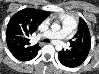
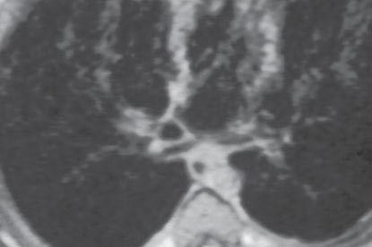
14 Q10,11,12 CT axial images on the 3 cases. For each case suggest the most likely diagnosis. a. No ring or sling b. Vascular ring very likely c. Vascular ring unlikely d. Pulmonary sling very likely e. Pulmonary sling unlikely

15 Case1: 16month girl with respiratory distress

16 Case 2: 16month girl with respiratory distress


17 ase 3: 2.5 month girl with respiratory distress
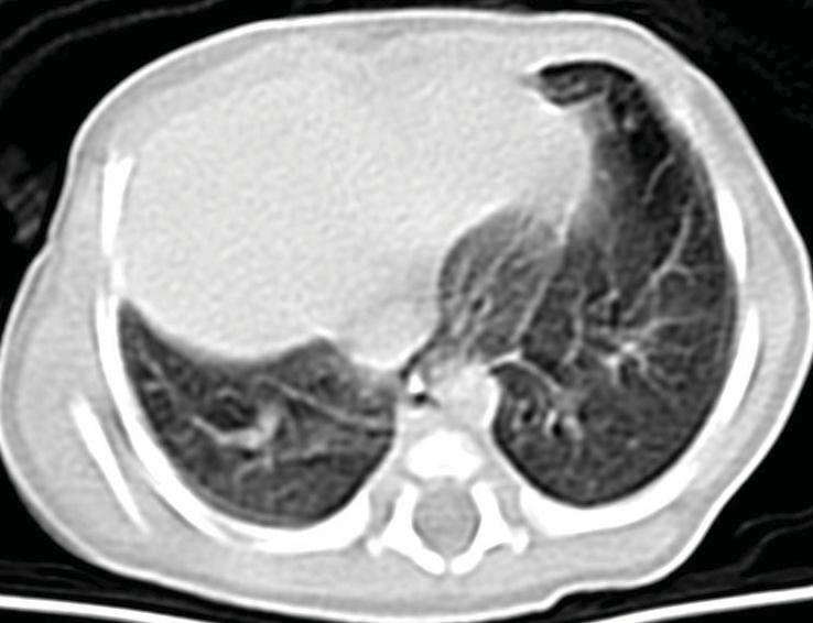
18 Chest Radiographic Findings Suggestive of a Vascular Ring Right Aortic arch Descending Aorta on opposite side of arch Anterior bowing of trachea (lateral view)


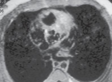
19 6 month old with recurrent respiratory infections
20 Vascular Ring ESOPHAGRAM Not necessary if plain film suggests ring Confirms likely presence of ring May suggest type but not definitive
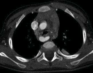
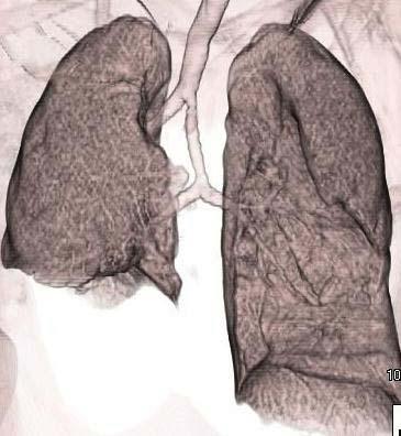
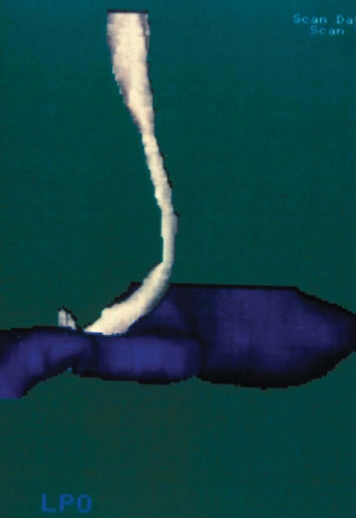
21 Imaging Vascular Rings CT Angiography Fast - decreased need for sedation.6mm-1mm axial spiral Timed/triggered dynamic IV contrast (2-3cc/Kg) 2 and 3-D reconstruction Pay attention to parameters and radiation dose Lower kvp (80-100) Automodulation of mas (ref mas 150) Breast shield in girls
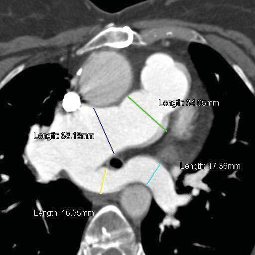
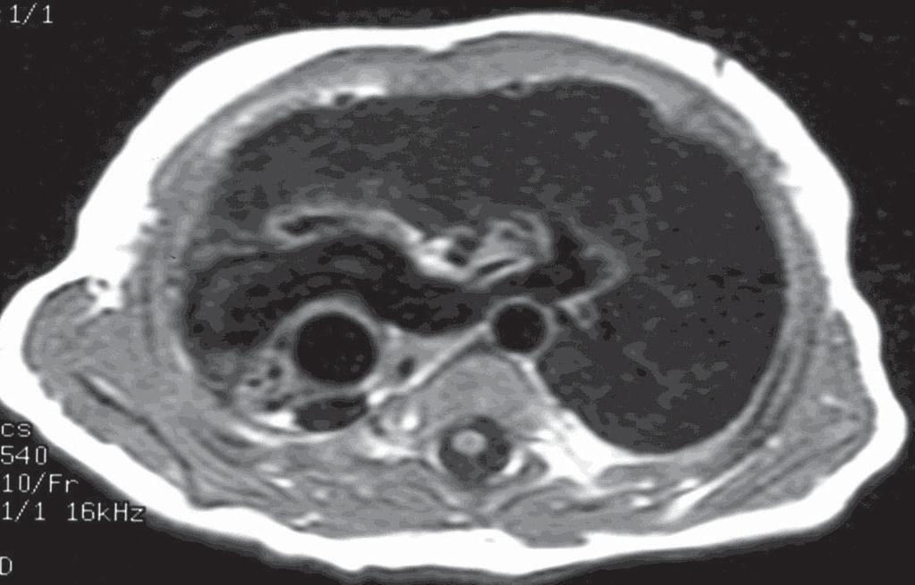
22 Imaging Vascular Rings MR Imaging/Angiography Multiplanar capability/many sequence options. Need specific sequences to optimally evaluate airway Timed/triggered dynamic IV contrast for MRA 2 and 3-D reconstruction of images No ionizing radiation Long study/sedation often required
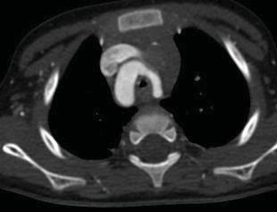
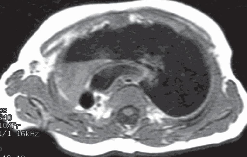
23 Vascular Ring combination of vascular/ ligamentous structures encircling the airway COMMON Double Aortic Arch Right arch, aberrant left subclavian artery + ductus UNCOMMON - Left arch, aberrant right subclavian artery + ductus - Right or left circumflex aorta and ductus aberrant subclavian artery - Mirror image right arch + ductus
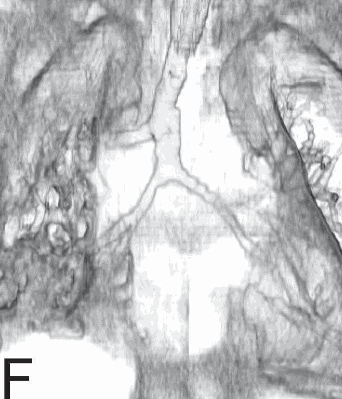
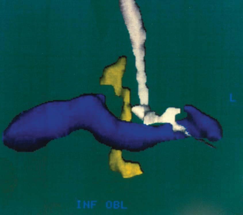
24 Imaging Vascular Rings KEY FEATURES Arch location Branching of aorta Compression of airway Diverticulum of Kommerel Descending aorta

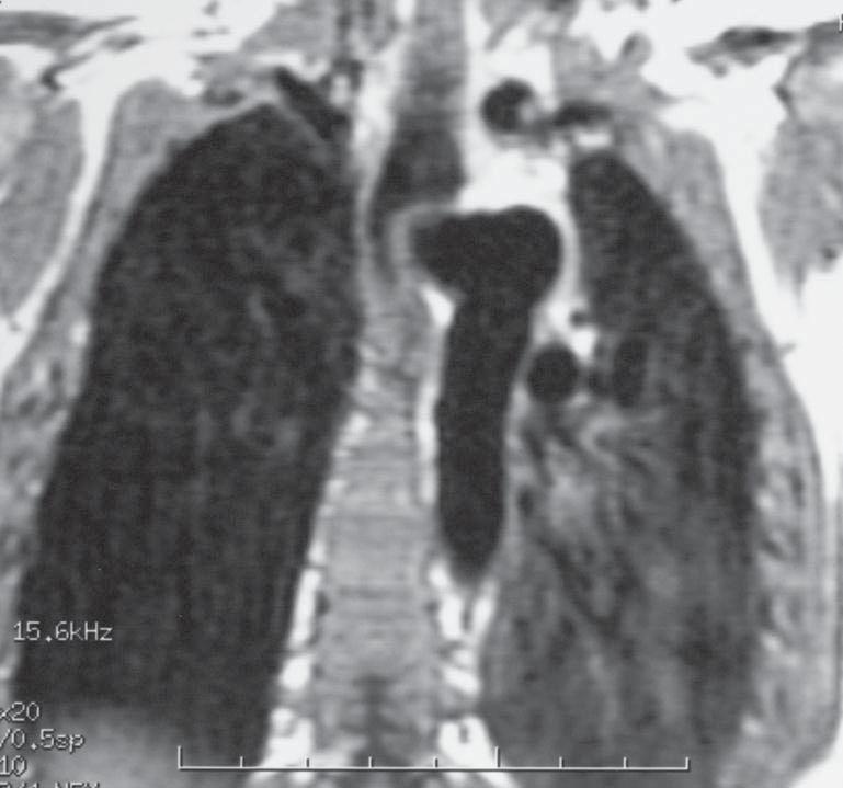
25 DOUBLE AORTIC ARCH DOUBLE AO ARCH WITH ATRETIC LEFT SEGMENT AA ANTERIOR AA C C S DA S DA POSTERIOR


26 DOUBLE AORTIC ARCH 4 symmetric vessels Bilateral Arches Ipsilateral carotid and subclavian Branches S c C S Compressed trachea


27 DOUBLE AORTIC ARCH L R R L Compressed airway Descending aorta on left

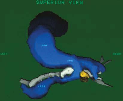
28 RIGHT ARCH ABERRANT LEFT SUBCLAVIAN ARTERY AA ANTERIOR DA DK


29 AAo T E DAo 2.5yo RAA ABERRANT LSCA BRANCH, Mild tracheal Compression, Diverticulum, Lt descending AO

30 LAA ABERRANT RSCA PLUS DIVERTICULUM


31 LAA ARSCA BRANCH Mild Tracheal Compression Diverticulum behind trachea ( prior TEF repair) Lt SVC
32 MIRROR IMAGE RIGHT ARCH RING AA ANTERIOR AA LT INNOM DA DA A


33 Mirror image right Arch Branching Mild tracheal Compression In Diverticulum Right descending aorta In


34 Mirror image Rt Arch Branching Mild tracheal Compression Diverticulum Circumflex aorta descending on left
35 Left Pulmonary Artery Sling Left pulmonary artery arises from RPA Courses posteriorly between the trachea and esophagus
36 Type I PA Sling Higher level of sling N bronchial branching or tracheal bronchus Associated compression/malacia (trachea/rmsb)

37 15yo girl with known VSD

38 15yo girl with known VSD

39 Type 1 PA sling
40 LPA Sling Chest Radiographic Findings May be normal Mass posterior to trachea on lateral view Unilateral hyperinflation (Type I) Bilateral hyperinflation or small right lung (Type II). Low T carina
41 Type II PA Sling ( Ring/Sling complex) Low position of PA sling Horizontal low carina Abnormal airway branching Associated long segment airway stenosis May be right lung hypoplasia/agenesis


42 5week old male with severe stridor

43 Newborn male with severe respiratory distress

44 Newborn male with severe respiratory distress 1 Type IIA LPAS 3 2


45 49 YO F PREVIOUS TOF REPAIR RIGHT DIAPHRAGMATIC PARALYSIS DECREASED EXERCISE TOLERANCE Previously unrecognized Type IIA PA sling and tracheal stenosis

46 Tracheal stenosis

47 LPA Sling Pre &Post

48 10 day old with tachypnea

49 10 day old with tachypnea


50 5 month old male with persistent respiratory symptoms
51
52 SUMMARY Vascular rings and slings are often first suspected on the basis of plain chest radiographs There is a wide spectrum of lesions and radiographic appearances Cross-sectional angiography provides the detailed anatomy needed for operative planning of vascular lesions compressing the airway Carefully evaluate and report vascular & airway appearance and relationships Multiple planes, 2D and 3D reconstructions are useful in understanding and communicating the important anatomy
53 References Vascular Ring/Pulmonary Artery Sling 1. Berdon WE. Rings, slings, and other things: Vascular compression of the infant trachea updated from the mid-century to the millennium the legacy of Robert E. Gross, MD, and Edward B.D. Neuhauser, MD. Radiology 2000; 216: Chan, MS, Chu WC, Cheung KL, Arifi AA, Lam WW. Angiography and dynamic airway evaluation with MDCT in the diagnosis of double aortic arch associated with tracheomalacia. AJR 2005; 185(5): Dodge-Khatami A, Tulevski II, Hitchcock JF, et al. Vascular rings and pulmonary arterial sling: from respiratory collapse to surgical cure, with emphasis on judicious imaging in the hi-tech era. Cardiol Young 2002; 12: Eichhorn J, Fink C, Delorme S, et al. Rings, slings and other vascular abnormalities. Ultra fast computed tomography and magnetic resonance angiography in pediatric cardiology. Z Kardiol 2004; 93:
54 References Vascular Rings (cont.) 5. Hernanz-Schulman M. Vascular rings: a practical approach to imaging diagnosis. Pediatric Radiology 2005; 35: Mahboubi S, Meyer JS, Hubbard AM, et al. Magnetic resonance imaging of airway obstruction resulting from vascular anomalies. International J of Pediatric Otorhinolaryngology 1994; 28: Dillman JR, Attili AK, Agarwal PP, Dorfman AL, Hernandez RJ, Strouse PJ. Common and uncommon vascular rings and slings: a multi-modality review Pediatr Radiol Nov;41(11): Newman B, Cho YA. Left Pulmonary Artery Sling-Anatomy and Imaging. Semin Ultrasound CT MR 2010; 31:
Aortic Arch Abnormalities
 Aortic Arch Abnormalities IPOK Norman H Silverman MD, D Sc (Med.). FACC, FAHA. Stanford University & Lucile Packard Children s Hospital E mail: norm.silverman@stanford.edu. NHS. www.md1world.com Abnormalities
9/8/2009 < 1 1,2 3,4 5,6 7,8 9,10 11,12 13,14 15,16 17,18 > 18. Tetralogy of Fallot. Complex Congenital Heart Disease.
 Current Indications for Pediatric CTA S Bruce Greenberg Professor of Radiology Arkansas Children s Hospital University of Arkansas for Medical Sciences greenbergsbruce@uams.edu 45 40 35 30 25 20 15 10
Aberrant Subclavian Arteries: Cross-Sectional Imaging Findings in Infants and Children Referred for Evaluation of Extrinsic Airway Compression
 Lane F. Donnelly 1 Robert J. Fleck 1, 2 Preeyacha Pacharn 1, 3 Matthew. Ziegler 1 radley L. Fricke 1 Robin T. Cotton 4 Received September 25, 2001; accepted after revision November 16, 2001. 1 Department
Original Report. Imaging Findings in Pediatric Patients with Persistent Airway Symptoms After Surgery for Double Aortic Arch
 Robert J. Fleck 1,2 Preeyacha Pacharn 1,3 Bradley L. Fricke 1 Matthew A. Ziegler 1 Robin T. Cotton 4 Lane F. Donnelly 1 Received August 30, 2001; accepted after revision October 22, 2001. 1 Department
Respiratory Symptoms due to Vascular Ring in Children
 HK J Paediatr (new series) 2016;21:14-21 Respiratory Symptoms due to Vascular Ring in Children GM ZHENG, XH WU, LF TANG Abstract Aim: To highlight the clinical features, signs and diagnosis of vascular
Original Article. Double Aortic Arch in Infants and Children CH XIE, FQ GONG, GP JIANG, SL FU. Key words. Background
 HK J Paediatr (new series) 2018;23:233-238 Original Article Double Aortic Arch in Infants and Children CH XIE, FQ GONG, GP JIANG, SL FU Abstract Key words Background: This study aimed to report the diagnosis,
Cardiopulmonary Syndromes: Conditions With Concomitant Cardiac and Pulmonary Abnormalities
 Cardiopulmonary Syndromes: Conditions With Concomitant Cardiac and Pulmonary Abnormalities Carlos S. Restrepo M.D. Professor of Radiology The University of Texas HSC at San Antonio Cardiopulmonary Syndromes
Aortic Coarctation: Evaluation with Computed Tomography Angiography in Pediatric Patients
 Med. J. Cairo Univ., Vol. 83, No. 2, June: 63-70, 2015 www.medicaljournalofcairouniversity.net Aortic Coarctation: Evaluation with Computed Tomography Angiography in Pediatric Patients MOHAMED ZAKI, M.D.
Patient Presenting with Dysphagia
 Patient Presenting with Dysphagia Radiology Elective Presentation Mansur Ghani 5/18/2018 S L I D E 0 Patient Presentation 86 y/o female with a past medical history of DM type II, diabetic neuropathy, and
Undergraduate Teaching
 Prof. James F Meaney Undergraduate Teaching Chest X-Ray Understanding the normal anatomical by reference to cross sectional imaging Radiology? It s FUN! Cryptic puzzle Sudoku (Minecraft?) It s completely
Chest and cardiovascular
 Module 1 Chest and cardiovascular A. Doss and M. J. Bull 1. Regarding the imaging modalities of the chest: High resolution computed tomography (HRCT) uses a slice thickness of 4 6 mm to identify mass lesions
Pulmonary vascular anatomy & anatomical variants
 Review Article Pulmonary vascular anatomy & anatomical variants Asha Kandathil, Murthy Chamarthy Department of Radiology, University of Texas Southwestern Medical Center, Dallas, TX, USA Contributions:
Right Sided Aortic Arch and its rare Associations- A Case Series
 DOI: 10.7860/IJARS/2018/35392:2410 Radiology Section Case Series Right Sided Aortic Arch and its rare Associations- A Case Series Nidhi Aggarwal, Narender kumar kardam, Kushal Babu Gehlot ABSTRACT Right
After the Chest X-Ray:
 After the Chest X-Ray: What To Do Next Alan S. Brody Professor of Radiology and Pediatrics Chief of Thoracic Imaging Cincinnati Children s Hospital Cincinnati, Ohio USA What Should We Do Next? CT scan?
Aortic arch pathology. Cerebral ischemia following carotid artery stenosis.
 Important: -Subclavian Steal Syndrome -Cerebral ischemia Aortic arch pathology. Cerebral ischemia following carotid artery stenosis. Mina Aubeed & Alba Hernández Pinilla Aortic arch pathology Common arch
Successful Resection of Esophageal Carcinoma Associated with Double Aortic Arch: A Case Report
 Successful Resection of Esophageal Carcinoma Associated with Double Aortic Arch: A Case Report NAOSHI KUBO 1, MASAICHI OHIRA 1, YOSHITO YAMASHITA 2, KATSUNOBU SAKURAI 1, HIROAKI TANAKA 1, KAZUYA MUGURUMA
Thoracic vascular anatomical variants
 Thoracic vascular anatomical variants Poster No.: C-0944 Congress: ECR 2010 Type: Educational Exhibit Topic: Chest - Vascular Authors: R. dos Santos, Â. Marques, H. M. R. Marques, N. Costa, O. Fernandes,
The Uncommon Vascular Ring - Not that Uncommon
 Cronicon OPEN ACCESS EC PAEDIATRICS Case Report The Uncommon Vascular Ring - Not that Uncommon Roshan D Souza MD 1, Deepa Prasad MD 1, James Strainic MD 1, Anoop Mohamed Iqbal MD 2 and Ravi Ashwath MD
Congenital Anomalies of the Aortic Arch: Evaluation with the Use of Multidetector Computed Tomography
 Congenital nomalies of the ortic rch: Evaluation with the Use of Multidetector Computed Tomography ysel Türkvatan, MD Fatma Gül üyükbayraktar, MD Tülay Ölçer, MD Turhan Cumhur, MD Index terms: ortic arch
Thoracoscopic division of vascular rings in infants and children
 Journal of Pediatric Surgery (2007) 42, 1357 1361 www.elsevier.com/locate/jpedsurg Thoracoscopic division of vascular rings in infants and children Abdulrahman Al-Bassam a, *, Mohammad Saquib Mallick a,
DOUBLE AORTIC ARCH SURGERY
 DOUBLE AORTIC ARCH SURGERY *Suraj Wasudeo Nagre 1 and Dwarkanath V. Kulkarni 2 1 Department of C.V.T.S., Grant Medical College, Mumbai 2 Department of C.V.T.S., G.S. Medical College, Mumbai *Author for
Intravascular Ultrasound (IVUS) and Optical Coherence Tomography (OCT)
 and Optical Coherence Tomography (OCT)") Intravascular Ultrasound (IVUS) and Optical Coherence Tomography (OCT) Clare McLaren Great Ormond Street Hospital London Introduction IVUS and OCT supplementary techniques to angiography provide information
Intravascular Ultrasound (IVUS) and Optical Coherence Tomography (OCT) Clare McLaren Great Ormond Street Hospital London Introduction IVUS and OCT supplementary techniques to angiography provide information
Neck Ultrasound. Faculty Info: Amy Kule, MD
 Neck Ultrasound Date: Friday, October 19, 2018 Time: 11:00 AM Location: SMALL GROUP LABORATORY SSOM L71 Watch: Ø Neck Ultrasound Scanning Protocol (4:00): https://www.youtube.com/watch?v=zozd2x2ll4q Faculty
Lung sequestration and Scimitar syndrome
 Lung sequestration and Scimitar syndrome Imaging approaches M. Mearadji International Foundation for Pediatric Imaging Aid Rotterdam, The Netherlands Pulmonary sequestration Pulmonary sequestration (PS)
CT Chest. Verification of an opacity seen on the straight chest X ray
 CT Chest Indications: To assess equivocal plain x-ray findings Staging of lung neoplasm Merastatic workup of extra thoraces malignancies Diagnosis of diffuse lung diseases with HRCT Assessment of bronchietasis
Tracheal stenosis in infants and children is typically characterized
 Slide Tracheoplasty for Congenital Tracheal Stenosis Peter B. Manning, MD Tracheal stenosis in infants and children is typically characterized by the presence of complete cartilaginous tracheal rings and
CT Versus MR for the Runoff
 CT Versus MR for the Runoff Robert R. Edelman, M.D. Dept. of Radiology NorthShore University HealthSystem Feinberg School of Medicine, Northwestern University Magnetic Resonance Computed Tomography Radio
IAEM Clinical Guideline 9 Laryngomalacia. Version 1 September, Author: Dr Farah Mustafa
 IAEM Clinical Guideline 9 Laryngomalacia Version 1 September, 2016 Author: Dr Farah Mustafa Guideline lead: Dr Áine Mitchell, in collaboration with IAEM Clinical Guideline committee and Our Lady s Children
Neonatal Airway Disorders, Treatments, and Outcomes. Steven Goudy, MD Pediatric Otolaryngology Emory University Medical Center
 Neonatal Airway Disorders, Treatments, and Outcomes Steven Goudy, MD Pediatric Otolaryngology Emory University Medical Center Disclosure I have nothing to disclose Neonatal and Pediatric Tracheostomy Tracheostomy
Congenital Heart Disease Systematic Interpretation of CT Suhny Abbara, MD
 Congenital Heart Disease Systematic Interpretation of CT Suhny Abbara, MD Chief, Cardiothoracic Imaging Division Professor of Radiology UT Southwestern Medical Center, Dallas, TX Suhny.Abbara@UTSouthwestern.edu
Chest X-ray Interpretation
 Chest X-ray Interpretation Introduction Routinely obtained Pulmonary specialist consultation Inherent physical exam limitations Chest x-ray limitations Physical exam and chest x-ray provide compliment
Postgraduate Student, Department of Radiodiagnosis, MVJ Medical College and Research Hospital, Hoskote, Bangalore. 2
 RIGHT-SIDED AORTIC ARCH WITH ABERRANT LEFT SUBCLAVIAN ARTERY AND DUPLICATION OF SUPERIOR VENA CAVA Parikhita Hazarika 1, Tejaswani Penmetsa 2, Narendranath Kudva 3 1 Postgraduate Student, Department of
Low-Tube-Current Multidetector CT for Children with Suspected Extrinsic Airway Compression
 Preeyacha Pacharn 1,2 Stacy A. Poe 3 Lane F. Donnelly 1,3 Received February 20, 2002; accepted after revision May 15, 2002. 1 Department of Radiology, Children s Hospital Medical Center, 3333 Burnet Ave.,
24. An infant with recurrent pneumonia underwent a frontal chest radiograph (Fig 24-A) followed by
 followed by") 24. An infant with recurrent pneumonia underwent a frontal chest radiograph (Fig 24-A) followed by diagnosis? ndings, what is the most likely A. Pulmonary sequestration B. Congenital pulmonary airway malformation
24. An infant with recurrent pneumonia underwent a frontal chest radiograph (Fig 24-A) followed by diagnosis? ndings, what is the most likely A. Pulmonary sequestration B. Congenital pulmonary airway malformation
Anomalous origin of the right subclavian artery from main pulmonary artery
 Anomalous origin of the right subclavian artery from main pulmonary artery Award: AOSR Best Exhibit Prize - Bronze Poster No.: R-0178 Congress: RANZCR-AOCR 2012 Type: Educational Exhibit Authors: U. Chaumrattanakul,
Case 47 Clinical Presentation
 93 Case 47 C Clinical Presentation 45-year-old man presents with chest pain and new onset of a murmur. Echocardiography shows severe aortic insufficiency. 94 RadCases Cardiac Imaging Imaging Findings C
Coronary Artery Anomalies from Birth to Adulthood; the Role of CT Coronary Angiography in Sudden Cardiac Death Screening
 Coronary Artery Anomalies from Birth to Adulthood; the Role of CT Coronary Angiography in Sudden Cardiac Death Screening E O Dwyer 1, C O Brien 1, B Loo 1, A Snow Hogan 1, O Buckley1 2, B 1. Department
Lung- and airway emergencies
 Lung- and airway emergencies Charlotte de Lange,MD,PhD Pediatric Radiology unit, Oslo University Hospital, Norway 5th Nordic course - Emergency Radiology Oslo 18-21.5.2015 clange@ous-hf.no How come pediatric
PULMONARY VENOLOBAR SYNDROME. Dr.C.Anandhi DNB Resident, Southern Railway Headquarters Hospital.
 PULMONARY VENOLOBAR SYNDROME Dr.C.Anandhi DNB Resident, Southern Railway Headquarters Hospital. Presenting complaint: 10 yrs old girl with recurrent episodes of lower respiratory tract infection from infancy.
Lab CT scan. Murad Kharabsheh Yaman Alali
 Lab CT scan Murad Kharabsheh Yaman Alali Some rules to read The CT Scan : 1. Remember that it s a transverse section across the body and we are looking at the inferior part of the section (not the superior),
Aortography in Fallot's Tetralogy and Variants
 Brit. Heart J., 1969, 31, 146. Aortography in Fallot's Tetralogy and Variants SIMON REES AND JANE SOMERVILLE From The Institute of Cardiology and National Heart Hospital, London W.J In patients with Fallot's
Monitor Images for Respiratory System Dissection
 Monitor Images for Respiratory System Dissection **This document includes extra images of the radiology of the bronchopulmonary segments. These imaged are an excellent way to review the three-dimensional
Debate: Should Ductal Stent Implantation be Considered for All Newborn Infants with Reduced Pulmonary Blood Flow?_Pros
 Debate: Should Ductal Stent Implantation be Considered for All Newborn Infants with Reduced Pulmonary Blood Flow?_Pros Mazeni Alwi Institut Jantung Negara Kuala Lumpur, Malaysia 5 th Asia Pacific Congenital
Chapter 3.14 Aortic arch interruption
 Chapter 3.14 Aortic arch interruption z Definition The aortic arch is described as three segments: proximal, distal and isthmus. The proximal component extends from the takeoff of the innominate artery
SAMPLE EDITION PELVIC AND LOWER EXTREMITY ARTERIES WITH ENDOVASCULAR REVASCULARIZATION. Cardiovascular Illustrations and Guidelines
 Cardiovascular Illustrations and Guidelines PELVIC AND LOWER EXTREMITY ARTERIES WITH ENDOVASCULAR REVASCULARIZATION ANGIOPLASTY INTRAVASCULAR STENT PLACEMENT ATHERECTOMY For Fem-Pop Territory Angioplasty
Pitfalls of the Pediatric Chest and Abdomen SPR 2017
 Pitfalls of the Pediatric Chest and Abdomen SPR 2017 Richard I. Markowitz, MD, FACR Children s Hospital of Philadelphia Perelman School of Medicine University of Pennsylvania No Disclosures Cognitive Perceptual
Correlation of Cardiac CTA to Conventional Cardiac Angiography in Diagnosing Coronary Artery Stenosis in a Community Based Center
 Correlation of Cardiac CTA to Conventional Cardiac Angiography in Diagnosing Coronary Artery Stenosis in a Community Based Center Mathieu Sabbagh, R3 Michigan State University Radiology Garden City Hospital
Imaging of Thoracic Trauma: Tips and Traps. Arun C. Nachiappan, MD Associate Professor of Clinical Radiology University of Pennsylvania
 Imaging of Thoracic Trauma: Tips and Traps Arun C. Nachiappan, MD Associate Professor of Clinical Radiology University of Pennsylvania None Disclosures Objectives Describe blunt and penetrating traumatic
Devendra V. Kulkarni, Rahul G. Hegde, Ankit Balani, and Anagha R. Joshi. 2. Case Report. 1. Introduction
 Case Reports in Radiology, Article ID 614647, 4 pages http://dx.doi.org/10.1155/2014/614647 Case Report A Rare Case of Pulmonary Atresia with Ventricular Septal Defect with a Right Sided Aortic Arch and
Chapter 124: Congenital Disorders of the Trachea. Bruce Benjamin
 Chapter 124: Congenital Disorders of the Trachea Bruce Benjamin Investigation of the larynx and pharynx may be incomplete in infants and children with congenital abnormalities without investigation of
Section V Cardiac Radiology
 Section V Cardiac Radiology Figure 1 89. Based on the diagram (Figure 1), which of the following vessels typically supplies the anterolateral cardiac segment? A. Left anterior descending B. Circumflex
Translocation of the Aortic Arch with Norwood Procedure for Hypoplastic Left Heart Syndrome Variant with Circumflex Retroesophageal Aortic Arch
 Korean J Thorac Cardiovasc Surg 2014;47:389-393 ISSN: 2233-601X (Print) ISSN: 2093-6516 (Online) Case Report http://dx.doi.org/10.5090/kjtcs.2014.47.4.389 Translocation of the Aortic Arch with Norwood
DISCLOSURE OBJECTIVES PULMONARY VEIN STENOSIS DIAGNOSTIC TOOLS. Echo with Doppler Catheterization with angiography CT angiography MRI
 1 2 ND INTERNATIONAL CONFERENCE: NEONATAL AND CHILDHOOD PULMONARY VASCULAR DISEASE, MARCH 13-14, 2009, SAN FRANCISCO, USA PATHOPHYSIOLOGY OF PULMONARY VEIN FLOW: IMAGING NORMAL AND ABNORMAL PULMONARY VEIN
Low-dose prospective ECG-triggering dual-source CT angiography in infants and children with complex congenital heart disease: first experience
 Low-dose prospective ECG-triggering dual-source CT angiography in infants and children with complex congenital heart disease: first experience Ximing Wang, M.D., Zhaoping Cheng, M.D., Dawei Wu, M.D., Lebin
Large veins of the thorax Brachiocephalic veins
 Large veins of the thorax Brachiocephalic veins Right brachiocephalic vein: formed at the root of the neck by the union of the right subclavian & the right internal jugular veins. Left brachiocephalic
Children are not small adults Children are Not Small Adults Anatomic considerations Pliable bony & cartilagenous structures - Significant thoracic inj
 PEDIATRIC CHEST TRAUMA Children are not small adults Role of imaging Spectrum of injury Children are not small adults Children are Not Small Adults Anatomic considerations Pliable bony & cartilagenous
Two Cases of Incidentally Picked Up Adult Unilateral Pulmonary Artery Atresia with Variable Imaging Features
 IOSR Journal of Dental and Medical Sciences (IOSR-JDMS) e-issn: 2279-0853, p-issn: 2279-0861.Volume 16, Issue 12 Ver. III (Dec. 2017), PP 45-49 www.iosrjournals.org Two Cases of Incidentally Picked Up
THE GOOFY ANATOMIST QUIZZES
 THE GOOFY ANATOMIST QUIZZES 7. LUNGS Q1. Fill in the blanks: the lung has lobes and fissures. A. Right, three, two. B. Right, two, one. C. Left, three, two. D. Left, two, three. Q2. The base of the lung
Surgical Treatment of Aortic Arch Hypoplasia
 Surgical Treatment of Aortic Arch Hypoplasia In the early 1990s, 25% of patients could face mortality related to complica-tions of hypertensive disease Early operations and better surgical techniques should
Case Report Recurrent Wheezing and Cough Caused by Double Aortic Arch, Not Asthma
 Hindawi Case Reports in Cardiology Volume 2017, Article ID 8079851, 4 pages https://doi.org/10.1155/2017/8079851 Case Report Recurrent Wheezing and Cough Caused by Double Aortic Arch, Not Asthma Qiao Zhang,
Cardiac Radiology In-Training Test Questions for Diagnostic Radiology Residents
 Cardiac Radiology In-Training Test Questions for Diagnostic Radiology Residents March, 2013 Sponsored by: Commission on Education Committee on Residency Training in Diagnostic Radiology 2013 by American
ROLE OF CONTRAST ENHANCED MR ANGIOGRAPHY IN AORTIC COARCTATION
 ROLE OF CONTRAST ENHANCED MR ANGIOGRAPHY IN AORTIC COARCTATION By Adel El Badrawy, Ahmed Abdel Razek, Nermin Soliman, Hala El Marsafawy *, Sameh Amer** From Radiodiagnosis, Pediatric Cardiology* & Cardiothoracic
90 th Annual Meeting The American Association for Thoracic Surgery May 1, 2010 Toronto, Ontario, Canada. Slide Tracheoplasty
 90 th Annual Meeting The American Association for Thoracic Surgery May 1, 2010 Toronto, Ontario, Canada Congenital Skills Course Slide Tracheoplasty Carl Lewis Backer, MD A.C. Buehler Professor of Surgery
Vascular compression of the airway in children
 PAEDIATRIC RESPIRATORY REVIEWS (2008) 9, 85 94 MINI-SYMPOSIUM: IMAGING AND INTERVENTIONAL RADIOLOGY Vascular compression of the airway in children Clare A. McLaren 2, *, Martin J. Elliott 1 and Derek J.
Developmental Aortic Arch Anomalies in Infants and Children Assessed With CT Angiography
 ardiopulmonary Imaging Pictorial Essay Ramos-Duran et al. T of Developmental ortic rch nomalies ardiopulmonary Imaging Pictorial Essay Downloaded from www.ajronline.org by 148.251.232.83 on 05/13/18 from
Hybrid Procedure of Bilateral Pulmonary Artery Banding and Bilateral Ductal Stenting in an Infant With Aortic Atresia and Interrupted Aortic Arch
 Catheterization and Cardiovascular Interventions 84:1157 1162 (2014) Hybrid Procedure of Bilateral Pulmonary Artery Banding and Bilateral Ductal Stenting in an Infant With Aortic Atresia and Interrupted
Lecturer: Ms DS Pillay ROOM 2P24 25 February 2013
 Lecturer: Ms DS Pillay ROOM 2P24 25 February 2013 Thoracic Wall Consists of thoracic cage Muscle Fascia Thoracic Cavity 3 Compartments of the Thorax (Great Vessels) (Heart) Superior thoracic aperture
Persistent right aortic arch (PRAA) is the most common
 is the most common") J Vet Intern Med 2004;18:510 514 Tracheal Signs and Associated Vascular Anomalies in Dogs with Persistent Right Aortic Arch James W. Buchanan Medical records of 55 dogs with 1 or more vascular rings around
J Vet Intern Med 2004;18:510 514 Tracheal Signs and Associated Vascular Anomalies in Dogs with Persistent Right Aortic Arch James W. Buchanan Medical records of 55 dogs with 1 or more vascular rings around
ULTRASOUND OF THE FETAL HEART
 ULTRASOUND OF THE FETAL HEART Cameron A. Manbeian, MD Disclosure Statement Today s faculty: Cameron Manbeian, MD does not have any relevant financial relationships with commercial interests or affiliations
B-I-2 CARDIAC AND VASCULAR RADIOLOGY
 (YEARS 1 3) CURRICULUM FOR RADIOLOGY 13 B-I-2 CARDIAC AND VASCULAR RADIOLOGY KNOWLEDGE To describe the normal anatomy of the heart and vessels including the lymphatic system as demonstrated by radiographs,
DR Turner, JA Vincent, and ML Epstein. Isolated right pulmonary artery discontinuity. Images Paediatr Cardiol Jul-Sep; 2(3):
:") IMAGES in PAEDIATRIC CARDIOLOGY Images PMCID: PMC3232486 Isolated right pulmonary artery discontinuity DR Turner, MD, * JA Vincent, ** and ML Epstein *** * Senior Fellow, Division of Cardiology, Children's
IMAGES in PAEDIATRIC CARDIOLOGY Images PMCID: PMC3232486 Isolated right pulmonary artery discontinuity DR Turner, MD, * JA Vincent, ** and ML Epstein *** * Senior Fellow, Division of Cardiology, Children's
Overview of developmental anomaly of the aorta
 Overview of developmental anomaly of the aorta Poster No.: C-1263 Congress: ECR 2012 Type: Educational Exhibit Authors: D. H. Yeom, M. J. Kang, W. H. Cho, J. H. Kim, T. K. Kang, J.-H. Lee, S. Y. Lee; Seoul/KR
Dr. Weyrich G07: Superior and Posterior Mediastina. Reading: 1. Gray s Anatomy for Students, chapter 3
 Dr. Weyrich G07: Superior and Posterior Mediastina Reading: 1. Gray s Anatomy for Students, chapter 3 Objectives: 1. Subdivisions of mediastinum 2. Structures in Superior mediastinum 3. Structures in Posterior
Cardiac CT Techniques in Neonates (and infants)
") Cardiac CT Techniques in Neonates (and infants) Siddharth P. Jadhav, MD Director, Body CT and MRI Edward B. Singleton Department of Pediatric Radiology Texas Children s Hospital Disclosures None Objectives
Cardiac CT Techniques in Neonates (and infants) Siddharth P. Jadhav, MD Director, Body CT and MRI Edward B. Singleton Department of Pediatric Radiology Texas Children s Hospital Disclosures None Objectives
Heart and Soul Evaluation of the Fetal Heart
 Heart and Soul Evaluation of the Fetal Heart Ivana M. Vettraino, M.D., M.B.A. Clinical Associate Professor, Michigan State University College of Human Medicine Objectives Review the embryology of the formation
PA/VSD/MAPCAs What a surgeon needs to know
 PA/VSD/MAPCAs What a surgeon needs to know Patrick McConnell, MD Nationwide Children s Hospital.... Name MRN Procedure Allergies Absolute Surgical Requirements Things needed to complete a Time out....
Case Report Computed Tomography Angiography Successfully Used to Diagnose Postoperative Systemic-Pulmonary Artery Shunt Narrowing
 Case Reports in Cardiology Volume 2011, Article ID 802643, 4 pages doi:10.1155/2011/802643 Case Report Computed Tomography Angiography Successfully Used to Diagnose Postoperative Systemic-Pulmonary Artery
Lab #3. Mohammad Hisham Al-Mohtaseb. Jumana Jihad. Ammar Ramadan. 0 P a g e
 Lab #3 Mohammad Hisham Al-Mohtaseb Jumana Jihad Ammar Ramadan 0 P a g e Last anatomy lab: Lungs and structure on the mediastinal surfs: 1-the right lung: How do we know it s the right lung??? -the 3 lobes
Preoperative Echocardiographic Assessment of Uni-ventricular Repair
 Preoperative Echocardiographic Assessment of Uni-ventricular Repair Salem Deraz, MD Pediatric Cardiologist, Aswan Heart Centre Magdi Yacoub Heart Foundation Uni-ventricular repair A single or series of
Superior and Posterior Mediastinum. Assoc. Prof. Jenny Hayes
 Superior and Posterior Mediastinum Assoc. Prof. Jenny Hayes WARNING This material has been provided to you pursuant to section 49 of the Copyright Act 1968 (the Act) for the purposes of research or study.
Individual Pulmonary Vein Atresia in Adults: Report of Two Cases
 Case Report DOI: 10.3348/kjr.2011.12.3.395 pissn 1229-6929 eissn 2005-8330 Korean J Radiol 2011;12(3):395-399 Individual Pulmonary Vein Atresia in Adults: Report of Two Cases Hyoung Nam Lee, MD, Young
US of Renovascular Hypertension. Jonathan R. Dillman, MD, MSc Associate Professor Director, Thoracoabdominal Imaging
 US of Renovascular Hypertension Jonathan R. Dillman, MD, MSc Associate Professor Director, Thoracoabdominal Imaging Disclosures Nothing Relevant Unrelated grant funding Siemens US Toshiba US Objectives
Cardiac MRI in ACHD What We. ACHD Patients
 Cardiac MRI in ACHD What We Have Learned to Apply to ACHD Patients Faris Al Mousily, MBChB, FAAC, FACC Consultant, Pediatric Cardiology, KFSH&RC/Jeddah Adjunct Faculty, Division of Pediatric Cardiology
Lines and tubes. 1 Nasogastric tubes Endotracheal tubes Central lines Permanent pacemakers Chest drains...
 Lines and tubes 1 Nasogastric tubes... 15 2 Endotracheal tubes.... 19 3 Central lines... 21 4 Permanent pacemakers.... 25 5 Chest drains... 30 This page intentionally left blank 1 Nasogastric tubes Background
Tubes and lines in neonatal chest radiograph
 Tubes and lines in neonatal chest radiograph Poster No.: C-1008 Congress: ECR 2014 Type: Educational Exhibit Authors: R. TUMMA, N. AHMED, V. Prasad ; Hyderabad/IN, 1 2 1 1 2 HYDERABAD, ANDHRA PRADESH/IN
Sectional Anatomy Quiz - III
 Sectional Anatomy - III Rashid Hashmi * Rural Clinical School, University of New South Wales (UNSW), Wagga Wagga, NSW, Australia A R T I C L E I N F O Article type: Article history: Received: 30 Jun 2018
DOUBLE AORTIC ARCH IN AN INFANT TREATED SURGICALLY
 Thorax (1957), 12, 214. DOUBLE AORTC ARCH N AN NFANT TREATED SURGCALLY BY J. APLEY, F. G. M. ROSS, AND R. MLNES WALKER From the United Bristol Hospitals (RECEVED FOR PUBLCATON FEBRUARY 26, 1957) Among
Advances in MDCT of Thoracic Trauma
 Baltic Congress of Radiology, Riga 2010 Advances in MDCT of Thoracic Trauma Robert A. Novelline, MD Professor of Radiology, Harvard Medical School Director of Emergency Radiology, Massachusetts General
Primary Resection of Kommerell Diverticulum and Left Subclavian Artery Transfer
 Primary Resection of Kommerell Diverticulum and Left Subclavian Artery Transfer Carl L. Backer, MD, Hyde M. Russell, MD, Katherine C. Wurlitzer, BA, Jeffrey C. Rastatter, MD, and Cynthia K. Rigsby, MD
HOW TO IMAGE AND DESCRIBE CONGENITAL LUNG MALFORMATIONS
 HOW TO IMAGE AND DESCRIBE CONGENITAL LUNG MALFORMATIONS Paul Thacker, MD Assistant Professor Departments of Radiology and Pediatrics Medical University of South Carolina DISCLOSURES I have no relevant
Right lung. -fissures:
 -Right lung is shorter and wider because it is compressed by the right copula of the diaphragm by the live.. 2 fissure, 3 lobes.. hilum : 2 bronchi ( ep-arterial, hyp-arterial ), one artery mediastinal
Case Report Prenatal Diagnosis of Down Syndrome Associated with Right Aortic Arch and Dilated Septum Cavi Pellucidi
 Case Reports in Obstetrics and Gynecology Volume 2012, Article I 329547, 4 pages doi:10.1155/2012/329547 Case Report renatal iagnosis of own Syndrome Associated with Right Aortic Arch and ilated Septum
Debate in Management of native COA; Balloon Versus Surgery
 Debate in Management of native COA; Balloon Versus Surgery Dr. Amira Esmat, El Tantawy, MD Professor of Pediatrics Consultant Pediatric Cardiac Interventionist Faculty of Medicine Cairo University 23/2/2017
ADI Procedure Codes. August 2016 Revised April 2017 Page 1 of 7 ADI Procedure Codes
 Code Description 70450 CT Head without contrast 70460 CT Head with contrast 70470 CT Head with & without contrast 70480 CT Orbit, et al without contrast 70481 CT Orbit, et al with contrast 70482 CT Orbit,
Chapter 2 Cardiac Interpretation of Pediatric Chest X-Ray
 Chapter 2 Cardiac Interpretation of Pediatric Chest X-Ray Ra-id Abdulla and Douglas M. Luxenberg Key Facts The cardiac silhouette occupies 50 55% of the chest width on an anterior posterior chest X-ray
Sheet lab 5 Anatomy: CT Scans
 Sheet lab 5 Anatomy: CT Scans In the orientation we see the picture from downward to upward. The first picture is a CT scan at the level of the heart. Left border of the heart is the left ventricle and
Isolated congenital coronary anomalies: Evaluation by multislice-ct or MRI
 Isolated congenital coronary anomalies: Evaluation by multislice-ct or MRI B.K. Velthuis, Dept. of Radiology UMC Utrecht, the Netherlands ESC 2010 Coronary artery anomalies CAA Uncommon 0.3-5% normal population
Wheeze. Dr Jo Harrison
 Wheeze Dr Jo Harrison 9.9.14 Wheeze - Physiology a continuous musical sound that lasts longer than 250 msec. can be high-pitched or low-pitched, consist of single or multiple notes, and occur during inspiration
SYMPOSIA. Coronary CTA. Indications, Patient Selection, and Clinical Implications
 SYMPOSIA Indications, Patient Selection, and Clinical Implications Christian Thilo, MD,* Mark Auler, MD,* Peter Zwerner, MD,w Philip Costello, MD,* and U. Joseph Schoepf, MD* Abstract: Recent technical
Congenital tracheal stenosis (CTS) in neonates and infants is an underdiagnosed,
 in neonates and infants is an underdiagnosed,") Simultaneous management of congenital tracheal stenosis and cardiac anomalies in infants Tsvetomir Loukanov, MD, a Christian Sebening, MD, a Wolfgang Springer, MD, b Herbert Ulmer, MD, PhD, b and Siegfried
Simultaneous management of congenital tracheal stenosis and cardiac anomalies in infants Tsvetomir Loukanov, MD, a Christian Sebening, MD, a Wolfgang Springer, MD, b Herbert Ulmer, MD, PhD, b and Siegfried