Aortic Arch Abnormalities
|
|
|
- Lucy Harper
- 6 years ago
- Views:
Transcription

. FACC, FAHA.")


1 Aortic Arch Abnormalities IPOK Norman H Silverman MD, D Sc (Med.). FACC, FAHA. Stanford University & Lucile Packard Children s Hospital E mail: norm.silverman@stanford.edu. NHS.

2 Abnormalities Supravalvar Aortic Stenosis The Aortic Root in Marfan s Syndrome Interruption Hypoplasia High Aortic Arch Arch Laterality Coarctation Vascular Rings

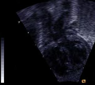
3 Examination Technique A variety views should be used including :- Suprasternal coronal and sagittal imaging, Parasternal and Subcostal views of the aorta and its relations. The Imaging should be complemented by various Doppler modalities including Color flow imaging.


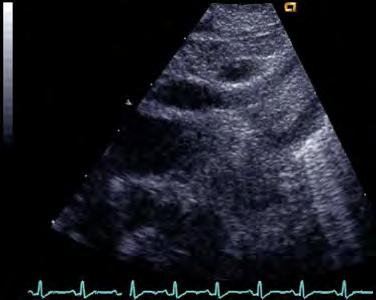
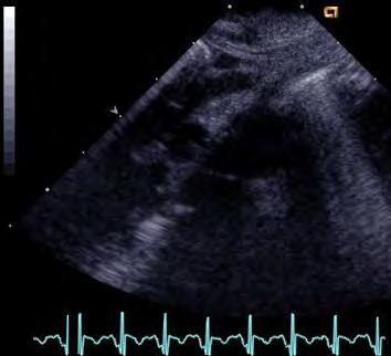
4 Normal Aortic Arch Echo

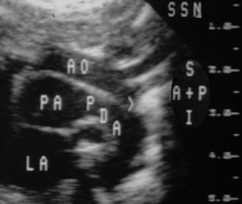
5 Normal Aortic Arch SSN Sagittal AAo IV RBr RPA D Ao

6 Aortic Arch Laterality Transducer alignment The arch direction follows the plane of the array Innominate Artery The innominate artery always bifurcates on the opposite side to the aortic arch. Lack of bifurcation may indicate that there is an anomalous subclavian artery Tracheal Rings or Esophagus The descending aorta can be recognized by the examiner either to the left (in left aortic arch) or to the right (in right aortic arch)


7 Innominate Artery Shows Arch Position Right Innominate, therefore left arch Left innominate, therefore right arch NHS.


8 Arch Laterality R Arch A Trachea A Left Arch


9 Tracheal Rings

10 Arch Laterality Left Arch Right Arch

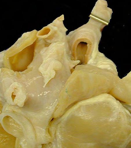
11 Routine Arch Sweep

12 Normal Aorta: Color Imaging IV THYMUS TAO LCA AAo LSA LA RPA DAo


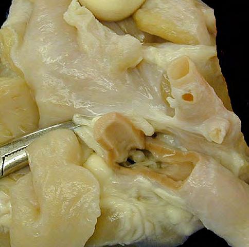
13 Supravalvar Aortic Stenosis


14 Supravalvar Aortic Stenosis


15 William s Syndrome

16 William s Syndrome

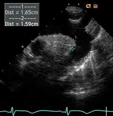
17 Supravalvar Aortic Stenosis RV LV Ao LA L Ax


18 Supravalvar Aortic Stenosis Ao RV LV Subcost. Coronal Ao P L Ax

19 Bicuspid Aortic Valve & Multiple Dissections


20 Aortic Dissection, Coarctation & Bicuspid Aortic Valve


21 Bicuspid Aortic Valve: Post Stenotic Dilatation AAo AAo LV


22 Marfan s Syndrome


23 Marfan s Syndrome Mariska Kemna


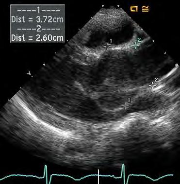
24 Marfan s Syndrome: Dilated Aortic Root RV P Sax. Ao RA LA DAo LX LA RV LA Ao


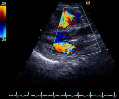
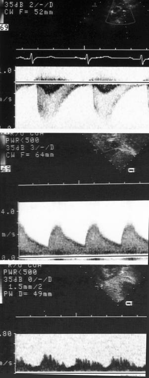
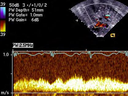
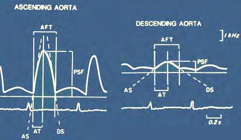
25 Marfan s Syndrome: Dilated Aortic Root Aortic Regurgitation


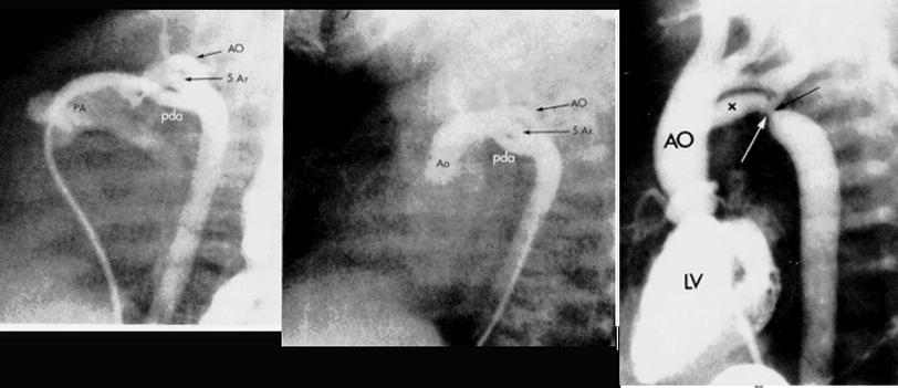
26 Marfan s Syndrome: Dilated Aortic Root


27 Marfan s Syndrome: Dilated Aortic Root


28 Coarctation


29 Coarctation Courtesy Diane Spicer


30 Coarctation


31 NHS. Pathological Varieties of Coarctation &Techniques of Display


32 DUCTUS SLING IN COARCTATION

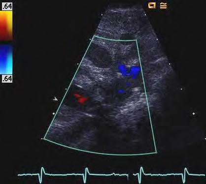
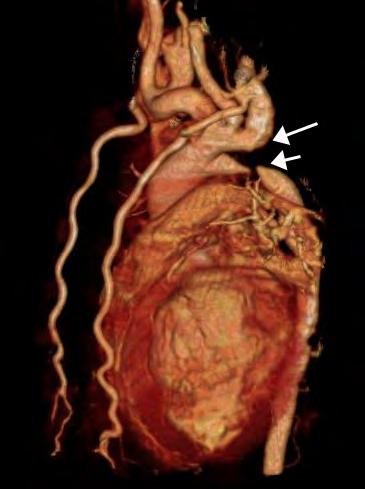
33 Doppler of Coarctation

34 Coarctation A Ao Duct Beak PA Shelf DAo

35 Color Angiogram in Coarctation Tr Ao Arch Innominate Artery RPA L Subclavian Coarctation DAO NHS.


36 HYPOPLASTIC ISTHMUS

37 Ductus &? Coarctation: On Prostaglandin MPA Duct Isthmus LA LPA DAo? Coarct NHS.


38 Coarctation of the Aorta PA TAO DAO OFF PGE1 On PGE1 NHS.


39 Taussig- Bing Malformation and Coarctation IV TAO AAo PA Coarct LA D Ao


40 The Diversity of Coarctation

41 Coarctation-MRI 3 D Reconstruction Courtesy of Dr. Keren Hasbani

42 Coarctation: End - to - Side Anastomosis SSN Cor. IV PA Suture Line D Ao


43 Fifth Aortic Arch


44 PMW 07

45 Interrupted Aortic Arch: Associations Ventricular septal defect Catch 22 Syndrome Truncus arteriosus Aortopulmonary Window Taussig Bing Malformation


46 Aortic Arch Interruption Found with:- 22 Q- Truncus Arteriosus Taussig Bing Aortopulmonary Window VSD C B A

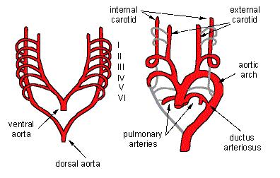
47 Aortic Arch Interruption

48 Type B Interruption AAo LSA PA D

49 Type B Interruption: Ductus & Descending Aorta MPA AAo LSA LA D A O Duct

50 Interrupted Aortic Arch Sagittal Plane Imaging MPA R L D LA D A O NHS.

51 INTERRUPTED ARCH TYPE B In. Vn In. A LSA AAo Duct D Ao


52 Interrupted Arch: 2 Different Doppler Signals a NHS.


53 Truncus with Interrupted Aortic Arch D A Ao LPA Tr RV * LV


54 The VSD in Interruption & Coarctation A4Ch. Ao P Lx RV LV RV LV Ao In Coarctation and interruption of the aortic arch, all the woe, lies below! Richard Van Praagh. LA

55 Arch Embryology

56 Arch Embryology


57 Right Aortic Arch Left Arch: Cervical Arch and Right Ductus


58 High ( Cervical ) Arch Right PA

 Arch")
59 Right PA High ( Cervical ) Arch


60 RAA- LPDA ALSCA DAA

61 Bronchoscopy RT Mainstem Bronchus Compression LT RT


62


63 Coronal Scan


64 Double Aortic Arch R L AAO


65 Double Aortic Arch

66 Fetal Double Aortic Arch


67 Double Aortic Arch

68 L Duct, Right Ao Arch


69 Interrupted Aortic Arch, R Desc. Ao & R Duct- Another Bronchial Vise

70 Right Arch with Aberrant Left Subclavian Left subclavian Left ductus Diverticulum of Kommerell's RING CP

71 Right Arch Mirror-image Branching and No Ring Right Arch Mirror Image Branching Left ductus From innominate NO RING CP


72 Right Aortic Arch Aberrant L subclavian artery and L arterial duct


73 Left Aortic Arch with Aberrant Right Subclavian Aberrant RT SCA CP


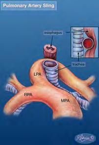
74 Left Duct with Right Aortic Arch


75 Resection of Kommerell s diverticulum and left subclavian artery transfer for recurrent symptoms after vascular ring division C.L. Backer, N. Hillman, C. Mavroudis, L.D. Holinger EJCTS Reoperations 7 Primary operations

76 Kommerell s Diverticulum

77 Kommerell s Diverticulum Aortic diverticulum


78 Left Ductus: Right Arch Potentially Isolated L subclav. Art NHS.


79 BILATERAL DUCTS WITH ABSENT CENTRAL PA S

80 Bilateral Ductus


81 Bilateral Ducts & Absent Central PA s Left Innominate and Duct Right Arch and Duct


82 MAPCA s LA D A O S Cost Sagittal S Sost Coronal P Sx


83 Pulmonary Artery Sling Front Back

84 CT Bronchial Tomography


85 Pulmonary artery Sling ECHO LPA


86 PA Sling


87 Pulmonary Artery Sling RPA LPA


88 Pulmonary Artery Sling PSX AO MPA RPA AO MPA LPA RPA LPA
89 Thank You!
VASCULAR RINGS A CASE - BASED REVIEW
 VASCULAR RINGS A CASE - BASED REVIEW Beverley Newman, BSc. MB.Bch. FACR Professor of Radiology Stanford University and Lucile Packard Children s Hospital Q1,2,3 Frontal chest radiographs on three different
Chapter 3.14 Aortic arch interruption
 Chapter 3.14 Aortic arch interruption z Definition The aortic arch is described as three segments: proximal, distal and isthmus. The proximal component extends from the takeoff of the innominate artery
Pediatric Echocardiography Examination Content Outline
 Pediatric Echocardiography Examination Content Outline (Outline Summary) # Domain Subdomain Percentage 1 Anatomy and Physiology Normal Anatomy and Physiology 10% 2 Abnormal Pathology and Pathophysiology
Cardiac Catheterization Cases Primary Cardiac Diagnoses Facility 12 month period from to PRIMARY DIAGNOSES (one per patient)
") PRIMARY DIAGNOSES (one per patient) Septal Defects ASD (Atrial Septal Defect) PFO (Patent Foramen Ovale) ASD, Secundum ASD, Sinus venosus ASD, Coronary sinus ASD, Common atrium (single atrium) VSD (Ventricular
PRIMARY DIAGNOSES (one per patient) Septal Defects ASD (Atrial Septal Defect) PFO (Patent Foramen Ovale) ASD, Secundum ASD, Sinus venosus ASD, Coronary sinus ASD, Common atrium (single atrium) VSD (Ventricular
Heart and Soul Evaluation of the Fetal Heart
 Heart and Soul Evaluation of the Fetal Heart Ivana M. Vettraino, M.D., M.B.A. Clinical Associate Professor, Michigan State University College of Human Medicine Objectives Review the embryology of the formation
List of Videos. Video 1.1
 Video 1.1 Video 1.2 Video 1.3 Video 1.4 Video 1.5 Video 1.6 Video 1.7 Video 1.8 The parasternal long-axis view of the left ventricle shows the left ventricular inflow and outflow tract. The left atrium
Making Sense of Cardiac Views and Imaging Characteristics for 13 Congenital Heart Defects (CHDs)
") Making Sense of Cardiac Views and Imaging Characteristics for 13 Congenital Heart Defects (CHDs) Manny Gaziano, MD, FACOG obimages.net obimages.net@gmail.com Acknowledgements: Krista Wald, RDMS, sonographer,
Making Sense of Cardiac Views and Imaging Characteristics for 13 Congenital Heart Defects (CHDs) Manny Gaziano, MD, FACOG obimages.net obimages.net@gmail.com Acknowledgements: Krista Wald, RDMS, sonographer,
Common Defects With Expected Adult Survival:
 Common Defects With Expected Adult Survival: Bicuspid aortic valve :Acyanotic Mitral valve prolapse Coarctation of aorta Pulmonary valve stenosis Atrial septal defect Patent ductus arteriosus (V.S.D.)
B O S S CHD prevalence rate at birth 6.16 per live births
 Coarctation of the Aorta (COA) B O S S 1980-1996 CHD prevalence rate at birth 6.16 per 1 000 live births COA coarctation of the Aorta LV Coarctation of the Aorta with PDA without PDA Prominent posterior
9/8/2009 < 1 1,2 3,4 5,6 7,8 9,10 11,12 13,14 15,16 17,18 > 18. Tetralogy of Fallot. Complex Congenital Heart Disease.
 Current Indications for Pediatric CTA S Bruce Greenberg Professor of Radiology Arkansas Children s Hospital University of Arkansas for Medical Sciences greenbergsbruce@uams.edu 45 40 35 30 25 20 15 10
Case Report DOUGLAS H. KING, MD, JAMES C. HUHTA, MD, HOWARD P. GUTGESELL, MD, FACC, DAVID A. OTT, MD*
 lacc Vol. 4, No.2 August 198'
Atrioventricular Canal (Septal) Defects. Norman H Silverman MD. D Sc (Med),FACC, FAHA
 Defects. Norman H Silverman MD. D Sc (Med),FACC, FAHA") Atrioventricular Canal (Septal) Defects Norman H Silverman MD. D Sc (Med),FACC, FAHA Embryology of the A-V Canal Looping NHS. Formation of the Atrial Septum Embryology of the A-V Canal NHS. Development
Atrioventricular Canal (Septal) Defects Norman H Silverman MD. D Sc (Med),FACC, FAHA Embryology of the A-V Canal Looping NHS. Formation of the Atrial Septum Embryology of the A-V Canal NHS. Development
September 28-30, 2018
 September 28-30, 2018 Course Director Optimizing Detection of Congenital Heart Disease: Important Anatomic Cardiac Regions The Top 5 Critical Anatomic Regions in Fetal Cardiac Imaging Alfred Abuhamad,
Data Collected: June 17, Reported: June 30, Survey Dates 05/24/ /07/2010
 Job Task Analysis for ARDMS Pediatric Echocardiography Data Collected: June 17, 2010 Reported: Analysis Summary For: Pediatric Echocardiography Exam Survey Dates 05/24/2010-06/07/2010 Invited Respondents
Respiratory Symptoms due to Vascular Ring in Children
 HK J Paediatr (new series) 2016;21:14-21 Respiratory Symptoms due to Vascular Ring in Children GM ZHENG, XH WU, LF TANG Abstract Aim: To highlight the clinical features, signs and diagnosis of vascular
Adult Congenital Heart Disease: What All Echocardiographers Should Know Sharon L. Roble, MD, FACC Echo Hawaii 2016
 1 Adult Congenital Heart Disease: What All Echocardiographers Should Know Sharon L. Roble, MD, FACC Echo Hawaii 2016 DISCLOSURES I have no disclosures relevant to today s talk 2 Why should all echocardiographers
Anomalous Systemic Venous Connection Systemic venous anomaly
 World Database for Pediatric and Congenital Heart Surgery Appendix B: Diagnosis (International Paediatric and Congenital Cardiac Codes (IPCCC) and definitions) Anomalous Systemic Venous Connection Systemic
Translocation of the Aortic Arch with Norwood Procedure for Hypoplastic Left Heart Syndrome Variant with Circumflex Retroesophageal Aortic Arch
 Korean J Thorac Cardiovasc Surg 2014;47:389-393 ISSN: 2233-601X (Print) ISSN: 2093-6516 (Online) Case Report http://dx.doi.org/10.5090/kjtcs.2014.47.4.389 Translocation of the Aortic Arch with Norwood
Congenital Heart Defects
 Normal Heart Congenital Heart Defects 1. Patent Ductus Arteriosus The ductus arteriosus connects the main pulmonary artery to the aorta. In utero, it allows the blood leaving the right ventricle to bypass
Congenital Heart Disease An Approach for Simple and Complex Anomalies
 Congenital Heart Disease An Approach for Simple and Complex Anomalies Michael D. Pettersen, MD Director, Echocardiography Rocky Mountain Hospital for Children Denver, CO None Disclosures 1 ASCeXAM Contains
Surgical Procedures. Direct suture of small ASDs Patch repair Transcatheter closure with a prosthetic device called occluder
 PEDIATRIC Review Surgical Procedures Atrial Septal Defect repair: Direct suture of small ASDs Patch repair Transcatheter closure with a prosthetic device called occluder Balloon atrial septostomy (Rashkind)
Echocardiography in Adult Congenital Heart Disease
 Echocardiography in Adult Congenital Heart Disease Michael Vogel Kinderherz-Praxis München CHD missed in childhood Subsequent lesions after repaired CHD Follow-up of cyanotic heart disease CHD missed in
Aortic arch anomalies Coarctation of the Aorta Interrupted Aortic Arch Echocardiography
 Aortic arch anomalies Coarctation of the Aorta Interrupted Aortic Arch Echocardiography V.Tomek, J. Marek, J. Škovránek, J. Gilík No disclosures Kardiocentrum, University Hospital Motol, Prague, Czech
NASCI 2012 Segmental Analysis
 NASCI 2012 Segmental Analysis Frandics Chan, M.D., Ph.D. Stanford University Medical Center Lucile Packard Department Children s of Radiology Hospital Menagerie of Congenital Cardiac Lesions 1. Absent
Surgical implications of right aortic arch with isolation of left subclavian artery'
 British Heart journal, I975, 37, 93I-936. Surgical implications of right aortic arch with isolation of left subclavian artery' L. Rodriguez,2 T. Izukawa, C. A. F. MoEs, G. A. Trusler, and W. G. Williams
Hypoplastic Left Heart Syndrome: Echocardiographic Assessment
 Hypoplastic Left Heart Syndrome: Echocardiographic Assessment Craig E Fleishman, MD, FACC, FASE Director, Non-invasive Cardiac Imaging The Hear Center at Arnold Palmer Hospital for Children, Orlando SCAI
Surgical Treatment of Aortic Arch Hypoplasia
 Surgical Treatment of Aortic Arch Hypoplasia In the early 1990s, 25% of patients could face mortality related to complica-tions of hypertensive disease Early operations and better surgical techniques should
The background of the Cardiac Sonographer Network News masthead is a diagnostic image:
 Number 5 Welcome Number 5 Welcome to the newsletter created just for you: sonographers who perform pediatric echocardiograms in primarily adult echo labs. Each issue features tips on echocardiography of
Transposition of the great arteries
 EuroEcho 2010 - Teaching course on CHD Transposition of the great arteries - Follow-up after the arterial switch Gertjan Tj. Sieswerda, MD PhD Nothing to disclose Interuniversitary Institute for Congenital
ROLE OF CONTRAST ENHANCED MR ANGIOGRAPHY IN AORTIC COARCTATION
 ROLE OF CONTRAST ENHANCED MR ANGIOGRAPHY IN AORTIC COARCTATION By Adel El Badrawy, Ahmed Abdel Razek, Nermin Soliman, Hala El Marsafawy *, Sameh Amer** From Radiodiagnosis, Pediatric Cardiology* & Cardiothoracic
Large veins of the thorax Brachiocephalic veins
 Large veins of the thorax Brachiocephalic veins Right brachiocephalic vein: formed at the root of the neck by the union of the right subclavian & the right internal jugular veins. Left brachiocephalic
Congenital tracheal stenosis (CTS) in neonates and infants is an underdiagnosed,
 in neonates and infants is an underdiagnosed,") Simultaneous management of congenital tracheal stenosis and cardiac anomalies in infants Tsvetomir Loukanov, MD, a Christian Sebening, MD, a Wolfgang Springer, MD, b Herbert Ulmer, MD, PhD, b and Siegfried
Simultaneous management of congenital tracheal stenosis and cardiac anomalies in infants Tsvetomir Loukanov, MD, a Christian Sebening, MD, a Wolfgang Springer, MD, b Herbert Ulmer, MD, PhD, b and Siegfried
SURGICAL TREATMENT AND OUTCOME OF CONGENITAL HEART DISEASE
 SURGICAL TREATMENT AND OUTCOME OF CONGENITAL HEART DISEASE Mr. W. Brawn Birmingham Children s Hospital. Aims of surgery The aim of surgery in congenital heart disease is to correct or palliate the heart
Case 47 Clinical Presentation
 93 Case 47 C Clinical Presentation 45-year-old man presents with chest pain and new onset of a murmur. Echocardiography shows severe aortic insufficiency. 94 RadCases Cardiac Imaging Imaging Findings C
Cardiopulmonary Syndromes: Conditions With Concomitant Cardiac and Pulmonary Abnormalities
 Cardiopulmonary Syndromes: Conditions With Concomitant Cardiac and Pulmonary Abnormalities Carlos S. Restrepo M.D. Professor of Radiology The University of Texas HSC at San Antonio Cardiopulmonary Syndromes
Cardiology Fellowship Manual. Goals & Objectives -Cardiac Imaging- 1 P a g e
 Cardiology Fellowship Manual Goals & Objectives -Cardiac Imaging- 1 P a g e UNIV. OF NEBRASKA CHILDREN S HOSPITAL & MEDICAL CENTER DIVISION OF CARDIOLOGY FELLOWSHIP PROGRAM CARDIAC IMAGING ROTATION GOALS
Diagnosis of the Double Aortic Arch and Its Differentiation from the Conotruncal Malformations
 Yonsei Med J 48(5):818-826, 2007 DOI 10.3349/ymj.2007.48.5.818 Diagnosis of the Double Aortic Arch and Its Differentiation from the Conotruncal Malformations Department of Pediatrics, Division of Pediatric
ADULT CONGENITAL HEART DISEASE. Stuart Lilley
 ADULT CONGENITAL HEART DISEASE Stuart Lilley More adults than children have congenital heart disease Huge variety of congenital lesions from minor to major Heart failure, re-operation and arrhythmia are
Transposition of the Great Arteries Preoperative Diagnostic Considerations. John Simpson Evelina Children s Hospital London, UK
 Transposition of the Great Arteries Preoperative Diagnostic Considerations John Simpson Evelina Children s Hospital London, UK Areas to be covered Definitions Scope of occurrence of transposition of the
Preoperative Echocardiographic Assessment of Uni-ventricular Repair
 Preoperative Echocardiographic Assessment of Uni-ventricular Repair Salem Deraz, MD Pediatric Cardiologist, Aswan Heart Centre Magdi Yacoub Heart Foundation Uni-ventricular repair A single or series of
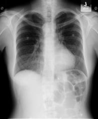
Chapter 2 Cardiac Interpretation of Pediatric Chest X-Ray
 Chapter 2 Cardiac Interpretation of Pediatric Chest X-Ray Ra-id Abdulla and Douglas M. Luxenberg Key Facts The cardiac silhouette occupies 50 55% of the chest width on an anterior posterior chest X-ray
ULTRASOUND OF THE FETAL HEART
 ULTRASOUND OF THE FETAL HEART Cameron A. Manbeian, MD Disclosure Statement Today s faculty: Cameron Manbeian, MD does not have any relevant financial relationships with commercial interests or affiliations
Amount of blood to be withdrawn slowly under cardiac monitoring, same volume replaced with normal saline or FFP.
 Pre Surgical Clinical and Echo Assessment of Tetralogy of Fallot S Khatri, S Shrivastava Director & Head of Department, Pediatric and Congenital Heart Diseases, Okhla Road, New Delhi, India. (Cardiovasc.
Foetal Cardiology: How to predict perinatal problems. Prof. I.Witters Prof.M.Gewillig UZ Leuven
 Foetal Cardiology: How to predict perinatal problems Prof. I.Witters Prof.M.Gewillig UZ Leuven Cardiopathies Incidence : 8-12 / 1000 births ( 1% ) Most frequent - Ventricle Septum Defect 20% - Atrium Septum
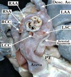
Right Sided Aortic Arch and its rare Associations- A Case Series
 DOI: 10.7860/IJARS/2018/35392:2410 Radiology Section Case Series Right Sided Aortic Arch and its rare Associations- A Case Series Nidhi Aggarwal, Narender kumar kardam, Kushal Babu Gehlot ABSTRACT Right
Most common fetal cardiac anomalies
 Most common fetal cardiac anomalies Common congenital heart defects CHD % of cardiac defects Chromosomal Infants Fetuses anomaly (%) 22q11 deletion (%) VSD 30 5~10 20~40 10 PS 9 5 (PA w/ VSD) HLHS 7~9
Appendix A.1: Tier 1 Surgical Procedure Terms and Definitions
 Appendix A.1: Tier 1 Surgical Procedure Terms and Definitions Tier 1 surgeries AV Canal Atrioventricular Septal Repair, Complete Repair of complete AV canal (AVSD) using one- or two-patch or other technique,
CMR for Congenital Heart Disease
 CMR for Congenital Heart Disease * Second-line tool after TTE * Strengths of CMR : tissue characterisation, comprehensive access and coverage, relatively accurate measurements of biventricular function/
Heart and Lungs. LUNG Coronal section demonstrates relationship of pulmonary parenchyma to heart and chest wall.
 Heart and Lungs Normal Sonographic Anatomy THORAX Axial and coronal sections demonstrate integrity of thorax, fetal breathing movements, and overall size and shape. LUNG Coronal section demonstrates relationship
Congenital Heart Disease Systematic Interpretation of CT Suhny Abbara, MD
 Congenital Heart Disease Systematic Interpretation of CT Suhny Abbara, MD Chief, Cardiothoracic Imaging Division Professor of Radiology UT Southwestern Medical Center, Dallas, TX Suhny.Abbara@UTSouthwestern.edu
ISUOG Basic Training. Obtaining & Interpreting Heart Views Correctly Alfred Abuhamad, USA. Basic training. Editable text here
 ISUOG Basic Training Obtaining & Interpreting Heart Views Correctly Alfred Abuhamad, USA Learning Objectives 6, 7 & 8 At the end of the lecture you will be able to: describe how to assess cardiac situs
By Dickens ATURWANAHO & ORIBA DAN LANGOYA MAKchs, MBchB CONGENTAL HEART DISEASE
 By Dickens ATURWANAHO & ORIBA DAN LANGOYA MAKchs, MBchB CONGENTAL HEART DISEASE Introduction CHDs are abnormalities of the heart or great vessels that are present at birth. Common type of heart disease
Tracheal Reconstruction in Children With Unilateral Lung Agenesis or Severe Hypoplasia
 Tracheal Reconstruction in Children With Unilateral Lung Agenesis or Severe Hypoplasia Carl Lewis Backer, MD, Angela M. Kelle, BS, Constantine Mavroudis, MD, Cynthia K. Rigsby, MD, Sunjay Kaushal, MD,
가천의대길병원소아심장과최덕영 PA C IVS THE EVALUATION AND PRINCIPLES OF TREATMENT STRATEGY
 가천의대길병원소아심장과최덕영 PA C IVS THE EVALUATION AND PRINCIPLES OF TREATMENT STRATEGY PA c IVS (not only pulmonary valve disease) Edwards JE. Pathologic Alteration of the right heart. In: Konstam MA, Isner M, eds.
When is Risky to Apply Oxygen for Congenital Heart Disease 부천세종병원 소아청소년과최은영
 When is Risky to Apply Oxygen for Congenital Heart Disease 부천세종병원 소아청소년과최은영 The Korean Society of Cardiology COI Disclosure Eun-Young Choi The author have no financial conflicts of interest to disclose
The Arterial Switch Operation for Transposition of the Great Arteries
 The Arterial Switch Operation for Transposition of the Great Arteries Jan M. Quaegebeur, M.D., Ph.D. A Journey of 60 Years Transposition of the Great Arteries First description: M. BAILLIE The morbid anatomy
3 Aortopulmonary Window
 0 0 0 0 0 Aortopulmonary Window Introduction Communications between the ascending aorta and pulmonary artery constitute a spectrum of malformations which is collectively designated aortopulmonary window,
MEDICAL MANAGEMENT WITH CAVEATS 1. In one study of 50 CHARGE patients with CHD, 75% required surgery. 2. Children with CHARGE may be resistant to chlo
 CARDIOLOGY IN CHARGE SYNDROME: FOR THE PHYSICIAN Angela E. Lin, M.D. Teratology Program/Active Malformation Surveillance, Brigham and Women's Hospital, Old PBBH-B501, 75 Francis St., Boston, MA 02115 alin@partners.org
3/14/2011 MANAGEMENT OF NEWBORNS CARDIAC INTENSIVE CARE CONFERENCE FOR HEALTH PROFESSIONALS IRVINE, CA. MARCH 7, 2011 WITH HEART DEFECTS
 CONFERENCE FOR HEALTH PROFESSIONALS IRVINE, CA. MARCH 7, 2011 MANAGEMENT OF NEWBORNS WITH HEART DEFECTS A NTHONY C. CHANG, MD, MBA, MPH M E D I C AL D I RE C T OR, HEART I N S T I T U T E C H I LDRE N
(i) Family 1. The male proband (1.III-1) from European descent was referred at
 Family 1. The male proband (1.III-1) from European descent was referred at") 1 Supplementary Note Clinical descriptions of families (i) Family 1. The male proband (1.III-1) from European descent was referred at age 14 because of scoliosis. He had normal development. Physical evaluation
1 Supplementary Note Clinical descriptions of families (i) Family 1. The male proband (1.III-1) from European descent was referred at age 14 because of scoliosis. He had normal development. Physical evaluation
Echocardiography in Congenital Heart Disease
 Chapter 44 Echocardiography in Congenital Heart Disease John L. Cotton and G. William Henry Multiple-plane cardiac imaging by echocardiography can noninvasively define the anatomy of the heart and the
Low-dose prospective ECG-triggering dual-source CT angiography in infants and children with complex congenital heart disease: first experience
 Low-dose prospective ECG-triggering dual-source CT angiography in infants and children with complex congenital heart disease: first experience Ximing Wang, M.D., Zhaoping Cheng, M.D., Dawei Wu, M.D., Lebin
AORTIC COARCTATION. Synonyms: - Coarctation of the aorta
 AORTIC COARCTATION Synonyms: - Coarctation of the aorta Definition: Aortic coarctation is a congenital narrowing of the aorta, usually located after the left subclavian artery, near the ductus or the ligamentum
Our Experiences With Adult Type Aortic Coarctation
 ISPUB.COM The Internet Journal of Thoracic and Cardiovascular Surgery Volume 7 Number 2 Our Experiences With Adult Type Aortic Coarctation E Duran, S Canbaz, M Acipayam, O Gur, O Karaca Citation E Duran,
Adult Echocardiography Examination Content Outline
 Adult Echocardiography Examination Content Outline (Outline Summary) # Domain Subdomain Percentage 1 2 3 4 5 Anatomy and Physiology Pathology Clinical Care and Safety Measurement Techniques, Maneuvers,
List by Terms Visceral anomalies
 1 List by Terms Visceral anomalies Dilated 10128 Dilated cerebral ventricle 11 7 2 0 20,00 10201 Dilated aorta 9 8 2 1 5,26 10207 Dilated aortic arch 9 8 3 0 5,00 10213 Dilated carotid 3 12 4 1-47,37 10218
Tetralogy of Fallot With Pulmonary Atresia Associated With Chromosome 22qll Deletion
 198 JACC Vol. 27, No. 1 Tetralogy of Fallot With Pulmonary Atresia Associated With Chromosome 22qll Deletion KAZUO MOMMA, MD, CHISATO KONDO, MD, RUMIKO MATSUOKA, MD Tokyo, Japan Objectives. The purpose
Case Report Recurrent Wheezing and Cough Caused by Double Aortic Arch, Not Asthma
 Hindawi Case Reports in Cardiology Volume 2017, Article ID 8079851, 4 pages https://doi.org/10.1155/2017/8079851 Case Report Recurrent Wheezing and Cough Caused by Double Aortic Arch, Not Asthma Qiao Zhang,
Section V Cardiac Radiology
 Section V Cardiac Radiology Figure 1 89. Based on the diagram (Figure 1), which of the following vessels typically supplies the anterolateral cardiac segment? A. Left anterior descending B. Circumflex
British Society of Echocardiography
 British Society of Echocardiography Affiliated to the British Cardiac Society A Minimum Dataset for a Standard Adult Transthoracic Echocardiogram From the British Society of Echocardiography Education
S. Bruce Greenberg, MD FNASCI and President, NASCI Professor of Radiology and Pediatrics University of Arkansas for Medical Sciences
 S. Bruce Greenberg, MD FNASCI and President, NASCI Professor of Radiology and Pediatrics University of Arkansas for Medical Sciences No financial disclosures Aorta Congenital aortic stenosis/insufficiency
90 th Annual Meeting The American Association for Thoracic Surgery May 1, 2010 Toronto, Ontario, Canada. Slide Tracheoplasty
 90 th Annual Meeting The American Association for Thoracic Surgery May 1, 2010 Toronto, Ontario, Canada Congenital Skills Course Slide Tracheoplasty Carl Lewis Backer, MD A.C. Buehler Professor of Surgery
Absent Pulmonary Valve Syndrome
 Absent Pulmonary Valve Syndrome Fact sheet on Absent Pulmonary Valve Syndrome In this condition, which has some similarities to Fallot's Tetralogy, there is a VSD with narrowing at the pulmonary valve.
Atrial Septal Defects
 Supplementary ACHD Echo Acquisition Protocol for Atrial Septal Defects The following protocol for echo in adult patients with atrial septal defects (ASDs) is a guide for performing a comprehensive assessment
CONGENITAL HEART DISEASE (CHD)
") CONGENITAL HEART DISEASE (CHD) DEFINITION It is the result of a structural or functional abnormality of the cardiovascular system at birth GENERAL FEATURES OF CHD Structural defects due to specific disturbance
CONGENITAL HEART DISEASE (CHD) DEFINITION It is the result of a structural or functional abnormality of the cardiovascular system at birth GENERAL FEATURES OF CHD Structural defects due to specific disturbance
The arterial switch operation has been the accepted procedure
 The Arterial Switch Procedure: Closed Coronary Artery Transfer Edward L. Bove, MD The arterial switch operation has been the accepted procedure for the repair of transposition of the great arteries (TGA)
Notes: 1)Membranous part contribute in the formation of small portion in the septal cusp.
Membranous part contribute in the formation of small portion in the septal cusp.") Embryology 9 : Slide 16 : There is a sulcus between primitive ventricular and bulbis cordis that will disappear gradually and lead to the formation of one chamber which is called bulboventricular chamber.
Embryology 9 : Slide 16 : There is a sulcus between primitive ventricular and bulbis cordis that will disappear gradually and lead to the formation of one chamber which is called bulboventricular chamber.
Scientific Exhibit Authors:
 Preoperative evaluation of infants and children with congenital heart disease with ECG triggered 128 Dual Source CT Angiography- Portugal- Angola collaboration Poster No.: C-0942 Congress: ECR 2013 Type:
Scientific Exhibit Authors: J. Costa, A. Proença Ramos, J. C. A. Costa, V. Silva, C.
 Preoperative evaluation of infants and children with congenital heart disease with ECG triggered 128 Dual Source CT Angiography- Portugal- Angola collaboration Poster No.: C-0942 Congress: ECR 2013 Type:
Radiological Point of View
 AASCIT Journal of Bioscience 2015; 1(5): 78-88 Published online October 30, 2015 (http://www.aascit.org/journal/bioscience) ISSN: 2381-1250 (Print); ISSN: 2381-1269 (Online) Type 3 Truncus Arteriosus with
Normal TTE/TEE Examinations
 Normal TTE/TEE Examinations Geoffrey A. Rose, MD FACC FASE Sanger Heart & Vascular Institute Before you begin imaging... Obtain the patient s Height Weight BP PLAX View PLAX View Is apex @ 9-10 o clock?
Slide 1. Slide 2. Slide 3 CONGENITAL HEART DISEASE. Papworth Hospital NHS Trust INTRODUCTION. Jakub Kadlec/Catherine Sudarshan INTRODUCTION
 Slide 1 CONGENITAL HEART DISEASE Jakub Kadlec/Catherine Sudarshan NHS Trust Slide 2 INTRODUCTION Most common congenital illness in the newborn Affects about 4 9 / 1000 full-term live births in the UK 1.5
Devendra V. Kulkarni, Rahul G. Hegde, Ankit Balani, and Anagha R. Joshi. 2. Case Report. 1. Introduction
 Case Reports in Radiology, Article ID 614647, 4 pages http://dx.doi.org/10.1155/2014/614647 Case Report A Rare Case of Pulmonary Atresia with Ventricular Septal Defect with a Right Sided Aortic Arch and
ORIGINAL RESEARCH PAPER
 ORIGINAL RESEARCH PAPER ROLE OF CT PULMONARY ANGIOGRAPHY IN CONGENITAL HEART DISEASES IN PAEDIATRIC POPULATION Radiology KEY WORDS: Congenital heart disease, CT pulmonary angiography, pediatric heart disease,
Debate: Should Ductal Stent Implantation be Considered for All Newborn Infants with Reduced Pulmonary Blood Flow?_Pros
 Debate: Should Ductal Stent Implantation be Considered for All Newborn Infants with Reduced Pulmonary Blood Flow?_Pros Mazeni Alwi Institut Jantung Negara Kuala Lumpur, Malaysia 5 th Asia Pacific Congenital
AbnormalThree-VesselView on Sonography: A Clue to the Diagnosis of Congenital Heart Disease in the Fetus
 rt Pictorial Essay bnormalthree-vesselview on Sonography: Clue to the Diagnosis of Congenital Heart Disease in the Fetus screening tool for major congenital heart diseases [I. 2J. However, anomalies of
Suggested Readings. Khonsiari S. Cardiac surgery: safeguards and pittfalls in operative techniques. Philadelphia: Lippincott Williams and
 Suggested Readings Adams FH, Emmanouilides GC, Riemenshneider TA. Heart disease in infants, children, and adolescent. Baltimore: Williams and Wilkins; 1987. Elliot LP. Cardiac imaging in infants, children,
List by Region - Visceral Anomalies
 1 List by Region - Visceral Anomalies General Terms 10127 Situs inversus 80,00 10125 Aneurysm 68,42 10126Fluid-filled abdomen -35,00 Brain 10131 Hydrocephaly 10128 Dilated cerebral ventricle 20,00 10132
Appendix II: ECHOCARDIOGRAPHY ANALYSIS
 Appendix II: ECHOCARDIOGRAPHY ANALYSIS Two-Dimensional (2D) imaging was performed using the Vivid 7 Advantage cardiovascular ultrasound system (GE Medical Systems, Milwaukee) with a frame rate of 400 frames
Aortic Coarctation: Evaluation with Computed Tomography Angiography in Pediatric Patients
 Med. J. Cairo Univ., Vol. 83, No. 2, June: 63-70, 2015 www.medicaljournalofcairouniversity.net Aortic Coarctation: Evaluation with Computed Tomography Angiography in Pediatric Patients MOHAMED ZAKI, M.D.
Screening for Critical Congenital Heart Disease
 Screening for Critical Congenital Heart Disease Caroline K. Lee, MD Pediatric Cardiology Disclosures I have no relevant financial relationships or conflicts of interest 1 Most Common Birth Defect Most
New ASE Guidelines: What you must know
 New ASE Guidelines: What you must know Federico M Asch MD, FASE, FACC Chair, ASE Guidelines and Standards Committee Medstar Washington Hospital Center Medstar Health Research Institute Georgetown University
Echocardiography in adult congenital heart disease
 S12 Department of Cardiology, Royal Hospital for Sick Children, Glasgow G3 8SJ, UK A Houston S Lilley T Richens University Department of Medicine and Therapeutics, Western Infirmary, Glasgow G11 6NT, UK
Primary Resection of Kommerell Diverticulum and Left Subclavian Artery Transfer
 Primary Resection of Kommerell Diverticulum and Left Subclavian Artery Transfer Carl L. Backer, MD, Hyde M. Russell, MD, Katherine C. Wurlitzer, BA, Jeffrey C. Rastatter, MD, and Cynthia K. Rigsby, MD
Pregnancy, Heart Disease and Imaging. Hemodynamics. Decreased systemic vascular resistance. Physiology anemia
 Pregnancy, Heart Disease and Imaging Sangeeta Shah, MD, FASE, FACC Associate Professor, Ochsner Clinical School of Medicine Advanced CV Imaging and Adult Congenital Heart Disease New Orleans, LA Hemodynamics
Uptofate Study Summary
 CONGENITAL HEART DISEASE Uptofate Study Summary Acyanotic Atrial septal defect Ventricular septal defect Patent foramen ovale Patent ductus arteriosus Aortic coartation Pulmonary stenosis Cyanotic Tetralogy
Double outlet right ventricle: navigation of surgeon to chose best treatment strategy
 Double outlet right ventricle: navigation of surgeon to chose best treatment strategy Jan Marek Great Ormond Street Hospital & Institute of Cardiovascular Sciences, University College London Double outlet
/b O. Figure 4.1 Tracing of a normal dorsoventral angiocardiogram. The. veins (PV) enter the left atrium (LA) well within the
 enter the left atrium (LA) well within the") /b O " Figure 4.1 Tracing of a normal dorsoventral angiocardiogram. The pu~nonary veins (PV) enter the left atrium (LA) well within the limits of the cardiac silhouette; the left atri~m does not contribute
/b O " Figure 4.1 Tracing of a normal dorsoventral angiocardiogram. The pu~nonary veins (PV) enter the left atrium (LA) well within the limits of the cardiac silhouette; the left atri~m does not contribute
The Dilated Pulmonary Artery: Is there a risk of Dissection?
 The Dilated Pulmonary Artery: Is there a risk of Dissection? Pastora Gallego, MD, PhD Inter-center Adult Congenital Heart Disease Unit Area del Corazón Seville, Spain No conflict of interest to disclose
Patent ductus arteriosus PDA
 Patent ductus arteriosus PDA Is connecting between the aortic end just distal to the origin of the LT sub clavian artery& the pulmonary artery at its bifurcation. Female/male ratio is 2:1 and it is more