By: Stephanie Bendtsen, Joseph Calderan, and Celeste Dupont Team 17 Client: Dr. Sun
|
|
|
- Jeffrey Brandon Short
- 6 years ago
- Views:
Transcription

1 By: Stephanie Bendtsen, Joseph Calderan, and Celeste Dupont Team 17 Client: Dr. Sun

2 Flow Loop Design
3 Introduction Purpose: to build a pulsatile flow loop that allows for implementation and testing of mechanical and bioprosthetic valves while accurately simulating the hemodynamic pressure and flow conditions of the left side of the heart. Includes an easier, more successful method for implementing a biological aortic root into the flow loop, as requested by Dr. Sun. The flow loop is oriented horizontally to prevent an increase in pressure throughout the system when the longer aortic root is implemented. The whole system is composed of Plexiglas to enable the mitral and aortic valve functions to be observed through viewing ports at both the inlet and outlet sides of the valves. The ventricle chamber is set up as a square with cameras mounted on both sides. It is important that this design does not affect the physiological waveforms produced by the function of the heart valves.

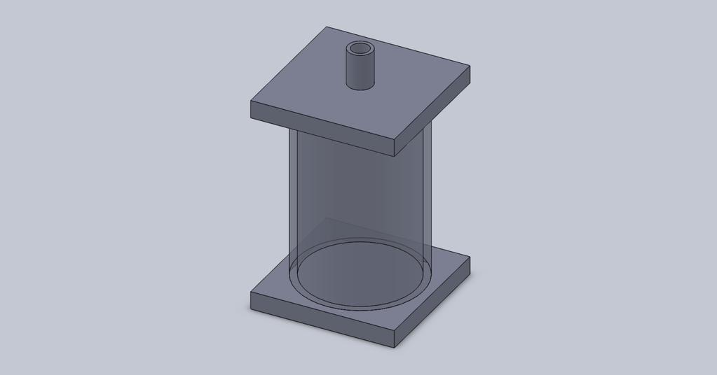
4 Atrium Chamber
5 Atrium Chamber Fluid first enters system through atrium Serves as a reservoir, holding excess water not currently used by the system 5.75 by by 3.25 open topped chamber of Plexiglas Open top allows for a fluctuation in volume without causing a drastic increase in pressure throughout the rest of the system Plan to incorporate a surrounding heat reservoir system at a temperature of 37 C, resulting in a more accurate representation of the in vivo environment Hole at the base of the atrium opens to a tube connecting to the mitral valve chamber. The valve chamber will be compression fit against the side of the output of the atrium. The compression will prevent leaks around the valve resulting in the correct pressurizations in the chambers
6 Mitral Valve
7 Mitral Valve One way valve- fluid from atrium to ventricle Fluid pulled into ventricle-2 leaflets open; venrticle contracts pushing fluid out- pushes leaflets back, closing path Goal- incorporate valves of different types and sizes into both mitral and aortic positions Valve housing- solid aluminum rectangle with a hole bored into itdiameter as large as the widest valve used. This hole will extend through the majority of the rectangle until the end where a smaller hole will be drilled, allowing the valve to rest against a flat surface
8 Mitral Valve Cont. Bioprosthetic valves- consists of a sewing ring or annulus at the base that is supporting three struts. Between these supports are three tissue leaflets that meet in the middle Fixture- a thin-walled steel cylinder with an outer diameter the same as the diameter of the larger hole will slide down onto the valve once it has been placed in the chamber. The cylinder will be pushed down to compress the valve in place to prevent sliding. Along the rectangle four holes will be drilled from each edge face. Set-screws will be used to hold the cylinder in place, holding the valve steady in the process. Mechanical valves- held in place using compression plates from the sides. Four small curved aluminum sections will be cut and fixed to sections of a compressible gasket. Each of these four sections will attach to one of the drilled holes for the set-screws. These sections will be attached to screws of their own and will be moved inwards towards the valve until the valve is compressed and stable.
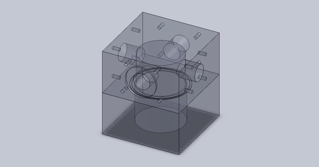
9 Ventricular Chamber
10 Ventricular Chamber Ventricle will pump fluid through the system powered by a pulsatile air pump Modeled from existing design: latex sack housed in airtight chamber Square design- flow entering and leaving at right angle which allows visualization of the mitral and aortic valves while the system is running. Cameras can be mounted on two of the sides of the ventricle which will allow visualization of both valves simultaneously without affecting the overall physiological flow waveforms. The two paths of view cross along the center of the ventricle and continue through the valve chambers until they reach view windows at either the exit from the atrium, or the entrance to the compliance chambers.
11 Ventricular Chamber Cont. Diastolic phase: the pump draws air from the ventricular housing chamber, creating a vacuum. the sack inflates to its original size and is filled with fluid from the atrium through the one way mitral valve Systolic phase: the pump pushes air through a tube into the airtight chamber housing the ventricle, directly influencing the internal pressure. the ventricle collapses on itself, pushing fluid through the aortic valve. contraction applies pressure to the mitral valve, closing it and preventing water from entering the atrium via this route.

12 Aortic Valve
13 Aortic Valve Fluid flows in from the ventricle through to the ascending aorta 2 interchangeable housing chambers: -1 for mechancial and bioprosthetic valves -1 to implement biological porcine or bovine valve with portion of connecting ascending aorta Portion of valve below native leaflets not uniform, very difficult to mount Still developing best method of attachment

14 Porcine Aortic Valve with Aortic Root
15 Ascending Aorta series of tubing to simulate the separation of the blood into different sections to travel throughout the body Urebrade Reinforced Polyurethane Tubing, -inner diameter outer diameter designed to resist weathering and tearing very tough and resilient material that can withstand high pressures but not very elastic Because the arteries in the body actually have an elastic component, the rigidity of the tubing is compensated for in the compliance chamber
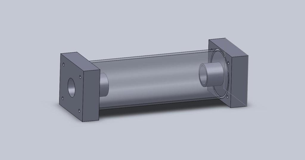
16 Compliance Chamber 1
17 Compliance Chamber 1 Rubber compartment within Plexiglas cylinder- -6 in long, ID- 3 OD- 3.5 connected to controllable pressure pump and gauge- as the pressure within the tube changes, the size of the rubber compartment changes accordingly the rubber component is attached to the tube at both ends so that the portion between the rubber and the tubing has no openings to the outside, other than the opening leading to the pressure pump and gauge
18 Compliance Chamber
19 Data Acquisition electronic flow meter will be mounted right before the aortic valve chamber detect regurgitation Pressure sensors throughout system to obtain waveforms
20 Progress Getting ready to order Plexiglas and aluminum Working with Dr. Sun to determine best method to house the aortic valve that allows for implementation of biological aortic valve with partial ascending aorta Developing ideal compliance set up
Optimal Design #17 Report
 Optimal Design #17 Report Development of a Pulsatile Left Heart Simulator By Stephanie Bendtsen, Joe Calderan, Celeste Dupont Team #17 Client Contact: Dr. Wei Sun, University of Connecticut, Tissue Mechanics
Final Report Development of a Pulsatile Left Heart Simulator
 0 Final Report Development of a Pulsatile Left Heart Simulator Stephanie Bendtsen, Joseph Calderan, Celeste Dupont Team #17 Client #25 Client Contact: Dr. Wei Sun, University of Connecticut Tissue Mechanics
Operator s Manual Development of a Pulsatile Left Heart Simulator
 0 Operator s Manual Development of a Pulsatile Left Heart Simulator Stephanie Bendtsen, Joseph Calderan, Celeste Dupont Team #17 Client #25 Client Contact: Dr. Wei Sun, University of Connecticut Tissue
Team 19 Kaitlyn Clarke Brittany DePoi Andrew Reynolds Adarsha Selvachandran. Client: Dr. Wei Sun
 Team 19 Kaitlyn Clarke Brittany DePoi Andrew Reynolds Adarsha Selvachandran Client: Dr. Wei Sun Dr. Sun has requested a testing device that will allow his research team to obtain physical evidence regarding
Health Science 20 Circulatory System Notes
 Health Science 20 Circulatory System Notes Functions of the Circulatory System The circulatory system functions mainly as the body s transport system. It transports: o Oxygen o Nutrients o Cell waste o
Heart Dissection. 5. Locate the tip of the heart or the apex. Only the left ventricle extends all the way to the apex.
 Heart Dissection Page 1 of 6 Background: The heart is a four-chambered, hollow organ composed primarily of cardiac muscle tissue. It is located in the center of the chest in between the lungs. It is the
PROSTHETIC VALVE BOARD REVIEW
 PROSTHETIC VALVE BOARD REVIEW The correct answer D This two chamber view shows a porcine mitral prosthesis with the typical appearance of the struts although the leaflets are not well seen. The valve
Radnoti Langendorff Constant Pressure Non-Recirculating Pump Driven Isolated Heart System For Mouse EZ
 Radnoti Langendorff Constant Pressure Non-Recirculating Pump Driven Isolated Heart System For Mouse Radnoti 2006 Description Qty Part # Base only, for 4-bar stand 1 159950-B4 Stabilizer Bar only, for 4-Bar
Proposal. Fabrication of Percutaneous Transvenous Mitral Annuloplasty Testing Device. Team 19
 Proposal Fabrication of Percutaneous Transvenous Mitral Annuloplasty Testing Device Team 19 Andrew Reynolds Kaitlyn Clarke Adarsha Selvachandran Brittany DePoi Project for Dr. Wei Sun Client Contact: Dr.
The Heart. Happy Friday! #takeoutyournotes #testnotgradedyet
 The Heart Happy Friday! #takeoutyournotes #testnotgradedyet Introduction Cardiovascular system distributes blood Pump (heart) Distribution areas (capillaries) Heart has 4 compartments 2 receive blood (atria)
IB TOPIC 6.2 THE BLOOD SYSTEM
 IB TOPIC 6.2 THE BLOOD SYSTEM TERMS TO KNOW circulation ventricle artery vein THE BLOOD SYSTEM 6.2.U1 - Arteries convey blood at high pressure from the ventricles to the tissues of the body Circulation
The Heart and Cardiovascular System
 The Heart and Cardiovascular System What you will learn The location of the heart 3 layers and covering of the heart Explain the function of the heart as 2 separate pumps Identify the 4 chambers of the
Cardiovascular System. I. Structures of the heart A. : Pericardium sack that surrounds the heart
 Cardiovascular System I. Structures of the heart A. : Pericardium sack that surrounds the heart 1. : Pericardial Cavity serous fluid filled space between the heart and the pericardium B. Heart Wall 1.
Cardiac Conduction System
 Cardiac Conduction System What causes the Heart to Beat? Heart contracts by electrical signals! Cardiac muscle tissue contracts on its own an electrical signal is sent out by the heart so that all cells
Topic 6: Human Physiology
 Topic 6: Human Physiology 6.2 The Blood System D.4 The Heart Essential Questions: 6.2 The blood system continuously transports substances to cells and simultaneously collects waste products. D.3 The chemical
Ch.15 Cardiovascular System Pgs {15-12} {15-13}
 Ch.15 Cardiovascular System Pgs {15-12} {15-13} E. Skeleton of the Heart 1. The skeleton of the heart is composed of rings of dense connective tissue and other masses of connective tissue in the interventricular
Image Analysis and Cytometry in Three-Dimensional Digital Reconstruction of Porcine Native Aortic Valve Leaflets
 Image Analysis and Cytometry in Three-Dimensional Digital Reconstruction of Porcine Native Aortic Valve Leaflets Introduction Chi Zheng, M1 - University of Pittsburgh; BSE - University of Michigan In association
THE HEART OBJECTIVES: LOCATION OF THE HEART IN THE THORACIC CAVITY CARDIOVASCULAR SYSTEM
 BIOLOGY II CARDIOVASCULAR SYSTEM ACTIVITY #3 NAME DATE HOUR THE HEART OBJECTIVES: Describe the anatomy of the heart and identify and give the functions of all parts. (pp. 356 363) Trace the flow of blood
Cardiovascular System. Heart Anatomy
 Cardiovascular System Heart Anatomy 1 The Heart Location & general description: Atria vs. ventricles Pulmonary vs. systemic circulation Coverings Walls The heart is found in the mediastinum, the medial
CJ Shuster A&P2 Lab Addenum Beef Heart Dissection 1. Heart Dissection. (taken from Johnson, Weipz and Savage Lab Book)
") CJ Shuster A&P2 Lab Addenum Beef Heart Dissection 1 Heart Dissection. (taken from Johnson, Weipz and Savage Lab Book) Introduction When you have finished examining the model, you are ready to begin your
CJ Shuster A&P2 Lab Addenum Beef Heart Dissection 1 Heart Dissection. (taken from Johnson, Weipz and Savage Lab Book) Introduction When you have finished examining the model, you are ready to begin your
The Circulatory System. Lesson Overview. Lesson Overview The Circulatory System
 33.1 THINK ABOUT IT More than one-third of the 1.2 million Americans who suffer a heart attack each year die. This grim evidence shows that the heart and the circulatory system it powers are vital to life.
Lab #3: Electrocardiogram (ECG / EKG)
") Lab #3: Electrocardiogram (ECG / EKG) An introduction to the recording and analysis of cardiac activity Introduction The beating of the heart is triggered by an electrical signal from the pacemaker. The
Lab #3: Electrocardiogram (ECG / EKG) An introduction to the recording and analysis of cardiac activity Introduction The beating of the heart is triggered by an electrical signal from the pacemaker. The
BME 5742 Bio-Systems Modeling and Control. Lecture 41 Heart & Blood Circulation Heart Function Basics
 BME 5742 Bio-Systems Modeling and Control Lecture 41 Heart & Blood Circulation Heart Function Basics Dr. Zvi Roth (FAU) 1 Pumps A pump is a device that accepts fluid at a low pressure P 1 and outputs the
Read Me. covering the Heart Anatomy. Labs. textbook. use. car: you
 Heart Anatomy Lab Pre-Lab Exercises Read Me These exercises should be done before coming to lab, after watching the videos covering the Heart Anatomy Labs. Answer the questions in this guide using the
Blood and Heart. Student Learning Objectives:
 Blood and Heart Student Learning Objectives: Identify the major components of the blood. Identify the primary structures associated with the heart Follow the blood through the path of the circulation.
the Cardiovascular System I
 the Cardiovascular System I By: Dr. Nabil A Khouri MD, MsC, Ph.D MEDIASTINUM 1. Superior Mediastinum 2. inferior Mediastinum Anterior mediastinum. Middle mediastinum. Posterior mediastinum Anatomy of
The cardiovascular system is composed of the heart and blood vessels that carry blood to and from the body s organs. There are 2 major circuits:
 1 The cardiovascular system is composed of the heart and blood vessels that carry blood to and from the body s organs. There are 2 major circuits: pulmonary and systemic. The pulmonary goes out to the
ENT 318/3 Artificial Organs
 ENT 318/3 Artificial Organs Modeling of cardiovascular system and VAD Lecturer Ahmad Nasrul bin Norali 1 What is modeling and why we need it? In designing product, sometimes we have to make sure that the
Blood flows away from the heart in arteries, to the capillaries and back to the heart in the veins
 Cardiovascular System Summary Notes The cardiovascular system includes: The heart, a muscular pump The blood, a fluid connective tissue The blood vessels, arteries, veins and capillaries Blood flows away
MICS & FEMORAL CANNULAE
 MICS & FEMORAL CANNULAE Cannulae that can High performance cannulae for direct and femoral cannulation, either in conventional or minimally invasive cardiac surgery 1. Pfeiffer et al. Interactive Cardiovascular
The Cardiovascular System
 11 PART A The Cardiovascular System PowerPoint Lecture Slide Presentation by Jerry L. Cook, Sam Houston University ESSENTIALS OF HUMAN ANATOMY & PHYSIOLOGY EIGHTH EDITION ELAINE N. MARIEB The Cardiovascular
Cardioplegia Circuit Products { CARDIOPLEGIA ADAPTERS}
 Cardioplegia Circuit Products { CARDIOPLEGIA ADAPTERS} Cardioplegia adapters are designed to permit customization of the cardioplegia circuit. Medtronic adapters can be used to connect multiple cardioplegia
Aortic root enlargement is an invaluable surgical technique
 Aortic Root Enlargement in the Adult Christopher M. Feindel, MD, CM, FRCS(C) Aortic root enlargement is an invaluable surgical technique with which every cardiac surgeon performing aortic valve replacement
Danil Hammoudi.MD 1/12/2009
 Danil Hammoudi.MD Aorta the biggest and longest artery (a blood vessel carrying blood away from the heart) in the body. It carries oxygen rich blood from the left ventricle of the heart to the body.inferior
Circulatory System 10.1
 1 Circulatory System 10.1 2 ARTERIES Arteries-blood vessels that carry blood away from the heart Thick walls Inner & Outer layers: connective tissue Middle layers are muscle and elastic connective tissue
IB TOPIC 6.2 THE BLOOD SYSTEM
 IB TOPIC 6.2 THE BLOOD SYSTEM THE BLOOD SYSTEM TERMS TO KNOW circulation ventricle artery vein 6.2.U1 - Arteries convey blood at high pressure from the ventricles to the tissues of the body Circulation
A. Incorrect! The left ventricle receives oxygenated blood from the lungs via the left atrium.
 DAT Biology - Problem Drill 10: The Circulatory System Question No. 1 of 10 1. What is the flow of deoxygenated blood through the heart as it returns from the body? Question #01 (A) Vena cava; right ventricle;
CVS Hemodynamics. Faisal I. Mohammed, MD,PhD.
 CVS Hemodynamics Faisal I. Mohammed, MD,PhD. Objectives point out the physical characteristics of the circulation: distribution of blood volume total cross sectional area velocity blood pressure List the
Blood Pressure Laboratory
 Introduction The blood that circulates throughout the body maintains a flow and pressure. The nervous system can change the flow and pressure based on the particular needs at a given time. For example,
The Heart. Size, Form, and Location of the Heart. 1. Blunt, rounded point; most inferior part of the heart.
 12 The Heart FOCUS: The heart is composed of cardiac muscle cells, which are elongated, branching cells that appear striated. Cardiac muscle cells behave as a single electrical unit, and the highly coordinated
Chapter 14. The Cardiovascular System
 Chapter 14 The Cardiovascular System Introduction Cardiovascular system - heart, blood and blood vessels Cardiac muscle makes up bulk of heart provides force to pump blood Function - transports blood 2
Figure 1 The Human Heart
 ARTIFICIAL HEART VALVE MODEL INTRODUCTION The heart is a muscular pump with four chambers and four heart valves. The upper chambers, the right atrium and left, are thin walled filling chambers. Blood flows
2. Langendorff Heart
 2. Langendorff Heart 2.1. Principle Langendorff heart is one type of isolated perfused heart which is widely used for biochemical, physiological, morphological and pharmacological researches. It provides
Collin County Community College
 Collin County Community College BIOL. 2402 Anatomy & Physiology WEEK 6 Blood Vessels 1 Anatomy of Blood Vessels Walls of blood vessels contain 3 distinct layers : Tunica intima innermost layer includes
Chapter 27 -The Heart & Blood Vessels
 Chapter 27 -The Heart & Blood Vessels 3.2 Learning Objectives 3.2.2 Organisational Complexity of the human 1. Describe the structures and organisation of tissues in the closed circulatory system. 2. Discuss
BMI Medical. Ventricular Catheter T
 Shunting System Ventricular Catheter 01101 01101T Barium-impregnated silicone catheter provides resistance to kinking and compression. Stainless steel stylet allows catheter to be directed during catheter
Heart phantom for testing of MRI guided catheter intervention
 Heart phantom for testing of MRI guided catheter intervention Erin Main, Peter Strohm, Fan Wu, Lacey Halfen, Jessica Hause ABSTRACT A recently developed intravenous catheter aims to treat atrial fibrillation
Cardiac Cycle MCQ. Professor of Cardiovascular Physiology. Cairo University 2007
 Cardiac Cycle MCQ Abdel Moniem Ibrahim Ahmed, MD Professor of Cardiovascular Physiology Cairo University 2007 1- Regarding the length of systole and diastole: a- At heart rate 75 b/min, the duration of
The Circulatory System
 The Circulatory System Key Questions What are the functions of the circulatory system? How does the heart pump blood through the body? What are three types of blood vessels? Vocabulary myocardium atrium
Physiology - 8 Hemodynamics - 1 M.jafar 24/3/2016 Turquoise Team
 21 Physiology - 8 Hemodynamics - 1 M.jafar 24/3/2016 Turquoise Team Hemodynamics Today we will take about hemodynamics which is the study of the movement of blood and of the forces concerned. Now how the
Ch 19: Cardiovascular System - The Heart -
 Ch 19: Cardiovascular System - The Heart - Give a detailed description of the superficial and internal anatomy of the heart, including the pericardium, the myocardium, and the cardiac muscle. Trace the
Mr. Epithelium s Anatomy and Physiology Test SSSS
 Mr. Epithelium s Anatomy and Physiology Test SSSS You have 50 minutes to complete this test packet. One 8.5 x 11 cheat sheet is allowed, along with 1 non-programmable calculator dedicated to computation.
Hemodynamic Assessment. Assessment of Systolic Function Doppler Hemodynamics
 Hemodynamic Assessment Matt M. Umland, RDCS, FASE Aurora Medical Group Milwaukee, WI Assessment of Systolic Function Doppler Hemodynamics Stroke Volume Cardiac Output Cardiac Index Tei Index/Index of myocardial
CIRCULATORY SYSTEM BLOOD VESSELS
 Name: Block: CIRCULATORY SYSTEM Multicellular organisms (above the level of roundworms) rely on a circulatory system to bring nutrients to, and take wastes away from, cells. In higher organisms such as
CHAPTER 34 LEFT VENTRICULAR FLOW 34-1
 CHAPTER 34 LEFT VENTRICULAR FLOW 34-1 CHAPTER 34 LEFT VENTRICULAR FLOW In Chapter 05 we studied the relationship of anterior leaflet shape to left ventricular pressure and flow from mid-diastole through
Cardiovascular System
 Cardiovascular System Purpose Transport oxygen and nutrients Take waste products away from tissues & organs Things we learned Blood pressure: the force of blood pushing against the walls of blood vessels
The Cardiovascular and Lymphatic Systems Cardiovascular System Blood Vessels Blood Vessels Arteries Arteries Arteries
 CH 12 The Cardiovascular and s The Cardiovascular and s OUTLINE: Cardiovascular System Blood Vessels Blood Pressure Cardiovascular System The cardiovascular system is composed of Blood vessels This system
A practical set-up to create an adjustable insufficiency in a porcine aortic root.
 A practical set-up to create an adjustable insufficiency in a porcine aortic root. Internship Hemolab BMTE07.13 T.L.Plantenga Supervision: P.H.M. Bovendeerd S.v.Tuijl M.Stijnen TU/e HemoLab HemoLab Eindhoven,
LECTURE 5. Anatomy of the heart
 LECTURE 5. Anatomy of the heart Main components of the CVS: Heart Blood circulatory system arterial compartment haemomicrocirculatory (=microvascular) compartment venous compartment Lymphatic circulatory
Cardiovascular Physiology
 Cardiovascular Physiology Lecture 1 objectives Explain the basic anatomy of the heart and its arrangement into 4 chambers. Appreciate that blood flows in series through the systemic and pulmonary circulations.
Dynamic Hollow Cylinder System Proposal
 Dynamic Hollow Cylinder System Proposal from Wykeham Farrance Limited Weston Road Slough SL1 4HW England Tel:+44 (0)1753 571241 Fax: +44 (0)1753 811313 E-mail: sales@wfi.co.uk Advanced Soils Testing 2
Do Now. Get out work from last class to be checked
 Do Now Get out work from last class to be checked Heart Actions Cardiac Cycle: One complete heartbeat. The contraction of a heart chamber is called systole and the relaxation of a chamber is called diastole.
Instructions for Use. IJ Catheter Ultrasound Model Version 2400 Series
 Instructions for Use IJ Catheter Ultrasound Model Version 2400 Series 3601 Sagamore Parkway North, Suite B Lafayette, IN 47904 Phone 765.742.4813 www.hemocleanse.com Instructions Revision 3.0 2014 Table
Systems Overview. Muscular System. Muscle System. PDF created with FinePrint pdffactory trial version
 Systems Overview Muscular System Functions: movement of body stabilizing in posture or joint generate heat support some tissues & organs guard exit & entrance of sphincters Muscle System 3 Types of muscle
Appendix II: ECHOCARDIOGRAPHY ANALYSIS
 Appendix II: ECHOCARDIOGRAPHY ANALYSIS Two-Dimensional (2D) imaging was performed using the Vivid 7 Advantage cardiovascular ultrasound system (GE Medical Systems, Milwaukee) with a frame rate of 400 frames
Circulatory and Respiratory
 Circulatory and Respiratory Systems Bởi: OpenStaxCollege Animals are complex multicellular organisms that require a mechanism for transporting nutrients throughout their bodies and removing wastes. The
Chapter 20 (1) The Heart
 The Heart") Chapter 20 (1) The Heart Learning Objectives Describe the location and structure of the heart Describe the path of a drop of blood from the superior vena cava or inferior vena cava through the heart out
Chapter 20 (1) The Heart Learning Objectives Describe the location and structure of the heart Describe the path of a drop of blood from the superior vena cava or inferior vena cava through the heart out
Circulation. Circulation = is a process used for the transport of oxygen, carbon! dioxide, nutrients and wastes through-out the body
 Circulation Circulation = is a process used for the transport of oxygen, carbon! dioxide, nutrients and wastes through-out the body Heart = muscular organ about the size of your fist which pumps blood.
The Cardiovascular and Lymphatic Systems
 BIOLOGY OF HUMANS Concepts, Applications, and Issues Fifth Edition Judith Goodenough Betty McGuire 12 The Cardiovascular and Lymphatic Systems Lecture Presentation Anne Gasc Hawaii Pacific University and
Functional anatomy of the aortic root. ΔΡΟΣΟΣ ΓΕΩΡΓΙΟΣ Διεσθσνηής Καρδιοθωρακοτειροσργικής Κλινικής Γ.Ν. «Γ. Παπανικολάοσ» Θεζζαλονίκη
 Functional anatomy of the aortic root ΔΡΟΣΟΣ ΓΕΩΡΓΙΟΣ Διεσθσνηής Καρδιοθωρακοτειροσργικής Κλινικής Γ.Ν. «Γ. Παπανικολάοσ» Θεζζαλονίκη What is the aortic root? represents the outflow tract from the LV provides
Anatomy of the Heart. Figure 20 2c
 Anatomy of the Heart Figure 20 2c Pericardium & Myocardium Remember, the heart sits in it s own cavity, known as the mediastinum. The heart is surrounded by the Pericardium, a double lining of the pericardial
All About the Heart. Structures of the heart. Layers. Chambers
 All About the Heart Your heart is a muscle. It is slightly larger than your fist and weighs less than a pound. It is located to the left of the middle of your chest. Your heart pumps blood to the lungs
The Cardiovascular System (Heart)
") The Cardiovascular System The Cardiovascular System (Heart) A closed system of the heart and blood vessels The heart pumps blood Blood vessels allow blood to circulate to all parts of the body The function
The Cardiovascular System The Cardiovascular System (Heart) A closed system of the heart and blood vessels The heart pumps blood Blood vessels allow blood to circulate to all parts of the body The function
2402 : Anatomy/Physiology
 Dr. Chris Doumen Lecture 1 2402 : Anatomy/Physiology Hemo Dynamics and Blood Vessels I nt r oduc t i on TextBook Readings Pages 721 through 734. Make use of the figures in your textbook ; a picture is
Cardiovascular System- Heart. Miss Wheeler Unit 8
 Cardiovascular System- Heart Miss Wheeler Unit 8 Overview CARDIOVASCULAR SYSTEM heart vessels Made up of heart, blood vessels, and blood Functions Heart- pump blood Vessels- (veins, arteries, capillaries)
CATCH A WAVE.. INTRODUCTION NONINVASIVE HEMODYNAMIC MONITORING 4/12/2018
 WAVES CATCH A WAVE.. W I S C O N S I N P A R A M E D I C S E M I N A R A P R I L 2 0 1 8 K E R I W Y D N E R K R A U S E R N, C C R N, E M T - P Have you considered that if you don't make waves, nobody
3D Printing & Echocardiography
 ASE SOTA Feb 19, 2018 3D Printing & Echocardiography Stephen H. Little, MD John S. Dunn Chair in Cardiovascular Research and Education, Associate professor, Weill Cornell Medicine Disclosures Personal
THE HEART. Unit 3: Transportation and Respiration
 THE HEART Unit 3: Transportation and Respiration The Circulatory System Also called the Cardiovascular System Circulates blood in the body Transports nutrients, oxygen, carbon dioxide, hormones, and blood
Simulating the Motion of Heart Valves Under Fluid Flows Induced by Cardiac Contraction
 Simulating the Motion of Heart Valves Under Fluid Flows Induced by Cardiac Contraction Eann A. Patterson Department of Mechanical Engineering, The University of Sheffield Mappin Street, Sheffield, S1 3JD
The heart & Cardiovascular system
 The heart & Cardiovascular system The heart s continuous pulse create a base for our understanding of rhythms in everyday life. Bonnie Bainbridge Cohen The heart constantly beats throughout our lives never
Pressurized Static Chamber to Determine Leaflet Strain
 Pressurized Static Chamber to Determine Leaflet Strain Team 10 Andrea Mandragouras, Michael Napolitano, and Victoria Fernandez Client: Dr. Wei Sun Overview Introduction Types of valves being tested Purpose
STRUCTURES OF THE CARDIOVASCULAR SYSTEM
 STRUCTURES OF THE CARDIOVASCULAR SYSTEM CARDIOVASCULAR SYSTEM Also called the circulatory system Consists of the heart, arteries, veins, and capillaries Main function is to pump/circulate oxygenated blood
Modeling Unloading of the Left Ventricle by the Levitronix CentriMag LVAS using a Cardiovascular Simulator
 Modeling Unloading of the Left Ventricle by the Levitronix CentriMag LVAS using a Cardiovascular Simulator BMTE 06.17 Lieke Cox Sandra Loerakker Internship at the Texas Heart Institute, Houston, USA Supervisors
KS4 Physical Education
 KS4 Physical Education The Circulatory System These icons indicate that teacher s notes or useful web addresses are available in the Notes Page. This icon indicates that the slide contains activities created
Human Anatomy, First Edition
 Human Anatomy, First Edition McKinley & O'Loughlin Chapter 22 : Heart 1 Functions of the Heart Center of the cardiovascular system, the heart. Connects to blood vessels that transport blood between the
#6 - Cardiovascular III Heart Sounds, Pulse Rate, Hemoglobin Saturation, and Blood Pressure
 #6 - Cardiovascular III Heart Sounds, Pulse Rate, Hemoglobin Saturation, and Blood Pressure Objectives: Observe slide of artery and vein cross-section Auscultate heart sounds using a stethoscope Measure
The Cardiovascular System
 Essentials of Human Anatomy & Physiology Elaine N. Marieb Seventh Edition Chapter 11 The Cardiovascular System Slides 11.1 11.19 Lecture Slides in PowerPoint by Jerry L. Cook The Cardiovascular System
Identify and describe the circulation system that is missing from the organizer above.
 Lesson 15.1 NOTES: The Circulatory System (Unlock) Essential Question: -What are the structures and functions of the circulatory system? Learning Target(s): -I can identify structures and explain functions
The Circulatory System. The Heart, Blood Vessels, Blood Types
 The Circulatory System The Heart, Blood Vessels, Blood Types The Closed Circulatory System Humans have a closed circulatory system, typical of all vertebrates, in which blood is confined to vessels and
Certificate in Clinician Performed Ultrasound (CCPU) Syllabus. Rapid Cardiac Echo (RCE)
 Syllabus. Rapid Cardiac Echo (RCE)") Certificate in Clinician Performed Ultrasound (CCPU) Syllabus Rapid Cardiac Echo (RCE) Purpose: Rapid Cardiac Echocardiography (RCE) This unit is designed to cover the theoretical and practical curriculum
Certificate in Clinician Performed Ultrasound (CCPU) Syllabus Rapid Cardiac Echo (RCE) Purpose: Rapid Cardiac Echocardiography (RCE) This unit is designed to cover the theoretical and practical curriculum
THE CIRCULATORY SYSTEM
 Biology 30S THE CIRCULATORY SYSTEM Name: This module adapted from bblearn.merlin.mb.ca 1 Introduction to Circulation The first organ to form, and the last organ to die. The heart is the pump of life. The
Unit 1: Human Systems. The Circulatory System
 Unit 1: Human Systems The Circulatory System nourish all cells with oxygen, glucose, amino acids and other nutrients and carry away carbon dioxide, urea and other wastes Purposes Transport chemical messengers
End of chapter exercises
 End of chapter exercises Problem 1: The following diagrams show the heart during the cardiac cycle. The arrows represent the flow of blood. Study the diagrams and answer the questions that follow: Figure
Cardiovascular Physiology. Heart Physiology. Introduction. The heart. Electrophysiology of the heart
 Cardiovascular Physiology Heart Physiology Introduction The cardiovascular system consists of the heart and two vascular systems, the systemic and pulmonary circulations. The heart pumps blood through
CP STENT. Large Diameter, Balloon Expandable Stent
 CP STENT Large, Expandable CP STENT OPTIONS 12mm to Expansion 26mm to Expansion CP Matrix (number of zigs) 1.6 2.2 2.8 3.4 3.9 4.5 5 5.5 6 12 14 15 16 18 20 22 24 26 28 30 CP Details: CP is composed of
PATIENT BOOKLET. Medtronic Transcatheter Aortic Valve Replacement (TAVR) System. medtronic.com/tavr
 System. medtronic.com/tavr") PATIENT BOOKLET Medtronic Transcatheter Aortic Valve Replacement (TAVR) System medtronic.com/tavr IS THE MEDTRONIC TAVR VALVE RIGHT FOR YOU? The Medtronic TAVR valve is for people with severe aortic stenosis
PATIENT BOOKLET Medtronic Transcatheter Aortic Valve Replacement (TAVR) System medtronic.com/tavr IS THE MEDTRONIC TAVR VALVE RIGHT FOR YOU? The Medtronic TAVR valve is for people with severe aortic stenosis
Two semilunar valves. Two atrioventricular valves. Valves of the heart. Left atrioventricular or bicuspid valve Mitral valve
 The Heart 3 Valves of the heart Two atrioventricular valves Two semilunar valves Right atrioventricular or tricuspid valve Left atrioventricular or bicuspid valve Mitral valve Aortic valve Pulmonary valve
CVS Hemodynamics. Change in blood pressure:
 CVS Hemodynamics -The distribution of blood inside the circulation: The major part of blood volume is found in the venous system 60% (2/3), that s why veins are called the capacitance vessels. -Arteries