Anatomy and Physiology of a Healthy Heart
|
|
|
- Bernice Barker
- 5 years ago
- Views:
Transcription

1 Anatomy and Physiology of a Healthy Heart The heart is a hollow muscle comprised of four chambers delineated by thick walls of tissue (septum). The atria are the two upper chambers whereas the ventricles are the two lower chambers. The left and right halves of the heart work together in teams to pump blood throughout the body. The right atrium (RA) and right ventricle (RV) pump deoxygenated blood to the lungs to be oxygenated after which the left atrium (LA) and left ventricle (LV) receive then pump the newly oxygenated blood throughout the rest of the body. Reverse blood flow is prevented by a valve between each pair of connected chambers. (Note: whenever referring to the left and right sides of the heart, you take the perspective of the patient). The two atria contract simultaneously, as do the ventricles, making the contractions of the heart go from top to bottom. Convention is that each beat begins in the RA. The LV is the largest and thickest-walled of the four chambers; it is also responsible for pumping the newly oxygenated blood to the rest of the body. Each of the cells of a healthy heart are able to generate an electrical pulse but it is the sinoatrial node (SA node) in the RA that creates the strong and organized rhythm for the rest of the cardiac cells to follow. This pulse then carries to the atrioventricular node (AV node), which is between the atria and ventricles. After remaining there briefly, it moves on to the Purkinje system, which is the group of cells that branches into the LV and RV stimulating them to contract and pump blood.
2 The pattern of pulses is called a sinus rhythm. It is the healthy, normal rhythm of the heart. Graphically (above), it is a wave showing the timing, polarity, and intensity of the electrical pulses. The resulting graph is known as an electrocardiogram (ECG or EKG). The stronger or weaker the charge, the further it will be from the isoelectric line. Positive charges are waves above the x-axis, whereas negative charges are those below it. EKGs are read left to right, with the most recent activity towards the right end of the graph. The first segment of the graph is the P wave which illustrates the contraction of the atria. Next is the QRS complex, showing the contraction of the ventricles. This is comprised of a drop, rise, and another drop on the graph. Last is the T wave which signifies th repolarization of the ventricles as they overcompensate and balance out awaiting the next pulse. The two audible beats of the heart (the atrial and ventricular beats) are individually represented on the graph as the P and R waves. Together, they are referred to as the PR interval. An abnormality in this could be crucial to diagnosing and assessing a cardiac problem as well as treatment. BLS Primary Survey and ACLS Secondary Survey Learning Objectives 1. Understand each of the steps of the BLS Primary Survey and the ACLS Secondary Survey 2. Understand specific assessment and management procedures during each step of the systematic approach 3. Understand and be able to apply the systematic approach to cardiopulmonary emergencies
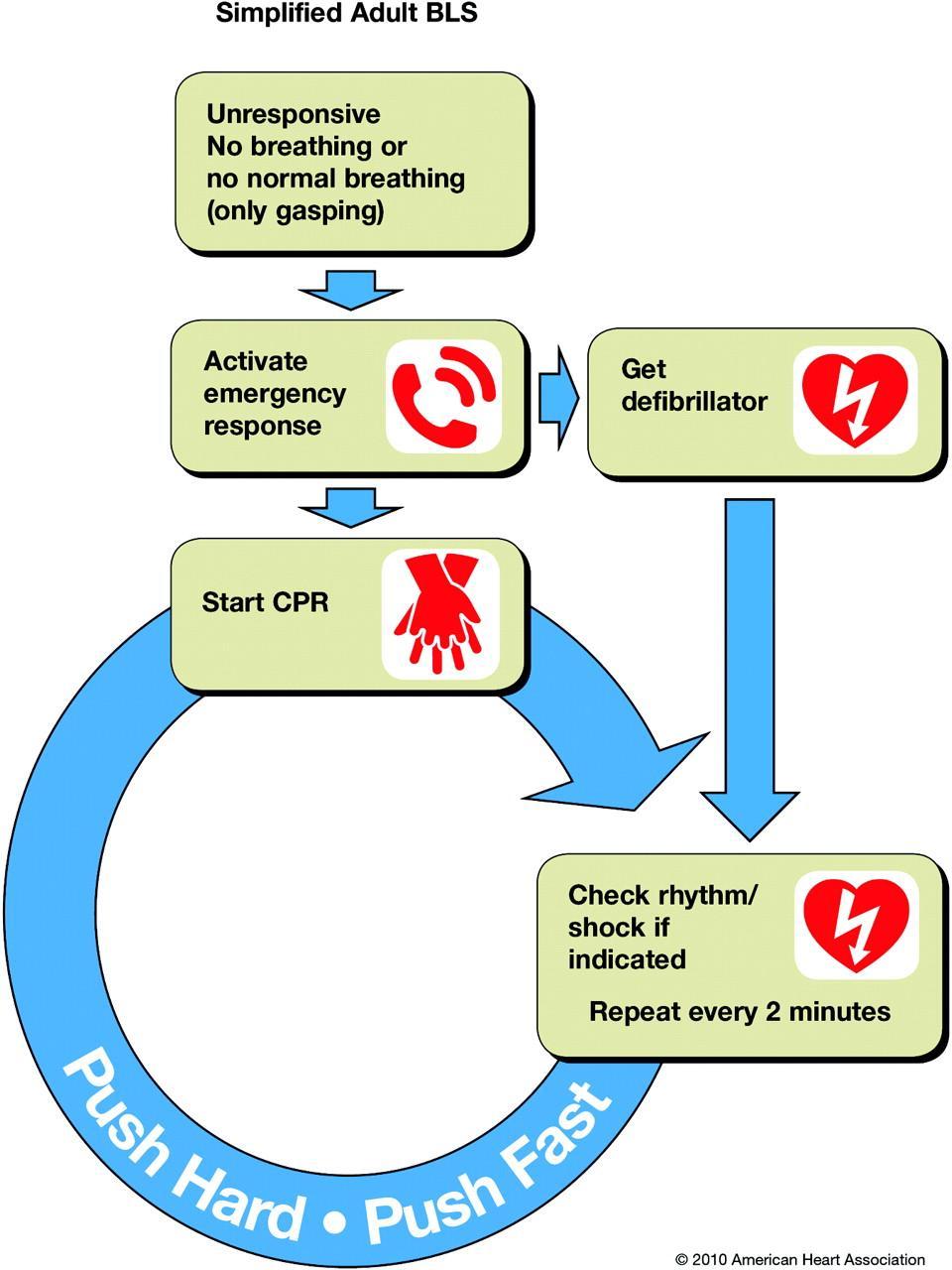
3 4. Note and understand the New 2010 Guidelines for the BLS Survey Introduction Resuscitation of a patient in cardiac or respiratory arrest revolves around the restoration of effective oxygenation, circulation, and ventilation. The initial goal should be the return of spontaneous circulation (ROSC) and the final circulation with intact neurological function. The systematic approach to the assessment of patients in cardiac or respiratory arrest uses two algorithms, the BLS Survey (using steps 1, 2, 3, and 4) and the ACLS Survey (steps A, B, C, and D). The BLS and ACLS Surveys If the patient APPEARS TO BE UNCONSCIOUS - Start with BLS Survey to gain initial assessment of patient condition - Use ACLS survey for more advanced patient management If the patient APPEARS TO BE CONSCIOUS go straight to ACLS survey for initial assessment. BLS Primary Survey - systematic approach to basic life support - aims to support effective ventilation, oxygenation, and circulation until ROSC or until ACLS interventions may be initiated - emphasizes early CPR and early defibrillation - increases patient s chance of survival and retention of intact neurological function - does not include advanced interventions, administration of drugs, or advanced airway techniques - before initiating BLS Primary Survey, ensure safe environment around patient - Assess first, then take appropriate action Assessment Action 1 Check responsiveness Ask Are you alright? Check breathing (absent/ abnormal/ gasping) Scan chest for movement for 5-10 seconds 2 Activate Emergency Response System Activate the emergency response system and get Get AED an AED OR send a bystander to do so 3 Circulation Check carotid pulse for >5 seconds IF NO PULSE detected within 10 seconds: - Perform CPR (30:2) beginning with CHEST COMPRESSIONS
4 - Compressions at the center of the chest (>100 compressions per minute; >2 inches) - Allow for complete chest recoil between each compression - Minimize interruptions in chest compressions (<10 seconds) - Switch CPR providers every 2 minutes to minimize fatigue - Avoid over ventilation IF PULSE detected start rescue breathing, once every 5-6 seconds or breaths per minute Check pulse every 2 minutes 4 Defibrillation IF NO PULSE check for shockable rhythm with manual defibrillator or AED Connect AED to patient; if shockable rhythm is present, administer shocks Resume CPR immediately after each shock BEGINNING WITH CHEST COMPRESSIONS ACLS Survey -start here if patient appears to be CONSCIOUS REMEMBER: assess first and then take appropriate action Assessment Airway -is it patent? -is an advanced airway indicated? -is the advanced airway placement confirmed? -secure and continuously confirm airway placement Action Ensure airway patency in unconscious patients with the following: -head tilt-chin lift -oropharyngeal airway (OPA) -nasopharyngeal airway (NPA) Use an advanced airway if necessary (LMA, combitude, endotracheal tube) Weigh benefits of using advanced airway vs risks of interrupting chest compressions. If bag-mask ventilation is adequate, consider delaying use of advanced airway until patient fails to respond to CPR/ defibrillation or ROSC is achieved. If using advanced airway: -time insertion of airway; minimize interruptions of compressions and ventilation
5 Breathing -is there adequate oxygenation and ventilation? -monitor quantitative waveform capnography and oxyhemoglobin saturation Circulation -Rhythm? -I V placement? -IV fluids? -Medications? Differential diagnosis -cause of the symptoms/ arrest? -reversible cause you can treat? -ensure proper placement of airway thru -physical examination -quantitative wave form capnography -generally: use class I for ET tube -reasonable for supraglottic airways -secure device -confirm placement with quantitative waveform capnography Use supplemental oxygen if needed -Use pure O 2 for patients with cardiac arrest -Titrate O 2 administration to achieve O 2 saturation of >94% (monitor with pulse oximetry) Monitor ventilation and oxygenation with clinical criteria (chest rise, cyanosis) -achieve IV or IO access -attach ECG leads -monitor patient for cardiac arrhythmia -give IV fluids if needed Diagnose and treat all reversible problems

6
7 Case 1: Respiratory Arrest (with pulse) Learning Objectives: 1. understand use of BLS and ACLS Survey in respiratory arrest 2. understand appropriate use of O 2 in management of patient with respiratory arrest 3. understand when to use the following airway devices: a. oropharyngeal airway (OPA) b. nasopharyngeal airway (NPA) c. bag-mask ventilation d. advanced airway Introduction -patient s breathing is either absent or insufficient for oxygenation and ventilation, but pulse is present -BLS and ACLS Survey are used when there is respiratory arrest even without cardiac abnormality -management is focused on effective airway control BLS and ACLS Surveys for Respiratory Arrest with a Pulse When managing patients in respiratory arrest with a pulse, use both the BLS and ACLS Surveys, the following may also be used: - administer O 2 - open the airway - Administer basic ventilation - Basic airway adjuncts - Suctioning BLS Survey for Respiratory Arrest -since pulse is present, do not defibrillate or initiate chest compressions. -assess with BLS Survey and follow with appropriate action Assess and Reassess Throughout the BLS Survey assess and reassess patient to account for change in patient condition.
8 Initiate chest compressions if pulse is lost Ventilation and Pulse Check Use bag-mask or advanced airway to administer 1 breath every 5-6 seconds (10-12 breaths per minute) Recheck pulse every 2 minutes for 5-10 seconds Assessment Action 1 Check responsiveness Ask Are you alright? Check breathing (absent/ abnormal/ gasping) Scan chest for movement for 5-10 seconds 2 Activate Emergency Response System Activate the emergency response system and get Get AED an AED OR send a bystander to do so 3 Circulation Check carotid pulse for >5 seconds IF NO PULSE detected within 10 seconds: - Perform CPR (30:2) beginning with CHEST COMPRESSIONS - Compressions at the center of the chest (>100 compressions per minute; >2 inches) - Allow for complete chest recoil between each compression - Minimize interruptions in chest compressions (<10 seconds) - Switch CPR providers every 2 minutes to minimize fatigue - Avoid over ventilation IF PULSE detected start rescue breathing, once every 5-6 seconds or breaths per minute Check pulse every 2 minutes 4 Defibrillation IF NO PULSE check for shockable rhythm with manual defibrillator or AED Connect AED to patient; if shockable rhythm is present, administer shocks Resume CPR immediately after each shock BEGINNING WITH CHEST COMPRESSIONS If patient does not respond to measures done in Primary Survey, proceed with ACLS Survey. If bag-mask ventilation provides sufficient oxygenation and ventilation, it may not be necessary to use advanced airway during ACLS Survey. Use assessment stages of the ACLS Survey to determine this.
9 ACLS Survey for Respiratory Arrest Assessment Airway -is it patent? -is an advanced airway indicated? -is the advanced airway placement confirmed? -secure and continuously confirm airway placement Breathing -is there adequate oxygenation and ventilation? -monitor quantitative waveform capnography and oxyhemoglobin saturation Circulation -Chest compressions effective? Action Ensure airway patency in unconscious patients with the following: -head tilt-chin lift -oropharyngeal airway (OPA) -nasopharyngeal airway (NPA) Use an advanced airway if necessary (LMA, combitude, endotracheal tube) Weigh benefits of using advanced airway vs risks of interrupting chest compressions. If bag-mask ventilation is adequate, consider delaying use of advanced airway until patient fails to respond to CPR/ defibrillation or ROSC is achieved. If using advanced airway: -time insertion of airway; minimize interruptions of compressions and ventilation -ensure proper placement of airway thru -physical examination -quantitative wave form capnography -generally: use class I for ET tube -reasonable for supraglottic airways -secure device -confirm placement with quantitative waveform capnography Use supplemental oxygen if needed -Use pure O 2 for patients with cardiac arrest -Titrate O 2 administration to achieve O 2 saturation of >94% (monitor with pulse oximetry) Monitor ventilation and oxygenation with clinical criteria (chest rise, cyanosis), quantitative waveform capnography, oxygen saturation Improve CPR IF: quantitative waveform capnography (if PETCO 2 is <10mm HG, improve CPR) -intra-arterial pressure (if diastolic pressure <20mm HG)
10 -Cardiac rhythm? -Defibrillation/ Cardioversion indicated? -IV/IO access? -ROSC? -Patient unstable with pulse? -any medications needed for rhythm/ blood pressure? -fluid resuscitation indicated? Differential diagnosis -cause of the symptoms/ arrest? -reversible cause you can treat? -attach monitor/defibrillator for arrhythmias/ cardiac arrest rhythms (VF, pulseless VT, asystole, PEA) -provide defibrillation/ cardioversion -obtain IV/IO access -administer drugs as needed -give IV fluids if needed Diagnose and treat all reversible problems Managing Respiratory Arrest ACLS and BLS interventions in managing respiratory arrest may include: - O 2 supplementation - Opening of the airway - Basic ventilation - Use of OPA and NPA basic airway adjuncts - Suctioning - Ventilation with advanced airways Ventilation (if patient is in respiratory arrest but pulse is maintained) - Supply every 5-6 seconds (10-12 breaths per minute) - Each should last >1 second and visibly raise patient s chest - Avoid excessive ventilation (both quantity and volume) NEW for 2010 Continuous waveform capnography is recommended in addition to clinical assessment to verify placement and monitor ET tube Supplementary O 2 - may be given to patients in respiratory distress or those with acute cardiac symptoms. - Monitor O 2 saturation (>94%), use supplementary O 2 Opening the Airway Manual Maneuvers

11 - Tongue may obstruct airway if patient is unconscious - Snoring respirations in breathing patients may indicate an obstructed airway - Airway obstruction by tongue may be resolved by repositioning head and jaw - Maintenance of an open airway is the priority in CPR, BLS, and ACLS - If patient is unconscious without gag reflex, insert OPA or NPA Head tilt-chin life maneuver (Do not perform if neck trauma is suspected) 1. Place patient in supine position 2. With one hand on patient s forehead, apply pressure with your palm to tilt patient s head 3. Place fingertips under the bony part of patient s chin. Lift and pull patient s jaw forward without applying pressure to soft tissue which may cause obstruction of airway 4. Open patient s mouth with thumb of hand used to lift chin by pulling downward on lower lip Jaw-thrust technique - Use when neck trauma is suspected - Opens airway without head extension - No longer taught to lay rescuers 1. Place patient in supine position. Use log-roll technique to avoid flexing of spinal column 2. Rest elbows on the same surface patient is lying on 3. Stabilize patient's head in neutral position. Gently grasp the angle of the mandible with two or three fingers 4. Gently lift jaw upward with three middle fingers; use thumbs to open jaw Basic Ventilation Skills 1. Bag Mask Ventilation o Device consists of ventilation bag attached to a facemask o Most common means of positive-pressure ventilation 2. Oropharyngeal Airway (OPA)


12 o Use on patients at risk for airway occlusion by the tongue o Use on unconscious patients when other airway opening techniques fail o J shaped device that fits over the tongue o Prevents tongue and soft palate from making contact with posterior wall of oropharynx o Should NOT be used if patient is conscious or semiconscious to prevent gagging and or vomiting - Use of OPA that is too big may obstruct pharynx and/or cause trauma to laryngeal structures - An OPA that is too small or inserted improperly may obstruct airway by pushing the base of the tongue posteriorly 3. OPA Insertion a. Clear mouth and pharynx of secretions, blood, vomit, etc with rigid pharyngeal suction tip b. Select appropriate OPA size (tip of OPA should be at the corner of the patient s mouth, phalange at the angle of the mandible) c. Insert OPA should be backwards entering the patient s mouth


13 d. Rotate OPA 180 as it passes through the oral cavity and approaches posterior wall of pharynx *after insertion of OPA, ensure head and jaw positions maintain clear airway. Suction as needed. The Nasopharyngeal Airway (NPA) -used for conscious or semiconscious patients -used when patient has sensitive gag reflex, trismus, massive trauma around the mouth, or wiring of the jaws -alternative to OPA for basic airway management adjunct -uses a soft rubber or plastic uncuffed tube

14 NPA Insertion 1. Select proper NPA size. - Outer circumference of NPA should be slightly smaller than the inner opening of the nares. - Avoid blanching of the nostrils - Patient s smallest finger may be used as a guide for proper NPA size 2. Lubricate airway with anesthetic jelly or other water-soluble lubricant 3. Insert NPA through nostril in posterior direction perpendicular to the plane of the face and thread along the floor of the nasopharynx 4. Monitor and maintain placement frequently. Maintain head tilt and anterior displacement of mandible with chin-lift or jaw-thrust techniques. *maintain airway patency after insertion via suctioning as NPA may become obstructed with blood, gastric contents, or soft tissue. *NPA may potentially irritate mucosal lining of the upper airway by lacerating adenoidal tissue. *NPA may cause laryngospasm and vomiting Suctioning - essential to maintaining patency of patient s airway - removes occlusions to upper airway - may be portable or wall-mounted o Portable suction devices easily transportable but less suction power ( mm HG)
 used on oropharynx or for thick fluids and particulate materials Oropharyngeal Suction 1.")
15 o Wall-mounted suction devices suction power of > mm HG Catheters a. Soft and flexible (image below, on the left) typically used for suctioning of the mouth or nose b. Rigid (on the right) used on oropharynx or for thick fluids and particulate materials Oropharyngeal Suction 1. Insert catheter into oropharynx but no further than the distance between tip of patient s nose and earlobe 2. Alternate suctioning and administration of pure O 2 - suction by covering side opening of catheter and withdrawing catheter in rotating motion - suction for no more than 10 seconds at a time Endotracheal Tube Suctioning 1. Sterilize whenever possible 2. Insert catheter into ET tube. - ensure non-occlusion of side openings of catheter 3. Alternate suctioning and administration of pure O 2 - suction by covering side opening of catheter and withdrawing catheter in rotating motion - suction for no more than 10 seconds at a time 4. Monitor heart rate, pulse, O 2 saturation, and clinical appearance

16 - stop suctioning if bradycardia, O 2 saturation levels drop, or if clinical deterioration occurs - administer high-flow O 2 until condition improves *remove occlusions from airway by instilling 1 2 ml sterile saline solution into patient s airway before suctioning; disperse via positive pressure ventilation Advanced Airway Devices 1. Combitude - dual-lumen tube used as an alternate to endotracheal intubation - has two balloon cuffs - only employ if you are experienced and trained to avoid potentially fatal complications 2. Laryngeal Mask Airway (LMA) - alternative to ET tube and facemask - only employ if you are experienced and trained to avoid potentially fatal complications 3. Endotracheal Tube (ET) - tube placed directly into trachea - only employ if you are experienced and trained to avoid potentially fatal complications a. Assemble necessary equipment b. Apply cricoid pressure (preferably by someone else) c. Intubate d. Inflate tube cuffs e. Attach ventilation bag f. Ensure correct placement by physically checking, listening for breath sounds over both lung fields and use of CO 2 detector
17 g. Secure ET with tape h. Monitor ET for displacement NEW for Cricoid pressure is no longer recommended for routine treatment of cardiac arrest - Cricoid pressure may help prevent aspiration and regurgitation of gastric contents when combined with bag-mask ventilation - Cricoid pressure may impede ventilation and appropriate positioning of advanced airway Precautions for Trauma Patients - Avoid unnecessary spine movement during ventilation especially when trauma is suspected or known - An estimated 2% of patients with blunt trauma examined with spinal imaging techniques also have spinal injuries - This goes up to 6% when combined with facial or head injuries - Patients with head, facial, or multiple traumas should be assumed to have cervical spine injury as well - When ventilating with suspected cervical spine trauma: o Use jaw thrust without head extension when opening airway o If jaw thrust elicits no response, employ head tilt-chin maneuver o Have second team member stabilize patient s head during ventilation. Manual spinal motion restriction is preferred to an immobilization device o Consider use of immobilization device during transport Case 2: Ventricular Fibrillation (VF) with CPR and AED Learning Objectives: 1. Be able to recognize and treat VF or pulseless VT with just AED and CPR 2. Understand BLS Primary Survey Algorithm 3. Learn 1-person CPR and AED operation

18 Introduction VF - Ventricular Fibrillation (VF) involves irregular, uncoordinated contraction of the ventricle cardiac muscles resulting in failure to pump blood to the pulmonary and systemic circuits - VF quickly degrades to asystole and sudden cardiac death - Patient in VF will have no detectable pulse at major pulse points - Treatment of patient with either VF or VT when you only have an AED with a pocket facemask - Treatment is feasible without other responders


19 Monomorphic VT Polymorphic VT Sustained VT
 Scan chest for movement for 5-10 seconds 2 Activate Emergency Response System Activate the emergency response system and get Get AED an AED OR send a")
20 Nonsustained VT BLS Primary Survey for VF or Pulseless VT Assessment Action 1 Check responsiveness Ask Are you alright? Check breathing (absent/ abnormal/ gasping) Scan chest for movement for 5-10 seconds 2 Activate Emergency Response System Activate the emergency response system and get Get AED an AED OR send a bystander to do so 3 Circulation Check carotid pulse for >5 seconds IF NO PULSE detected within 10 seconds: - Perform CPR (30:2) beginning with CHEST COMPRESSIONS - Compressions at the center of the chest (>100 compressions per minute; >2 inches) - Allow for complete chest recoil between each compression - Minimize interruptions in chest compressions (<10 seconds) - Switch CPR providers every 2 minutes to minimize fatigue - Avoid over ventilation IF PULSE detected start rescue breathing, once every 5-6 seconds or breaths per minute Check pulse every 2 minutes 4 Defibrillation IF NO PULSE check for shockable rhythm with manual defibrillator or AED Connect AED to patient; if shockable rhythm is present, administer shocks Resume CPR immediately after each shock BEGINNING WITH CHEST COMPRESSIONS

21
22
23 Defibrillation - Definitive treatment for VF and pulseless VT - Does not restart the heart - Stops arrhythmia, allowing heart to resume normal rhythm - Process of shocking the heart with an electrical impulse which causes a brief stop in cardiac activity - Normal sinus rhythm resumes if heart is still viable - Follow defibrillation with CPR (even when it is successful) Importance of Early Defibrillation during VF - Early defibrillation is crucial for survival from sudden cardiac arrest - VF and pulseless VT are the most common initial rhythms during this time - Pulseless VT quickly degrades to VF and then asystole and cardiac death if not treated with defibrillation - CPR alone, although allows for some blood flow to the brain and heart, does not help it resume normal rhythm and blood flow to systemic and pulmonary circuits - Survival rate decreases 7-10%/min between collapse and defibrillation - Survival rate decreases 3-4% /min when there is immediate CPR - CPR between defibrillations may double or triple chances of survival AED Operation 1. Turn on AED 2. Attach electrode pads to patient s chest a. Choose correct pads (children pads if patient is <8 years; adult if 8 or older) b. Peel backing off of each pad c. Ensure patient s chest is dry d. Attach electrodes to patient s chest i. Anterolateral Placement (standard) 1. One pad on upper right side of chest, directly below the collarbone and right of the breastbone 2. The other lateral to patient s left nipple with top margin a few inches below left armpit 3. Attach AED cables to AED if they are not already preconnected ii. Anteroposterior iii. Anterior-left infrascapular iv. Anterior-right infrascapular
 c. Visually double-check d. Press shock button e.")
24 3. Analyze cardiac rhythm a. Always clear patient first b. Some AEDs automatically do analysis; some require the pressing of a button c. AED will determine if shock is required 4. Clear the patient if AED advises electric shock a. Ensure no one is touching patient b. Loudly announce a clear the patient message. (Clear!) c. Visually double-check d. Press shock button e. Wait for muscle contraction to end 5. Resume CPR STARTING WITH CHEST COMPRESSIONS (30:2) a. Do not perform rhythm or pulse check b. Repeat 3 and 4 after 2 minutes of CPR AED Use for Special Situations Hairy Chested Patient Water - Excessive chest hair may impede connection between electrodes and skin resulting in improper/ lack of readings 1. If AED says Check Electrode error, firmly depress each electrode again 2. If AED still displays error message, remove each pad which will also pull out hair 3. Shave off any remaining hair that may continue to interfere with AED 4. Replace pads on electrodes - Presence of water may diffuse effectivity of AED shock - if patient is immersed in water, remove patient from water - if chest is wet, wipe it dry Implanted Pacemaker
25 - check patient for implanted pacemaker or defibrillator - patient will have a well-defined rectangular lump with a scar on either the chest or the abdomen - use of AED when patient already has a pacemaker is not dangerous but putting the pads over the pacemaker may prevent administration of adequate shock - place electrode >1 inch away from either side of the pacemaker Transdermal Medication Patch - transdermal patch may block shock - remove patch first and wipe off area - use AED as normal Case 3: VF/ Pulseless VT Introduction - Treatment of VF/ pulseless VT where cardiac event is witnessed and either resistant to initial schock or recurrent - Requires manual defibrillator instead of AED - Requires teamwork - Combines CPR, bag-mask ventilation, and defibrillation - Team leader must o Conduct ACLS Secondary Survey o Perform rhythm analysis and recognition o Determine if rhythm is shockable or not o Administer shocks o Administer necessary drugs o Discuss IV/IO access and use of advanced airway - Case to be used for the following rhythms: o VF


26 o VT o o - Drugs o o o ECG artifact resembling VF New left bundle branch block Vasopressor drugs Epinephrine vasopressin Anti-arrythmic drugs Amiodarone Lidocaine Magnesium sulfate For treatment of hypotension Epinephrine Norepinephrine Dopamine ACLS Adult Cardiac Arrest Algorithm - Used when adult patient is pulseless and fails to respond to BLS primary survey - Treats pulseless patients in VF/VT, asystole, and PEA - Algorithm has two pathways
27 o o Left side (for VF/VT patients or patients who are shockable) Right side (for asystole/ PEA patients; nonshockable) NEW for AHA Adult Cardiac Arrest Algorithm has been changed to Cardiac Arrest Algorithm which highlights the importance of high-quality CPR with minimal interruptions and beginning with chest compressions
28
29 VF/ Pulseless VT (Left Side of the ACLS Adult Cardiac Arrest Algorithm) - VF and pulseless VT are to be treated in the same manner - Conduct BLS Survey FIRST (activate emergency response system as well) - Ensure safe environment around patient before starting - Continue CPR - Initial shock should be done via manual defibrillator - REMEMBER: chest compressions should only be interrupted for ventilation without advanced airway, rhythm check, and shock transmission. This is vital for resuscitation. Delay checking pulse until organized rhythm is re-established - Use manual defibrillator provided it is available and team leader is comfortable interpreting rhythm - Continue CPR (high quality chest compressions) while defibrillator is charging; the shorter the interval between compressions and shock, the more effective the shock becomes and this also improves patient outcome Box 3 Deliver Initial shock - Be sure patient is clear before administering shock - Energy dosage depends on type of device a. Monophasic model: use 360 J b. Biphasic model a. Truncated exponential waveform use initial charge between 150 and 200 J b. Rectilinear waveform initial charge of 120 J c. If range of device is unknown, use initial charge of 200 J - Though VF may be terminated by shock, it may return; re-administer shock at recent successful charge level - Always follow shock with quality CPR (30:2) - Conductive materials (electrode paste, gel pads, self-adhesive pads) decrease electrical resistance towards patient. Research findings do not show that one is more effective than another however self-adhesive pads may reduce arcing, allow for faster shock delivery, and facilitate continued rhythm monitoring Box 4 Resume CPR - Remember to always start with compressions - Delay rhythm or pulse check - Have another team member get IV/ IO access without interrupting CPR Boxes 5 and 12 Check Rhythm - Perform rhythm check after 2 minutes or 5 cycles of CPR (30:2)
30 - Check pulse if organized rhythm is detected - Immediately resume CPR if no pulse is detected or if there is doubt on pulse o If rhythm is unshockable with no pulse, proceed to box 9 on the RIGHT SIDE of the ACLS Adult Cardiac Arrest Algorithm o If rhythm is shockable, resume CPR while charging defibrillator and proceed to box 3 o Start post-cardiac arrest care if rhythm is organized with detectable pulse Box 6 Deliver 1 shock and Vasopressors - Clear patient before administering shock - Resume CPR immediately after shocking patient - Administer vasopressor (epinephrine or vasopressin) during CPR, before or after the shock - Dosages: o 1 mg IV/IO epinephrine every 3-5 minutes o 40 U IV/IO vasopressin Use vasopressin ONLY ONCE: as substitution for the first or second dose of epinephrine - Prepare drugs early to save time Box 7 Check Rhythm - Perform rhythm check after 2 minutes or 5 cycles of CPR (30:2) - If organized rhythm is found, check pulse during rhythm analysis - If no pulse is found or there is doubt, resume CPR - If rhythm is UNSHOCKABLE WITH NO PULSE, go to BOX 10 on the RIGHT SIDE of ACLS Cardiac Arrest Algorithm - If rhythm is SHOCKABLE, resume CPR while charging defibrillator Box 8 Deliver 1 Shock and Administer Anti-arrhythmics - Clear patient before administering shock - Resume CPR immediately after shocking patient - Administer anti-arrhythmic (amiodarone, lidocaine, or magnesium sulfate) during CPR, before or after the shock - Dosages: o Amiodarone: 300 mg IV/IO once followed by 150 mg IV/IO if needed o Lidocaine: mg/kg IV/IO once followed by mg/kg IV/IO every 5-10 minutes *observe maximum dosage of 3 mg/kg o Magnesium sulfate: 1-2 g IV/IO diluted in 10 ml D5W administered over 5-20 minutes; used for torsades de pointes - Prepare drugs early to save time
31 NEW for Cardiac Arrest Treatment Sequence has been replaced with the 2010 Cardiac Arrest Circular Algorithm - This emphasizes importance of high-quality CPR with minimal interruptions Treatment of VF/VT Patients with Hypothermia - hypothermic patient (temperature <30 C/ 86 F) may not respond to defibrillation - if this occurs, continue normally with BLS algorithm while raising core body temperature above hypothermic levels - delay administration of drugs to severely hypothermic patients to avoid buildup of toxic levels of medication caused by decreased metabolism. Focus intervention on raising core temperature - increase time interval between doses of medicine for moderately hypothermic patients (30-34 C) - there is no evidence to support giving antiarrhythmic drugs to hypothermic patients; consider instead vasopressor (ACLS algorithm) while rewarming the patient
32 Drug Administration during Cardiac Arrest do not allow drug administration to disrupt CPR and defibrillation 1. Intravenous (IV) a. Central IV Line i. Uses large vein in chest or abdomen ii. Usually superior vena cava, inferior vena cava, or right atrium of heart iii. Allows for immediate drug delivery iv. Insertion and access to central IV line may interrupt CPR b. Peripheral IV line i. Any vein not in chest or abdomen ii. Preferred method (minimizes interruption to CPR) iii. Drugs administered in this manner take approx. 1-2 minutes to get to central circulatory system iv. Steps 1. Administer via bolus injection (unless otherwise instructed) 2. Follow with 20 ml bolus of IV fluid 3. Elevate extremity above heart level for seconds to hasten drug delivery 2. Intraosseous (IO) a. Injection of medication directly into bone marrow b. Can be used on patients of all ages (most common with pediatrics) c. Can be established in seconds d. If drug can be IV then it can be administered IO e. IO preferred over ET 3. Endotracheal (ET) a. Injection of medication into trachea b. ONLY USE when IV/IO cannot be established c. CONSIDERATIONS i. Drugs are absorbed poorly ii. Optional drug dosing is unknown iii. Dosages must be times IV/IO dosage iv. Use for epinephrine, vasopressin, atropine, lidocaine, and naloxone v. Must dilute drug with 5-10 ml H 2 O/ saline Vasopressors - have been shown to increase probability of initial resuscitation with ROSC - have NOT been shown to increase survival rate leading to discharge - increase cardiac output and blood pressure - epinephrine
33 o 1 mg IV/IO every 3-5 minutes o if peripheral IV is used, remember to flush and elevate o high/ increasing dosages are not shown to improve ROSC and not recommended - vasopressin o 40 U in lieu of 1 st or 2 nd dose of epinephrine o increases arterial blood pressure o if continued use is indicated, resume epi 3-5 minutes after vasopressin - if ET administration is unavoidable: dilute mg epi in 5-10 ml sterile H 2 O/ saline and inject into ET tube. Antiarrhythmic Agents - have not been shown to increase survival to hospital discharge - amiodarone o has been shown to increase short-term survival o use if VF/pulseless VT is nonresponsive to shock, CPR, and vasopressor o first dose: 300 mg IV/IO push o consider second dose of 150 mg after 3-5 minutes if still unresponsive - lidocaine o consider if amiodarone is unavailable o has not been shown to produce short or long term effectiveness in cardiac arrest o first dose: mg/kg IV/IO o repeat every 5-10 minutes (if needed) at mg/kg IV/IO maintaining maximum doseage of 3 mg/kg o if ET administration is unavoidable: use 2-4 mg/kg - magnesium sulfate o may prevent/ terminate recurrent torsades de pointes in patients with prolonged QT interval with normal sinus rhythm, IF delivered IV o dose: dilute 1-2 g in 10 ml D5W IV/IO every 5-10 minutes o helps counter low serum magnesium levels (in alcoholism, malnutrition, etc) Immediate Post-Cardiac Arrest Care - high quality Post-cardiac arrest care after ROSC increases patient survival and quality of life - GOALS o Optimizes homodynamic and ventilation status o Initiates therapeutic hypothermia o Immediate coronary reperfusion with PCI o Institutes glycemic control o Anticipates/ provides for neurological care and other interventions - Begin after achieving ROSC

34 - Continue ventilation and oxygenation Box 1 ROSC - Patient has been successfully resuscitated with BLS/ACLS Surveys - Rhythm check (Box 12 of Adult Cardiac Arrest Algorithm) shows organized rhythm with pulse Box 2 Optimize Ventilation and Oxygenation - Is patient is unresponsive/ unconscious, advanced airway device may be required\ - For ET Tube o Employ continuous waveform capnography to confirm and monitor placement of ET tube o Maintain oxyhemoglobin concentration of >94% with lowest inspired O 2 concentration o If you cannot determine O 2 concentration, give patient pure O 2 until able to do so
35 o Avoid hyperventilation breaths / minute Maintain PETCO 2 of mm HG OR PaCO 2 of mm HG Do not tie/ secure advanced airway around patient s neck; this may inhibit blood return from brain - Waveform capnography o Allows for monitoring of CPR quality o Maximizes chest compressions o Allows for detection of ROSC NEW for 2010 Waveform capnography and clinical assessment are recommended to confirm and monitor proper placement of ET tube Box 3 Hypotension Treatment (SBP < 90 mm Hg) - Obtain IV access - Continue use of ECG to monitor patient after ROSC - Drug dosages (refer to diagram) Box 5 Therapeutic hypothermia - Can protect brain and other organs if patient is unresponsive - If ROSC is achieved after VF cadiac arrest out-of-hospital, start cooling process and then transport - Lowering of core body temperature to C for hours via: o Rapid infusion of anice-cold, isotonic, non-glucose containing fluid (30mL/kg) o Surface cooling device o Ice bags - Also consider this if: o In-hospital cardiac arrest with initial rhythm present o Out-of-hospital cardiac arrest with PEA/ asystole initial rhythm - CONSIDERATIONS: o Avoid rewarming patiend for hours after ROSC o This is the only intervention proven to improve recovery of neurological function o Recommended duration: hours o Monitor core body temperature via esophageal thermometer, bladder catheter, or pulmonary artery catheter o Maintenance with PCl is safe
36 Box 6 STEMI / high suspicion of AMI - Obtain 12-lead ECG ASAP after ROSC - Use this to identify STEMI or high suspicion of AMI - Transport patient to hospital that can provide coronary reperfusion (Box 7) if either is detected Box 7 Coronary reperfusion - Aggressive treatment for STEMI or AMI - Done in-hospital via PCl - Notify hospital of this need beforehand Learning objectives Notes Case 4: Pulseless Electrical Activity (PEA) 1. Be able to recognize and treat PEA with the ACLS Pulseless Arrest Algorithm 2. Understand and be able to perform ACLS Pulseless Arrest Algorithm 3. Integration of new 2010 guidelines - PEA is a set of heart rhythms that produce organized cardiac rhythms while the patient is unresponsive without a discernible pulse - There is electrical activity in the heart, but no mechanical activity - Includes the following rhythms o Idioventricular rhythms o Ventricular escape rhythms o Post-defibrillation idioventricular rhythms o Bradyasystolic rhythms o Any pulseless rhythm except VF, VT, and asystole - Formerly referred to as EMD (electromechanical dissociation) - Treat with ACLS Pulseless Arrest Algorithm - REMEMBER: the probability of resuscitation is dependent on uninterrupted high quality CPR - Teamwork is still critical for treatment - Commonly caused by a primary condition (H s and T s) o Hypovolemia decreased blood volume o Hypoxia decreased partial pressure of O 2 in blood o Hydrogenion (acidosis) increase in the concentration of hydrogen ions in blood o Hyper/hypokalemia abnormally high/ low potassium concentration in blood o Hypoglycemia abnormally low blood glucose concentration in blood
37 o Hypothermia core body temperature < 30 C (86 F) o Toxins poisonous substances o Tamponade (cardiac) compression of the heart caused by excess fluids in chest cavity o Tension pneumothorax lung displacement, compression of the heart, and possible lung collapse due to air in pleural cavity aroung the lungs o Thrombosis (coronary/ pulmonary) blockage (partial or full) of a blood vessel caused by a blood clot o Trauma o Hypovolemia and hypoxia are the most common and easiest to treat NEW for 2010 DO NOT administer atropine during cardiac arrest

38
39 Treatment of PEA with ACLS Adult Cardiac Arrest Algorithm - Performed BLS survey - Activated emergency response system as well - Ensured safe environment around patient before starting - Step 1 of ACLS Adult Cardiac Arrest Algorithm has been done - Monitor/ defibrillator displayed PEA rhythm - Continued high-quality CPR (30:2) ensuring chest compressions are not interrupted for >10 seconds (box 10 of ACLS Adult Cardiac Arrest Algorithm) - If shockable rhythm is found, resume CPR while charging defibrillator and go to Box 3 Box 10 Administration of Vasopressors - Administer vasopressors while still providing high quality CPR - Prioritize IV/IO access over advanced airway UNLESS: o Ventilation via bag mask device is ineffective OR o Patient is hypoxic - 1 mg IV/IO epinephrine every 3-5 minutes - 40 U IV/IO vasopressin o Use vasopressin ONLY ONCE: as substitution for the first or second dose of epinephrine - Consider advanced airway and capnography if necessary Rhythm check - After giving drugs, administer CPR for 2 minutes / 5 cycles (30:2) - Perform rhythm check o If organized rhythm is found, follow with pulse check o If NOT, resume CPR o If in doubt, resume CPR - If rhythm is NONSCHOCKABLE: continue on RIGHT side of ACLS Cardiac Arrest Algorithm - If rhythm is SCHOCKABLE: go over to LEFT side of ACLS Cardiac Arrest Algorithm Box 11 / 12 Nonshockable rhythm - If no electrical activity (asystolic), return to Box 10 or 11 and continue on asystolic pathway - If electrical activity found, check pulse o If no pulse is found, continue CPR (30:2) for 2 minutes and return to box 10 o If pulse is found with organized rhythm, move on to post-cardiac arrest care Box 11 Shockable rhythm
40 - Resume CPR while charging defibrillator - Return to step 5 or 7 (shock) NEW for Cardiac Arrest Treatment Sequence has been replaced with the 2010 Cardiac Arrest Circular Algorithm - This emphasizes importance of high-quality CPR with minimal interruptions Learning objectives Case 5: Asystole 1. Be able to recognize and treat asystole with ACLS Adult Cardiac Arrest Algorithm 2. Familiarization with 2010 Cardiac Arrest Circular Algorithm and it s use
41 Notes 3. Know when to cease resuscitation 4. Integration of new 2010 guidelines - No electrical activity on ECG + no pulse = asystole - Ensure ECG flat line is due to asystole and not user error before starting treatment via checking o Leads are properly connected o Power / unit is on o Signal gain/ amplitude/ strength is not turned down o That flat line is not another pattern - Uses the right side of ACLS Cardiac Arrest Algorithm because it is a nonshockable rhythm - Use ACLS Pulseless Arrest Algorithm (right side/ nonshockable) - Consider stopping resuscitation efforts if: o There are clear signs of irreversible death (rigor mortis, etc) o DNAR Status (do not attempt resuscitation status) as indicated by bracelet, anklet, or written document o Living will/ family refuse o Situation is dangerous for rescuers NEW for 2010 DO NOT administer atropine during cardiac arrest Managing Asystole with ACLS Adult Cardiac Arrest Algorithm - Step 1 of ACLS Adult Cardiac Arrest Algorithm has been performed - Monitor displays asystole - Continue uninterrupted CPR (30:2) - Obtain IV/IO access (highest priority) - Team leader coordinates ACLS Adult Cardiac Arrest Algorithm starting at Step 10 Looking for underlying causes of asystole - If (possible) underlying cause is resolved, chances for successful resuscitation increase - Check H s and T s o Hypovolemia decreased blood volume o Hypoxia decreased partial pressure of O 2 in blood o Hydrogenion (acidosis) increase in the concentration of hydrogen ions in blood o Hyper/hypokalemia abnormally high/ low potassium concentration in blood o Hypoglycemia abnormally low blood glucose concentration in blood o Hypothermia core body temperature < 30 C (86 F) o Toxins poisonous substances
42 o o o o Tamponade (cardiac) compression of the heart caused by excess fluids in chest cavity Tension pneumothorax lung displacement, compression of the heart, and possible lung collapse due to air in pleural cavity aroung the lungs Thrombosis (coronary/ pulmonary) blockage (partial or full) of a blood vessel caused by a blood clot Trauma Box 10 Administration of Vasopressors - Administer vasopressors while still providing high quality CPR - 1 mg IV/IO epinephrine every 3-5 minutes - 40 U IV/IO vasopressin o Use vasopressin ONLY ONCE: as substitution for the first or second dose of epinephrine Rhythm check - Administer CPR for 2 minutes / 5 cycles (30:2) - Perform rhythm check o If organized rhythm is found, follow with pulse check o If NOT, resume CPR o If in doubt, resume CPR - If rhythm is NONSCHOCKABLE: continue on RIGHT side of ACLS Cardiac Arrest Algorithm - If rhythm is SCHOCKABLE: go over to LEFT side of ACLS Cardiac Arrest Algorithm Box 11 / 12 Nonshockable rhythm - If no electrical activity (asystolic), return to Box 10 or 11 and continue on asystolic pathway - If electrical activity found, check pulse o If no pulse is found, continue CPR (30:2) for 2 minutes and return to box 10 o If pulse is found with organized rhythm, move on to post-cardiac arrest care Box 11 Shockable rhythm - Resume CPR while charging defibrillator - Return to step 5 or 7 (shock) *do not stop chest compressions for >10 seconds *transcutaneous pacing is not recommended for asystole *shock treatment is not recommended for asystole *if unsure if rhythm is asystole or fine VF, attempt initial defibrillation

43 *if fine VF is suspected, 5 cycles of CPR may help before defibrillation NEW for Cardiac Arrest Treatment Sequence has been replaced with the 2010 Cardiac Arrest Circular Algorithm - This emphasizes importance of high-quality CPR with minimal interruptions When to Terminate Resuscitation Attempts - Attending physician s responsibility in the hospital - If no underlying cause can be determined (H s and T s) - If patient does not respond to BLS or ACLS Survey - Other factors to consider o How long did it take to start CPR? o How long did it take before patient was defibrillated? o Pre-existing disease or condition? o What condition was the patient in before arrest? o What was the preliminary arrest rhythm?
44 o Did the patient respond to resuscitation attempts? - Patient has perfusing rhythm with organized, regular rhythm is noted with detectable blood pressure - Transfer to the care of a senior medical professional is available - Irreversible death is obvious - Situation becomes dangerous or rescuers are exhausted - DNAR exists - Online notification to do so is given by senior medical professtional Bear in mind that as time to resuscitate increases, probability of the patient surviving with minimal neurological problems decreases Consider extenuating circumstances like drug overdose or hypothermia: these increase likelihood that extended resuscitation may succeed Be aware of ACLS protocol of local EMS or hospital Extended asystole - Agonal rhythm is common in irreversibly dead patients - Asystole is common as o Final rhythm for patient who started arrest in VF/ VT o Initial rhythm for patient in prolonged arrest - Usually causes extensive, irreversible damage to patient due to lack of blood flow to organs - Results in severe cardiac damage due to myocardial ischemia - 15 minutes usually ends in patient becoming brain dead - Successful recovery is more likely in-hospital Learning Objectives Notes Case 6: Acute Coronary Syndrome 1. Understand the use of AHA Acute Coronary Syndromes Algorithm in treating ischemia, UA, and STEMI 2. Familiarization with Fibrinoloytic checklist and it s use 3. Integration of new 2010 guidelines
45 - ACS comprises conditions resulting in acute myocardial ischemia - Acute myocardial ischemia is chest pain caused by insufficient blood supply to the heart due to CAD / CHD - Composed of 3 categories of disease, grouped by risk and ECG pattern o STEMI - ST segment elevation myocardial infarction o High-risk unstable angina (UA) or Non-ST segment elevation myocardial infarction (NSTEMI) o Intermediate/ Low-Risk Unstable Angina - Often first sign of CAD - By 60 years, 1 in 5 men and 1 in 17 women with have some form of CAD - Estimated 13 M emergency department ACS cases per year - Treatments used in the ACLS algorithm for ACS o Oxygen o Aspirin o Nitroglycerin o Morphine o Fibrinolytic (thrombolytic) therapy o Heparin - Primary goal in treating ACS is quick identification and treatment - STEMI carries the highest risk; acute treatment includes early reperfusion therapy - Early reperfusion therapy re-opening an occluded coronary artery using one of the following: o thrombolytic (fibrinolytic) therapy must be done within 12 hours of onset of symptoms most successful during first 2 hours risks outweigh benefit after 12 hours o bypass surgery o percutaneous coronary intervention (PCI) coronary angiogram to find occluded vessel balloon angioplasty stent placement (optional) - Conditions leading to ACS o Unstable angina (features) Sever pain new in onset Occurs with crescendo pattern (more severe, prolonged, or frequent than before) Occurs at rest / with minimal exertion; lasting >20 minutes o Microemboli composed of cholesterol, calcium, and platelets from nearby atherosclerotic plaques
46 o o dislodgment may occlude coronary microvasculature causing small elevations in cardiac troponin occlusive thrombus myocardial ischemia results when myocardial demand exceeds supply, possibly caused by large enough thrombus unstable plaque NEW for 2010 AHA Acute Coronary Syndromes Algorithm allows for maximum benefit for myocardial salvage New algorithm allows early management of UA and STEMI Box 1 Identifying Signs and Symptoms of Ischemia - uncomfortable feeling of pressure, tightening, fullness, squeezing, or pain in the middle of the chest lasting several minutes - discomfort originating in chest, spreading to shoulders, neck, arm/s, or jaw - discomfort originating in chest, spreading to back or between shoulder blades - discomfort in chest with lightheadedness, fainting, nausea, or sweating - unusual episodes of shortness of breath, with/out chest pain - after identifying ischemia, go to Box 2 and prepare patient for transport to an ED Box 2 EMS Assessment and care and hospital preparation - possible medications (MONA) o Morphine - use when patient is unresponsive to nitroglycerin o Oxygen proven to reduce ST segment elevation during anterior infarction o Nitroglycerin 3 sublingual tablets 3-5 minutes apart Only allow if patient is hemodynamically stable Contraindicated if SBP <90 mm Hg or 30 mm Hg below patient s baseline Contraindicated for patients with o Inferior MI and RV infarction Hypotension, bradycardia, tachycardia Viagra or phosphodiesterase inhibitor aspirin 325 mg (ASA), chewed Relative contraindications: aspirin/ salicylate allergy, history of GI bleed, kidney disease, hyperuricemia, and gout
47 ASA suppository may be given to patients with nausea, vomiting, peptic ulcer, or other upper GI disorder - Patient Assessment (under 10 minutes) o Attach cardiorespiratory monitor o Obtain IV access o Review 12-lead ECG o Complete fibrinolytic checklist and check for contraindications o Get blood sample for initial cardiac marker, electrolyte, and coagulation levels (should not interfere with reperfusion therapy unless clinically necessary) o History and physical exam o Obtain/ review chest x-ray within 30 minutes of arrival at ED (should not interfere with reperfusion therapy) ACS Categories 1. STEMI o ST Segment Elevation Myocardial Infarction o ST segment elevation >1 mm (0.1 mv) in 2 or more contiguous leads, 2 or more adjacent limb leads, or new and presumed new left bundle branch block 2. High-risk unstable angina (UA) or Non-ST Segment Elevation Myocardial Infarction (NSTEMI) o ST depression of 0.5 mm (0.005 mv) or greater dynamic T-wave inversion o Associated with chest pain and discomfort o Also includes non-persistent/ transient ST segment elevation (0.5 mm or mv) 3. Intermediate/ Low-Risk Unstable Angina o Includes patients with normal ECGs o ST segment deviation <0.5 mm (0.05 mv) or T-wave inversion of <2 mm (0.02 mv)

48 Boxes 5-8: STEMI - Class of ACS carrying highest risk
 Treatment should commence within 30 minutes of arrival at hospital o PCI used")
49 - Patients usually have complete occlusion of an epicardial coronary artery - EARLY reperfusion therapy is crucial o Fibrinolytic therapy When >1 mm ST segment elevation in 2 contiguous leads on ECG Results in reperfusion 50% of the time Check first for contra/indications Use fibrinolytic checklist (see diagram below) Treatment should commence within 30 minutes of arrival at hospital o PCI used on patients who do not reperfuse after fibrinolytic therapy been found to lead to better patient outcomes
50 Adjunctive Treatment for STEMI - Angiotensin converting enzyme (ACE) inhibitors - Beta blockers - Clopidogrel - Heparin - HMG coenzyme inhibitor therapy - IV nitroglycerin o Indicated for: STEMI complicated by hypertension CTEMI complicated by pulmonary edema Chest discomfort that is unresponsive to spray nitroglycerin, morphine, or SL Learning Objectives Notes Case 7: Bradycardia 1. Be able to treat bradycardia with ACLS Adult Bradycardia (with pulse) Algorithm 2. Be able to recognize bradycardia in paitents 3. Integration of new 2010 guidelines - Bradycardia = unusually slow heart rate (<60 bpm), dangerous to patient s health that may result in o Fainting o Shortness of breath o Cardiac arrest o Death - Relative bradycardia bradycardia with bpm > 60 due to factors increasing patient s heart rate - Management includes: Differentiation of bradycardic symptoms from those that are unrelated Recognition and identification of possible AV block Administration of atropine Initiation of transcutaneous pacing (TCP) Maintenance of heart rate and blood pressure by administration of epinephrine and/ or dopamine when needed


51 Bradycardic ECG Rhythms Sinus bradycardia 1 st degree AV block PR interval >0.2 seconds 2 nd degree AV block Type I (Wenckebach-Mobitz II) PR interval progressively lengthens and drops a beat

 Pulmonary congestion/ edema Ventricular")
52 2 nd degree AV block Type II (Mobitz II) PR interval remains constant and drops a beat 3 rd degree AV block no connection between P waves and QRS complexes Symptoms Chest pain/ discomfort Dizziness Fatigue Presyncope Reduced level of consciousness Shortness of breath syncope Weakness Signs Congestive heart failure Diaphoresis Hypotension Orthostatic hypotension Premature ventricular contractions (PVCs) Pulmonary congestion/ edema Ventricular tachycardia
53 NEW for Initial treatment has become atropine - If patient is unresponsive to atropine, proceed to IV infusion of dopamine or epinephrine - TCP may be effective for patient being prepped for emergent transvenous temporary pacing Box 1: Verify symptomatic bradycardia Box 2: Perform BLS and ACLS Surveys - Administer and monitor O 2 - Check rhythm and blood pressure - Establish IV access Box 3: Check for poor perfusion - Check for signs and symptoms of bradycardia (see table above) - If patient presents adequate perfusion, proceed to Box 4 - If NOT, proceed to Box 5 Box 5: Treatment for Inadequate Perfusion - Administer 0.5 mg IV atropine, repeat if needed (up to 3 mg) - If patient does not respond to atropine try o Transcutaneous pacing (TCP) OR o Dopamine (2-10 mcg/kg /min IV) OR o Epinephrine (2-10 mcg/kg /min IV) - REMEMBER: Patients who present with symptomatic bradycardia and inadequate profusion may degrade to cardiac arrest - Commence TCP IF: o Patient does not respond to atropine o Atropine is predetermined to be ineffective o IV access is delayed o Patient s condition is rapidly degrading - After TCP is initiated o Confirm correct placement and functioning o Check for improved perfusion and condition o Administer analgesics and sedatives as necessary o If TCP does not work, begin infusion of dopamine/ epinephrine and prepare for transvenous pacing - REMEMBER: o Medications for bradycardia may produce confusion and altered state of consciousness o TCP may be painful
 Algorithm Case 8: Unstable")
54 o TCP may not work, especially if there is underlying cause ACLS Adult Bradycardia (with Pulse) Algorithm Case 8: Unstable Tachycardia

55 Learning Objectives Notes 1. Be able to recognize different rhythms classified as unstable tachycardia 2. Familiarization with different heart rhythms 3. Familiarization with ACLS Adult Tachycardia Algorithm 4. Integration of 2010 guidelines - Tachycardia (HR >100bpm) leads to less blood flow to systemic and pulmonary circuits and less O 2 to organs - Increase in O 2 demand and lack of supply may lead to ischemia then myocardial infarction - Both rapid and unstable heart rate Rhythms Classified as Unstable Tachycardia 1. Atrial Fibrillation - caused by trembling atrium muscles 2. Atrial flutter originating in atrium, often leading to atrial fibrillation 3. Reentry supraventricular tachycardia (SVT) any rapid rhythm starting in atrium 4. Monomorphic VT rapid VT with similar peaks on ECG 5. Polymorphic VT rapid VT, irregular ECG 6. Wide-complex tachycardia of an uncertain type Atrial Fibrillation (AFib) - Results from altered rapid firing of impulses in atria - Causes atrial muscles to fibrillate (tremble) - Hinders full atrial contractions, leads to decrease in stroke volume - Decreased cardiac output - Increases risk for stroke since blood is not properly pumped out, potential for clotting - Associated with: o Advanced age o Cardiomyopathy o Congenital heart failure o Congestive heart failure o Diabetes

56 o Electrocution o Excess caffeine o Hypertension o Hyperthyroidism o Hypoglycemia o Hypokalemia o Hypoxia o Ischemic heart disease o Recovery from surgery o Rheumatic heart disease o Stress o Sympathomimetics o Systemic infection - Characteristics o Rate Ventricular: may vary Atrial: bpm o Rhythm irregularly irregular o P waves not identifiable with fibrillatory waves present; wavy, erratic baseline o PR interval not measurable o QRS duration usually <0.1 second - Treatment o Depends on: Duration of rhythm Patient s health Ventricular rate Other factors o May focus on Controlling ventricular rate Reestablishing sinus rhythm if AFib is associated with rapid ventricular rate and patient is symptomatic yet stable o Consider synchronized cardioversion when patient has rapid ventricular rate and other serious symptom ie heart failure, low blood pressure, or shock Atrial Flutter
57 - Ectopic atrial rhythm wherein irritable sites fire at regular, rapid rates - Flutter waves are evident waveforms that look like teeth of a saw - Associated with: o Cardiac surgery o Cardiomyopathy o Chronic lung disease o Complications of myocardial infarction o Digitalism/ quinidine toxicity o Hyperthyroidism o Hypoxia o Ischemic heart disease o Mitral/ tricuspid valve stenosis/ regurgitation o myocarditis o Pericarditis o Pneumonia o Pulmonary embolism - Characteristics o Rate Type I: bpm Type II: bpm o Rhythm Atrial regular Ventricular ir/regular, depends on AV conduction o P waves not identifiable with fibrillatory waves present o PR interval not measurable o QRS duration usually <0.1 second - Treatment o Depends on: Duration of rhythm Patient s health Ventricular rate Other factors o May focus on Controlling ventricular rate Reestablishing sinus rhythm if flutter is associated with rapid ventricular rate and patient is symptomatic yet stable o Consider synchronized cardioversion when patient has rapid ventricular rate and other serious symptom ie heart failure, low blood pressure, or shock - Vagal maneuvers may help identify rhythm and slow down AV conduction to reveal flutter - Considered uncontrolled when ventricular rate >100 bpm
 - Any tachycardic rhythm originating above the ventriculator tissue in the atria or AV node - Atrioventricular Nodal")
58 - Considered controlled when ventricular rate <100 bpm o May result from Normal AV node protecting ventricles from fast atrial impulses Drugs used to control impulse conduction through AV node reducing number of impulses reaching ventricles Supraventricular Tachycardia (SVT) - Any tachycardic rhythm originating above the ventriculator tissue in the atria or AV node - Atrioventricular Nodal Reentrant Tachycardia (AVNRT) o More common type of SVT o results from a reentry circuit that forms the AV node (or next to it) o more common in women (75%) than men o can occur at any age and in patients without heart disease o associated with chronic obstructive pulmonary disease coronary artery disease valvular heart diseases other o may cause myocardial infarction in patients with coronary artery disease or angina o may be triggered by anxiety caffeine hypoxia lack of sleep other medications smoking
 Rhythm - regular P waves Hidden in QRS complex Inverted P wave appears after QRS leads II, III, and VF Occurs when")
59 o o o signs and symptoms chest pain or pressure congestive heart failure dyspnea lightheadedness nausea neck vein pulsations nervousness and anxiety palpitations signs of shock syncope or near-syncope weakness Characteristics Rate bpm (usually bpm) Rhythm - regular P waves Hidden in QRS complex Inverted P wave appears after QRS leads II, III, and VF Occurs when ventricles are stimulated before atria End of QRS complex becomes distorted by P wave if atria are depolarized after ventricles PR interval not measurable QRS duration usually <0.1 second, barring intraventricular conduction delay treatment oxygen vagal maneuvers if this fails, follow with adenosine treatment IV access Follow successful treatment with synchronized cardioversion Wolff-Parkinson White Syndrome (WPW)
60 - Occurs when the bundle of Kent pre-excites the ventricles - WPW Syndrome is type of AVNRT - Occurs in between 0.9 and 3% of the population, more commonly in men - One of the most common causes of tachydysrhythmias in infants and children - Patients are usually asymptomatic but with risk of sudden death Ventricular Tachycardia (VT) - Occurs when 3 or more premature ventricular contractions occur immediately in quick succession at >100 bpm - May last <30 seconds and be unsustained - Usually lasts >30 seconds (sustained) - May occur with or without a pulse - Patient may be stable or unstable - May originate from an ectopic focus in one of the ventricles - Becomes monomorphic VT when QRS complexes are the same size and shape - Becomes polymorphic VT when QRS complexes differ each beat - Associated with o Acid-base imbalance o Acute coronary syndromes o Cardiomyopathy o Cocaine abuse o Digitalis toxicity o Hyperkalemia o Hypokalemia o Mitral valve prolapse o Trauma (invasive heart procedures/ myocardial contusion) o Valvular heart disease - Signs and symptoms o Vary o May occur with or without a pulse
61 o Sustained monomorphic VT may remain stable for extended periods of time o Monomorphic VT may degenerate to polymorphic VT or VFib if ventricular rate is very fast or myocardial ischemia occurs o Syncope or near-syncope may occur with sudden onset of VT - Treatment of monomorphic VT o Treat and search for cause of VT simultaneously o If without pulse, use ACLS Cardiac Arrest Algorithm o If stable but symptomatic, suppress rhythm with IV, O 2, and ventricular arrhythmics o If unstable with sustained heart rate of >150 bpm, treat with IV access, O 2, and sedate patient; follow with synchronized cardioversion - SVT with intraventricular conduction delay is difficult to distinguish from VT - If there is doubt whether a rhythm is VT or SVT, treat it as VT, and use 12-lead ECG to determine later on Monomorphic VT - occurs when QRS complexes on ECG are the same shape and size - common in patients with structural heart disease - scarring may have resulted from an area of slow conduction - occasionally seen in patients with normal hearts - may be triggered by o acid-base imbalance o acute coronary syndroms o cardiomyopathy o cocaine abuse o digitalis toxicity o hyperkalemia o hypokalemia o mitral valve prolapse o trauma o tricyclic antidepressant overdose o valvular heart disease - may degrade to polymorphic VT or VFib - there may be brief lightheadedness followed by near/ syncope when VT occurs abruptly
62 - Characteristics o Rate bpm o Rhythm - regular o P waves usually absent o PR interval not measurable o QRS duration usually <0.1 second, usually difficult to differentiate between QRS and T wave Polymorphic VT - caused by slow heart rate - Associated with electrolyte disturbances or medications prolonging QT interval - QRS complexes go from upright to negative and negative to positive - Symptoms related to decreased cardiac output resulting from increased ventricular rate - Characteristics o Rate bpm (usually bpm) o Rhythm regular or irregular o P waves - none o PR interval not measurable o QRS duration usually <0.12 seconds, with irregular peaks and changing directions of QRS complexes Uncertain Wide-Complex Tachycardia

63 - usually caused by VT or SVT with unusually wide QRS complex - Sustained rate of >100 bpm - QRS complexes last >0.12 seconds - Treatment o Find pulse if patient presents with rapid heart rate o If pulse is found, determine stability of rhythm o Follow ACLS Tachycardia Algorithm o Consider H s and T s as causes of tachycardia with pulse Sinus Tachycardia - Rapid heart rate in response to stimulus - Normal rhythm starting in sinoatrial node but at an increased rate - Other causes include o Anemia
64 o o Dehydration Hypovolemia NEW for Focus on identifying tachycardia as symptom or underlying cause - Algorithm has been simplified and reorganized to facilitate retention - ACLS Tachycardia Algorithm may be used in treatment of stable or unstable tachycardia Box 1 Assess Appropriateness of Algorithm to Clinical Situation Box 3 - Determine tachycardic rhythm first (>150 bpm) - Find pulse - If pulse cannot be found or is lost, follow ACLS Cardiac Arrest Algorithm - Decide whether persistent tachycardia is causing significant signs/ symptoms o YES, then patient is unstable, go to box 4 o NO, patient is stable, go to Box 5 - Signs of unstable tachycardic patient o Acute heart failure o Acutely altered mental state o Hypotension o Ongoing chest pain/ discomfort o Presyncope o Shock o Shortness of breath o Syncope Box 4 Synchronized Cardioversion - Uses electric shocks to reestablish normal rhythm after tachycardia - Establish IV access first - Sedate patient - Consider adenosine if patient is not hypotensive with regular narrow-complex SVT or monomorphic wide-complex tachycardia - Synchronized cardioversion o Shocks are delivered simultaneously with regular intervals (at QRS complex peaks) o Uses the lower energy setting o Benefit: prevents shock from being administered during cardiac repolarization (which may lead to VF) o Employ when:
65 Unstable atrial fibrillation Unstable atrial flutter Unstable regular monomorphic tachycardia with pulse Unstable SVT o Preferred method of treatment with symptomatic reentry SVT or VT with pulses - Unsynchronized cardioversion o Shocks are delivered on demand of responder o Uses high-energy setting o Employ with Patient who may become pulseless Patient with critical deterioration (severe shock/ polymorphic VT) Tachycardic patient without pulse Unsure if patient has monomorphic/ polymorphic VT - Shock dosages o Unstable atrial fibrillation 200 J o Unstable monomorphic VT 100 J o Other unstable SVT, atrial flutter 200 J o Polymorphic VT (irregular and unstable) treat as VF with high-energy shock - Procedure o Sedate patient unless condition is rapidly deteriorating/ unstable o Turn on defibrillator o Attach leads/ electrodes o Press SYNC o Look for indications of sync mode (usually on R wave) o Sync with R wave if necessary o Set energy level o Announce CLEAR the patient o Press charge button o CLEAR patient o Hit SHOCK button o Determine if tachycardia is still present, if yes, increase energy o Press sync button * may not be effective in treating junctionatachycardia or ectopic or multifocal atrial tachycardia Learning Objectives Case 9: Stable Tachycardia

66 1. Be able to recognize stable narrow-complex and wide-complex tachycardia 2. Know when treatment with ACLS Adult Tachycardia Algorithm is appropriate Notes - Caused by cardiac electrical problem - If patient has a pulse, proceed with ACLS Tachycardia Algorithm - If NOT, use ACLS Pulseless Arrest Algorithm - May be evidenced by o Sinus tachycardia Heart rate between bpm (>200 bpm in infants, >160 in children over 5) Caused by increase in O 2 demand Symptoms Palpitations Racing heart Pounding in chest Related causes: Acute myocardial infarction Caffeine Congestive heart failure Dehydration Drugs (amphetamines, cannabis) Exercise Fear/ anxiety Fever Hyperthyroidism Hypovolemia Hypoxia Infection Medications (epinephrine, dopamine, atropine, dobutamine) Nicotine Pain Pulmonary embolism


67 Shock Sympathetic stimulation o Atrial fibrillation o Caused by inappropriate firing in the atria or reentry involving one or more circuits in atria Rapid impulses inhibit atria from contracting properly Results in decreased cardiac output; loss of atrial kick Treatment Atrial flutter Depends on: o Duration of rhythm o Patient s health o Ventricular rate o Other factors May focus on o Controlling ventricular rate o o Reestablishing sinus rhythm if AFib is associated with rapid ventricular rate and patient is symptomatic yet stable Ectopic atrial rhythm wherein irritable sites fire at regular, rapid rates Flutter waves are evident waveforms that look like teeth of a saw May be observed in leads II, III, avf 1, and V 1


68 o Multifocal atrial tachycardia o Aka chaotic atrial tachycardia Exhibits wandering atrial pacemaker rhythm and >100 bpm ventricular rate Wandering rhythm P waves differ, sometimes from beat to beat AVNRT o Caused by the presence of two pathways in AV node conducting impulses and recovering at different rates Monomorphic VT Evidenced by QRS complexes of the same shape and size May arise from 3 or more PVCs occurring in succession at a rate >100 bpm Causes include Acute coronary syndroms Tricyclic antidepressant overdose Cocaine abuse Cardiomyopathy
69 o Valvular heart disease Mitral valve prolapse Acid-base imbalance Trauma (myocardial contusion, invasive cardiac procedures) Electrolyte imbalance Polymorphic VT Evidenced by varying QRS complexes Waves usually go from upright to negative and vice cersa May degrade to VFib
70 Box 1: Assess Appropriateness of Algorithm to clinical situation - Determine Tachycardic rhythm (>150 bpm) - Find pulse - If pulse cannot be found/ is suddenly lost, proceed to ACLS Cardiac Arrest Algorithm Box 2: Identify and Treat Underlying Causes - Perform BLS and ACLS Surveys - Administer O 2 - Check rhythm and blood pressure - Establish IV access - Identify and treat primary/ reversible causes - If tachycardia persists, proceed to Box 3 Box 3: Decision Point
71 - Decide whether persistent tachycardia is causing significant signs/ symptoms o YES, then patient is unstable, go to box 4 o NO, patient is stable, go to Box 5 - Signs of unstable tachycardic patient o Acute heart failure o Acutely altered mental state o Hypotension o Ongoing chest pain/ discomfort o Presyncope o Shock o Shortness of breath o Syncope Box 5: IV Access and ECG Analysis - Once patient is stable, start IV line - Obtain 12-lead ECG or use rhythm strip - Examine QRS region o If QRS region is wide, proceed to Box 6 o If QRS region is narrow, proceed to Box 7 Protocol for narrow QRS region with regular rhythm - Apply vagal techniques (successful 20-25% of cases) o Coughing o Holding of breath o Squatting o Carotid sinus pressure o Cold stimulus application to the face o Valsalva s maneuver Have patient blow through partially occluded straw Have patient bear down as if moving bowels for 10 seconds o Gagging o Maintain 12-lead ECG during procedure o Monitor ECG, mark beginning and end of procedure on strip o Have O 2, suction, defibrillator, and emergency medications ready before commencing procedure o Do not use carotid massage in children o Do not apply external ocular pressure - Administer adenosine (successful in 90% of cases) o 6 mg rapid IV push
 if patient is taking dipyrimadole")
72 o o o o o o 12 mg rapid IV push is SVT persists after 1-2 minutes Follow push with 20 ml saline flush Will not stop atrial flutter or atrial fibrillation but will help in their identification Safe for expectant mothers if there are no counter indications Increase dosage if patient has high levels of theophylline, caffeine, or theobromide Reduce dosage (3 mg) if patient is taking dipyrimadole or carbamazepine Case 10: Acute Stroke Learning Objectives 1. Familiarization with signs, symptoms, and treatment of acute stroke 2. Understand ACLS Adult Suspected Stroke Algorithm Notes - Rapid response is critical for successful rescue of stroke patient - 2 major types o Ischemic stroke (85% of cases) blockage of artery in brain o Hemorrhagic stroke (15% of cases) rupture of blood vessel in brain - 700,000 people / year have strokes in the US - 1 in 15 deaths are stroke related Stroke Chain of Survival - Designed to reduce brain injury and chance of death - Recognize and react to the signs/ symptoms of a stroke - EMS response/ dispatch - Quick EMS transport - Diagnosis and treatment in hospital 7 D s of Stroke Care
73 - Detection of signs and symptoms - Dispatch of EMS thru 911 or emergency response system - Delivery and pre-arrival notification to facility that can treat stroke - Door of ED - Data including CT scan - Drug administration Goals of Stroke Care - Immediate general assessment within 10 minutes of patient arrival, non-contrast CT scan - Neurological assessment by stroke team, CT scan within 25 minutes of arrival - CT scan interpretation within 45 minutes of arrival - Fibrinolytic therapy within 1 hour of arrival and 3 hours from onset of symptoms - Door to admission time of 3 hours Signs and Symptoms - Weakness and numbness of face, arm, leg (possibly only on one side) - Mental confusion - Trouble speaking or understanding conversation - Visual impairment - Difficulty walking - Dizziness - Lack of balance/ coordination - Sudden, severe headache Treatment 1. Perform BLS survey 2. Administer O 2 if necessary 3. Perform pre-hospital stroke assessment 4. Determine onset of symptoms if possible 5. Transport patient to stroke center 6. Alert hospital 7. Check glucose level 8. GOAL: arrive at ED within 10 minutes
74 Critical time limits Goal Immediate general assessment Neurological assessment Head CT CT scan interpretation Fibrinolytic therapy Admission Time limit Within 10 minutes Within 25 minutes Within 25 minutes Within 45 minutes Within 60 minutes-3 hours Within 3 hours

75
ACLS Review. Pulse Oximetry to be between 94 99% to avoid hyperoxia (high oxygen tension can lead to tissue death
 ACLS Review BLS CPR BLS CPR changed in 2010. The primary change is from the ABC format to CAB. After establishing unresponsiveness and calling for a code, check for a pulse less than 10 seconds then begin
Michigan Pediatric Cardiac Protocols. Date: November 15, 2012 Page 1 of 1 TABLE OF CONTENTS
 Date: November 15, 2012 Page 1 of 1 TABLE OF CONTENTS Pediatric Asystole Section 4-1 Pediatric Bradycardia Section 4-2 Pediatric Cardiac Arrest General Section 4-3 Pediatric Narrow Complex Tachycardia
SUMMARY OF MAJOR CHANGES 2010 AHA GUIDELINES FOR CPR & ECC
 SUMMARY OF MAJOR CHANGES 2010 AHA GUIDELINES FOR CPR & ECC The following is a summary of the key issues and changes in the AHA 2010 Guidelines for Cardiopulmonary Resuscitation (CPR) and Emergency Cardiac
Michigan Pediatric Cardiac Protocols. Date: November 15, 2012 Page 1 of 1 TABLE OF CONTENTS
 Date: November 15, 2012 Page 1 of 1 TABLE OF CONTENTS Pediatric Asystole Section 4-1 Pediatric Bradycardia Section 4-2 Pediatric Cardiac Arrest General Section 4-3 Pediatric Narrow Complex Tachycardia
Scene Safety First always first, your safety is above everything else, hands only CPR (use pocket
 BLS BASICS: Scene Safety First always first, your safety is above everything else, hands only CPR (use pocket facemask or AMBU bag) Adults call it in, start CPR, get AED Child CPR First, Phone call second
ACLS Prep. Preparation is key to a successful ACLS experience. Please complete the ACLS Pretest and Please complete this ACLS Prep.
 November, 2013 ACLS Prep Preparation is key to a successful ACLS experience. Please complete the ACLS Pretest and Please complete this ACLS Prep. ACLS Prep Preparation is key to a successful ACLS experience.
Adult Advanced Cardiovascular Life Support 2015 American Heart Association Guidelines for Cardiopulmonary Resuscitation and Emergency Cardiovascular
 Adult Advanced Cardiovascular Life Support 2015 American Heart Association Guidelines for Cardiopulmonary Resuscitation and Emergency Cardiovascular Care 1 DR. Alireza Abootalebi Assistant Professor Of
Lesson 4-3: Cardiac Emergencies. CARDIAC EMERGENCIES Angina, AMI, CHF and AED
 Lesson 4-3: Cardiac Emergencies CARDIAC EMERGENCIES Angina, AMI, CHF and AED THREE FAMILIAR CARDIAC CONDITIONS Angina Pectoris Acute Myocardial Infarction Congestive Heart Failure ANGINA PECTORIS Chest
Advanced Cardiac Life Support (ACLS) Science Update 2015
 Science Update 2015") 1 2 3 4 5 6 7 8 9 Advanced Cardiac Life Support (ACLS) Science Update 2015 What s New in ACLS for 2015? Adult CPR CPR remains (Compressions, Airway, Breathing Chest compressions has priority over all other
1 2 3 4 5 6 7 8 9 Advanced Cardiac Life Support (ACLS) Science Update 2015 What s New in ACLS for 2015? Adult CPR CPR remains (Compressions, Airway, Breathing Chest compressions has priority over all other
Advanced Cardiac Life Support ACLS
 Essential Medical Training, LLC Providing Quality, Professional Training Advanced Cardiac Life Support ACLS Course Study Guide and Agenda 772-781-9249 office 772-382-0607 fax Email: treasurecoastcpr@gmail.com
Final Written Exam ASHI ACLS
 Final Written Exam ASHI ACLS Instructions: Identify the choice that best completes the statement or answers the question. Questions 1 and 2 pertain to the following scenario: A 54-year-old man has experienced
MICHIGAN. State Protocols. Pediatric Cardiac Table of Contents 6.1 General Pediatric Cardiac Arrest 6.2 Bradycardia 6.
 MICHIGAN State Protocols Protocol Number Protocol Name Pediatric Cardiac Table of Contents 6.1 General Pediatric Cardiac Arrest 6.2 Bradycardia 6.3 Tachycardia PEDIATRIC CARDIAC PEDIATRIC CARDIAC ARREST
Adult Advanced Cardiovascular Life Support. Emergency Procedures in PT
 Adult Advanced Cardiovascular Life Support Emergency Procedures in PT BLS Can be learned & practiced by the general public Includes: CPR First Aid (e.g. choking relief) Use of AED ACLS Used by healthcare
Management of Cardiac Arrest Based on : 2010 American Heart Association Guidelines
 Management of Cardiac Arrest Based on : 2010 American Heart Association Guidelines www.circ.ahajournals.org Elham Pishbin. M.D Assistant Professor of Emergency Medicine MUMS C H E S Advanced Life Support
Unstable: Hypotension/Shock, Fever, Altered Mental Status, Chest discomfort, Acute Heart Failure Saturation <94%, Systolic BP < 90mmHg
 Bradycardia Heart Rate less than 50/min Stable: Monitor Seek expert help Treat Reversible Causes Unstable Signs and Symptoms: chest pain, shortness of breath, altered mental status, weak, Hypotension,
Cardiopulmonary Resuscitation in Adults
 Cardiopulmonary Resuscitation in Adults Fatma Özdemir, MD Emergency Deparment of Uludag University Faculty of Medicine OVERVIEW Introduction Pathophysiology BLS algorithm ALS algorithm Post resuscitation
ACLS. Advanced Cardiac Life Support Practice Test Questions. 1. The following is included in the ACLS Survey?
 1. The following is included in the ACLS Survey? a. Airway, Breathing, Circulation, Differential Diagnosis b. Airway, Breathing, Circulation, Defibrillation c. Assessment, Breathing, Circulation, Defibrillation
Lecture. ALS Algorithm
 Lecture ALS Algorithm 1 Learning outcomes The ALS algorithm Treatment of shockable and non-shockable rhythms Potentially reversible causes of cardiac arrest 2 Adult ALS Algorithm 3 To confirm cardiac arrest
Preparing for your upcoming PALS course
 IU Health PALS Study Guide Preparing for your upcoming PALS course UPDATED November 2016 Course Curriculum: 2015 American Heart Association (AHA) Guidelines for Pediatric Advanced Life Support (PALS) AHA
national CPR committee Saudi Heart Association (SHA). International Liason Commission Of Resuscitation (ILCOR)
. International Liason Commission Of Resuscitation (ILCOR)") 2 It is our pleasure to present to you this work as a result of team work of the national CPR committee at the Saudi Heart Association (SHA). We adapted the 2010 guidelines as per International Liason
2 It is our pleasure to present to you this work as a result of team work of the national CPR committee at the Saudi Heart Association (SHA). We adapted the 2010 guidelines as per International Liason
Johnson County Emergency Medical Services Page 23
 Non-resuscitation Situations: Resuscitation should not be initiated in the following situations: Prolonged arrest as evidenced by lividity in dependent parts, rigor mortis, tissue decomposition, or generalized
OBJECTIVE. 1. Define defibrillation. 2. Describe Need and history of defibrillation. 3. Describe the principle and mechanism of defibrillation.
 Defibrillators OBJECTIVE 1. Define defibrillation. 2. Describe Need and history of defibrillation. 3. Describe the principle and mechanism of defibrillation. 4. Types and classes of defibrillator 5. Describe
Cardiovascular Emergencies. Chapter 12
 Cardiovascular Emergencies Chapter 12 Cardiovascular Emergencies Cardiovascular disease (CVD) claimed 931,108 lives in the US during 2001. 2,551 per day Almost two people per minute! CVD accounts for 38.5%
Chain of Survival. Highlights of 2010 American Heart Guidelines CPR
 Highlights of 2010 American Heart Guidelines CPR Compressions rate of at least 100/min. allow for complete chest recoil Adult CPR depth of at least 2 inches Child/Infant CPR depth of 1/3 anterior/posterior
THE FOLLOWING QUESTIONS RELATE TO THE RESUSCITATION COUNCIL (UK) RESUSCITATION GUIDELINES 2005
 RESUSCITATION GUIDELINES 2005") THE FOLLOWING QUESTIONS RELATE TO THE RESUSCITATION COUNCIL (UK) RESUSCITATION GUIDELINES 2005 1. The guidelines suggest that in out-of-hospital cardiac arrests, attended but unwitnessed by health care
THE FOLLOWING QUESTIONS RELATE TO THE RESUSCITATION COUNCIL (UK) RESUSCITATION GUIDELINES 2005 1. The guidelines suggest that in out-of-hospital cardiac arrests, attended but unwitnessed by health care
MICHIGAN. State Protocols
 MICHIGAN State Protocols Protocol Number 5.1 5.2 5.3 5.4 5.5 Protocol Name Adult Cardiac Table of Contents General Cardiac Arrest Bradycardia Tachycardia Pulmonary Edema/CHF Chest Pain/Acute Coronary Syndrome
Chapter 9. Learning Objectives. Learning Objectives 9/11/2012. Cardiac Arrhythmias. Define electrical therapy
 Chapter 9 Cardiac Arrhythmias Learning Objectives Define electrical therapy Explain why electrical therapy is preferred initial therapy over drug administration for cardiac arrest and some arrhythmias
INSTITUTE FOR MEDICAL SIMULATION & EDUCATION ACLS PRACTICAL SCENARIOS
 Practical Teaching for Respiratory Arrest with a Pulse (Case 1) You are a medical officer doing a pre-operative round when 60-year old patient started coughing violently and becomes unconscious. Fortunately
1 Pediatric Advanced Life Support Science Update What s New for 2010? 3 CPR. 4 4 Steps of BLS Survey 5 CPR 6 CPR.
 1 Pediatric Advanced Life Support Science Update 2010 2 What s New for 2010? 3 CPR Take no longer than seconds for pulse check Rate at least on per minute (instead of around 100 per minute ) Depth change:
Cardiac arrest Cardiac arrest (CA) occurs when the heart ceases to produce an effective pulse and circulate blood It includes four conditions:
 occurs when the heart ceases to produce an effective pulse and circulate blood It includes four conditions:") Basic Life Support: Cardiopulmonary Resuscitation (CPR). 2017 Lecture prepared by, Amer A. Hasanien RN, CNS, PhD Cardiac arrest Cardiac arrest (CA) occurs when the heart ceases to produce an effective
Basic Life Support: Cardiopulmonary Resuscitation (CPR). 2017 Lecture prepared by, Amer A. Hasanien RN, CNS, PhD Cardiac arrest Cardiac arrest (CA) occurs when the heart ceases to produce an effective
The ALS Algorithm and Post Resuscitation Care
 The ALS Algorithm and Post Resuscitation Care CET - Ballarat Health Services Valid from 1 st July 2018 to 30 th June 2020 2 Defibrillation Produces simultaneous mass depolarisation of myocardial cells
Advanced Resuscitation - Adult
 C02A Resuscitation 2017-03-23 17 years & older Office of the Medical Director Advanced Resuscitation - Adult Intermediate Advanced Critical From PRIMARY ASSESSMENT Known or suspected hypothermia Algorithm
Routine Patient Care Guidelines - Adult
 Routine Patient Care Guidelines - Adult All levels of provider will complete an initial & focused assessment on every patient, and as standing order, use necessary and appropriate skills and procedures
Portage County EMS Patient Care Guidelines. Cardiac Arrest
 Portage County EMS Patient Care Guidelines Cardiac Arrest Note: These guidelines are based on (or adapted from) the current American Heart Association Guidelines for Cardiopulmonary Resuscitation and Emergency
Pediatric Cardiac Arrest General
 Date: November 15, 2012 Page 1 of 5 Pediatric Cardiac Arrest General This protocol should be followed for all pediatric cardiac arrests. If an arrest is of a known traumatic origin refer to the Dead on
San Benito County EMS Agency Section 700: Patient Care Procedures
 Purpose: To outline the steps EMTs & paramedics will take to manage possible life threats in any child or adult patient they encounter. This policy is in effect for all treatment protocols & is to be referred
HeartCode PALS. PALS Actions Overview > Legend. Contents
 HeartCode PALS PALS Actions Overview > Legend Action buttons (round buttons) Clicking a round button initiates an action. Clicking this button, for example, checks the child s carotid pulse. Menu buttons
Advanced Resuscitation - Adolescent
 C02B Resuscitation 2017-03-23 10 up to 17 years Office of the Medical Director Advanced Resuscitation - Adolescent Intermediate Advanced Critical From PRIMARY ASSESSMENT Known or suspected hypothermia
HigHligHts of the 2018 Focused In 2015 Updates to the American Heart Association Guidelines for CPR and ECC: Advanced Cardiovascular Life
 Highlights of the 2018 Focused Updates to the American Heart Association Guidelines for CPR and ECC: Advanced Cardiovascular Life Support and Pediatric Advanced Life Support - Heart and Stroke Foundation
Advanced Resuscitation - Child
 C02C Resuscitation 2017-03-23 1 up to 10 years Office of the Medical Director Advanced Resuscitation - Child Intermediate Advanced Critical From PRIMARY ASSESSMENT Known or suspected hypothermia Algorithm
CHANHASSEN FIRE DEPARTMENT MEDICAL / RESCUE SKILLS
 CHANHASSEN FIRE DEPARTMENT MEDICAL / RESCUE SKILLS PRACTICAL STATIONS CHANHASSEN FIRE DEPARTMENT MEDICAL / RESCUE SKILLS 1. CARDIAC ARREST MANAGEMENT 2. AIRWAY & RESPIRATORY MANAGEMENT 3. SPINAL IMMOBILIZATION
ADVANCED LIFE SUPPORT
 ANSWERS IN ITALICS WITH REFERENCES 1. The guidelines suggest that in out-of-hospital cardiac arrests, attended but unwitnessed by health care professionals equipped with a manual defibrillator, the providers
Cardiac Emergencies. Jim Bennett Paramedic and Clinical Education Coordinator American Medical Response Spokane, Washington
 Cardiac Emergencies Jim Bennett Paramedic and Clinical Education Coordinator American Medical Response Spokane, Washington The Heart -------Aorta Pulmonary Veins---- Superior Vena Cava------ Right Atrium-----
CSI Skills Lab #5: Arrhythmia Interpretation and Treatment
 CSI 202 - Skills Lab #5: Arrhythmia Interpretation and Treatment Origins of the ACLS Approach: CSI 202 - Skills Lab 5 Notes ACLS training originated in Nebraska in the early 1970 s. Its purpose was to
HealthCare Training Service
 HealthCare Training Service Advanced Life Support Exam Time: Perusal Time: 20 minutes 5 minutes Total Marks: 25 Instructions: Read each question carefully. Using a pencil, record your response to each
Adult Basic Life Support
 Adult Basic Life Support UNRESPONSIVE? Shout for help Open airway NOT BREATHING NORMALLY? Call 112* 30 chest compressions 2 rescue breaths 30 compressions *or national emergency number Fig 1.2_Adult BLS
TABLE OF CONTENTS. Page 2 of 52
 Page 1 of 52 TABLE OF CONTENTS Table of Contents...2 List of Figures...4 List of Tables...4 Unit One: ACLS Overview...5 Preparing for ACLS...5 Organization of the ACLS Course...5 2015 ACLS Guideline Changes...6
Michigan Adult Cardiac Protocols TABLE OF CONTENTS
 Date: November 15, 2012 Page 1 of 1 TABLE OF CONTENTS Asystole Section 2-1 Bradycardia Section 2-2 Cardiac Arrest General Section 2-3 Cardiac Arrest ROSC Section 2-4 Chest Pain Acute Coronary Syndrome
Hanna K. Al-Makhamreh, M.D., FACC Interventional Cardiologist
 Hanna K. Al-Makhamreh, M.D., FACC Interventional Cardiologist Introduction. Basic Life Support (BLS). Advanced Cardiac Life Support (ACLS). Cardiovascular diseases (CVDs) are the number one cause of death
Objectives: This presentation will help you to:
 emergency Drugs Objectives: This presentation will help you to: Five rights for medication administration Recognize different cardiac arrhythmias and determine the common drugs used for each one List the
Introduction to Emergency Medical Care 1
 Introduction to Emergency Medical Care 1 OBJECTIVES 8.1 Define key terms introduced in this chapter. Slides 12 15, 21, 24, 31-34, 39, 40, 54 8.2 Describe the anatomy and physiology of the upper and lower
1 Chapter 40 Advanced Airway Management 2 Advanced Airway Management The advanced airway management techniques discussed in this chapter are to
 1 Chapter 40 Advanced Airway Management 2 Advanced Airway Management The advanced airway management techniques discussed in this chapter are to introduce the EMT-B student to these procedures only. In
Copyright 2017 Satori Continuum Publishing
 Copyright 2017 Satori Continuum Publishing All rights reserved. Except as permitted under U.S. Copyright Act of 1976, no part of this publication can be reproduced, distributed, or transmitted in any form
THE HEART THE CIRCULATORY SYSTEM
 THE HEART THE CIRCULATORY SYSTEM There are three primary closed cycles: 1) Cardiac circulation pathway of blood within the heart 2) Pulmonary circulation blood from the heart to lungs and back 3) Systemic
Chapter 40 Advanced Airway Management
 1 2 3 4 5 Chapter 40 Advanced Airway Management Advanced Airway Management The advanced airway management techniques discussed in this chapter are to introduce the EMT-B student to these procedures only.
TEACHING BASIC LIFE SUPPORT (& ALS)
") TEACHING BASIC LIFE SUPPORT (& ALS) Anton Koželj, R.N., B. Sc., lecturer Faculty of Health Sciences, University of Maribor Žitna ulica 15, 2000 Maribor, Slovenia Fact s To know-how to perform basic life
TEACHING BASIC LIFE SUPPORT (& ALS) Anton Koželj, R.N., B. Sc., lecturer Faculty of Health Sciences, University of Maribor Žitna ulica 15, 2000 Maribor, Slovenia Fact s To know-how to perform basic life
ACLS Study Guide Key guidelines recommendations for healthcare professionals:
 1 ACLS Study Guide 0.849. Key guidelines recommendations for healthcare professionals: Effective teamwork techniques should be learned and practiced regularly. Professional rescuers should use quantitative
PALS Review 2015 Guidelines
 PALS Review 2015 Guidelines BLS CPR BLS CPR changed in 2010. The primary change is from the ABC format to CAB. 1. Scene Safety 2. Establish Unresponsiveness 3. Check for breathing if absent or agonal (No
VENTRICULAR FIBRILLATION. 1. Safe scene, standard precautions. 2. Establish unresponsiveness, apnea, and pulselessness. 3. Quick look (monitor)
") LUCAS COUNTY EMS SUMMARY PAGES VENTRICULAR FIBRILLATION 2. Establish unresponsiveness, apnea, and pulselessness 3. Quick look (monitor) 4. Identify rhythm 5. Provide 2 minutes CPR if unwitnessed by EMS
LUCAS COUNTY EMS SUMMARY PAGES VENTRICULAR FIBRILLATION 2. Establish unresponsiveness, apnea, and pulselessness 3. Quick look (monitor) 4. Identify rhythm 5. Provide 2 minutes CPR if unwitnessed by EMS
Department of Paediatrics Clinical Guideline. Advanced Paediatric Life Support. Sequence of actions. 1. Establish basic life support
 Advanced Paediatric Life Support Sequence of actions 1. Establish basic life support 2. Oxygenate, ventilate, and start chest compression: - Provide positive-pressure ventilation with high-concentration
Consider Treatable Underlying Causes Early
 Page 1 of 8 Cardiac Arrest Timeout Checklist Assign roles for Pit Crew CPR o Compressors x 2 o Airway o Lead responsible for coordinating team, making decisions o Medications Continuous compressions at
Cardiac Arrest and CPR
 Definition Cardiac arrest Cardiac Arrest and CPR Dr. B. Paudel l Sudden and complete loss of cardiac fn l No pulse, LOC, respi ceases immediately l Death-if no effective t/t Sudden cardiac death (SCD)
Adult Intubation Skill Sheet
 Adult Intubation 2. Opens the airway manually and inserts an oral airway *** 3. Ventilates the patient with BVM attached to oxygen at 15 lpm *** 4. Directs assistant to oxygenate the patient 5. Selects
Yolo County Health & Human Services Agency
 Yolo County Health & Human Services Agency Kristin Weivoda EMS Administrator John S. Rose, MD, FACEP Medical Director DATE: December 28, 2017 TO: Yolo County Providers and Agencies FROM: Yolo County EMS
2015 Interim Training Materials
 2015 Interim Training Materials ACLS Manual and ACLS EP Manual Comparison Chart Assessment sequence Manual, Part 2: The Systematic Approach, and Part BLS Changes The HCP should check for response while
Portage County EMS Annual Skills Labs
 Portage County EMS Annual Skills Labs Scope: Provide skills labs for all Emergency Medical Responders and First Response EMTs to assure proficiency of skills and satisfy the Wisconsin State approved Operational
Pediatric Advanced Life Support Overview Judy Haluka BS, RCIS, EMT-P
 Pediatric Advanced Life Support Overview 2006 Judy Haluka BS, RCIS, EMT-P General Our Database is lacking in pediatrics Pediatrics are DIFFERENT than Adults not just smaller The same procedure may require
CRC 431 ECG Basics. Bill Pruitt, MBA, RRT, CPFT, AE-C
 CRC 431 ECG Basics Bill Pruitt, MBA, RRT, CPFT, AE-C Resources White s 5 th ed. Ch 6 Electrocardiography Einthoven s Triangle Chest leads and limb leads Egan s 10 th ed. Ch 17 Interpreting the Electrocardiogram
Prehospital Resuscitation for the 21 st Century Simulation Case. VF/Asystole
 Prehospital Resuscitation for the 21 st Century Simulation Case VF/Asystole Case History 1 (hypovolemic cardiac arrest secondary to massive upper GI bleed) 56 year-old male patient who fainted in the presence
Automated External Defibrillation
 Automated External Defibrillation American Heart Association FROM BLS FOR HEALTH CARE PROVIDERS Authors : Edward Stapleton EMT-P, Tom P. Aufderheide MD, Mary Fran Hazinski RN, MSN, Richard O. Cummins MD,
Resuscitation Checklist
 Resuscitation Checklist Actions if multiple responders are on scene Is resuscitation appropriate? Conditions incompatible with life Advanced decision in place Based on the information available, the senior
1. Normal sinus rhythm 2. SINUS BRADYCARDIA
 1. Normal sinus rhythm 2. SINUS BRADYCARDIA No signs and symptoms observe There are severe signs or symptoms o What are the signs and symptom Hypotension
Basic Airway Management
 Basic Airway Management Dr. Madhurita Singh, Assoc. Professor, Dept. of Critical Care, CMC Vellore. This is the first module in a series on management of airway and ventilation in critically ill patients.
CARDIAC ARREST GENERAL CONSIDERATION
 CARDIAC ARREST GENERAL CONSIDERATION A. Age delineation: Infant CPR guidelines apply to victims less than one year of age; Child CPR guidelines apply to victims one year of age to the onset of adolescence
What works? What doesn t? What s new? Terry M. Foster, RN
 What works? What doesn t? What s new? Terry M. Foster, RN 2016 Changes Updated every 5 years Last update was 2010 All recommendations have been heavily researched with studies involving large number of
Emergency Cardiovascular Care: EMT-Intermediate Treatment Algorithms. Introduction to the Algorithms
 Emergency Cardiovascular Care: EMT-Intermediate Treatment Algorithms Introduction to the Algorithms Cardiac Arrest Algorithms Prehospital Medication Profiles Perspective regarding the EMT- Intermediate
Update of CPR AHA Guidelines
 Update of CPR AHA Guidelines Donald Hal Shaffner Course objective is to have an updated understanding of the American Heart Association s treatment algorithms for the management of cardiac decompensation
Cardiovascular Nursing Practice: A Comprehensive Resource Manual and Study Guide for Clinical Nurses 2 nd Edition
 Cardiovascular Nursing Practice: A Comprehensive Resource Manual and Study Guide for Clinical Nurses 2 nd Edition Table of Contents Volume 1 Chapter 1: Cardiovascular Anatomy and Physiology Basic Cardiac
Updated Policies and Procedures # s 606, 607, 610, 611, 612, 613, 625, 628, 630, 631, and 633 (ACLS Protocols and Policies)
") SLO County Emergency Medical Services Agency Bulletin 2012-09 PLEASE POST Updated Policies and Procedures # s 606, 607, 610, 611, 612, 613, 625, 628, 630, 631, and 633 (ACLS Protocols and Policies) July
SLO County Emergency Medical Services Agency Bulletin 2012-09 PLEASE POST Updated Policies and Procedures # s 606, 607, 610, 611, 612, 613, 625, 628, 630, 631, and 633 (ACLS Protocols and Policies) July
Medical First Responder Program Protocols
 Medical Scene Safety Protocol Verify Scene Safety with Police or Dispatch UNKWN Scene Safe? Enter Continue to Appropriate Protocol Possible to Make Safe Make Safe Then Continue Exit Area and Stage Outside
Simulation 15: 51 Year-Old Woman Undergoing Resuscitation
 Simulation 15: 51 Year-Old Woman Undergoing Resuscitation Flow Chart Flow Chart Opening Scenario Section 1 Type: DM Arrive after 5-6 min in-progress resuscitation 51 YO female; no pulse or BP, just received
Waitin In The Wings. Esophageal/Tracheal Double Lumen Airway (Combitube ) Indications and Use for the Pre-Hospital Provider
 Indications and Use for the Pre-Hospital Provider") Waitin In The Wings Esophageal/Tracheal Double Lumen Airway (Combitube ) Indications and Use for the Pre-Hospital Provider 1 CombiTube Kit General Description The CombiTube is A double-lumen tube with
Waitin In The Wings Esophageal/Tracheal Double Lumen Airway (Combitube ) Indications and Use for the Pre-Hospital Provider 1 CombiTube Kit General Description The CombiTube is A double-lumen tube with
How to Perform CPR on a Cardiac Arrest Victim (Adult Victim Only)
") 1 How to Perform CPR on a Cardiac Arrest Victim (Adult Victim Only) A cardiac arrest occurs when the heart stops pumping blood throughout the human body, either because it stopped beating or it is beating
1 How to Perform CPR on a Cardiac Arrest Victim (Adult Victim Only) A cardiac arrest occurs when the heart stops pumping blood throughout the human body, either because it stopped beating or it is beating
Your heart is a muscular pump about the size of your fist, located
 How Your Heart Works Your heart is a muscular pump about the size of your fist, located slightly to the left and behind your breastbone. Its function is to pump blood throughout your body. As your heart
Nassau Regional Emergency Medical Services. Advanced Life Support Pediatric Protocol Manual
 Nassau Regional Emergency Medical Services Advanced Life Support Pediatric Protocol Manual 2014 PEDIATRIC ADVANCED LIFE SUPPORT PROTOCOLS TABLE OF CONTENTS Approved Effective Newborn Resuscitation P 1
Other methods for maintaining the airway (not definitive airway as still unprotected):
:") Page 56 Where anaesthetic skills and drugs are available, endotracheal intubation is the preferred method of securing a definitive airway. This technique comprises: rapid sequence induction of anaesthesia
Page 56 Where anaesthetic skills and drugs are available, endotracheal intubation is the preferred method of securing a definitive airway. This technique comprises: rapid sequence induction of anaesthesia
ADULT TREATMENT GUIDELINES
 A1 Adult Patient Care A2 Chest Pain / Suspected ACS A3 Cardiac Arrest Initial Care and CPR A4 Ventricular Fibrillation / Ventricular Tachycardia A5 PEA / Asystole A6 Symptomatic Bradycardia A7 Ventricular
37 1 The Circulatory System
 H T H E E A R T 37 1 The Circulatory System The circulatory system and respiratory system work together to supply cells with the nutrients and oxygen they need to stay alive. a) The respiratory system:
Learning Station Competency Checklists
 Learning Station Competency Checklists Cardiac Arrest: Shockable Rhythm Team Dynamics Practice Demonstrates effective team dynamics (see, below) Performs manual maneuvers to open airway* Initiates assisted
Module 2: Facilitator instructions for Airway & Breathing Skills Station
 Module 2: Facilitator instructions for Airway & Breathing Skills Station 1. Preparation a. Assemble equipment beforehand. b. Make sure that you have what you need and that it is functioning properly. 2.
1-Epinephrine 2-Atropine 3-Amiodarone 4-Lidocaine 5-Magnesium
 ١ 1-Epinephrine 2-Atropine 3-Amiodarone 4-Lidocaine 5-Magnesium ٢ When VF/pulseless VT cardiac arrest is associated with torsades de pointes, providers may administer magnesium sulfate at a dose of 1-2
Chapter 14. Cardiovascular Emergencies
 Chapter 14 Cardiovascular Emergencies Introduction (1 of 2) Cardiovascular disease has been leading killer of Americans since 1900. Accounts for 1 of every 2.8 deaths Introduction (2 of 2) EMS can help
THE EVIDENCED BASED 2015 CPR GUIDELINES
 SAUDI HEART ASSOCIATION NATIONAL CPR COMMITTEE THE EVIDENCED BASED 2015 CPR GUIDELINES (EXECUTIVE SUMMARY) Page 1 FORWARD Since 2000, the International Liaison Committee on Resuscitation (ILCOR) has published
COUNTY OF SACRAMENTO EMERGENCY MEDICAL SERVICES AGENCY
 COUNTY OF SACRAMENTO EMERGENCY MEDICAL SERVICES AGENCY Document # 8024.31 PROGRAM DOCUMENT: Initial Date: 10/26/94 Cardiac Dysrhythmias Last Approval Date: 11/01/16 Effective Date: 11/01/18 Next Review
COALINGA STATE HOSPITAL. NURSING POLICY AND PROCEDURE MANUAL SECTION Emergency Procedures POLICY NUMBER: 716. Effective Date: March 3, 2007
 COALINGA STATE HOSPITAL NURSING POLICY AND PROCEDURE MANUAL SECTION Emergency Procedures POLICY NUMBER: 716 Effective Date: March 3, 2007 SUBJECT: MONITOR/DEFIBRILLATOR 1. PURPOSE: This policy and procedure
Airway management. Dr. Dóra Ujvárosy Medical Unversity of Debrecen Emergency Department
 Airway management Dr. Dóra Ujvárosy Medical Unversity of Debrecen Emergency Department Airway management Airway management is the medical process of ensuring there is an open pathway between a patient
Wake County EMS System Peer Review/Clinical Data/System Performance
 P a g e 1 Wake County EMS System Peer Review/Clinical Data/System Performance Explanations and Definitions for Reports Wake County EMS engages in regular external review. The System makes quarterly reports
Pediatric Advanced Life Support Essentials
 chapter 14 Pediatric Advanced Life Support Essentials Sharon E. Mace, MD, FACEP, FAAP 1Discuss the new developments in resuscitation science. Review new 2010 2 American Heart Association Guidelines for
Presents American Heart Association BLS / AED Training for the Neighbors Saving Neighbors Program
 Presents American Heart Association BLS / AED Training for the Neighbors Saving Neighbors Program WELCOME! Local Neighbors Saving Neighbors Partnership: Lake Jovita, Sponsor Lew Simon, Founder, Neighbors