AMBULATORY GYNAECOLOGY: GUIDELINES AND ECONOMIC ANALYSIS
|
|
|
- Kerrie Hill
- 6 years ago
- Views:
Transcription
1 AMBULATORY GYNAECOLOGY: GUIDELINES AND ECONOMIC ANALYSIS Natalie Ann MacKinnon Cooper A thesis submitted to the University of Birmingham for the degree of DOCTOR OF PHILOSOPHY School of Clinical and Experimental Medicine College of Medical and Dental Sciences University of Birmingham June 2013
2 University of Birmingham Research Archive e-theses repository This unpublished thesis/dissertation is copyright of the author and/or third parties. The intellectual property rights of the author or third parties in respect of this work are as defined by The Copyright Designs and Patents Act 1988 or as modified by any successor legislation. Any use made of information contained in this thesis/dissertation must be in accordance with that legislation and must be properly acknowledged. Further distribution or reproduction in any format is prohibited without the permission of the copyright holder.
3 ABSTRACT The aim of this thesis was to investigate the role of outpatient hysteroscopy in modern gynaecological care by conducting a series of systematic reviews and meta-analyses to examine how the procedure can be optimised to reduce pain and by performing a cost effectiveness analysis. The systematic reviews concluded that women undergoing outpatient hysteroscopy should take simple analgesia beforehand and that the hysteroscopist should adopt a vaginoscopic approach using a small diameter, rigid hysteroscope and normal saline as the distension medium. If dilatation of the cervix is required this should be done under a paracervical block. These findings were incorporated into a clinical guideline and the quality of the evidence that the reviews provided was assessed using the SIGN and GRADE methods. A comparison of the assessments found that they gave varying estimates of the quality of evidence and that neither offered a perfect solution to the assessment of evidence quality when writing clinical guidance. The economic analysis found that initial testing with outpatient hysteroscopy was the most cost-effective testing strategy for investigation of heavy menstrual bleeding when compared to other diagnostic tests, regardless of a woman s wish for future fertility or prior treatment with a levonorgestrel intrauterine system.
4 DEDICATION To my family
5 ACKNOWLEDGEMENTS The work for this thesis was conducted between February 2009 and June 2013 whilst I was working at Birmingham Women s Hospital, initially as a Research Fellow (funded by the Outpatient Polyp Treatment (OPT) Trial, which was funded by the National Institute for Health Research, Health Technology Assessment Programme) and then as a clinical trainee in obstetrics and gynaecology. There are many people who have helped and supported me and I would like to acknowledge them for this. Thank you to Paul Smith for acting as a second reviewer for the systematic reviews. Thank you to Lee Middleton and Jon Deeks for their advice and help with statistical analysis. Thank you to Chris Kremer who was a co-author on the Best Practice in Outpatient Hysteroscopy guideline. Pelham Barton provided invaluable support for the cost-effectiveness analysis by advising on the design of the decision model and managing the economic analyses. Thank you to the following gynaecologists for acting as my expert panel and responding to my surveys of opinion regarding the decision model and clinical management scenarios: Dr Mary Connor, Royal Hallamshire Hospital, Sheffield, UK Prof. Sian Jones, Bradford Teaching Hospitals Foundation Trust, Bradford, UK Mr. Tyrone Carpenter, Poole Hospital NHS Foundation Trust, Poole, UK Mr Matthew Parsons, Birmingham Women s Hospital, Birmingham, UK
6 Ms Mamta Patak, Worcestershire Royal Hospital, Worcester, UK Mr Kevin Phillips, Castle Hill Hospital, Hull, UK Dr Hanny Pijnenborg, TweeSteden Ziekenhuis, Tilburg, NL Mr Nick Raine-Fenning, University of Nottingham, Nottingham, UK Mr Robert Richardson, Chelsea and Westminster Hospital, London, UK Dr Anthony Roberts, Queens Hospital, Burton-upon-Trent, UK Ms Manjeet Shehmar, Birmingham Women s Hospital, Birmingham, UK Dr Andreas Thurkow, Saint Lucas Andreas Ziekenhuis, Amsterdam, NL Dr Sebastiaan Veersema, St Antonius Hospital, Utrecht, NL Dr. Lucet van der Voet, Deventer Ziekenhuis, NL Dr. Maria Breijer, Amsterdam Medical Centre, Amsterdam, NL Prof. Ben Willem Mol, University of Amsterdam, Amsterdam NL Thank you to Dr. Raji Ganesan for providing advice regarding plausible false diagnoses for endometrial biopsy samples and to Dr. Ioannis Gallos and Prof. Janesh Gupta for providing their follow up data for women with endometrial disease being treated with the levonorgestrel intrauterine system. Thank you to my colleagues Siobhan O Connor, Tracy Bingham, Laura Gennard and Avril Williams for their cups of tea, pep talks and friendship. Khalid Khan taught me how to use meta-analysis software and provided meta-regression analysis for the systematic review of local anaesthetic. As a supervisor he taught me methodology for conducting systematic reviews and helped me to optimise my work for
7 publication. Thank you to Arri Coomarasamy who stepped in as supervisor when Khalid left the University of Birmingham for the bright lights of London. My greatest thanks have to go to my lead supervisor Justin Clark for introducing me to research and for his continual encouragement, advice and support. He has an inexhaustible enthusiasm for teaching and improving gynaecology care and has given me numerous opportunities that have allowed me to develop as an academic and a clinician. Finally I would like to thank my parents, Bruce and Catherine Cooper, my sister, Alexandra Cooper and my husband Lexi Cherniavsky for their love and encouragement, for allowing me to spend hours in front of my computer and for being so very proud of me.
8 CONTENTS CHAPTER 1 THESIS OVERVIEW AND OBJECTIVES 1 PROJECT OBJECTIVES 2 CHAPTER 2 INTRODUCTION TO A SERIES OF SYSTEMATIC REVIEWS AND META-ANALYSES WHICH INVESTIGATE PAIN REDUCTION IN OUTPATIENT HYSTEROSCOPY 3 CHAPTER 3 METHODS FOR THE SEVEN SYSTEMATIC REVIEWS AND META- ANALYSES WHICH INVESTIGATE PAIN REDUCTION IN OUTPATIENT HYSTEROSCOPY 9 TOPICS FOR INVESTIGATION 9 DATA SOURCES, SEARCHES AND STUDY SELECTION 10 DATA EXTRACTION 15 DATA SYNTHESIS 24 CHAPTER 4 RESULTS OF THE SEVEN SYSTEMATIC REVIEWS AND META- ANALYSES WHICH INVESTIGATE PAIN REDUCTION IN OUTPATIENT HYSTEROSCOPY 26 RESULTS OF THE SYSTEMATIC REVIEW AND META-ANALYSIS OF LOCAL ANAESTHESIA FOR PAIN CONTROL DURING OUTPATIENT HYSTEROSCOPY 26 RESULTS OF THE SYSTEMATIC REVIEW OF ANALGESIA FOR PAIN CONTROL DURING OUTPATIENT HYSTEROSCOPY 43 RESULTS OF THE SYSTEMATIC REVIEW OF CONSCIOUS SEDATION FOR PAIN CONTROL DURING OUTPATIENT HYSTEROSCOPY 49 RESULTS OF THE SYSTEMATIC REVIEW OF THE EFFECT OF CERVICAL PREPARATION ON PAIN DURING OUTPATIENT HYSTEROSCOPY 51 RESULTS OF THE SYSTEMATIC REVIEW OF THE EFFECT ON PAIN OF THE VAGINOSCOPIC APPROACH TO OUTPATIENT HYSTEROSCOPY 59 RESULTS OF THE SYSTEMATIC REVIEW OF THE EFFECT OF DISTENSION MEDIA ON PAIN DURING OUTPATIENT HYSTEROSCOPY 66 RESULTS OF THE SYSTEMATIC REVIEW OF THE EFFECT ON PAIN OF THE TYPE OF HYSTEROSCOPE USED FOR OUTPATIENT HYSTEROSCOPY 75
9 CHAPTER 5 DISCUSSION REGARDING THE RESULTS OF THE SYSTEMATIC REVIEWS AND META-ANALYSES WHICH INVESTIGATE PAIN REDUCTION IN OUTPATIENT HYSTEROSCOPY 79 PRINCIPAL FINDINGS OF THE REVIEWS 79 STRENGTHS AND LIMITATIONS OF THE REVIEWS 82 COMPARISON WITH OTHER STUDIES 85 CLINICAL IMPLICATIONS OF THE REVIEWS 89 UNANSWERED QUESTIONS AND FUTURE RESEARCH 93 SUMMARY 96 CHAPTER 6 EVIDENCE QUALITY IN CLINICAL GUIDELINES: A COMPARISON OF TWO METHODS 97 METHODS FOR COMPARING SIGN AND GRADE FOR ASSESSING QUALITY OF EVIDENCE 98 RESULTS OF THE COMPARISON OF SIGN AND GRADE FOR ASSESSING QUALITY OF EVIDENCE 104 DISCUSSION OF THE COMPARISON OF SIGN AND GRADE FOR ASSESSING QUALITY OF EVIDENCE 106 CHAPTER 7 INTRODUCTION TO THE ECONOMIC ANALYSIS OF DIAGNOSTIC STRATEGIES FOR THE INVESTIGATION OF HEAVY MENSTRUAL BLEEDING 111 HEAVY MENSTRUAL BLEEDING 111 CAUSES OF HEAVY MENSTRUAL BLEEDING 112 DIAGNOSIS OF HEAVY MENSTRUAL BLEEDING 113 CURRENT DIAGNOSTIC PATHWAYS FOR HEAVY MENSTRUAL BLEEDING 115 LITERATURE REVIEW OF COST-EFFECTIVENESS STUDIES FOR THE DIAGNOSTIC WORK UP OF HEAVY MENSTRUAL BLEEDING 116 CURRENT TREATMENT OF HEAVY MENSTRUAL BLEEDING 119 DEFINING TREATMENT SUCCESS IN HEAVY MENSTRUAL BLEEDING 122 CHAPTER 8 METHODS FOR THE ECONOMIC ANALYSIS OF DIAGNOSTIC STRATEGIES FOR THE INVESTIGATION OF HEAVY MENSTRUAL BLEEDING 124 CONSTRUCTION OF THE DECISION MODEL 124 ADAPTATIONS OF THE BASE CASE TREE TO ASSESS ALTERNATIVE CLINICAL SCENARIOS 139 CLINICAL DATA COLLECTION 142 RESULTS OF CLINICAL DATA COLLECTION 148 COSTS 175 METHODS FOR THE COST-EFFECTIVENESS ANALYSIS 180
10 CHAPTER 9 RESULTS OF THE ECONOMIC ANALYSIS OF DIAGNOSTIC STRATEGIES FOR THE INVESTIGATION OF HEAVY MENSTRUAL BLEEDING 188 DETERMINISTIC RESULTS: BASE CASE 188 PROBABILISTIC SENSITIVITY ANALYSIS RESULTS: BASE CASE 194 DETERMINISTIC SENSITIVITY ANALYSIS RESULTS 203 DETERMINISTIC RESULTS: WOMEN BEING MANAGED DURING MULTIPLE CLINIC APPOINTMENTS 206 PROBABILISTIC SENSITIVITY ANALYSIS RESULTS: WOMEN BEING MANAGED DURING MULTIPLE CLINIC APPOINTMENTS 209 DETERMINISTIC RESULTS: PRIOR TREATMENT WITH THE LNG-IUS 219 PROBABILISTIC SENSITIVITY ANALYSIS RESULTS: PRIOR TREATMENT WITH THE LNG-IUS 224 DETERMINISTIC ANALYSIS: WOMEN WISHING TO PRESERVE THEIR FERTILITY 229 PROBABILISTIC SENSITIVITY ANALYSIS RESULTS: WOMEN WISHING TO PRESERVE THEIR FERTILITY 233 CHAPTER 10 DISCUSSION OF THE ECONOMIC ANALYSIS OF DIAGNOSTIC STRATEGIES FOR THE INVESTIGATION OF HEAVY MENSTRUAL BLEEDING 240 DISCUSSION OF THE MAIN FINDINGS 240 STRENGTHS AND LIMITATIONS 248 COMPARISONS WITH EXISTING GUIDANCE 257 CHAPTER 11 CONCLUSIONS 258 SUMMARY OF THE FINDINGS OF THE SYSTEMATIC REVIEWS OF OUTPATIENT HYSTEROSCOPY 258 SUMMARY OF THE COMPARISON OF THE SIGN AND GRADE METHODS FOR ASSESSING EVIDENCE QUALITY 260 SUMMARY OF FINDINGS OF THE ECONOMIC ANALYSIS OF HMB 260 SUMMARY OF THE ROLE OF OUTPATIENT HYSTEROSCOPY AS INVESTIGATED BY THIS THESIS 262 APPENDIX 1 SEARCH STRATEGIES FOR THE SYSTEMATIC REVIEWS OF OUTPATIENT HYSTEROSCOPY 263 APPENDIX 2 DATA RETRIEVAL FORM FOR SYSTEMATIC REVIEW AND META- ANALYSIS OF LOCAL ANAESTHESIA FOR PAIN CONTROL DURING OUTPATIENT HYSTEROSCOPY 277 APPENDIX 3 JADAD METHOD FOR SCORING THE QUALITY OF RANDOMISED CONTROLLED TRIALS 282 APPENDIX 4 BEST PRACTICE IN OUTPATIENT HYSTEROSCOPY GUIDELINE 284
11 APPENDIX 5 GRADE TABLES FOR BEST PRACTICE IN HYSTEROSCOPY GUIDELINE RECOMMENDATIONS WITH CORRESPONDING SIGN GRADINGS 305 APPENDIX 6 DECISION TREES FOR THE ECONOMIC ANALYSIS OF HEAVY MENSTRUAL BLEEDING 318 APPENDIX 7 SEARCH STRATEGIES FOR COLLECTION OF DATA TO POPULATE THE DECISION TREES FOR THE ECONOMIC ANALYSIS OF HEAVY MENSTRUAL BLEEDING 351 REFERENCES 371
12 LIST OF FIGURES Chapter 2 Figure 2.1 Diagram showing the different methods of local anaesthetic administration for outpatient hysteroscopy 4 Chapter 4 Figure 4.1 Study selection process for systematic review of local anaesthetic for pain relief during outpatient hysteroscopy 27 Figure 4.2 Jadad Quality Assessment of Studies Using Local Anaesthetic for Out-patient Hysteroscopy 36 Figure 4.3 Forest plot showing the results of meta-analysis of studies that examine the use of local anaesthetic for reducing pain during out-patient hysteroscopy. Results overall and sub-grouped according to method of administration and quality 38 Figure 4.4 Forest plot showing the results of meta-analysis of studies that examine the use of intracervical injection without the study whose data were transformed 39 Figure 4.5 Forest plot showing the results of meta-analysis of studies that examine the use anaesthetics sub-grouped into injectable and topical application 41 Figure 4.6 Forest plot showing the results of meta-analysis examining the incidence of vasovagal episodes in studies examining the use of local anaesthetic for out-patient hysteroscopy 42 Figure 4.7 Study selection process for the systematic reviews of the effect of analgesia on pain during outpatient hysteroscopy 43 Figure 4.8. Methodological quality assessment of studies using analgesia for outpatient hysteroscopy 46 Figure 4.9 Study selection process for the systematic reviews of the effect of conscious sedation on pain during outpatient hysteroscopy 49 Figure 4.10 Study selection process for the systematic review of the effect of cervical preparation on pain during outpatient hysteroscopy 51 Figure 4.11 Forest plots showing the results of meta-analysis of adverse effects and failed procedures when prostaglandins are given prior to outpatient hysteroscopy 58
13 Figure 4.12 Study selection process for systematic review of the effect on pain of the vaginoscopic approach to outpatient hysteroscopy 59 Figure 4.13 Methodological Quality Assessment of Studies Examining the effect of the vaginoscopic approach on pain during outpatient hysteroscopy 60 Figure 4.14 Forest plot showing the results of meta-analysis of studies that examine the use of a vaginoscopic approach to outpatient hysteroscopy 64 Figure 4.15 Forest plot showing the results of meta-analysis of the high quality studies that examine the use of a vaginoscopic approach to outpatient hysteroscopy 65 Figure 4.16 Forest plot showing the results of meta-analysis of the studies that report feasibility of the vaginoscopic approach to outpatient hysteroscopy 65 Figure 4.17 Study selection process for systematic review of the effect of distension media on pain during outpatient hysteroscopy 67 Figure 4.18 Forest plot showing the effect of distension media on procedural pain during outpatient hysteroscopy 71 Figure 4.19 Forest plot showing the effect of distension media on shoulder tip pain during hysteroscopy 72 Figure 4.20 Forest plot showing the effect of distension media on vasovagal episodes during outpatient hysteroscopy 73 Figure 4.21 Forest plot showing the effect of distension media on procedural time for outpatient hysteroscopy 73 Figure 4.22 Study selection process for systematic review of the effect on pain of the type of hysteroscope used for outpatient hysteroscopy 75 Chapter 6 Figure 6.1. Quality rating according to SIGN and GRADE methodology and the difference between the two scores. 104 Figure 6.2. Reasons for downgrading the quality of evidence for guideline recommendations when using the GRADE system. 105
14 Chapter 8 Figure 8.1 Example decision tree for evaluating the cost-effectiveness of diagnostic testing in heavy menstrual bleeding 127 Chapter 9 Figure 9.1 Cost-effectiveness plane showing the results of deterministic analysis of the strategies for investigation of women with heavy menstrual bleeding 192 Figure 9.2 Cost-effectiveness plane showing the results of deterministic analysis of the strategies for investigation of women with heavy menstrual bleedinghysterectomy excluded 193 Figure 9.3 Scatterplot showing the uncertainty in costs and effectiveness within the model for each of the individual strategies for investigation of women with heavy menstrual bleeding 194 Figure 9.4 Cost-effectiveness acceptability frontier showing the optimal investigative strategy across a range of willingness to pay thresholds for the sensitivity analysis of strategies to investigate women with heavy menstrual bleeding 195 Figure 9.5 Cost-effectiveness plane (a) and cost-effectiveness acceptability curve (b): Outpatient hysteroscopy alone strategy relative to the LNG-IUS alone strategy 197 Figure 9.6 Cost-effectiveness plane (a) and cost-effectiveness acceptability curve (b): Outpatient hysteroscopy and endometrial biopsy strategy relative to the outpatient hysteroscopy alone strategy 198 Figure 9.7 Cost-effectiveness plane (a) and cost-effectiveness acceptability curve (b): Transvaginal ultrasound scan alone strategy relative to the LNG-IUS alone strategy 199 Figure 9.8 Cost-effectiveness plane (a) and cost-effectiveness acceptability curve (b): Saline infusion sonography alone strategy relative to the LNG-IUS alone strategy 200 Figure 9.9 Cost-effectiveness plane (a) and cost-effectiveness acceptability curve (b): Outpatient hysteroscopy alone strategy relative to the transvaginal ultrasound scan alone strategy 201 Figure 9.10 Cost-effectiveness plane (a) and cost-effectiveness acceptability curve (b): Outpatient hysteroscopy alone strategy relative to the saline infusion sonography alone strategy 202
15 Figure 9.11 Incremental cost-effectiveness ratios between outpatient hysteroscopy and saline infusion sonography when the cost of saline infusion sonography is varied 204 Figure 9.12 Incremental cost-effectiveness ratios between non-dominated options at a variety of prevalence values for polyps / submucous fibroids and dysfunctional uterine bleeding 205 Figure 9.13 Cost-effectiveness plane showing the results of deterministic analysis for strategies to investigate women presenting with heavy menstrual bleeding managed over multiple clinic appointments. (Hysterectomy removed). 208 Figure 9.14 Scatterplot showing the uncertainty in costs and effectiveness within the model for each of the individual strategies for investigating women with heavy menstrual bleeding managed over multiple clinic appointments. 209 Figure 9.15 Cost-effectiveness acceptability frontier showing the optimal investigative strategies for women with heavy menstrual bleeding managed over multiple clinic appointments. 210 Figure 9.16 Cost-effectiveness plane (a) and cost-effectiveness acceptability curve (b): transvaginal ultrasound scan alone strategy relative to the LNG-IUS alone strategy for women with heavy menstrual bleeding managed over multiple clinic appointments. 211 Figure 9.17 Cost-effectiveness plane (a) and cost-effectiveness acceptability curve (b): saline infusion sonography alone strategy relative to the LNG-IUS alone strategy for women with heavy menstrual bleeding managed over multiple clinic appointments. 212 Figure 9.18 Cost-effectiveness plane (a) and cost-effectiveness acceptability curve (b): outpatient hysteroscopy alone strategy relative to the saline infusion sonography alone strategy for women with heavy menstrual bleeding managed over multiple clinic appointments. 213 Figure 9.19 Cost-effectiveness plane (a) and cost-effectiveness acceptability curve (b): outpatient hysteroscopy with endometrial biopsy strategy relative to the outpatient hysteroscopy alone strategy for women with heavy menstrual bleeding managed over multiple clinic appointments 214 Figure 9.20 Cost-effectiveness plane (a) and cost-effectiveness acceptability curve (b): saline infusion sonography alone strategy relative to the transvaginal scan alone strategy for women with heavy menstrual bleeding managed over multiple clinic appointments. 215
16 Figure 9.21 Cost-effectiveness plane (a) and cost-effectiveness acceptability curve (b): saline infusion sonography with endometrial biopsy strategy relative to the saline infusion sonography alone strategy for women with heavy menstrual bleeding managed over multiple clinic appointments 216 Figure 9.22 Cost-effectiveness plane (a) and cost-effectiveness acceptability curve (b): outpatient hysteroscopy with endometrial biopsy strategy relative to the saline infusion sonography with endometrial biopsy strategy for women with heavy menstrual bleeding managed over multiple clinic appointments. 217 Figure 9.23 Cost-effectiveness plane (a) and cost-effectiveness acceptability curve (b): outpatient hysteroscopy with endometrial biopsy strategy relative to the saline infusion sonography alone strategy for women with heavy menstrual bleeding managed over multiple clinic appointments. 218 Figure 9.24 Cost-effectiveness plane showing the results of deterministic analysis for strategies to investigate women with heavy menstrual bleeding with a LNG-IUS in situ 222 Figure 9.25 Cost-effectiveness plane showing the results of deterministic analysis for strategies to investigate women with heavy menstrual bleeding with a LNG-IUS in situ. (Hysterectomy alone and LNG-IUS alone not shown) 223 Figure 9.26 Scatterplot showing the uncertainty in costs and effectiveness within the model for each of the individual strategies for investigating women with heavy menstrual bleeding with a LNG-IUS 224 Figure 9.27 Cost-effectiveness acceptability frontier showing the optimal investigative strategies for women with heavy menstrual bleeding with a LNG- IUS in situ 225 Figure 9.28 Cost-effectiveness plane (a) and cost-effectiveness acceptability curve (b): Outpatient hysteroscopy alone strategy relative to the LNG-IUS alone strategy for women with heavy menstrual bleeding with a LNG-IUS in situ 226 Figure 9.29 Cost-effectiveness plane (a) and cost-effectiveness acceptability curve (b): Transvaginal scan and endometrial biopsy strategy relative to the outpatient hysteroscopy alone strategy for women with heavy menstrual bleeding with a LNG-IUS in situ 227 Figure 9.30 Cost-effectiveness plane (a) and cost-effectiveness acceptability curve (b): Saline infusion scan alone strategy relative to the outpatient hysteroscopy alone strategy for women with heavy menstrual bleeding with a LNG-IUS in situ 228
17 Figure 9.31 Total costs and effectiveness of the alternative strategies for the diagnostic work up of HMB for women wishing to preserve their fertility. (Hysterectomy has been excluded). 231 Figure 9.32 Scatterplot showing the uncertainty in costs and effectiveness within the model for each of the individual strategies for investigating women with heavy menstrual bleeding who wish to preserve their fertility 233 Figure 9.33 Cost-effectiveness acceptability frontier showing the preferred diagnostic strategy over a range of WTP thresholds for women who wish to preserve their fertility 234 Figure 9.34 Cost-effectiveness plane (a) and cost-effectiveness acceptability curve (b): Saline infusion sonography alone strategy relative to the LNG-IUS alone strategy for women wishing to preserve their fertility 235 Figure 9.35 Cost-effectiveness plane (a) and cost-effectiveness acceptability curve (b): Outpatient hysteroscopy alone strategy relative to the saline infusion sonography alone strategy for women wishing to preserve their fertility 236 Figure 9.36 Cost-effectiveness plane (a) and cost-effectiveness acceptability curve (b): Saline infusion sonography and outpatient hysteroscopy strategy relative to the outpatient hysteroscopy alone strategy for women wishing to preserve their fertility 237 Figure 9.37 Cost-effectiveness plane (a) and cost-effectiveness acceptability curve (b): Transvaginal ultrasound scan alone strategy relative to the LNG-IUS alone strategy for women wishing to preserve their fertility 238 Figure 9.38 Cost-effectiveness plane (a) and cost-effectiveness acceptability curve (b): Saline infusion sonography alone strategy relative to the transvaginal ultrasound scan alone strategy for women wishing to preserve their fertility 239
18 LIST OF TABLES Chapter 3 Table 3.1. Search terms and selection criteria for the seven systematic reviews and meta-analyses which investigate pain reduction in outpatient hysteroscopy 12 Table 3.2. Methodological quality assessment of the studies included in the systematic review of use of local anaesthetic for outpatient hysteroscopy 16 Table 3.3 Methodological quality assessment of the studies included in the systematic review analgesia for pain control during outpatient hysteroscopy 18 Table 3.4. Methodological quality assessment of the studies included in the systematic review of conscious sedation for pain control during outpatient hysteroscopy 19 Table 3.5. Methodological quality assessment of the studies included in the systematic review of the effect of cervical preparation on pain during outpatient hysteroscopy 19 Table 3.6 Methodological quality assessment of the studies included in the systematic review of the effect on pain of the vaginoscopic approach to outpatient hysteroscopy. 21 Table 3.7. Methodological quality assessment of the studies included in the systematic review of the effect of the distension medium used on pain during outpatient hysteroscopy. 22 Table 3.8. Methodological quality assessment of the studies included in the systematic review of the effect on pain of the type of hysteroscope used for outpatient hysteroscopy 23 Chapter 4 Tables 4.1a-c Characteristics of the studies included in the systematic review of use of local anaesthetic during outpatient hysteroscopy, subgrouped according to distension media. 29 Table 4.2. Characteristics of the selected studies included in the systematic reviews of analgesia for pain control during outpatient hysteroscopy 44 Table 4.3. Methods, interventions and results for the studies included in the systematic review of the effect of cervical preparation on pain during outpatient hysteroscopy 53
19 Table 4.4. Characteristics of the selected studies included in the systematic review of the effect of the vaginoscopic approach on pain during outpatient hysteroscopy. 61 Table 4.5 Characteristics of the selected studies included in the systematic review of the effect of the distension medium used on pain during outpatient hysteroscopy 68 Table 4.6 Overview of the studies included in the systematic review of the effect on pain of the type of hysteroscope used for outpatient hysteroscopy including interventions and results 77 Chapter 6 Table 6.1 SIGN method for classification of evidence for clinical guidelines 99 Table 6.2 GRADE evidence table examining the evidence for using different types of distension medium for outpatient hysteroscopy 102 Table 6.3 Score allocated to the quality of evidence gradings as determined by SIGN and GRADE 103 Table 6.4 Scores applied to the downgrading aspects of the GRADE profile elements 104 Chapter 7 Table 7.1 Causes of heavy menstrual bleeding 113 Table 7.2 Description of currently used tests for the diagnosis of uterine pathology 114 Chapter 8 Table 8.1 False diagnoses according to testing modality 135 Table 8.2 First and second line treatments for heavy menstrual bleeding according to underlying diagnosis in women with no desire to retain fertility 139 Table 8.3 Gold standard diagnostic tests for uterine pathology 143 Table 8.4 Quality criteria for disease prevalence studies 143 Table 8.5 Quality criteria for assessing test accuracy studies 145
20 Table 8.6 Accuracy value calculation using sensitivity and specificity 146 Table 8.7 Estimated prevalence of uterine pathologies in heavy menstrual bleeding: Base case 151 Table 8.8 Recalculated disease prevalence for HMB in women refractory to treatment with a LNG-IUS 152 Table 8.9 Diagnostic test success rate data 155 Table 8.10 Test sensitivity for specific pathologies 161 Table 8.11 Manipulation of false positive rates and their use within the decision tree 164 Table 8.12 Costs for investigation and treatment of women presenting with heavy menstrual bleeding 178 Table 8.13 Beta distribution parameters for analysis of the base case decision tree for investigating women with heavy menstrual bleeding 184 Chapter 9 Table 9.1 Determinist analysis results for the investigation of women with heavy menstrual bleeding 189 Table 9.2 Deterministic results for the non-dominated strategies for investigation of women with heavy menstrual bleeding 191 Table 9.3 Incremental cost-effectiveness ratio value for saline infusion sonography when the cost is varied 203 Table 9.4 Deterministic results of cost-effectiveness analysis for women with heavy menstrual bleeding managed over multiple clinic appointments 206 Table 9.5 Non-dominated strategies from the analysis of women presenting with heavy menstrual bleeding managed over multiple clinic appointments 207 Table 9.6 Deterministic results of cost-effectiveness analysis for women with heavy menstrual bleeding with a LNG-IUS in situ 220 Table 9.7 Non-dominated strategies from the analysis of women presenting with heavy menstrual bleeding with a LNG-IUS in situ 221 Table 9.8 Deterministic results of cost-effectiveness analysis for women with heavy menstrual bleeding who wish to preserve their fertility 229
21 Table 9.9 Non-dominated strategies from the analysis of women presenting with heavy menstrual bleeding who wish to preserve their fertility 231
22 LIST OF ABBREVIATIONS AUB BMI BSO BWH CEAC CEAF CI D&C DUB EA EBx FIGO FPR GnRH-a HMB HRG HRQL ICER IPD LNG-IUS NICE NHS Abnormal uterine bleeding Body mass index Bilateral salpingo-oophorectomy Birmingham Women s Hospital Cost-effectiveness acceptability curve Cost-effectiveness acceptability frontier Confidence interval Dilatation and curettage Dysfunctional uterine bleeding Endometrial ablation Endometrial biopsy International Federation of Gynaecology and Obstetrics False positive rate Gonadatrophin releasing hormone antagonist Heavy menstrual bleeding Health resources group Health related quality of life Incremental cost-effectiveness ratio Individual patient data Levonorgestrel intrauterine system National institute for health and clinical excellence National Health Service
23 NSAIDs OPH QALY RCT SIS SR TAH TPR TVS UAE WTP Non-steroidal anti-inflammatories Outpatient hysteroscopy Quality adjusted life years Randomised controlled trial Saline infusion sonography /scan Systematic review Total abdominal hysterectomy True positive rate Transvaginal ultrasound scan Uterine artery embolisation Willingness to pay
24 CHAPTER 1 THESIS OVERVIEW AND OBJECTIVES In recent years the classical inpatient hysteroscopy, which allows gynaecologists to visualise the lining of the womb has evolved into a diagnostic and therapeutic outpatient procedure. The Royal College of Obstetricians and Gynaecologists (RCOG) has published standards of care stating that outpatient-based diagnostic services should be available in the community and hospital setting, including operative procedures for carefully selected cases (1). For this to be possible, outpatient hysteroscopy (OPH) needs to be acceptable to patients and to be cost-effective when compared to alternative outpatient diagnostic testing strategies such as transvaginal ultrasound scan (TVS) and global endometrial biopsy (EBx). The aim of this thesis was to investigate the role of outpatient hysteroscopy in modern gynaecological care. There are two themes within the thesis. The first examines how outpatient hysteroscopy can be optimised using systematic reviews. A clinical guideline was then produced to recommend best-practice for outpatient hysteroscopy. The quality of the evidence behind the guideline was then graded and examined using two guideline methods. The second theme within the thesis looks at the cost-effectiveness of outpatient hysteroscopy and other diagnostic tests in the investigation of heavy menstrual bleeding. The aim of this analysis is 1
25 to determine the most cost-effective diagnostic strategy for investigating women who present to secondary care with HMB. Project objectives The thesis objectives are as follows: to examine different aspects of outpatient hysteroscopy and perform systematic reviews to determine how best they can be adapted to make the procedure as tolerable as possible for patients. to use the data from the systematic reviews to write a Green-top style guideline and a compare the evidence gradings produced with those allocated by the GRADE guideline process. to examine strategies for investigating HMB to see if outpatient hysteroscopy is a costeffective option when compared to other outpatient diagnostic tests. 2
26 CHAPTER 2 INTRODUCTION TO A SERIES OF SYSTEMATIC REVIEWS AND META-ANALYSES WHICH INVESTIGATE PAIN REDUCTION IN OUTPATIENT HYSTEROSCOPY Diagnostic and operative outpatient hysteroscopy is feasible, safe and acceptable to women (2-5), however, the outpatient setting can present many challenges, with the conscious patient being less forgiving of induced discomfort (6). To examine how outpatient hysteroscopy can be optimised to reduce pain, different aspects of the procedure can be examined and the available alternative options evaluated. For example, is using normal saline as a distension medium less painful than using carbon dioxide? What method of anaesthetising the cervix is the least painful? Can the procedure be performed without passing a vaginal speculum? The obvious method of controlling pain is to use pharmaceuticals, whether they are anaesthetics or analgesics. Pain from the body of the uterus is relayed by T12 to L2 nerve roots which are not accessible vaginally (7) and thus cannot be anaesthetised, however the splanchnic plexus nerves (S2-S4) (7) which sense pain from the cervix and lower uterus can be anaesthetised by administering local anaesthetic to the cervix. Vasovagal reactions during outpatient hysteroscopy are reported to occur in 20% of women in the general population (8). Nerve stimulation during passage of instruments through the cervical canal has been suggested as the cause of vasovagal 3
27 episodes. It has been theorised that blocking the nerves with local anaesthetic may reduce the incidence of attacks (9-11) but there is also a study which has shown conflicting results (12). Individual studies examining the effect of local anaesthetics are often imprecise and hence provide contradictory results (13). Local anaesthetic can be applied as an injection directly into the body of the cervix (intracervical or direct cervical block) or to the surrounding tissues (paracervical block), or topically to the uterine cavity (transcervical application) or the ectocervix (topical application), these methods are shown in Figure 2.1. Figure 2.1 Diagram showing the different methods of local anaesthetic administration for outpatient hysteroscopy A recent review (14) examined the use of paracervical injection for cervical dilatation and uterine interventions in a variety of obstetric and gynaecological procedures, however, there is no 4
28 comprehensive review evaluating comparative effectiveness of the whole range of local anaesthetic modalities for specific procedures. Analgesics block pain receptors and prevent pain signals being relayed to the central nervous system. They can be administered before, during or after procedures to reduce pain. Opiate and non-steroidal inflammatory drugs (NSAID s) have been used to reduce pain during outpatient hysteroscopy as has conscious sedation (6), however no guidance exists on use of analgesics for outpatient hysteroscopy, resulting in eclectic practice. A compromise between general anaesthesia and analgesics might be to use conscious sedation (i.e. depression of the central nervous system enabling treatment to be carried out, but during which verbal contact with the patient is maintained (15)) which is widely used in endoscopic procedures of the gastrointestinal system and in dentistry to make unpleasant interventions more acceptable but it is less commonly employed in outpatient hysteroscopy. The Royal College of Anaesthetists has issued guidance on the safe use of conscious sedation by other medical specialties, recommending that a designated trained staff member be responsible for monitoring the patient throughout the sedation and that resuscitation facilities be available (15). This has obvious cost implications on staffing, equipment and recovery areas. To justify these costs there would need to be a large added benefit of using conscious sedation over less compromising medication. A final group of drugs which may alleviate discomfort during outpatient hysteroscopy are those which can dilate the cervix and potentially aid the passage of the hysteroscope into the uterine 5
29 cavity. Hysteroscopy is traditionally an inpatient procedure that requires dilatation of the cervix so that large diameter instruments can be passed into the uterine cavity. Blind mechanical dilatation risks perforation of the uterus, especially where the cervical canal provides more resistance i.e. postmenopausal or nulliparous women or those who have had previous cervical surgery or caesarean section (16;17). Studies examining the use of pharmaceutical (antiprogestogens, prostaglandins) and mechanical (laminaria) dilatation of the cervix prior to inpatient hysteroscopy under general anaesthesia have produced conflicting results regarding their effect on dilatation and trauma during the procedure (18-22). Prostaglandins are commonly used in gynaecology for inducing dilatation of the cervix for termination of pregnancy and for removing retained products of conception (23). The natural progression from this has been for clinicians to use prostaglandins in non-pregnant women to dilate the cervix and ease access to the uterine cavity for transcervical procedures (24). However, the evidence is unclear as to whether the dilatory effect is apparent in a non-pregnant cervix and prostaglandins have unpleasant side effects (nausea, vomiting and excessive bleeding) (25). Similarly antiprogestogens have also been used to soften the cervix (26) as well as dilatory materials which when inserted into the cervical canal absorb water and cause a mechanical dilatation (18;27;28). Again these methods have associated side effects.thus any benefit may become outweighed by the associated adverse effects. The technological advances that have resulted in miniaturisation of endoscopic instruments have done away with the need for routine, painful cervical dilatation. However, a significant minority of women undergoing an outpatient procedure will require cervical dilatation with the risks of excessive pain and potential trauma to the genital tract. 6
30 As well as investigating pharmaceuticals that can play a role in pain there are technical aspects of the outpatient hysteroscopy procedure that can be adapted to be less painful. The first method explored is vaginoscopy, also known as the no-touch technique which is an alternative method for performing hysteroscopy without the need for a vaginal speculum to view the cervix (29) or cervical instrumentation to grasp and steady it (6). The hysteroscope is inserted into the vagina which is distended with the selected medium, the external cervical os identified and the hysteroscope steered into the cervical canal so that the hysteroscopy can be performed. Distension of the vagina to aid identification of the cervix can be facilitated by a Trendelenburg tilt and manual closure of the labia minora. Individual papers have suggested that use of the vaginoscopic technique is feasible and may reduce the pain of outpatient hysteroscopy (30-33) but no clear, collated summaries of evidence exist. Once the cervix is traversed and the cavity entered a distending medium needs to be instilled to visualise the uterine cavity. A variety of fluid (normal saline, water, sorbitol, dextran and glycine (34-37)) and gaseous (carbon dioxide (38;39)) media have been used. As with any hollow viscous, distension of the uterine cavity causes pain (irritation of T10 L2 nerve roots). Moreover, spilling of distension medium into the abdominal cavity can be associated with phrenic (C3-5) nerve irritation and referred pain to the shoulder tip and cervical manipulation may cause vagal stimulation resulting in fainting episodes (6). Image quality is an important consideration and may vary according to medium used. The final technical aspect of the procedure to explore is the type of hysteroscope used for the procedure. Flexible endoscopes are used to investigate the gastrointestinal and respiratory tracts 7
31 as they allow the operator to negotiate the convoluted anatomy. Flexible hysteroscopes are able to exploit this advantage when negotiating the cervical canal and viewing the cornuae (40). Studies have shown that outpatient flexible hysteroscopy is well tolerated (41;42) so this technique was compared to outpatient hysteroscopy using rigid hysteroscopes. 8
32 CHAPTER 3 METHODS FOR THE SEVEN SYSTEMATIC REVIEWS AND META-ANALYSES WHICH INVESTIGATE PAIN REDUCTION IN OUTPATIENT HYSTEROSCOPY Topics for investigation Seven different aspects of outpatient hysteroscopy were evaluated: 1. the use of local anaesthesia 2. the use of analgesics 3. the use of conscious sedation 4. the use of cervical preparation 5. the vaginoscopic approach 6. different distension media 7. the type of hysteroscope All of the systematic reviews were conducted prospectively, devising a protocol based upon widely documented methods (43;44). 9
33 Data Sources, Searches and Study Selection A comprehensive literature search was conducted for each of the seven topics to identify relevant studies. The databases searched included Medline (from 1950 to September 2008), EMBASE (from 1980 to September 2008), CINAHL (from 1981 to September 2008) and the Cochrane library. The search terms used are shown in Table 3.1 and the full search strategies are shown in Appendix 1. There were no limits or filters placed on the searches to ensure maximal sensitivity. The reference sections of selected original articles were checked for relevant papers that had not already been retrieved by the database searches. The contents lists from two specialist journals (Gynaecological Surgery and The Journal of Minimally Invasive Gynecology) from November 1993 until 2008 were scrutinised for relevant studies. The titles and abstracts from the electronic literature searches were reviewed in duplicate (see acknowledgements). The citations were selected if they appeared to fulfil the selection criteria. The selection criteria for each systematic review are shown in Table 3.1. The complete manuscripts of selected citations were then reviewed in full to reach the final decision on inclusion or exclusion. Studies were excluded if numerical data assessing pain were not presented explicitly (e.g. some papers displayed results graphically such that the mean pain scores could only be estimated from the graph and given this ambiguity were excluded from further analysis). An attempt was made to contact authors for missing data but in some instances this was not possible (11) and in others no reply was received (45-47). When duplicate data were 10
34 published, only the most up to date, larger series were included. Any disagreements about study eligibility were resolved by consensus. Inter-rater agreement for study selection was assessed using the kappa statistic (48) 11
35 Table 3.1. Search terms and selection criteria for the seven systematic reviews and meta-analyses which investigate pain reduction in outpatient hysteroscopy Topic Search terms Study selection criteria Medline, EMBASE, CINAHL Cochrane Library Population Intervention Outcome Study Design Local anaesthesia hysteroscopy, vaginoscopy, local anaesthetic and associated Medical Subject Headings hysteroscopy, anaesthetic Women undergoing diagnostic or operative hysteroscopy in the outpatient setting i.e. without general anaesthesia Use of local anaesthetic for pain relief during the procedure (e.g. intracervical block, paracervical block, local anaesthetic instilled into the cavity or applied to the ectocervix - see figure 2.1), compared to no intervention, placebo, oral analgesics or conscious sedation. Assessment of pain (primary outcome) and vasovagal episodes (secondary outcome) associated with the procedure Randomised controlled trials Analgesia hysteroscopy, vaginoscopy, analges*and associated Medical Subject Headings hysteroscopy, analgesia As above. Use of analgesics for pain relief during the procedure compared to no intervention or placebo. Assessment of pain associated with the procedure (primary outcome) and medication side effects (secondary outcome). Randomised controlled trials 12
36 Table 3.1 continued Topic Search terms Study selection criteria Medline, EMBASE, CINAHL Cochrane Library Population Intervention Outcome Study Design Conscious sedation hysteroscopy, vaginoscopy, conscious sedation, sedative, sedati*, pain and associated Medical Subject Headings hysteroscopy, sedation As above Use of conscious sedation for pain relief during the procedure compared to no intervention or placebo. Assessment of pain associated with the procedure (primary outcome) and medication side effects (secondary outcome). Randomised controlled trials Cervical preparation hysteroscopy, vaginoscopy, cervical ripening, laminaria, progest*, prostaglandin, oestrogen cervical preparation and associated Medical Subject Headings hysteroscopy, cervical As above Use of cervical preparation prior to the procedure, compared to no intervention, or placebo. Assessment of pain associated with the procedure. Randomised controlled trials Vaginoscopic approach hysteroscopy, vaginoscopy, vaginoscop*, notouch and associated Medical Subject Headings hysteroscopy, vaginoscopy, vaginoscopic, no-touch As above Comparison of the vaginoscopic technique versus hysteroscopy using a vaginal speculum. Assessment of pain associated with the procedure and feasibility (secondary outcome). Randomised controlled trials * is used in the search terms to identify all possible suffixes e.g. analges* will identify analgesia, analgesic, and analgesics 13
37 Table 3.1 continued Topic Search terms Study selection criteria Medline, EMBASE, CINAHL Cochrane Library Population Intervention Outcome Study Design Distension media hysteroscopy, vaginoscopy, vaginoscop*, (uter* AND disten*), distension media, sodium chloride, normal saline, carbon dioxide, dextran, mannitol and associated Medical Subject Headings hysteroscopy, distension As above Comparison of the use of carbon dioxide versus another distending medium for the outpatient hysteroscopy. Assessment of pain associated with the procedure. Randomised controlled trials Type of hysteroscope hysteroscopy, vaginoscopy, flexible, rigid, pain and associated Medical Subject Headings hysteroscopy, flexible, rigid As above Comparison of the use of flexible versus rigid hysteroscope Assessment of pain associated with the procedure. Randomised controlled trials * is used in the search terms to identify all possible suffixes e.g. analges* will identify analgesia, analgesic, and analgesics 14
38 Data extraction For each systematic review data were extracted from the selected studies using a piloted data extraction form (see Appendix 2 for an example). Data were extracted independently by two reviewers. Data were collected from each trial for study quality (the confidence that the trial design, conduct, and analysis has minimized or avoided biases in its treatment comparisons) (49), the intervention, technical aspects of the outpatient hysteroscopy, assessment of pain and for the relevant secondary outcomes (feasibility, vasovagal episodes, effectiveness, side-effects). Jadad s scoring method (Appendix 3) which allowed a quality score on a 5-point scale to be calculated (50;51) was used to assess the quality of the selected studies in the systematic reviews of local anaesthetic, analgesia, conscious sedation and cervical preparation. Papers that scored more than three points were considered to be of high quality. For the systematic reviews of vaginoscopy, distension media and type of hysteroscope the scoring system was adapted to allow for the fact that blinding would not have been possible in the studies. Further information regarding the seven different topics reviewed is detailed in the sections below. Local anaesthetic Table 3.2 shows the quality assessment of the studies selected for use in the systematic review of the use of local anaesthetic for outpatient hysteroscopy. 15
39 Table 3.2. Methodological quality assessment of the studies included in the systematic review of use of local anaesthetic for outpatient hysteroscopy Study Randomised ±1 Double blind? ±1 Withdrawals and dropouts Total Quality (>3 = high) Al-sunaidi(52) Low Bellati(53) Low Broadbent(54) High Cicinelli 1997 (9) High Cicinelli 1998 (10) High Costello(55) High Davies (56) High Esteve (57) High Finikiotis (58) Low Giorda (59) Low Guida (60) Low Kabli (61) Low Lau 1999(12) High Lau 2000 (62) High Makris (63) Low Sagiv (33) Low Shankar(64) Low Soriano(65) High Vercellini (66) Low Wong (67) High See Appendix 3 for explanation of scoring method. 16
40 Studies varied in how pain was assessed. Some studies gave an overall pain score for the procedure. Others scored each of the steps separately (i.e. tenaculum application, administration of anaesthetic or placebo, insertion of the scope, inspection of uterine cavity, during the biopsy and at intervals after the end of the procedure). When an overall pain score was given, this was used for the meta-analysis but when the individual steps were scored, and no overall score was given, the score relating to inspection of the uterine cavity was deemed most appropriate to use rather than the scores for other aspects of the procedure (e.g. cervical dilatation, endometrial biopsy). When scores were only given after the procedure, the most immediate score was used. Vasovagal reactions during outpatient hysteroscopy are reported to occur in 20% of women in the general population (8). Parasympathetic nerve stimulation during passage of instruments through the cervical canal has been suggested as the cause of vasovagal episodes. To examine the incidence of vasovagal episodes in relation to use of local anaesthetics, data were extracted as 2 x 2 contingency tables (occurrence versus non-occurrence). 17
41 Analgesia Table 3.3 shows the quality assessment of studies that were selected for use in the systematic review of analgesia for pain control during outpatient hysteroscopy. Table 3.3 Methodological quality assessment of the studies included in the systematic review analgesia for pain control during outpatient hysteroscopy Study Randomised ±1 Double blind? ±1 Withdrawals and dropouts Total Quality (>3 = high) Bellati (53) Low Caligiani (68) Low Floris (45) High Lin (69) Low Nagele (46) High Tam (70) High See Appendix 3 for explanation of scoring method. Again, studies varied in how pain was assessed so the same rules were applied to this review as for the local anaesthetic review (see page 17). Side effects are an important consideration when administering drugs and it must be established whether the benefit of the medication outweighs any adverse effects experienced by the patient, hence the incidence of reported side effects was collected. 18
42 Conscious sedation Only one study was selected from the abstracts of studies which looked at the use of conscious sedation for outpatient hysteroscopy. This was assessed as a low quality study (see Table 3.4). Table 3.4 Methodological quality assessment of the studies included in the systematic review of conscious sedation for pain control during outpatient hysteroscopy Study Randomised ±1 Double blind? ±1 Withdrawals and dropouts Total Quality (>3 = high) Guida (60) Low See Appendix 3 for explanation of scoring method. Cervical preparation The quality assessment of the studies selected for the systematic review of cervical preparation is detailed in.table 3.5. Table 3.5 Methodological quality assessment of the studies included in the systematic review of the effect of cervical preparation on pain during outpatient hysteroscopy Study Randomised ±1 Double blind? ±1 Withdrawals and dropouts Total Quality (>3 = high) Atay (71) Low Ben-Chetrit (72) High Da Costa (73) High Singh (74) Low Valente (75) High Waddell (76) High See Appendix 3 for explanation of scoring method. 19
43 Data regarding pain were reported in an inconsistent manner so were collected for the most appropriate time point reported. When an overall pain score was given, this was used but when the individual steps were scored, and no overall score was given, the score relating to inspection of the uterine cavity was deemed most appropriate to use rather than the scores for other aspects of the procedure (e.g. cervical dilatation, endometrial biopsy). When scores were only given after the procedure, the most immediate score was used. The evidence is unclear as to whether the dilatory effect of prostaglandins is apparent in a non-pregnant cervix, moreover, any potential benefit has to be weighed against unpleasant side effects (nausea, vomiting, excessive bleeding) and the costs associated with prostaglandin use. Therefore data regarding the effect on dilatation and the presence of side effects were collected in 2 x 2 contingency tables. Vaginoscopy The quality assessment of the data for the systematic review of the vaginoscopic technique is shown in Table
44 Table 3.6 Methodological quality assessment of the studies included in the systematic review of the effect on pain of the vaginoscopic approach to outpatient hysteroscopy Study Randomisation sequence a Allocation Follow-up c Total Adequate Concealment b Almeida (77) Adequate Inadequate 100% 2 Garbin (31) Adequate Inadequate 100% 2 Guida (78) Adequate Adequate 100% 3 Paschopoulos (79) Not reported Not reported 100% 1 Sagiv (33) Adequate Not reported 100% 2 Sharma (32) Adequate Inadequate 100% 2 a Randomisation adequate if computer generated random number sequence. b Concealment considered adequate if third party e.g. nursing staff. Inadequate if sealed envelopes used. c If the total number of patients entering the trial are accounted for in follow-up it was considered 100%. This is because if the patients have not had the procedure they would be unable to contribute to the results but the authors are able to explain why data are missing for these patients. The rules regarding pain score data were applied as in the previous reviews. Data regarding feasibility of the procedure were extracted as 2 x 2 contingency tables (successful versus failed procedures). Distension media The quality assessment of the data for the systematic review of distension media is shown in Table
45 Table 3.7. Methodological quality assessment of the studies included in the systematic review of the effect of the distension medium used on pain during outpatient hysteroscopy. Study Randomisation sequence a Allocation Follow-up c Total Adequate concealment b Brusco (80) Inadequate Inadequate 100% 1 Lavitola (81) Adequate Adequate 100% 3 Litta (82) Inadequate Inadequate 100% 1 Nagele (83) Inadequate Inadequate 100% 1 Paschopoulos (35) Inadequate Inadequate 100% 1 Shankar (64) Inadequate Inadequate 100% 1 a Randomisation adequate if computer generated random number sequence. b Concealment considered adequate if third party e.g. nursing staff. Inadequate if sealed envelopes used. c If the total number of patients entering the trial are accounted for in follow-up it was considered 100%. This is because if the patients have not had the procedure they would be unable to contribute to the results but the authors are able to explain why data are missing for these patients. The majority of studies gave an overall pain score for the procedure. One scored the steps separately (i.e. insertion of the endoscope, inspection of the uterine cavity, during endometrial biopsy and at intervals after the end of the procedure) so the score relating to inspection of the uterine cavity was used as it seemed the most appropriate. Data were collected regarding image quality as this may be adversely affected by the type of distension medium used. Normal saline has a higher refractive index than air which causes magnification and reduces the visual field (35). Carbon dioxide does not create a lavage and so blood, mucus and bubbles may obscure the image. The different mechanisms of administration (insufflators for carbon dioxide and pressure bags for normal saline) may affect the length of the procedure which prompted the collection of data regarding procedural time. Data regarding shoulder tip pain (a common side effect of gas insufflations caused by gas leaking from the 22
46 fallopian tubes and irritating the phrenic nerve) and vasovagal episodes (a common side effect of hysteroscopy) were also extracted. Type of hysteroscope The quality assessment of the data for the systematic review of flexible versus rigid hysteroscopes is shown in Table 3.8. Table 3.8. Methodological quality assessment of the studies included in the systematic review of the effect on pain of the type of hysteroscope used for outpatient hysteroscopy Study Randomisation sequence a Allocation Concealment b Blinding c Follow-up d Total Adequate Baxter (84) Adequate Not reported Single blind 100% 3 Unfried (85) Adequate Not reported Not reported 100% 2 a Randomisation adequate if computer generated random number sequence. b Concealment considered adequate if third party e.g. nursing staff. Inadequate if sealed envelopes used. c If the total number of patients entering the trial are accounted for in follow-up it was considered 100%. This is because if the patients have not had the procedure they would be unable to contribute to the results but the authors are able to explain why data are missing for these patients. Pain data were collected and data regarding failed procedures were collected in 2x2 contingency tables. 23
47 Data Synthesis Meta-analysis of pain data was conducted in the systematic reviews of local anaesthetic, distension media and use of the vaginoscopic approach. The standardised mean difference (SMD) was used because it allowed comparison of outcome data from studies using different scales to quantify pain (43). Heterogeneity was assessed by examining forest plots and the I 2 statistic, which if greater than 75% suggests considerable heterogeneity (43). Studies were weighted by the inverse of the variance and random effects models were used as standard as they give conservative estimates of effect (43). This method was also used to assess the secondary outcome of procedure time in the systematic review of distension media. Data assessing pain with the use of analgesics, conscious sedation, cervical preparation and type of hysteroscope were not suitable for meta-analysis. Meta-analysis was possible for secondary outcomes in the systematic reviews of local anaesthetic, cervical preparation, vaginoscopy and distension media. For dichotomous outcomes the Peto method was used due to a low incidence of outcome events in the studies (86). Analyses were performed using RevMan software (87). In the systematic review of local anaesthesia, subgroup meta-analysis was performed for data grouped according to method of local anaesthetic administration (intracervical, paracervical, transcervical and topical) because it was felt that the different methods were not directly 24
48 comparable. Meta-regression analysis (88) was then used to explore if one of the four types of local anaesthetic techniques was superior. Meta-regression was performed using Stata (89). Subgroup analysis was not performed in the six other systematic reviews. 25
49 CHAPTER 4 RESULTS OF THE SEVEN SYSTEMATIC REVIEWS AND META-ANALYSES WHICH INVESTIGATE PAIN REDUCTION IN OUTPATIENT HYSTEROSCOPY Results of the systematic review and meta-analysis of local anaesthesia for pain control during outpatient hysteroscopy Study Selection, Details and Quality The literature search yielded 245 citations. Reviewing the reference lists yielded two further citations. Of these, 20 studies were considered eligible for inclusion in the review (Figure 4.1). The inter-rater reliability for the study selection was very good (kappa=0.9). Details of the study populations, intervention, outcome assessment and data reporting are shown in Tables 4.1a- 4.1c. The quality of the studies varied with deficiencies in randomisation and blinding (Figure 4.2). 26
50 Figure 4.1 Study selection process for systematic review of local anaesthetic for pain relief during outpatient hysteroscopy 27
51 Of the 20 selected studies, 18 reported data comparing local anaesthetic to placebo or nothing. One of these studies also reported data for a third randomised group of patients who received opiate analgesia (tramadol) (53). Of the remaining two studies, one compared use of local anaesthetic to conscious sedation (midazolam) (60) and the other compared different local anaesthetic regimens (paracervical injection versus uterosacral ligament injection) (58). Of the 18 papers reporting data for pain relief, three were excluded from meta-analysis; two because data were reported as the median value (56) or the mean but without standard deviation or standard error (63) precluding calculation of the SMD and another because of differences in intervention between the groups in addition to the use of local anaesthetic (33). The majority of the papers used continuous visual analogue scales (VAS) to assess pain, other studies used ordinal numerical or descriptive scales (Tables 4.1a-4.1c). 28
52 Tables 4.1a-c Characteristics of the studies included in the systematic review of use of local anaesthetic during outpatient hysteroscopy, subgrouped according to distension media. Table 4.1a Studies using carbon dioxide as the distension medium Study Participants Intervention Comparison Outcome measure Data reported Bellati (53) (Study written in Italian, abstract in English also) Women undergoing diagnostic out-patient hysteroscopy and endometrial biopsy. Intracervical injection of 4ml 2% mepivicaine, 5 minutes before the procedure. N=40 2 groups: 1. Tramadol 100mg i.m. 50 mins preprocedure. N=40 2. Nil. N=40 Ordinal score 0-20 during the hysteroscopy. Mean and Standard deviation calculated from raw data. Broadbent (54) Women undergoing diagnostic out-patient hysteroscopy for abnormal uterine bleeding. Exclusions: patients who were unable to tolerate the procedure. Intracervical injection of 10ml 1% lignocaine with 1:200,000 adrenaline, at least 5 minutes before the procedure. N=49 Intracervical injection with 10ml 0.9% saline, at least 5 minutes before the procedure. N=48 Pain defined by selecting a category from none, mild, moderate and severe. Graded before, during, immediately and 30min after the procedure. Mean and Standard deviation calculated by assigning a numerical value to the groups. Cicinelli 1997 (9) Post-menopausal women undergoing diagnostic hysteroscopy and endometrial biopsy because of endometrial bleeding. 2ml 2% mepivicaine injected transcervically through the os into the uterine cavity 5 minutes before the procedure. N=40 2ml 0.9% saline injected transcervically through the os into the uterine cavity 5 minutes before the procedure. N=40 VAS 0-20 completed before, during and 15 minutes after the procedure and during the endometrial biopsy. Mean and standard deviation reported. Cicinelli 1998 (10) Post-menopausal women undergoing diagnostic hysteroscopy and endometrial biopsy because of endometrial bleeding. Paracervical block of 10ml 1.5% mepivicaine 10minutes before the procedure. N=36 Paracervical injection of 10ml 0.9% saline 10minutes before the procedure. N=36 VAS 0-20 completed before, during and 15 minutes after the procedure and during the endometrial biopsy. Mean and standard deviation reported. 29
53 Table 4.1a continued Study Participants Intervention Comparison Outcome measure Data reported Costello (55) Women referred for out-patient hysteroscopy Scope passed into cervical os until snug. 5ml 2% lignocaine was then injected through the operating channel of the scope. 2 minutes then passed before the procedure continued. N=49 Scope passed into cervical os until snug. 5ml 0.9% saline was then injected through the operating channel of the scope. 2 minutes then passed before the procedure continued. N=50 VAS 0-10cm to score pain during the procedure. Mean and standard deviation reported. Davies (56) Women requiring out-patient hysteroscopy. Exclusions: known sensitivity to lignocaine, epilepsy, significantly impaired respiratory or cardiac function, liver disease, treatment with tricyclic antidepressants or monoamine oxidase inhibitors. 10% lignocaine sprayed onto the endocervix and through the cervical os into the uterine cavity, 10 sprays in total. N=60 Placebo sprayed onto the endocervix and through the cervical os into the uterine cavity, 10 sprays in total. N=60 VAS 10cm to score pain as the tenaculum was applied, the nozzle of the spray inserted into the canal, insertion of the scope, during the procedure, during the biopsy and 5 minutes after the end of the procedure. Median VAS and interquartile ranges reported. Esteve (57) Women attending for out-patient hysteroscopy. Intracervical injection of 8ml 2% lignocaine. N=34 Intracervical injection of 8ml 0.9% saline. N=28 VAS 0-10 cm to score pain during the hysteroscopy, during the biopsy, at the end of the procedure and 30 minutes after the end of the procedure. Mean and standard deviation reported. 30
54 Table 4.1a continued Study Participants Intervention Comparison Outcome measure Data reported Giorda (59) All post-menopausal women referred for diagnostic out-patient hysteroscopy. Exclusions: patient refused to partake, allergy to anaesthesia, previous hysteroscopy, and previous severe vagal reaction to a blind endometrial biopsy. Paracervical injection of 20ml 1% mepivicaine at least 5 minutes before the procedure. Hysteroscopy performed with a 5mm diameter scope. N=121 2 groups: 1. No paracervical injection. Hysteroscopy performed with a 5mm scope. 2. No paracervical injection. Hysteroscopy performed with a 3.5mm diameter scope. N=119 Visual numerical rating scale ranging from 0 to 10 to score pain during the procedure only (patients who received a paracervical block were asked to discount the pain from the injection from their assessment.) Mean reported. Standard deviation calculated from standard error. Lau 1999 (12) Women undergoing diagnostic outpatient hysteroscopy for abnormal uterine bleeding. Paracervical injection of 10ml 2% lignocaine 5 minutes before the procedure. N=49 Paracervical injection of 10ml 0.9% saline 5 minutes before the procedure. N=50 VAS 10cm used to score the pain when the tenaculum was applied, after the paracervical injection, at hysteroscopy insertion, during hysteroscopy, after endometrial biopsy and 30 minutes after the procedure. Mean and standard deviation reported. Lau 2000 (62) Women scheduled for diagnostic outpatient hysteroscopy. 5ml 2% lignocaine instilled transcervically into the uterine cavity. N=45 5ml 0.9% saline instilled transcervically into the uterine cavity. N=44 VAS 10cm used to score the pain when the tenaculum was applied, after the paracervical injection, at hysteroscopy insertion, during hysteroscopy, after endometrial biopsy and 30 minutes after the procedure. Mean and standard deviation reported. 31
55 Table 4.1a continued Study Participants Intervention Comparison Outcome measure Data reported Makris (63) Women undergoing diagnostic outpatient hysteroscopy ± endometrial biopsy. Intracervical injection of 1-3ml 3% mepivicaine, 3 minutes before the procedure. N=100 Intracervical injection of 1-3ml 0.9% saline, 3 minutes before the procedure. N=100 Ordinal scale Patients asked to rate pain experienced during hysteroscopy and at 30 and 60 minutes after the procedure by circling one of the numbers. Mean reported. Unable to calculate standard deviation. Wong (67) Women referred for investigation of abnormal uterine bleeding or suspected endometrial pathology. Exclusions: women who spoke a dialect (study carried out in China) or had other communication problems. 4ml of 2% lignocaine rubbed over the cervix for 20 seconds immediately before the hysteroscopy. N=250 4ml of KY Jelly (Johnson and Johnson Medical, UK) rubbed over the cervix for 20 seconds immediately before the hysteroscopy. N=250 Patients asked to grade the severity of pain at 1 minute intervals using the PPI scale. The mean pain score, peak pain score and overall pain score were all calculated as were mean pain scores for each of the individual steps of the procedure. Mean and standard deviation reported. 32
56 Table 4.1b. Studies using normal saline as the distension medium Study Participants Intervention Comparison Outcome measure Data reported Al-sunaidi (52) Guida (60) Women undergoing diagnostic outpatient hysteroscopy for evaluation of uterine cavity. Exclusions: women needing operative hysteroscopy under GA, positive Chlamydia culture, pregnancy or allergy to local anaesthetic. Women undergoing operative outpatient hysteroscopy for surgically treatable lesions associated with infertility or abnormal uterine bleeding. Intracervical injection of 2ml 0.5% bupivacaine and paracervical injection of 8ml 0.5% bupivacaine, 5 minutes before the procedure. N=42 Paracervical injection of 10ml 1% mepivicaine. N=82 Intracervical injection of 2ml 0.5% bupivacaine, 5 minutes before the procedure. N=42 Conscious sedation with o.5mg atropine i.v., 0.25mg fentanyl i.v. and 2mg midazolam i.v. N=84 VAS 0-10, completed during the procedure and at 10, 30 and 60 minutes post procedure. 5cm VAS used during, immediately after, 15 and 60 minutes after and 24 and 72 hours after the procedure. Mean and standard deviation reported. Mean and standard deviation reported. Kabli (61) Infertile women undergoing outpatient hysteroscopy. Exclusions: women needing operative hysteroscopy under GA, positive Chlamydia culture, pregnancy or allergy to local anaesthetic. Intracervical injection of 2ml 1% lignocaine and distension media with 18ml lignocaine per 250ml saline. N=42 Intracervical injection of 2ml 1% lignocaine. N=36 VAS 0-10 used to score pain after the hysteroscopy, after endometrial biopsy and at 10, 30 and 60 minutes after the procedure. Mean and standard deviation reported. Sagiv (33) Women undergoing diagnostic outpatient hysteroscopy. Intracervical injection of 10ml 3% mepivicaine. N=47 Vaginoscopy (procedure performed without a speculum or anaesthesia). N=83 VAS 0-10cm used to score the pain immediately and 15 minutes after the hysteroscopy. Mean and standard deviation reported. Shankar (64) Women with abnormal uterine bleeding referred by their general practitioner for diagnostic outpatient hysteroscopy. Exclusions: unable to visualize the cervix or severe cervical stenosis. Distension media containing 40ml 2% lignocaine per 500ml 0.9% saline. N=100 Distension media of 0.9% saline only. N=100 Pain scored with VAS 0-10, and PPI. Mean and standard deviation reported. 33
57 Table 4.1b continued Study Participants Intervention Comparison Outcome measure Data reported Soriano (65) Women undergoing diagnostic hysteroscopy for abnormal uterine bleeding or infertility. Exclusions: menorrhagia at the time of the procedure, sensitivity to lignocaine, epilepsy, significantly impaired respiratory or cardiac function and active liver disease. 5% lignocaine sprayed onto the endocervix and into the cervical canal, (3 sprays in total) 5 minutes before the procedure. N=62 Placebo sprayed on the endocervix and into the cervical canal, (3 sprays in total) 5 minutes before the procedure. N=56 VAS 0-10cm to score pain experienced during the procedure. Mean and standard deviation reported. 34
58 Table 4.1c Studies using other distension media Study Participants Intervention Comparison Outcome measure Data reported Studies using 1.5% glycine as the distension medium Vercellini (66) Premenopausal (FSH <30mIU/ml) non-pregnant (negative β-hcg test) women referred for investigation of excessive uterine bleeding of 3 months duration. Exclusion: genital infection, previous cervical surgery or hysteroscopy, severe cardiac disease and known sensitivity to local anaesthetics. Paracervical injection of 1% mepivicaine more than 5 minutes before the procedure. N=87 No anaesthesia. N=90 10 point visual analogue scale used to score pain during the hysteroscopy and the endometrial biopsy. Mean and standard deviation reported. Distension medium not stated Finikiotis (58) Patients referred from general practitioners and from other gynaecologists for the investigation of a variety of gynaecological complaints. Paracervical injection of 16-20ml 1% lignocaine. N=60 Uterosacral injection of 2ml 2% lignocaine with 1:80,000 adrenaline. N=60 VAS 0-10cm to score pain during the procedure. Reported as the number of patient selecting VAS between 0 and 3.3, 3.4 and 6.3 and 6.4 to Mean and standard deviation calculated using the mean value of each category. NB. For consistency the group receiving local anaesthetic (or a combination of anaesthetics) are considered as the intervention group even if that was not the case in the original study. GA= general anaesthetic, VAS= visual analogue scale, i.m.= intra muscular, i.v.= intra vascular, HR= heart rate, BP= blood pressure, PPI= Present pain intensity scale (verbal descriptors of pain ranked from 0-5 on a numeric scale). 35
,")
 from which the mean and standard deviation were calculated (90).")
59 Figure 4.2 Jadad Quality Assessment of Studies Using Local Anaesthetic for Out-patient Hysteroscopy Results were reported as mean or median pain scores, but for the one study using a descriptive scale (54), numerical values were applied to each category (none=1, mild=2, moderate=3 and severe=4) and used to calculate the mean scores and standard deviations (90). One study reported raw patient data (53) from which the mean and standard deviation were calculated (90). The populations in the two studies (53;54) for which the mean and standard deviation were calculated were sufficiently large for them to be approximated to a normal distribution according to central limit theorem (91). Another study reported the standard error (59) which was converted into the standard deviation (92). 36
60 Nine of the selected studies had data on vasovagal episodes. Four of the studies reported vasovagal attacks according to a strict definition based upon heart rate, blood pressure and symptoms (9;10;12;62), four of them reported vasovagal symptoms (e.g. faintness, nausea, pallor) (59;60;63;64) and one reported a vasovagal attack in the complications but did not give any a-priori definition of symptoms or signs (65). Effect of local anaesthetic on pain Meta-analysis of 15 studies showed that the use of local anaesthetic reduced the amount of pain experienced during outpatient hysteroscopy (SMD = -0.54, 95%CI to -0.23, I 2 = 91%) (Figure 4.3). Meta-analysis of the studies sub-grouped according to quality found that both the poor and the high quality studies demonstrated a significant benefit of using local anaesthetic for outpatient hysteroscopy (SMD = -0.77, 95%CI to -0.08, I 2 = 95% and SMD = -0.43, 95%CI to- 0.12, I 2 = 83% respectively) (Figure 4.3). 37
61 Figure 4.3 Forest plot showing the results of meta-analysis of studies that examine the use of local anaesthetic for reducing pain during out-patient hysteroscopy. Results overall and sub-grouped according to method of administration and quality. When divided into subgroups there were three studies examining intracervical injection (53;54;57), five which used paracervical injection, (10;12;52;59;66), five that used transcervical application (topical into the uterine cavity) (9;55;61;62;64) and two that applied the anaesthetic topically (topical to the cervix only) (65;67). 38
62 The use of an intracervical injection of local anaesthetic significantly reduced pain during outpatient hysteroscopy (SMD = -0.36, 95%CI to -0.10, I 2 = 0%) (Figure 4.3). This finding however, contrasted with a single study included in the review but not in the meta-analysis because of insufficient data (63), which found no significant effect of intracervical local anaesthetic on pain relief for out-patient hysteroscopy. To examine this conflicting result, a sensitivity analysis was performed excluding from the meta-analysis the study where categorical data had been transformed (54). No significant reduction in pain was observed with intracervical injection (SMD =-0.35, 95% CI to 0.12, I 2 =48%) (Figure 4.4). Figure 4.4 Forest plot showing the results of meta-analysis of studies that examine the use of intracervical injection without the study whose data were transformed The use of paracervical injection was associated with a significant reduction of pain during outpatient hysteroscopy (SMD = -1.28, 95%CI to -0.38, I 2 =97%) (Figure 4.3). The use of topically administered local anaesthetic did not ameliorate pain during outpatient hysteroscopy. Specifically, transcervical local anaesthetic was not found to significantly reduce the amount of pain experienced during hysteroscopy (SMD= -0.11, 95%CI to 0.10, I 2 =27%) (Figure 4.3). Similarly, there was no significant alleviation of pain when local anaesthetic 39
63 was applied topically to the cervix (SMD = -0.32, 95%CI to 0.33, I 2 = 90%) (Figure 4.3), although meta-analysis demonstrated substantial heterogeneity. A further study included in the review that could not be used for the meta-analysis because it reported median VAS scores, showed no significant difference between topical cervical local anaesthetic and placebo for the hysteroscopy, but it did show a significant reduction in pain in the local anaesthetic group during application of a cervical tenaculum (p=0.005) (56). A further meta-analysis was performed to compare injectable administration of local anaesthetic (intracervical and paracervical) against topical application (transcervical to uterine cavity and topical to the cervix). This showed a benefit of using injectable local anaesthetics (SMD = -0.92, 95% CI to -0.33, I 2 = 94%) but not topical ones (SMD =-0.17, 95%CI to 0.03, I 2 = 62%). (Figure 4.5). Meta-regression analysis showed that paracervical injection was significantly more effective than the other anaesthetic modalities in reducing the pain of diagnostic outpatient hysteroscopy (p = 0.048). 40
64 Figure 4.5 Forest plot showing the results of meta-analysis of studies that examine the use anaesthetics sub-grouped into injectable and topical application A single study compared two methods of cervical block (58) and found no significant difference in pain between a paracervical and an uterosacral ligament local block p<0.65. Two studies compared local anaesthetic to other medication (53;60). The first compared intracervical local anaesthetic to a control group (data used in meta-analysis) and to intramuscular injection of 100mg tramadol. Tramadol was significantly better at reducing the amount of pain experienced during hysteroscopy (p=0.001) compared to intracervical block (53). The second study compared paracervical injection of local anaesthetic to the use of conscious sedation for operative hysteroscopy and found no significant difference in the pain experienced between the two groups (60). 41
65 Effect on vasovagal episodes There was no significant difference in the incidence of vasovagal episodes between local anaesthetic and control (nil, normal saline, placebo, conscious sedation) groups p=0.09 (Figure 4.6), regardless of how a vasovagal reaction was defined. Figure 4.6 Forest plot showing the results of meta-analysis examining the incidence of vasovagal episodes in studies examining the use of local anaesthetic for out-patient hysteroscopy 42
. The inter-rater reliability for the study selection was good (kappa 0.67). Figure 4.")
66 Results of the systematic review of analgesia for pain control during outpatient hysteroscopy Study Selection, Details and Quality The analgesia literature search yielded 185 citations. Of these, 6 studies were considered eligible for inclusion in the review (Figure 4.7). The inter-rater reliability for the study selection was good (kappa 0.67). Figure 4.7 Study selection process for the systematic reviews of the effect of analgesia on pain during outpatient hysteroscopy. Details of the study populations, intervention, outcome assessment and data reporting are shown in Table
67 Table 4.2. Characteristics of the selected studies included in the systematic reviews of analgesia for pain control during outpatient hysteroscopy Study Participants Intervention Comparison Outcome measure Data reported Bellati (53) (Study written in Italian, abstract in English also) Women undergoing diagnostic out-patient hysteroscopy and endometrial biopsy. 2 groups: 1. Tramadol 100mg i.m. 50 mins pre-procedure. N= Nil. N=40. Rigid 5mm hysteroscope and carbon dioxide for distension. Intracervical injection of 4ml 2% mepivicaine, 5 minutes before hysteroscopy with a rigid 5mm hysteroscope and carbon dioxide for distension. N=40 Rigid 5mm hysteroscope and carbon dioxide for distension. Ordinal score 0-20 during the hysteroscopy. Mean and Standard deviation calculated from raw data. Caligiani (68) (Study written in Italian, abstract in English also) Women undergoing diagnostic out-patient hysteroscopy and endometrial biopsy to investigate postmenopausal bleeding. Intracervical injection of 4ml 3% mepivicaine, 5 minutes before the procedure and ketorolac 30mg i.m. and atropine 0.5mg i.m. 45 minutes before the procedure. Hysteroscopy not described. N=12. Intracervical injection of 4ml 3% mepivicaine, 5 minutes before the procedure and atropine 0.5mg i.m. 45 minutes before the procedure. Hysteroscopy not described. N=12. VAS 0-10cm to score pain at insertion of the speculum, during the hysteroscopy, during endometrial biopsy and 5 minutes after the end of the procedure. Mean and standard error. Floris (45) Perimenopausal women undergoing outpatient diagnostic hysteroscopy and endometrial biopsy. Exclusions: known uterine malformation, previous uterine surgery. Tramadol 100mg i.v. over 20 minutes, followed 30 minutes later by the outpatient hysteroscopy with a 2.9mm, rigid, 30 degree fore-oblique hysteroscope using carbon dioxide as the distension medium. N = 25. Placebo infusion i.v. over 20 minutes, followed 30 minutes later by the outpatient hysteroscopy with a 2.9mm, rigid, 30 degree fore-oblique hysteroscope using carbon dioxide as the distension medium. N = 25. VAS 0-10cm to score pain during and 15 minutes after the end of the procedure. ACTH and cortisol levels were also assayed from blood samples pre and post procedure. Mean reported graphically, no values given. 44
68 Table 4.2 continued Study Participants Intervention Comparison Outcome measure Data reported Lin (69) Pre- and postmenopausal women undergoing outpatient hysteroscopy to investigate abnormal bleeding, infertility and suspected intracavity lesion. Buprenorphine 0.2mg sublingually, 40 minutes before the hysteroscopy, which was performed a 3.1mm flexible hysteroscope, using 5% dextrose as the distension medium. N= 80. Placebo tablet, sublingually, 40 minutes before the hysteroscopy, which was performed with a 3.1mm flexible hysteroscope, using 5% dextrose as the distension medium. N=84. VAS 0-10cm to score the worst pain experienced during the procedure. Mean and standard deviation. Nagele (46) Pre- and postmenopausal women undergoing outpatient hysteroscopy. Exclusions: sensitivity to NSAID s and prostaglandin synthesis inhibitors, medication that interacts with NSAID s, inflammatory bowel disease, porphyria and those unable to consent. Mefenamic acid 500mg orally, 1 hour pre-procedure. Hysteroscopy with 4mm, rigid, 30 degree fore-oblique hysteroscope with a 5mm sheath using carbon dioxide as the distension medium. N= 49. Placebo orally, 1 hour preprocedure. Hysteroscopy with 4mm, rigid, 30 degree fore-oblique hysteroscope with a 5mm sheath using carbon dioxide as the distension medium. N- 46. Women asked to score the worst pain experienced during the hysteroscopy and at 30 and 60 minutes after the procedure using a 4 point ordinal scale (0=none, 1=mild, 2=moderate, 3=severe). No values given for pain during the procedure, results expressed graphically. Pain after the procedure reported as medians with interquartile ranges Tam (70) Pre- and postmenopausal women undergoing outpatient diagnostic hysteroscopy. Diclofenac 50mg, orally, 1-2 hours preprocedure. Hysteroscopy with 2.7mm, rigid, 25 degree fore-oblique hysteroscope with a 5mm sheath and carbon dioxide for distension. N=92. Placebo tablet, orally, 1-2 hours pre-procedure. Hysteroscopy with 2.7mm, rigid, 25 degree fore-oblique hysteroscope with a 5mm sheath and carbon dioxide for distension. N=89. VAS (length not stated) to score pain before the procedure, when tenaculum applied, insertion of hysteroscope, examination of the uterine cavity, after biopsy and 30 minutes post procedure. Mean and standard deviation. 45

69 The quality of the studies varied with deficiencies in randomisation and blinding (Figure 4.8). Figure 4.8. Methodological quality assessment of studies using analgesia for outpatient hysteroscopy Of the 6 selected analgesia studies, three examined the use of opiate drugs (45;53;69) and three examined non-steroidal anti-inflammatory drugs (NSAIDs) (46;68;70). All six studies examined diagnostic outpatient hysteroscopy. Four studies reported data comparing analgesia to placebo (45;46;69;70), one compared ketorolac and intracervical block to intracervical block alone (68) and the final study compared tramadol to intracervical block (53). Two of the selected studies reported their data graphically without any specific numerical values (45;46); the authors were contacted to try and obtain the values but no replies were received. Four of the papers used 46
70 continuous visual analogue scales (VAS) to assess pain, and two used ordinal numerical or descriptive scales (Table 4.2). Effect of analgesics on pain Meta-analysis was not performed as the studies used different drugs, in different doses, making them non-comparable. When divided into medication subgroups, there were three studies examining the use of opiate analgesics (45;53;69) and three which studied NSAID s (46;68;70). Two of the opiate studies examined the use of 100mg tramadol administered approximately 50 minutes before the outpatient hysteroscopy, one study giving it intramuscularly (53) and the second giving it as an intravenous infusion (45). The first study found that the women who had received tramadol had significantly less pain at the end of the procedure than women in the intracervical block group and the women who received no medication ( p =0.001 and p<0.001 respectively) (53). Although this was a low quality study the result was supported by those from the second, high quality study that reported significantly lower pain scores in the tramadol group when compared to placebo both during (p<0.012) and 15 minutes after ( p<0.008) the procedure (45). The third opiate study examined the use of sublingual buprenorphine 0.2mg, 40 minutes before the procedure, versus placebo. There was no significant pain reduction with the use of buprenorphine overall and when stratified for menopausal status and parity (69). The three NSAID studies all examined different drugs. The first, high quality study compared the use of 500mg of oral mefenamic acid, one hour prior to the procedure, to placebo. It found that mefenamic acid did not significantly reduce the pain of the hysteroscopy, however it did significantly reduce the pain experienced at 30 minutes (p<0.01) and 60 minutes (p<0.05) post- 47
71 procedure. The second high quality study examined oral diclofenac 50mg, given one to two hours before the procedure (70). Pain was assessed at a number of points during the procedure (see Table 4.2) but no significant difference between the intervention and control groups was found (p values not reported) (70). The final NSAID study was low quality. It assessed the use of oral ketorolac 30mg with an intracervical block compared to intracervical block alone and found that the addition of ketorolac significantly reduced pain at all stages of the procedure (no p values reported) (68). Side effects Two of the opiate studies reported side effects (45;70). The tramadol study found no significant difference between the groups in incidence of nausea, vomiting or bradycardia (45). Conversely, in the buprenorphine study there was a high incidence of adverse effects (nausea, vomiting, drowsiness) in the intervention group (38.8%) and none in the control group (69). The diclofenac study was the only NSAID study to report side effects (70). Two patients (2.2%) in the diclofenac group reported side effects (one reported epigastric pain and one reported a rash) which were mild and self-limiting (70). 48
. Figure 4.")
72 Results of the systematic review of conscious sedation for pain control during outpatient hysteroscopy Study Selection, Details and Quality The conscious sedation literature review yielded 39 citations with just one being eligible for inclusion in the review (Figure 4.9). Figure 4.9 Study selection process for the systematic reviews of the effect of conscious sedation on pain during outpatient hysteroscopy 49
73 There was complete agreement between the reviewers and so the kappa score was 1. According to the Jadad quality assessment this paper was of low quality, however this was due to the study not scoring any points for blinding. It would be very difficult to blind conscious sedation versus intracervical injection and so blinding was removed as a quality consideration. The paper received full marks for randomisation and follow-up of withdrawals and dropouts, thus was ultimately deemed to be of high quality. The conscious sedation study (60) compared midazolam to a paracervical anaesthetic block for operative outpatient hysteroscopy using a bipolar electrode. A VAS marked with pain descriptions was used to evaluate the pain experienced and the results were reported as means and standard deviations. The trial examined the use of conscious sedation immediately before operative outpatient hysteroscopy (polypectomy, myomectomy, septoplasty and adhesiolysis) using the Versapoint bipolar electrosurgical system (Gynecare, Ethicon Inc. Menlo Park, CA, USA). It compared 0.25 mg of fentanyl i.v., 0.5 mg of atropine and 2mg of midazolam, to paracervical anaesthesia with 10mL 1% mepivicaine hydrochloride without sedation. There were no significant differences between local anaesthesia and conscious sedation in terms of pain control during the procedure, postoperative pain, side effects or patient satisfaction (60). 50
 (71-76). The inter-rater reliability for the study selection was very good (kappa=0.86). Figure 4.")
74 Results of the systematic review of the effect of cervical preparation on pain during outpatient hysteroscopy Study Selection, Details and Quality The literature search yielded 585 citations. Of these, six studies were considered eligible for inclusion in the review (Figure 4.10) (71-76). The inter-rater reliability for the study selection was very good (kappa=0.86). Figure 4.10 Study selection process for the systematic review of the effect of cervical preparation on pain during outpatient hysteroscopy 51
75 Four of the six papers were deemed to be of high quality (72;73;75;76) (Table 4.3). Details of the study populations, intervention, outcome assessment and data reporting are shown in Table 4.3. A single study (72) examined the use of oral mifepristone for cervical dilatation in premenopausal women given 30 hours pre-procedure. The five remaining studies examined the use of vaginal misoprostol administered between four and 24 hours before the hysteroscopy. The dose used was 400µg in all but one study (73) which used 200µg. Two studies used only premenopausal women (71;75), one, only postmenopausal women (73) and the remaining two a combination of both (74;76). All studies reported results for pain and five (71-74;76) reported results for cervical dilatation. One paper used an ordinal scale from zero to ten to assess pain (71) whilst the other studies all used continuous visual analogue scales (VAS). Results were reported in a variety of ways (see Table 4.3) that could not be compared, making meta-analysis impossible. Cervical dilatation was assessed in different ways (adequate if scope passes into os, need for dilatation, force needed to dilate) and so the results were once again unsuitable for meta-analysis. Gastrointestinal side effects were reported by two studies (75;76), bleeding by four studies (71;74-76), cervical lacerations by three studies (71;73;76) and failed procedures by three studies (72;73;75). These results were meta-analysed. 52
76 Table 4.3. Methods, interventions and results for the studies included in the systematic review of the effect of cervical preparation on pain during outpatient hysteroscopy Antiprogestogen Prostaglandins Premenopausal Premenopausal Postmenopausal Pre and postmenopausal Ben Chetrit (72) Atay (71) Valente (75) Da Costa (73) Singh (74) Waddell (76) Intervention Mifepristone 200mg PO 30 hours pre hysteroscopy Misoprostol 400µg PV 4 hours pre hysteroscopy Misoprostol 400µg PV 6 hours pre hysteroscopy Misoprostol 200µg PV 8 hours pre hysteroscopy Misoprostol 400µg PV 4-6 hours pre hysteroscopy Misoprostol 400µg PV hours pre hysteroscopy Control method Placebo Placebo Placebo Placebo Nil Placebo Number in intervention group vs. control group 28 vs vs vs vs vs. 50 (only 8 postmenopausal women in the intervention group and 1 in the control group). 50 vs. 51 Routine use of cervical dilatation No If 7mm resectoscope sheath or 6 Hegar not easily passed into the cervical canal. No No No Yes Hysteroscope size 2.9mm rigid hysteroscope 4mm rigid hysteroscope with 30 fore oblique lens for diagnostic and 7mm resectoscope for operative 4mm hysteroscope with 30 fore oblique lens 4mm rigid hysteroscope with 30 fore oblique lens and 5mm sheath 4mm rigid hysteroscope with 30 fore oblique lens 4mm rigid hysteroscope with 30 fore oblique lens and 5.5mm sheath 53
77 Table 4.3 continued Ben Chetrit (72) Atay (71) Valente (75) Da Costa (73) Singh (74) Waddell (76) Quality High Low High High Low High Operative or Diagnostic Diagnostic Suggests all operative but not clear. Diagnostic Diagnostic Diagnostic Diagnostic Method of assessing cervical dilatation Dilatation measured using Hegar dilators before administration of mifepristone and when patient returned for hysteroscopy. The change in Hegar is reported as mean (standard deviation). Adequate if 7mm hysteroscope sheath or Hegar 6 dilator passes into cervical canal. Number assessed adequate in each group. Not reported. Need for dilatation. Percentages. Need for dilatation. Number (percentage). Force needed to dilate at 3, 4, 5 and 6mm measured with dilators attached to a tonometer. Mean (standard deviation). Dilatation in intervention group vs. control group 1.28 (1.4) Hegar vs (1.4) Hegar p= vs. 6 adequate p< vs. 20.3% p= (30%) vs. 11 (22%) p=0.36 3mm= 1.7 (1.7)vs. 1.8 (2.1) p=0.82 4mm= 2.6 (3.5) vs. 3.0 (4.5) p= mm= 4.3 (6.0) vs. 4.0 (3.1) p= mm= 5.0 (4.2) vs. 7.5 (5.9) p= 0.02 Scores separated into pre and postmenopausal are all nonsignificant (p>0.05) 54
78 Table 4.3 continued Ben Chetrit (72) Atay (71) Valente (75) Da Costa (73) Singh (74) Waddell (76) Method of pain assessment 100mm VAS Score from 0-10 (0= no pain, 10= worst menstrual pain). 0-10cm VAS with faces symbolising the amount of pain. VAS (length not reported but assumed to be 10cm). 10cm VAS Sliding visual analogue scale of 1 to 100. Timing of pain assessment Pain as hysteroscope passes through the cervical os. Mean (standard deviation). Pain during cervical dilatation. Mean (range). Pain during the hysteroscopy, biopsy and after the hysteroscopy. Number (percentage), selecting between 0-5 and 6-10 Pain as scope passes through internal cervical os, as the cervix is clamped and the biopsy is taken. Median. Pain at the end of the procedure. Median (standard deviation). Pain assessed during assessment of cervical dilatation at baseline (before administration of misoprostol) and after cervix dilated to 6mm before hysteroscopy. Mean (standard deviation). Pain score in intervention group vs. control group 33.4mm (23.5)vs. 37.0mm (30.0) p= (4-10) vs. 9.3 (6-10) p<0.05 Selecting 0-5: 7 (53.8%) vs. 11 (47.8%) Selecting 6-10: 6 (46.2%) vs. 12 (52.2%) p=0.72 During procedure medians: 5 vs. 7, p=0.02 Comparing number in group selecting Vas>6 : Clamping p = Hysteroscopy p= Endometrial biopsy p= cm (2.0) vs. 5.0cm (1.8) p= (23.1) vs (24.9), p= If adjusted for baseline pain: 43.2 (3.7) vs (3.4), p= 0.01 Premenopausal: Overall p = 0.56 or if adjusted for baseline pain p= 0.77 Postmenopausal: Overall p=0.004 or if adjusted for baseline pain p= Presence of pain: Clamping p = 0.74 Hysteroscopy p=0.32 Endometrial biopsy p=
79 Effect of cervical preparation on pain Premenopausal women Mifepristone given 30 hours before hysteroscopy did not reduce the pain of the outpatient procedure for pre-menopausal women (p=0.60) (72) (Table 4.3). Two studies examined the use of misoprostol 400µg given vaginally before the hysteroscopy to premenopausal women. The drugs were administered 4 hours before in one of the studies (71) and 6 hours before in the other (75). The low quality study (71) found that pain during cervical dilatation was significantly reduced after the use of prostaglandin compared to placebo (p<0.05) however the other study which, was considered to be high quality (75), found no significant reduction in pain during the hysteroscopy with the use of misoprostol (p= 0.72) (Table 4.3). Postmenopausal women One study (73) examined the use of misoprostol 200µg given vaginally 8 hours before the hysteroscopy to postmenopausal women. The median pain scores as the hysteroscope passed through the cervical os were five in the intervention group and seven in the placebo group (p=0.02). When the pain severity was assessed by comparing the number of patients scoring more than six on the VAS (i.e. considerable pain) there were significantly fewer in the intervention group (p=0.0132). However no significant difference between the groups was identified when assessing the presence of pain during clamping of the cervix (p= 0.74), during the examination (p= 0.32) or during the endometrial biopsy (p=0.19) (Table 4.3). Pre- and postmenopausal women Two studies included pre- and post-menopausal women in their study populations (74;76). One of these studies gave misoprostol 400µg vaginally four to six hours before the 56
80 hysteroscopy (74) and found that pain at the end of the procedure was significantly less in the intervention group when compared to no medication (p=0.03). This was judged to be a low quality study due to the lack of blinding. The second study gave the same dose of misoprostol twelve to twenty-four hours before the procedure and assessed pain after the cervix was dilated to 6mm (76). Pain was found to be significantly less in the misoprostol group (p=0.004 and when adjusted for baseline pain score p=0.01). This study subgrouped the patients according to menopausal status and found that there was a significant reduction in pain for postmenopausal women given misoprostol (p= and when adjusted for baseline scores p=0.006) but not for premenopausal women (p=0.56, and when adjusted for baseline scores p=0.77). This was a high quality study (Table 4.3). Effect of cervical preparation on dilatation of the cervix Five studies evaluated the effect of cervical priming with antiprogestogens or prostaglandins on cervical dilatation. No significant differences were seen between priming and placebo as regards the need for or degree of cervical dilatation. The only significant results were for the amount of force required to dilate the cervix above 6mm in a mixed pre and postmenopausal population (76) and the number of premenopausal women requiring dilatation above 6mm (p=0.001) (71) (Table 4.3). Side effects No significant differences in the prevalence of gastrointestinal side effects, unscheduled vaginal bleeding, cervical laceration or failed procedures were found (Figure 4.11). 57


81 Figure 4.11 Forest plots showing the results of meta-analysis of adverse effects and failed procedures when prostaglandins are given prior to outpatient hysteroscopy 58
82 Results of the systematic review of the effect on pain of the vaginoscopic approach to outpatient hysteroscopy Study Selection, Details and Quality The literature search yielded 1167 citations. Of these, 6 studies (31-33;77-79) were considered eligible for inclusion in the review (Figure 4.12). The inter-rater reliability for the study selection was very good ( =0.81). Figure 4.12 Study selection process for systematic review of the effect on pain of the vaginoscopic approach to outpatient hysteroscopy. Potentially relevant citations identified and screened for retrieval Medline, EMBASE and CINAHL 1157 Cochrane Library 10 Total n = 1167 Citations excluded Inappropriate population, intervention or outcome measure n=1060 Duplicates n= 88 Citations retrieved for more detailed evaluation Total n = 1048 Total n = 19 Publications excluded Not randomized controlled trial n =11 Duplicates n = 2 Publications selected for appraisal Total n =13 n = 6 59
83 Details of the study populations, intervention, outcome assessment and data reporting are shown in Table 4.4. The quality of the studies was high but with deficiencies in allocation concealment (Figure 4.13). Figure 4.13 Methodological Quality Assessment of Studies Examining the effect of the vaginoscopic approach on pain during outpatient hysteroscopy Allocation concealment Randomisation sequence Adequate Inadequate Unreported Follow-up >90% 0% 20% 40% 60% 80% 100% All six papers (31-33;77-79) used a visual analogue scale (VAS) to assess the patients level of pain. Of the six papers reporting data for pain experience three reported means and standard deviations (31;33;77), two reported medians (32;78) and one reported the pain in categories (79). The data reported as median pain scores could not be used to calculate the SMD, so were excluded from the analysis. For data reported categorically, the midpoint of each category and the number of people selecting that category were used to calculate the mean and standard deviation. 60
84 Table 4.4. Characteristics of the selected studies included in the systematic review of the effect of the vaginoscopic approach on pain during outpatient hysteroscopy Study Participants Intervention Comparison Outcome measure Data reported Almeida (77) Pre- and post-menopausal women undergoing diagnostic outpatient hysteroscopy. Exclusions: pelvic inflammatory disease, pregnancy, uterine perforation within the last 30 days, active uterine bleeding and use of hormonal vaginal cream. Patients positioned in lithotomy, vagina cleaned and the rigid, 2.7mm (3.7mm with the sheath) hysteroscope introduced into the vagina. Holding the labia minora together the vagina was distended with normal saline and the hysteroscope steered into the external cervical os and along the canal into the uterine cavity. The hysteroscopy then continued as normal. Endometrial biopsies were also taken without the use of a speculum. N =91. Patients positioned in lithotomy, Collins speculum inserted, vagina and cervix cleaned and the cervix grasped with a tenaculum. The rigid, 2.7mm (3.7mm with sheath) hysteroscope was then inserted into the external cervical canal and advanced into the uterine cavity which was then distended with carbon dioxide. The hysteroscopy then continued as normal and endometrial biopsies were taken. N=93. VAS 0-10 with five faces drawn above the line indicating the amount of pain. Scored during the procedure and 5, 10, 15 and 20 minutes afterwards. Mean and standard deviation reported. Garbin (31) Pre- and post-menopausal women undergoing diagnostic outpatient hysteroscopy. Exclusions: age < 18 years, current genital infection, refusal to participate. Vagina cleaned and the rigid, 2.7mm (3.5mm with the sheath) hysteroscope introduced into the vagina, which was distended with normal saline so that the hysteroscope could be steered into the external cervical os and along the canal into the uterine cavity. The hysteroscopy then continued as normal. N =200. Speculum inserted, vagina and cervix cleaned and the cervix grasped with a tenaculum. The rigid, 2.7mm (3.5mm with sheath) hysteroscope was then inserted into the external cervical canal and advanced into the uterine cavity, using normal saline as the distension medium. The hysteroscopy then continued as normal. N=200. VAS 0-10cm, pain rated immediately after the procedure. Mean and standard deviation and median and range reported. 61
85 Table 4.4 continued Study Participants Intervention Comparison Outcome measure Data reported Guida (78) Pre- and post-menopausal women undergoing diagnostic outpatient hysteroscopy. Exclusions: active genital tract infection, cervical cancer, heavy bleeding, severe cardiovascular disease and suspected pregnancy. Rigid, 3.5mm minihysteroscope introduced into the vagina, which was distended with normal saline so that the hysteroscope could be steered into the external cervical os and along the canal into the uterine cavity. The hysteroscopy then continued as normal. Endometrial biopsies were taken when indicated. N =145. Speculum inserted in to the vagina and if necessary the cervix was grasped with a tenaculum. The 3.5mm minihysteroscope was then inserted into the external cervical canal and advanced into the uterine cavity, using normal saline as the distension medium. The hysteroscopy then continued as normal. Endometrial biopsies were taken when indicated. N=145. VAS 0-5cm marked no, slight, tolerable, severe and intolerable pain. Asked to rate pain at introduction of the hysteroscope or speculum into the vagina, progression through the cervical canal, inspection of the cavity and during the endometrial biopsy. Median and 95% confidence intervals. Paschopoulos (79) Pre- and post-menopausal women undergoing diagnostic outpatient hysteroscopy. Rigid, 3.5mm hysteroscope introduced into the vagina. Holding the labia minora together the vagina was distended with normal saline and the hysteroscope steered into the external cervical os and along the canal into the uterine cavity. The hysteroscopy then continued as normal. N=98. Conventional hysteroscopy. N=99. VAS 0-10cm Divided into groups scoring <3, 3 5, >5. 62
86 Table 4.4 continued Study Participants Intervention Comparison Outcome measure Data reported Sagiv (33) Pre- and post-menopausal women undergoing diagnostic outpatient hysteroscopy. Patients positioned in lithotomy, vagina cleaned and the rigid, 3.7mm hysteroscope introduced into the vagina which was then distended with normal saline and the hysteroscope steered into the external cervical os and along the canal into the uterine cavity. The hysteroscopy then continued as normal. Endometrial biopsies were performed as necessary. N=83. Traditional hysteroscopy using a rigid, 3.7mm hysteroscope and normal saline, with an intracervical injection of local anaesthetic. N=47. VAS 0-10cm used to score the pain immediately and 15 minutes after the hysteroscopy. Mean and standard deviation reported. Sharma (32) Pre- and post-menopausal women undergoing diagnostic outpatient hysteroscopy. Rigid, 3.5mm or 5mm hysteroscope introduced into the vagina, which was distended with normal saline so that the hysteroscope could be steered into the external cervical os and along the canal into the uterine cavity. The hysteroscopy then continued as normal. Endometrial biopsies were taken when indicated. N =60. Speculum inserted in to the vagina and the cervix was grasped with forceps. Cervical canal dilated if necessary and local anaesthetic given if requested by the patient. Either a 3.5mm or 5mm rigid hysteroscope was then inserted into the external cervical canal and advanced into the uterine cavity, using normal saline as the distension medium. The hysteroscopy then continued as normal. Endometrial biopsies were taken when indicated N=60. VAS 0-10cm, rated at insertion of hysteroscope, inspection of cavity, insertion of speculum, administration of local anaesthetic, endometrial biopsy, at the end of the procedure and 30mins after the procedure. Medians and ranges. 63
87 Five papers reported data on the feasibility of the vaginoscopic procedure versus the traditional hysteroscopy (31-33;77;78). Feasibility was assessed using the number of failed procedures (i.e. failure to adequately visualise the uterine cavity) in each of the study arms. Effect of vaginoscopy on pain Meta-analysis of four studies showed that in the outpatient setting, the use of the vaginoscopic approach to hysteroscopy was less painful than using the traditional technique with a vaginal speculum (SMD -0.44, 95%CI to -0.22, I 2 = 58%) (Figure 4.14). Figure 4.14 Forest plot showing the results of meta-analysis of studies that examine the use of a vaginoscopic approach to outpatient hysteroscopy One of these studies was considered poor quality (79) as it only met one of the three quality criteria. When this study was excluded from meta-analysis, the magnitude of reduction in pain favouring vaginoscopy was not significantly altered and results were more homogenous (SMD -0.52, 95%CI to -0.33, I 2 = 30%) (Figure 4.15). Neither of the two papers included in the systematic review that could not be included in the meta-analysis (32;78) reported any significant differences in the mean pain scores between the vaginoscopic and the traditional hysteroscopy groups. However one of the studies (78) found that the 95% 64
88 confidence interval in the vaginoscopic group was significantly lower than in the traditional hysteroscopy group (p<0.05). Figure 4.15 Forest plot showing the results of meta-analysis of the high quality studies that examine the use of a vaginoscopic approach to outpatient hysteroscopy Feasibility There was no significant difference in the number of failed procedures between the vaginoscopic and traditional approaches to hysteroscopy p=0.38 (Figure 4.16). Figure 4.16 Forest plot showing the results of meta-analysis of the studies that report feasibility of the vaginoscopic approach to outpatient hysteroscopy 65
89 Results of the systematic review of the effect of distension media on pain during outpatient hysteroscopy Study Selection, Details and Quality The literature search yielded 703 citations; twenty were retrieved for further evaluation. Eleven studies were rejected because they did not meet the inclusion criteria (34;36-38;60;93-98) and two were rejected as duplicates (64;93). The seven remaining studies were selected as eligible for inclusion in the review (35;60;64;80-83); however it became apparent that two of these papers may have reported the same study (81;99). The authors were contacted to confirm this, however no answer was received. To prevent probable duplication of data only the earliest published paper was used (81) resulting in six papers being eligible for the review (Figure 4.17). The inter-rater reliability for the study selection was very good (kappa=0.85). Details of the study populations, interventions, outcome assessment and data reporting are shown in Table 4.5. The quality of the studies was poor with only one considered to have adequate randomisation and concealment (81) this study was randomised by computer and so it was assumed that it was also concealed. 66
90 Figure 4.17 Study selection process for systematic review of the effect of distension media on pain during outpatient hysteroscopy 67
91 Table 4.5 Characteristics of the selected studies included in the systematic review of the effect of the distension medium used on pain during outpatient hysteroscopy. Study Participants Intervention Comparison Outcome measures Data reported Brusco (80) Women attending for hysteroscopy at an artificial insemination and sterility clinic. External genitalia cleaned with disinfectant. Outpatient hysteroscopy performed with a 4mm rigid hysteroscope and a 7mm operative sheath by a vaginoscopic approach. Normal saline delivered by a pressure bag (at 50mmHg) used to distend the uterine cavity. A paracervical block was used when necessary. N =45. External genitalia cleaned with disinfectant. Outpatient hysteroscopy performed with a 4mm rigid hysteroscope and a 7mm operative sheath by a traditional approach using a speculum and tenaculum. Carbon dioxide delivered by a uterine insufflator at 40ml/min used to distend the uterine cavity. A paracervical block was used when necessary. N =29. Scale of 0-5 to score pain during the procedure. Also, presence of shoulder pain was recorded. Operators graded the quality of the image on a scale from 0-5 (5= good image quality). Time from introduction of the hysteroscope until the removal at the end of the procedure. Pain and procedure time reported as mean and standard deviation. Number and percentage of women experiencing shoulder tip pain reported. Image quality reported as percentages. Lavitola (81) Infertile women undergoing diagnostic outpatient hysteroscopy. Vaginoscopic hysteroscopy with a 30 degree continuous flow hysteroscope, using normal saline as the distension medium introduced by a pressure bag at mmHg measured by a manometer. An endometrial biopsy was taken when indicated. N =97. Vaginoscopic hysteroscopy with a 30 degree continuous flow hysteroscope, using carbon dioxide as the distension medium introduced by uterine insufflator at a pressure of 100mmHg. An endometrial biopsy was taken when indicated. N =92. VAS 0-10 used to score pain during progression of the scope through the canal, during inspection of the cavity, and the intensity of any shoulder pain. The image quality was scored by the operator on a scale of 0-5 (5 = excellent). The incidence of vasovagal episodes. Mean and standard deviation for pain and procedural time. Number and percentage of women experiencing vasovagal episodes and having examinations with mediocre image quality. 68
92 Table 4.5 continued Study Participants Intervention Comparison Outcome measures Data reported Litta (82) Pre- and post-menopausal women undergoing diagnostic outpatient hysteroscopy. Speculum inserted in to the vagina to visualise the cervix. Rigid, 2.9mm, 30 degree hysteroscope, introduced into the cervical os and along the canal into the uterine cavity which was distended with normal saline infused by a 100mmHg pressure bag. Endometrial biopsies were taken when indicated. N =214. Speculum inserted in to the vagina to visualise the cervix. Rigid, 2.9mm, 30 degree hysteroscope, introduced into the cervical os and along the canal into the uterine cavity which was distended with carbon dioxide administered by a hysterosufflator with a pressure of 100mmHg and flow rate of 40ml/min. Endometrial biopsies were taken when indicated. N =201. VAS 0-10 completed approximately 10 minutes after the end of the procedure to rate the pain experienced. Procedure duration. Presence of shoulder tip pain. Mean and standard deviation for pain and procedural time. Number and percentage of women experiencing shoulder tip pain. Nagele (83) Pre- and post-menopausal women undergoing diagnostic outpatient hysteroscopy. Speculum inserted in to the vagina. Os probed and if thought to be tight,dilated under local anaesthesia. Rigid, 5.5mm, 30 degree hysteroscopy with normal saline infused by a mmHg pressure bag. Targeted endometrial biopsies and minor surgical procedures were performed when indicated using a 7mm operative sheath. N =78. Speculum inserted in to the vagina. Os probed and if thought to be tight,dilated under local anaesthesia.rigid, 5.5mm, 30 degree hysteroscopy with carbon dioxide distension media administered by a hysterosufflator at 100mmHg. Targeted endometrial biopsies and minor surgical procedures were performed when indicated using a 7mm operative sheath. N =79. Abdominal pain and shoulder tip pain ranked on a scale of 0-4. (0= no pain,, 4= very severe pain). Image quality was graded on a scale of 0-4 (0= none, 1= good, 2 = adequate, 3= poor, 4= very poor). Incidence of vasovagal episodes. Procedure duration. Abdominal pain, shoulder pain, hysteroscopic vision and procedural time were all reported as means and standard deviations. The number of women experiencing shoulder pain and vasovagal episodes were also reported as numbers. The number of poor or very poor image examinations was reported. 69
93 Table 4.5 continued Study Participants Intervention Comparison Outcome measures Data reported Paschopoulos (35) Women admitted for total abdominal hysterectomy who agreed to undergo outpatient diagnostic hysteroscopy hours prior to surgery. Exclusions: suspicion of endometrial cancer Hysteroscopy performed with a 2.8mm, 30 degree, rigid hysteroscope by a vaginoscopic approach. Normal saline delivered by a pressure bag (40-80mmHg) used to distend the uterine cavity. N =35. Hysteroscopy performed with a 2.8mm, 30 degree, rigid hysteroscope by a vaginoscopic approach. Carbon dioxide was delivered by a microhysteroflator with a maximum pressure of 200mmHg and a flow rate of 25ml/min to distend the uterine cavity. N =39. Completed a questionnaire after the hysteroscopy which asked patients to rate shoulder pain and pelvic pain on a 4 point scale (0= none, 1= mild, 2=severe, 3= pain which did not allow the procedure to continue). Number and percentage of women in each group selecting each of the four categories. Values were allocated to the categories i.e. 0, 1, 2, 3 and calculated the mean and standard deviation. Shankar (64) Pre- and post-menopausal women undergoing diagnostic outpatient hysteroscopy for abnormal uterine bleeding. Exclusions: procedure not feasible as unable to visualise the cervix or severe cervical stenosis. Speculum inserted in to the vagina to visualise the cervix. Vulsellum applied. Cervix dilated if necessary. Rigid, 5.5mm (with sheath), 30 degree hysteroscope, introduced into the uterine cavity which was distended with normal saline infused by a mmHg pressure bag. Endometrial biopsies were taken from all patients. N= 100. Another saline group had lignocaine added to the distension medium. This group was not used in the analysis. N=100 Speculum inserted in to the vagina to visualise the cervix. Vulsellum applied. Cervix dilated if necessary. Rigid, 5.5mm (with sheath), 30 degree hysteroscope, introduced uterine cavity which was distended with carbon dioxide, delivered by a hysteron-insufflator at a maximum pressure of 100mmHg with a variable flow rate of up to 100ml/min. Endometrial biopsies were taken from all patients. N= 100. PPI= Present pain intensity scale (verbal descriptors of pain ranked from 0-5 on a numeric scale). VAS= visual analogue scale Pelvic pain was scored with VAS 0-10, and PPI. Shoulder tip pain scored with VAS The image quality was ranked as very satisfactory, satisfactory or unsatisfactory. Pelvic pain reported as mean and standard deviation. Shoulder tip pain reported as percentages with 95% confidence intervals. Percentages were converted to numbers. Image quality reported as number of examinations falling into each category. 70
94 Effect of distension media on pain All six studies reported data for pain when comparing normal saline versus carbon dioxide as the distension media for the procedure. Five studies used scales to collect the data (64;80-83) and reported them as means and standard deviations. One study reported the number of people selecting each of four numbered categories (0= none, 1= mild, 2=severe, 3= pain which did not allow the procedure to continue) (35). The category number was used as a value and the mean and standard deviation were calculated (90) for the pain experienced during the procedure. Meta-analysis showed that there was no significant difference in pain scores when carbon dioxide or normal saline were used as the distension medium for outpatient hysteroscopy (SMD= -0.05, 95% CI to 0.07, I² = 92%) (Figure 4.18). Figure 4.18 Forest plot showing the effect of distension media on procedural pain during outpatient hysteroscopy Effect of distension media on shoulder tip pain Shoulder tip pain was reported by all six studies. One study used a visual analogue scale to assess the severity of the pain and reported a mean and standard deviation (81). The remaining five studies (35;64;80;82;83) reported the number of events or the percentage of women that experienced shoulder tip pain. If the number of events were not reported (64;82) 71
95 the value was calculated from the percentage. Meta-analysis of five studies found that shoulder tip pain was significantly reduced when using normal saline as compared to carbon dioxide as the distension medium (OR = 0.19, 95% CI 0.09 to 0.40, I²= 41%) (Figure 4.19). Figure 4.19 Forest plot showing the effect of distension media on shoulder tip pain during hysteroscopy Effect on vasovagal episodes Vasovagal reactions were specifically reported by four studies (35;81-83), one of which reported no vasovagal episodes (82). The two studies that didn t report vasovagal episodes did report symptoms that may be attributed to vasovagal episodes (nausea, dizziness, hypotension) (64;80) but did not specifically state them to be vasovagal reactions. The number of events from the three studies (35;81;83) that stated the patients had suffered vasovagal episodes were used for meta-analysis and it was found that there were significantly fewer vasovagal episodes when using normal saline as the distension medium (OR = 0.31, 95% CI 0.12 to 0.82, I²= 0%) (Figure 4.20). 72
96 Figure 4.20 Forest plot showing the effect of distension media on vasovagal episodes during outpatient hysteroscopy Effect on duration of the procedure Procedural time was reported by four studies (80-83) as means and standard deviations. Metaanalysis found that outpatient hysteroscopy using normal saline was significantly shorter than when using carbon dioxide (SMD=-1.32, 95% CI to -1.17, I² =98%) (Figure 4.21). Figure 4.21 Forest plot showing the effect of distension media on procedural time for outpatient hysteroscopy Effect of distension media on image quality Image quality was reported by four studies (64;80;81;83). Three studies used scales with categories (e.g. 0= no view, 1= poor view etc.) (80;81;83) and asked the operators to select the appropriate number. One study used this data to calculate a mean and standard deviation 73
97 (83). The remaining two studies (80;81) reported the number of operators selecting from certain categories but did not give data for all of the categories. The final study (64) used unnumbered categories (very satisfactory, satisfactory, unsatisfactory) and asked the operators to select an appropriate one. Data for the very satisfactory category were reported in the text and for the unsatisfactory category in a table, thus allowing calculation of the number of operators selecting the satisfactory category. Data from the studies could not be metaanalysed as they were not adequately reported and outcome assessments were not comparable. Three studies reported no significant difference in image quality between carbon dioxide and normal saline (80;81;83), however one of these studies (83) reported changing the distension media from carbon dioxide to normal saline in 8 (10.1%) patients. One study found a statistically significant increased risk of unsatisfactory view with the use of carbon dioxide (Relative risk (RR) =4.75, 95% CI 1.61, 16.4) attributing it to bubbles, and bleeding (64). Of the 19 patients who had an unsatisfactory view at hysteroscopy using carbon dioxide, 17 were changed to normal saline and an improved view reported in 11 (64.7%). 74


98 Results of the systematic review of the effect on pain of the type of hysteroscope used for outpatient hysteroscopy Study Selection, Details and Quality From 214 citations, 212 were rejected on methodological grounds or because they were duplicates. That left just two studies (84;85) which were selected for review (Figure 4.22). Figure 4.22 Study selection process for systematic review of the effect on pain of the type of hysteroscope used for outpatient hysteroscopy 75
99 Both selected studies were randomised controlled trials that compared flexible with rigid hysteroscopes, using normal saline as the distension medium. The inter-rater reliability for the study selection was very good (kappa=0.84). Pain scores were assessed on 10cm visual analogue scales in both studies. Both studies assessed pain immediately after the procedure (84;85) and one assessed pain during the procedure (85). The study characteristics and results are shown in Table 4.6. Pain during the procedure was found to be significantly less when the flexible hysteroscope was used (flexible median pain score 1.2, rigid median pain scores 3.1, p = (85)). Immediately after the hysteroscopy the studies reported different results, with one finding a significantly lower pain score when a flexible hysteroscope was used (flexible pain scores mean 1.8, median 1.2 versus rigid pain scores mean 4.0, median 3.6, p= ) (84) and the second finding no difference between pain scores (median 0 in flexible and rigid groups) (85). No vasovagal episodes occurred in the single study reporting this outcome (84). Image quality was assessed by the hysteroscopists and reported in both studies. One study (84) reported that the view was good to excellent in all examinations whereas the other study (85) reported a significantly better view with the use of the rigid hysteroscope (p< 0.001). There were no failures in the rigid hysteroscopy groups, however one of the studies (85) reported five failures in the flexible hysteroscopy group. This is reported as (5/40) 12.5% in the paper, however there were 70 rather than 40 patients in the group which results in a 7.2% failure rate. One of the studies found use of a rigid hysteroscope to be significantly quicker than using a flexible scope (p = 0.003) (85) in contrast to the second paper which reported similar procedure times in both groups (flexible 5.9 minutes, rigid 6.1 minutes) (84). 76
100 Table 4.6 Overview of the studies included in the systematic review of the effect on pain of the type of hysteroscope used for outpatient hysteroscopy including interventions and results Baxter (84) Unfried (85) Design Randomised controlled trial Randomised controlled trial Randomisation Computer generated random number sequence Computer generated random permutation list with 36 blocks of length four Blinding Single blinded Not reported Follow-up 100% 100% No. in flexible group No. in rigid group Intervention After a vaginal examination to assess uterine size and position, a Cusco s speculum was inserted and the cervix was visualized and cleaned. When necessary, the anterior lip of the cervix was grasped gently with a tenaculum. Occasionally a local anaesthetic block was used. Hysteroscopy was then performed with either a 3mm flexible hysteroscope (Olympus Keymed, Southend-on Sea, UK) or a 3.3mm rigid scope, including sheath (Storz, Slough, UK). The distension medium was warmed normal saline at a pressure of up to 100 mmhg. The procedure was performed according to a standard protocol which is not described in the paper. Lignocaine spray was applied to the cervix before the hysteroscopy and occasionally an additional local anaesthetic block was used. Hysteroscopy was then performed with either a 3.6mm flexible hysteroscope with 100 bending section (Olympus HYF type P, Hamburg, Germany) or a 3.7mm rigid hysteroscope with a 30 fore oblique lens (WISAP, Munich, Germany). The distension medium was normal saline at a pressure of mmhg. 77
101 Table 4.6 continued Baxter (84) Unfried (85) Method of scoring pain Pain score in flexible group Pain score in rigid group P value of pain score difference Analogue pain scale measuring 10 cm long, with a score of 0 at one end corresponding to having no pain at all and 10 at the other being the worst pain they have ever had. Completed immediately and 30 minutes after the hysteroscopy. During hysteroscopy- not reported Immediately after- mean 1.8, median 1.2 During hysteroscopy- not reported Immediately after- mean 4.0, median 3.6 During hysteroscopy- not reported Immediately after p= A 10cm visual analogue scale completed before the test, at insertion of the hysteroscope, during and immediately after the hysteroscopy and at endometrial biopsy. During hysteroscopy- Median 1.2 Immediately after 0 During hysteroscopy- Median 3.1 Immediately after 0 During hysteroscopy- p<0.001 Immediately after- p= NS Vasovagal episodes in flexible group 0 Not reported Vasovagal episodes in rigid group 0 Not reported Failures in flexible group 0 5 (7.2%) reported as 12.5% in the paper. Failures in rigid group 0 0 Quality of image in flexible group Excellent to good 66% excellent to good Quality of image in rigid group Excellent to good 100% excellent to good 78
102 CHAPTER 5 DISCUSSION REGARDING THE RESULTS OF THE SYSTEMATIC REVIEWS AND META-ANALYSES WHICH INVESTIGATE PAIN REDUCTION IN OUTPATIENT HYSTEROSCOPY Principal findings of the reviews The systematic review of local anaesthetic found that pain is reduced during outpatient hysteroscopy with paracervical and intracervical injections of anaesthetic but not with transcervical and topical application. Paracervical injection appears to be the most effective method of administering local anaesthetic for the procedure. Local anaesthetic did not significantly reduce the incidence of vasovagal attacks during outpatient hysteroscopy but there was a beneficial trend. The analgesia systematic review found that opiates may reduce the pain of outpatient hysteroscopy during the procedure but not without significant side effects, although some of the symptoms experienced (nausea, vomiting, dizziness) could be attributed to the procedure. In one study the side effects attributed to buprenorphine caused high rates of patient dissatisfaction, demonstrating the importance of considering whether the benefit of pain relief outweighs medication side effects. A study, examining mefenamic acid found a significant reduction in post-operative pain at 30 and 60 minutes. 79
103 Conscious sedation with midazolam was found to be as effective as intracervical local anaesthetic for reducing pain during outpatient hysteroscopy. Sedation requires regular monitoring and may be associated with inadvertent overdose, requiring reversal. Hysteroscopists will be experienced in administering an intracervical block but will have far less experience in the use of conscious sedation and are unlikely to feel confident to manage the sedation without an anaesthetist or a second clinician. Additionally, ambulatory gynaecologists who perform colposcopy are already familiar with techniques for anesthetising the cervix when taking biopsies. Thus to ensure patient safety and reduce the number of trained staff required, alternatives to conscious sedation should be used for outpatient hysteroscopy. The studies used in the systematic review of cervical preparation examined different drug doses and administration times and assessed pain at different time points. Similarly, effectiveness of cervical priming was assessed in a variety of ways. This heterogeneity made meta-analysis unfeasible and interpretation of the results problematic. However the following conclusions were drawn from the results. Mifepristone given before hysteroscopy does not have any significant effect on the pain experienced during the procedure or dilatation of the cervix in premenopausal women. Cervical priming with prostaglandins is beneficial in reducing pain when dilating the cervix beyond 6mm but this is likely to be greater in postmenopausal women. Pain on inserting the hysteroscope and during the hysteroscopy is reduced when using large diameter hysteroscopic systems (4mm endoscopes with 5.5mm sheath) but miniaturisation of the instruments used in the outpatient setting means that there is rarely any need to dilate the cervix above six millimetres thus these findings are, in the most, clinically irrelevant. It was demonstrated that prostaglandins do not have significant adverse 80
104 effects when compared to placebo and do not increase the number of failed procedures, however the negligible benefit means that the routine use of cervical priming prior to outpatient hysteroscopy cannot be recommend. The review of vaginoscopy for outpatient hysteroscopy found that the vaginoscopic approach to hysteroscopy significantly reduces the pain experienced by patients during the procedure. The vaginoscopic technique is successful as demonstrated by the majority of hysteroscopies being completed successfully (83-98%) and there being no significant difference in the number of failed procedures between groups of patients undergoing a vaginoscopic hysteroscopy and a traditional outpatient hysteroscopy using a vaginal speculum. However whilst a reduction in pain is clearly advantageous in outpatient procedures to optimise acceptability to patients, the review did not demonstrate any improvement in procedural feasibility (i.e. successful completion of hysteroscopy) as a consequence of minimising discomfort. There is no significant difference in pain experienced between carbon dioxide and normal saline when used as the distension medium for outpatient hysteroscopy. However, metaanalysis found that vasovagal episodes, shoulder tip pain and procedural time were all significantly reduced when using normal saline. Image quality may be better with normal saline as it causes a lavage and so prevents blood and bubbles obscuring the view. Flexible hysteroscopy causes less pain than rigid hysteroscopy in the outpatient setting. Rigid hysteroscopy may provide a better image, resulting in fewer failed procedures and be quicker to perform although these results were not consistently reported. Mean pain scores were low 81
105 with both types of hysteroscope ( 4) and so without further qualitative assessment it is difficult to assess the clinical relevance of the lower pain score associated with flexible hysteroscopy, especially when procedure times may be more prolonged (85) and more likely to fail (85) with a non-rigid endoscope. Whilst the current review did not evaluate test accuracy, the superior image quality with rigid hysteroscopy reported in one study (85) may further offset any clinical advantage of a less painful procedure using a flexible instrument. Strengths and limitations of the reviews Many aspects of the reviews support their results being valid. Firstly, clinically focused questions were formulated. Comprehensive searches were then performed which encompassed multiple online databases as well as searching of the reference sections of relevant studies. The searches were not restricted to the English language and broad search terms were used to avoid making the question too specific to be adequately sensitive. Unpublished data were not sought and therefore there is a risk of publication bias. Data were restricted by the study design to randomised controlled trials to minimise selection bias. Excepting the review of local anaesthesia, all of the reviews contained small numbers of studies which limits the conclusions. However, although there was paucity of studies the reviews did contain the totality of published, randomised data. Meta-analysis was not always possible making it difficult to draw conclusions for the examined elements of hysteroscopy in some of the reviews. For the systematic review of local anaesthetic the studies were sub-grouped according to quality to examine for the overall heterogeneity. Heterogeneity was reduced in the high 82
106 quality sub-group, but I 2 was still 83%. Meta-analysis of the high quality studies found a significant reduction in pain with the use of local anaesthetic, a finding which was consistent with and thus supportive of the overall findings. Intracervical injection of local anaesthetic was associated with a significant reduction in pain during outpatient hysteroscopy, but the strength of this finding is limited. This is because a sensitivity analysis excluding a study in which categorical data had been transformed, demonstrated no beneficial effect of intracervical injection, although this finding was associated with increased heterogeneity. Reasons for heterogeneity could not be examined in the method of administration sub-groups due to the paucity of studies. Out-patient hysteroscopy is a multi-faceted procedure and so there are many factors that contribute towards pain. These can be categorised into patient factors (e.g. menopausal status, reason for hysteroscopy) and procedural factors (e.g. the type of distension media, use of a speculum, use of a rigid or flexible hysteroscope). The small number of studies in the review of local anaesthesia meant that there were not enough data to sub-group the patients according to menopausal status and indication for hysteroscopy. Confounding due to procedural factors should be eradicated on the basis of the study designs being restricted to randomised controlled trials in this review. There were further limitations in the study of the cervical preparation, mainly due to the heterogeneity of the studies. Although the majority of studies gave 400µg misoprostol (71;74-76) the administration times varied between the studies from four, to twenty-four hours before the procedure. One study used mifepristone as a priming agent (72) and so was incomparable with the other five studies The studies varied in the timing of pain assessment with some 83
107 assessing pain during cervical dilatation whilst others assessed pain during or after the procedure. Similarly, cervical dilatation was assessed in a non-uniform manner, some studies assessing the force needed to dilate and others assessing change in dilatation from baseline or dilatation pre-procedure. The study populations differed so that conclusions regarding menopausal status and the use of cervical priming agents were limited. Only the abstract was available for one of the studies included in the vaginoscopy systematic review (79) and only limited information about the study method was available hence it was graded as low quality. However, the five remaining RCT s were all considered to be of high quality. All of the studies used a tenaculum in the traditional hysteroscopy arm and thus it was not assessed whether there was a significant pain reduction when there is no cervical instrumentation. Whilst all studies included in the review compared vaginoscopy with a traditional approach to hysteroscopy utilising a vaginal speculum, there were some minor procedural differences between the studies (e.g. use of intracervical local anaesthetic (33), varying distension media between the arms of the study (77)), however heterogeneity was low in all analyses, showing consistency of findings across studies in favour of vaginoscopy. Effectiveness of the procedure in sub-groups of patients (e.g. pre- versus post-menopausal, nulliparous versus parous) could not be assessed as all of the studies included mixed populations. There were no studies which used flexible hysteroscopes for vaginoscopy or that compared vaginoscopy to flexible hysteroscopy. There was considerable heterogeneity (I 2 =92%) in the meta-analysis of procedural pain when comparing distension media for outpatient hysteroscopy. Because the random effects model gives more weight to small studies, one small study (80) with an outlying result was excluded 84
108 to explore heterogeneity. As well as being small this study used a vaginoscopic approach (without a speculum and tenaculum) in the normal saline group and a traditional approach with a speculum in the carbon dioxide group and so had more than one variable. Analysis without this study reduced the heterogeneity to 82% but still produced a non-significant result overall (SMD = 0.06, 95% CI to 0.37). No causes for heterogeneity were identified but may be attributable to the small number of studies used for the meta-analysis. Heterogeneity was also high in the meta-analysis of procedural time (I 2 =98%), again this was difficult to explain. All of the studies in this analysis found individual significant results in favour of the use of normal saline, which were in keeping with the meta-analysis result. In contrast, heterogeneity was low in the meta-analyses exploring vasovagal episodes and shoulder tip pain. A weakness of this distension media review is that it only contains six studies, five of which were assessed to be of low quality due to inadequate randomisation and concealment and two of which have small populations. However, as already mentioned, these data reflect the totality of the published trials in this area. Although statistically significant results were found it was not possible to assess whether they were clinically significant. Comparison with other studies The systematic reviews that examine local anaesthetic, vaginoscopy, cervical preparation and distension media have been published in peer reviewed journals ( ) since this thesis was undertaken. Prior to that there were no published systematic reviews that addressed methods to relieve pain during outpatient or office hysteroscopy. The conclusions of these reviews were unique in having been drawn from the available primary studies. 85
109 A Cochrane review had assessed the use of paracervical injection for a variety of obstetric and gynaecological procedures including hysteroscopy, endometrial biopsy, fractional curettage, vacuum aspiration, suction termination of pregnancy or evacuation of retained products of conception and bimanual removal of retained placenta (14). Only three studies involving hysteroscopy were included in the review and only two in the meta-analysis so their conclusion that the use of paracervical injection does not reduce the pain of uterine intervention cannot be applied specifically to hysteroscopy. The meta-analysis of paracervical injection in this systematic review contains five studies assessing paracervical anaesthesia in hysteroscopy and so has greater power, adding weight to the findings. Moreover, intracervical and topical administration of local anaesthesia is more commonly practised in ambulatory hysteroscopy than paracervical approaches (6). The systematic review of local anaesthesia described in this dissertation assesses all routes of local anaesthetic administration thereby providing relevant guidance to clinicians for one of the most commonly practised interventions in gynaecology. Similarly, a Cochrane review published in 2012 examined analgesics and local anaesthetics (104) but did not separate the anaesthetics according to method of administration. This review agreed that local anaesthetics were successful at reducing pain during outpatient hysteroscopy but by failing to look at method of administration it is difficult to make clinical inferences and decide how to administer the anaesthetic for maximum effect. In fact, the review of local anaesthetic in this thesis was able to demonstrate that the topical methods of administration do not have any benefit in pain reduction and thus provide clinically useful information. 86
110 The Cochrane review of local anaesthetics and analgesia (104) concluded that there was insufficient evidence for the use of analgesics prior to outpatient hysteroscopy which is in keeping with the review conducted for this thesis. A review that assessed the use of analgesia for hysterosalpingogram (HSG) found that when using analgesia versus placebo there was no benefit of using the analgesia during the procedure, however there was a benefit 30 minutes post-procedure (105). This is in keeping with the results of the outpatient analgesia review and although it refers to a different procedure, HSG is an outpatient test which also involves distension of the uterus in a similar way to hysteroscopy. A systematic review of the use of paracervical local anaesthesia for cervical dilatation and uterine intervention (14) assessed conscious sedation versus cervical block for hysteroscopy and bimanual removal of placenta and found there to be no difference in pain between the sedated patients and those receiving local anaesthetic. This is in keeping with the findings of the only study retrieved in the review of conscious sedation. A review that examined the use of misoprostol prior to hysteroscopy (studies examining procedures under general anaesthetic and in the office) (24) found that misoprostol given to premenopausal women reduced the need for cervical dilatation as well as the incidence of cervical laceration. However, vaginal bleeding, abdominal cramping and fever were significantly increased in the misoprostol groups. No significant difference was found between the control and intervention groups in postmenopausal women. These results conflict with the findings of the review of cervical preparation for outpatient hysteroscopy, mainly because the majority of the studies examined were assessing the use of misoprostol for inpatient hysteroscopy which routinely requires cervical dilatation because of the use of larger 87
111 instruments. Secondly as most of the patients were anaesthetised the review was unable to assess pain during the procedure. The systematic review of cervical dilatation for outpatient hysteroscopy was only able to meta-analyse a small number of studies to assess the adverse effect of prostaglandins and thus the result may underestimate their true incidence. Two recent systematic reviews have examined the use of misoprostol given prior to hysteroscopy (106;107). The first examined the benefits and harms of misoprostol given prior to operative hysteroscopy with hysteroscopes of greater than 9mm in diameter. These procedures were not carried out in the outpatient setting but despite the use of much larger hysteroscopes, there was still no benefit of using misoprostol for dilatation of the cervix or in reducing complications. Similarly to the review by Crane et al. (24), side effects were significantly increased in the group of women who had taken misoprostol. This is not in keeping with the results found in this thesis as discussed above. The second review (107) investigated the feasibility of using misoprostol to facilitate the passage of the hysteroscope and the side effects associated with the medication. Pain during the hysteroscopy was not assessed. Studies that included inpatient and outpatient procedures were included, resulting in 17 studies being eligible. However, this will have caused heterogeneity as the size of hysteroscopes used will vary. The authors do not discuss this. This review found that misoprostol significantly reduced the need for cervical dilatation in the total population but when the women were analysed according to menopausal status there was no significant difference for either pre or postmenopausal women. The difference found for women overall may have been driven by the studies which used large hysteroscopes and although the differences were statistically significant they may not be clinically significant. Once again, the presence of side effects was significantly increased in the misoprostol group. The authors 88
112 concluded that there was not enough evidence to recommend routine use of misoprostol prior to hysteroscopy. A large case series of five thousand patients found that carbon dioxide and normal saline were both feasible distending media, with no difference in success rate but that there were significantly more vasovagal episodes and shoulder tip pain when carbon dioxide was used (93). This is in keeping with the findings of the distension media review. Clinical implications of the reviews Injectable, preferably paracervical, administration of local anaesthetic should be used for outpatient hysteroscopy to reduce the amount of pain that women experience. Topical application of local anaesthetic does not reduce the pain of the hysteroscopy but should be used when applying a tenaculum to the cervix. Only one study examined the use of local anaesthetic for operative hysteroscopy (60) and therefore the results do not adequately address the benefit of local anaesthetic in this variation of the procedure. Although these conclusions show a benefit of using local anaesthetic, data reviewing the harms could not be reviewed. This is because harms were not explicitly reported by the majority of studies except when referring to symptoms caused by vagal stimulation (hypotension, bradycardia, nausea, vomiting etc.) although it is possible that similar symptoms may arise from intravasation of injected local anaesthetic. Therefore side effects resulting from the use of local anaesthetic for outpatient hysteroscopy are likely to be underestimated. For example, the time taken to perform the block prolongs the procedure and 89
113 the pain scores may not take into account the pain experienced during injection of local anaesthetic, in itself a painful procedure. In fact it was found in one study that a vaginoscopic approach to hysteroscopy was significantly less painful than having the procedure done traditionally using a vaginal speculum and a local anaesthetic block (33). Good pain control is important to ensure an efficient outpatient ambulatory hysteroscopy service with rapid discharge and high levels of patient satisfaction (6;108). When prescribing analgesia non-steroidal anti-inflammatories should be preferred to opioids as they have fewer side effects but should be avoided in certain patient groups (asthmatics, history of peptic ulceration, allergy). For investigation of the gastrointestinal tract, which cannot be anaesthetised and may require quashing of the gag reflex, conscious sedation is an invaluable resource. However, conscious sedation requires an experienced clinician to monitor the degree of sedation and possibly reverse the medication (15) and this is an unfamiliar entity to the gynaecologist. As there is no benefit of conscious sedation over paracervical block (a technique familiar to the hysteroscopist and administrable by them) it would seem that conscious sedation should be avoided for outpatient hysteroscopy. The review of cervical preparation suggests that routine use of prostaglandins prior to outpatient hysteroscopy would not provide a clinically significant change in dilatation or pain and although no significant adverse effects were found, other systematic reviews of their use suggest that this may have been underestimated. A further clinical consideration is the added cost of using cervical preparations. As well as the cost of the drugs there may be increased 90
114 costs related to the added intervention, such as extra clinic appointments and management of adverse effects. There may need to be a facility for administering the vaginal drugs, especially in elderly women. Whether they are admitted for the prostaglandin and stay until the procedure or they go home between, there will be significant inconvenience for the patient and a possible impact on hospital beds. The final consideration is patient acceptability. Patients may prefer to have a short period of discomfort during the procedure, rather than abdominal cramping and vaginal bleeding for hours beforehand. The findings that vaginoscopy is associated with significantly reduced procedural pain and comparable feasibility, compared with traditional speculum assisted and cervical instrumentation approaches to office hysteroscopy have clinical implications for clinical practice. Outpatient hysteroscopy is a common diagnostic and increasingly therapeutic intervention (e.g. polypectomy, sterilisation) and vaginoscopy is not widely taught or practised (6). The findings of this review however, strongly support vaginoscopic outpatient hysteroscopy as the technique of choice. Although it was not examined by this review, it is proposed that the vaginoscopic approach allows greater intracavity manoeuvrability of the hysteroscope as there is no restriction on external movement caused by the presence of a vaginal speculum. This is particularly advantageous in certain patient groups (e.g. restricted hip abduction / flexion, inability to lie supine due to respiratory disease, obesity, enlarged or acutely flexed uteri) and when access to the tubal ostia is required, for example, during hysteroscopic sterilisation (109). In addition, nulliparous women, those who have not been sexually active, have atrophy of the genital tract or who find intimate examinations distressing are likely to find the vaginoscopic approach more acceptable as it obviates the need for stretching the vagina by a bivalve speculum. Traditional hysteroscopy, with a vaginal 91
115 speculum and possibly cervical instrumentation, will still be necessary in the minority of cases where visualisation of the cervical canal is difficult (e.g. deficient intravaginal cervix, previous cone biopsy) or cervical stenosis is encountered. A vaginal speculum is also required when a global endometrial biopsy is indicated although there are new developments in global hysteroscopic endometrial sampling (110). Adopting a first-line approach of vaginoscopy allows visualisation of the uterine cavity with a subsequent recourse to either directed hysteroscopic biopsy or global blind office sampling of the endometrium. Normal saline and carbon dioxide are both suitable for outpatient hysteroscopy, as pain experienced during the diagnostic procedure is comparable between media. However, normal saline is the distension medium of choice in the office setting because it is associated with significantly fewer vasovagal episodes and incidences of shoulder tip pain. Furthermore, distension of the uterine cavity with normal saline as opposed to carbon dioxide results in a quicker hysteroscopic procedure, which is especially advantageous in conscious patients undergoing an invasive test. Although image quality could not adequately be assessed two of the studies reported changing from carbon dioxide to normal saline when the image was poor or obscured by blood (64;83) thus suggesting that normal saline is a more practical medium and can cause an improvement in view over carbon dioxide. A further consideration is the specialist equipment that is required to use carbon dioxide to distend the uterus. A hysterosufflator is required to control insufflation of carbon dioxide into the uterus however normal saline can be administered simply with a giving set and gravity or a pressure bag, although this can result in leakage of fluid and a more messy procedure, compared with carbon dioxide. This review did not examine cost-effectiveness of the distension media but the increased duration of the procedure and capital outlay for specialist equipment associated 92
116 with the use of carbon dioxide, for no apparent benefit over normal saline, may not support the routine use of carbon dioxide in an outpatient setting from an economic perspective. A final, important consideration when choosing distension medium refers to the increasing use of therapeutic procedures in outpatient hysteroscopy. These procedures include the use of bipolar electrosurgery (6;111) for removing fibroids, polyps, synechiae (112) and septae (113;114) which require a conducting liquid medium (i.e. normal saline). Moreover, other office interventional procedures using mechanical equipment (115) or where there is a likelihood of endometrial fragmentation, necessitate the use of a liquid distension medium to maintain visualisation by clearing both blood and debris. Thus performing a diagnostic hysteroscopy with normal saline will negate the need to change distending medium should a subsequent operative procedure be required (i.e. the see and treat ethos). The review of hysteroscope type was not able to address the differences between the types of scope with regards to cost and maintenance. It has been reported that flexible hysteroscopes are approximately twice as expensive to purchase (40;116) as rigid hysteroscopes and more expensive to sterilise and maintain (116). The clinical implications of this review are that when purchasing hysteroscopes, clinicians should consider the advantage of flexible hysteroscopy as regards to reduced procedural pain against the potential disadvantages of procedure duration, feasibility and cost. Unanswered questions and future research These reviews have implications for future research. They highlight the need for large trials comparing how the different hysteroscopic techniques (e.g. vaginoscopy versus a traditional 93
117 hysteroscopy with or without local anaesthesia, type of distension media, use of a flexible or rigid scope, cervical preparation etc.) affect pain, feasibility and the incidence of vasovagal episodes during outpatient hysteroscopy. Such trials should explicitly define and standardise the procedure and systematically examine acceptability and quality of life, in addition to alleviation of pain. These qualitative outcomes can then be correlated with pain scores to see if any reported reduction in pain during outpatient hysteroscopy is actually clinically meaningful. Is the pain that women feel during a hysteroscopy enough to warrant the use of anaesthetic? It may cause a significant reduction in the amount of pain experienced, however in the majority of studies the mean pain scores felt in the intervention and control groups showed little variation and tended to be low anyway. Then it must be considered that case selection is important when assessing who will benefit the most from the use of local anaesthetic. Parous, pre-menopausal women are less likely to benefit as it is presumed that they have less narrowing of the cervical canal, whereas nulliparous post-menopausal women who will almost certainly have a degree of cervical stenosis may benefit greatly. The results from these reviews cannot quantify this benefit. It may be that altering different aspects of the procedure (e.g. vaginoscopy, warming liquid distension media, distension pressures etc.) can reduce the pain significantly without having to use local anaesthetic injection. There are obvious cost implications (for equipment and medication, as well as reducing the number of people seen in clinic) of using local anaesthetic for every patient who undergoes an outpatient hysteroscopy. Research trials should also evaluate administration of local anaesthetic, analgesics and hysteroscopic technique in operative outpatient hysteroscopic surgery, which is becoming increasingly prevalent with technological advances in endoscopic instrumentation 94
118 (6;117;118). The timing of administration needs to be assessed as well as the different available drugs and doses and the incidence of adverse effects. It would be prudent to assess the benefit of simple analgesics regimens (e.g. paracetamol or ibuprofen) self-administered by the patient prior to attending for their examination. The use of rescue analgesia once the patient has left the hospital may be indicative of the medium term benefit of analgesics and this information could also be collected along with qualitative data referring to the patient experience and rates of satisfaction. Additionally, the role of simply administered, short acting inhalational sedation (e.g. Entonox TM - nitrous oxide with oxygen) should be examined with randomised studies, particularly for therapeutic outpatient hysteroscopic procedures. It is during blind dilatation of the cervix that perforation of the uterus is most likely to occur. There are subgroups of women (previous caesarean section, nulliparous, postmenopausal, previous loop biopsy) who more often need dilatation of the cervix to facilitate passage of the hysteroscope. A well designed clinical trial should concentrate on these subgroups to assess whether the use of prostaglandins provides a clinically significant reduction in pain, reduces the need for dilatation and incidence of genital tract trauma and whether these benefits outweigh the potential harms (bleeding, laceration, abdominal cramping, fever). As well as focussing on certain populations the study should use miniature hysteroscopes to make the results clinically relevant to modern practice. If positive results were found, further research would be needed to determine optimum dose and timing of administration of the prostaglandins. Large trials are needed to compare vaginoscopy to traditional hysteroscopy using both rigid and flexible hysteroscopes. Such trials should explicitly define and standardise the procedure 95
119 and systematically examine acceptability and quality of life, in addition to alleviation of pain, the risk of ascending infection and the incidence of vasovagal episodes. In addition, studies need to look a priori at patient factors, such as parity, menopausal status, previous cervical biopsy or caesarean section and body mass index to identify patient sub-groups in whom the vaginoscopic procedure is less likely to succeed. An economic analysis should be performed to analyse whether there is any true discrepancy regarding cost when different distension media or flexible or rigid hysteroscopes are used for outpatient hysteroscopy. Personal experience is that the majority of clinicians within the UK are using normal saline and rigid hysteroscopes and it would be interesting to examine the reasons for this with qualitative research. Summary The ideal outpatient hysteroscopy would be pain free. However this is impossible and so the utmost should be done to minimise pain. The seven systematic reviews conducted suggest that in order to optimise success and minimise pain, women undergoing outpatient hysteroscopy should take simple analgesia a short time prior to the procedure and the hysteroscopist should adopt a vaginoscopic approach using a small diameter, rigid hysteroscope and normal saline as the distension medium. If dilatation of the cervix is required, this should be done under a paracervical block. The use of cervical preparation and conscious sedation should not be routinely used. 96
120 CHAPTER 6 EVIDENCE QUALITY IN CLINICAL GUIDELINES: A COMPARISON OF TWO METHODS In the 21 st century we no longer practice medicine based on our own experience and prejudices but try to find real evidence that our practice is not only beneficial but that it does not harm and is cost-effective. This process is called evidence based medicine and should be the foundation of modern medical care. Guidelines and protocols are developed using evidence relevant to their topics and this should be critically appraised during development. The Royal College of Obstetricians and Gynaecologists use the SIGN methodology (119;120) for their clinical, Green-top guidelines, which are used to inform practice in Obstetrics and Gynaecology throughout the UK and internationally. Quality of evidence has been defined as reflecting the extent to which confidence in the estimate of an effect is adequate to support recommendations (121). Guidelines report the quality of the evidence used when formulating recommendations so that clinicians can decide how reliable the recommendation might be. Unfortunately, different organisations use different methods of grading the quality of evidence which can make interpretation difficult (122), particularly if multiple guidelines are being considered. In view of this, the Grading of Recommendations Assessment, Development and Evaluation (GRADE) working group have 97
121 developed the GRADE system which is intended as a common, sensible and transparent approach to grading the quality of evidence (123) for use in clinical guidelines. The GRADE format (122) claims to make it easier for users to assess the judgments behind recommendations by tabulating the evidence. Using the systematic reviews of outpatient hysteroscopy presented in this thesis, a guideline was written for the British Society of Gynaecological Endoscopists (BSGE) in the format of an RCOG Green-top guideline, entitled Best Practice in Outpatient Hysteroscopy. This guideline has since been published by the RCOG as a formal Green-top guideline (124) and is available to clinicians in the UK and abroad (Appendix 4). To investigate how the evidence quality would have been graded if the GRADE method was used, evidence profiles were created for the outcomes from the outpatient hysteroscopy reviews and compared to the gradings in the original guideline. This chapter examines how assessment of the quality of evidence differs between the GRADE and SIGN guideline methods and discusses whether the GRADE method offers significant advantages that might benefit the RCOG Green-top guidance. Methods for comparing SIGN and GRADE for assessing quality of evidence Using the seven systematic reviews of outpatient hysteroscopy (described in previous chapters), a guideline was written entitled Best Practice in Outpatient Hysteroscopy on behalf of the British Society of Gynaecological Endoscopists (BSGE). The guideline was developed with two NHS consultants and used the RCOG methods of guideline development 98
122 (119) which grades the quality of evidence and recommendations using the Scottish Intercollegiate Guideline Network (SIGN) model (125) (Table 6.1). The final guideline was published as a greentop guideline by the RCOG in association with the BSGE. The full guideline can be reviewed in Appendix 4 Table 6.1 SIGN method for classification of evidence for clinical guidelines (120) Classification of evidence levels 1++ High-quality meta-analyses, systematic reviews of randomised controlled trials or randomised controlled trials with a very low risk of bias 1+ Well-conducted meta-analyses, systematic reviews of randomised controlled trials or randomised controlled trials with a low risk of bias 1- Meta-analyses, systematic reviews of randomised controlled trials or randomised controlled trials with a high risk of bias 2++ High-quality systematic reviews of case control or cohort studies or highquality case control or cohort studies with a very low risk of confounding, bias or chance and a high probability that the relationship is causal 2+ Well-conducted case control or cohort studies with a low risk of confounding, bias or chance and a moderate probability that the relationship is causal 2- Case control or cohort studies with a high risk of confounding, bias or chance and a significant risk that the relationship is not causal. 3 Non-analytical studies, e.g. case reports, case series 4 Expert opinion Grades of Recommendation A At least one meta-analysis, systematic reviews or RCT rated as 1++, and directly applicable to the target population; or A systematic review of RCTs or a body of evidence consisting principally of studies rated as 1+, directly applicable to the target population and demonstrating overall consistency of results. B A body of evidence including studies rated as 2++ directly applicable to the target population, and demonstrating overall consistency of results; or extrapolated evidence from studies rated as 1++ or 1+ C A body of evidence including studies rated as 2+ directly applicable to the target population, and demonstrating overall consistency of results; or extrapolated evidence from studies rated as 2++ D Evidence level 3 or 4; or extrapolated evidence from studies rated as 2+ Recommended best practice based on the clinical experience of the guideline development group 99
123 On reviewing Table 6.1 it is clear that by using the SIGN classification method, even the most poorly conducted randomised controlled trial (RCT) can at worst be classified as 1- and any recommendations that arise from this evidence will be graded as an A or B. Similarly, a rigorous observational study can at best be judged as 2++ and recommendations arising from it classified as C level evidence. This system puts the emphasis on trial design and not on trial quality. The GRADE guideline method examines eight determinants of quality; five which can lead to downgrading of evidence ( limitations of the study design, inconsistency, indirectness, imprecision and publication bias) and three which can lead to upgrading of the evidence (strong association, evidence of a dose-response gradient and possible dilution due to confounding). Randomised controlled trials start as high quality and are downgraded as appropriate and observational studies start as low quality and are upgraded as appropriate. The results of this evaluation are then tabulated as GRADE evidence profiles. The studies within the meta-analysis or the individual study that provides the evidence for recommendations must be evaluated for each of the outcomes and a decision made whether there is enough concern to downgrade the quality of the evidence. The hysteroscopy guideline only used randomised controlled trials and so in this evaluation the emphasis was on downgrading. Limitations of the study design relates to the methodology of the studies and downgrades the quality if there are concerns about randomisation method, allocation concealment, blinding, reporting of pre-stated outcomes and loss to follow-up or inappropriate analysis (e.g. not analysed as intention to treat). When inconsistency is considered, studies are 100
124 downgraded if there is heterogeneity that cannot be explained or if the point estimates vary widely with little overlap of the confidence intervals. When the systematic review or studies assess a different population, use surrogate outcomes or comparisons are not head-to-head, the evidence should be downgraded for indirectness. Imprecision considers the size of the study population to assess whether it is adequate to answer the point in question and examines the width of confidence intervals to assess if the clinical action would differ if the true point estimate lies at the upper or lower limit of the confidence interval. The final criterion for downgrading is publication bias which is likely if there are only a few small studies, duplicate publications or the studies are unrepresentative (all in English, all find a significant effect). Studies can be upgraded if there is a strong association (relative risk greater than 2 or less than 0.5), if there is evidence of a dose-response gradient or if the presence of confounding factors may have diluted the perceived result. However, it is recommended that studies with limitations are not upgraded. To examine how the quality of evidence in the hysteroscopy guideline would be graded if the GRADE system was used evidence profiles were drawn for each of the recommendations in the hysteroscopy guideline, that had been directly drawn from the systematic reviews (chapters 2-5) using GradeProfiler (126). The GRADE tables are displayed in Appendix 5 with the recommendations and quality gradings from the original guideline. Each profile was made up of a number of elements which addressed different aspects of the clinical question e.g. when asking which type of distension media to use, five elements were investigated, (i) pain, (ii) shoulder tip pain, (iii) vasovagal episodes, (iv) procedure time and (v) unsatisfactory view. This GRADE table is displayed below as an example (Table 6.2) 101
125 Table 6.2 GRADE evidence table examining the evidence for using different types of distension medium for outpatient hysteroscopy Quality assessment No of patients Effect Quality Importance No of studies Design Risk of bias Pain score (Better indicated by lower values) 6 randomised trials Shoulder tip pain 5 randomised trials Vasovagal episodes 3 randomised trials Inconsistency Indirectness Imprecision very serious 2 no serious serious 1 indirectness very serious 1 very serious 1 no serious inconsistency no serious inconsistency Procedure time (Better indicated by lower values) 4 randomised trials very serious 1 Unsatisfactory hysteroscopic view 1 randomised trials very serious 1 no serious inconsistency 4 no serious inconsistency no serious indirectness no serious indirectness no serious indirectness no serious indirectness no serious imprecision no serious imprecision no serious imprecision no serious imprecision Other considerations Normal saline Carbon dioxide Relative (95% CI) Absolute none SMD 0.34 higher (0.12 lower to 0.8 higher) none 9/472 (1.9%) reporting bias 3 5/210 (2.4%) 55/448 (12.3%) 18/210 (8.6%) OR 0.19 (0.09 to 0.4) OR 0.31 (0.12 to 0.82) 97 fewer per 1000 (from 70 fewer to 110 fewer) 57 fewer per 1000 (from 14 fewer to 75 fewer) none SMD 1.32 lower (1.48 to 1.17 lower) serious 5 reporting bias 6 4/100 (4%) 19/100 (19%) RR 4.75 (1.61 to 16.4) 712 more per 1000 (from 116 more to 1000 more) 1 Poor randomisation methods (quasi-randomised in some cases) and lack of allocation concealment in most studies. Neither patient nor operator was blinded but this would not be possible. 2 High heterogeneity that cannot be explained by differences in technique. 3 Outcome reported in just three of the six identified trials. 4 High heterogeneity but all studies in favour of normal saline and heterogeneity can be explained by the two studies that don't using a speculum showing the largest effect size. 5 Single study therefore imprecise. 6 This is the only study that reports a difference. VERY LOW LOW VERY LOW LOW VERY LOW CRITICAL CRITICAL IMPORTANT IMPORTANT CRITICAL 102
126 The importance of each aspect was determined by clinicians and not patients; however this judgment does not contribute to the assessment of evidence quality and therefore was not felt to be a significant omission. A score was then allocated to the quality assessment in its GRADE and SIGN forms, with the highest quality rating awarded four points and the lowest (see Table 6.3), one point. The difference between the two methods could then be quantified. Table 6.3 Score allocated to the quality of evidence gradings as determined by SIGN and GRADE Score SIGN GRADE 4 A High 3 B Moderate 2 C Low 1 D Very low To investigate which of the five aspects of the GRADE method (study limitations, indirectness, inconsistency, imprecision and publication bias) were most often chosen as reasons to downgrade the quality of the evidence, points were allocated to each aspect in the GRADE evidence profiles as detailed in Table 6.4. When there was no risk of bias the score allocated was 0, When there was serious risk of bias a score of 1 was allocated and if there was very serious risk of bias a score of 2 was given. The aspect publication bias can only be downgraded to serious and so in this category a score of 2 was not possible. There were 37 elements examined using evidence profiles and so the maximum point allocation for each aspect was 74 (37 x 2), except for publication bias, which was just
127 Table 6.4 Scores applied to the downgrading aspects of the GRADE profile elements GRADE aspect Risk Score Study limitations No serious risk Serious Very serious Inconsistency Indirectness Imprecision As for study limitations As for study limitations As for study limitations Publication bias Undetected Strongly suspected 0 1 Results of the comparison of SIGN and GRADE for assessing quality of evidence The differences between the allocated quality scores for the evidence behind each recommendation were plotted graphically (Figure 6.1). Figure 6.1. Quality rating according to SIGN and GRADE methodology and the difference between the two scores. 104
128 Eleven of the recommendations from the SIGN guideline were converted into GRADE evidence profile tables with 37 elements. Figure 6.1 above shows the points allocated to each recommendation by SIGN and GRADE. It then shows the value when the GRADE rating is subtracted from the SIGN rating to show the difference between the two methods. A positive results shows that SIGN estimates the quality as higher than GRADE, a result of zero shows that the assessment was the same in both systems and a negative result shows that GRADE assessed the evidence quality as higher than SIGN. The figure illustrates that SIGN has a tendency to grade evidence as higher quality than GRADE because all of the recommendations were allocated a positive results when the scores were subtracted. None of the recommendations were assessed as being the same level of quality by both of the guidelines. The points difference ranged from one to three points. Figure 6.2. Reasons for downgrading the quality of evidence for guideline recommendations when using the GRADE system. 100% 90% 80% 70% 60% 50% 40% 30% 20% 10% 0% % not selected % selected Figure 6.2 shows the contribution of each quality aspect to downgrading of the evidence. It gives an overview of the quality of the evidence used throughout the guideline. It is clear that 105
129 the most common reasons for downgrading evidence were study limitations, imprecision and publication bias. This is consistent with studies which have small populations and are poorly conducted and also reflects the paucity of data available. However, Figure 6.2 also suggests that the studies addressed the right questions and were reasonably homogenous as indirectness and inconsistency were rarely selected as reasons to downgrade the evidence. Evidence for all recommendations was downgraded and so upgrading was not appropriate. Discussion of the comparison of SIGN and GRADE for assessing quality of evidence Guidelines need to make clear recommendations. If the recommendations are open to interpretation they become unfit for purpose and rather than guiding practice can cause confusion and hesitancy. However, these recommendations can only be based upon the available evidence and it is important that the quality of the evidence is taken into account when making recommendations about health care. This evaluation of two different systems of assessing available evidence to formulate guideline recommendations showed that the GRADE and SIGN methods produced varying estimates of the quality of evidence. This may not be surprising given that the systems adopt different methodologies. The GRADE approach aims to demonstrate the available evidence in a transparent fashion that allows the users of the guideline to understand why recommendations are afforded their weight. In contrast the SIGN guideline system uses an alternative method of grading the quality of recommendations which is based on the hierarchy of published data, with systematic reviews and meta-analysis at the top, followed by randomised controlled trials and then observational studies. 106
130 Guideline developers have focused upon the quality of available evidence to inform recommendations but other considerations need to be taken into account especially as often the evidence available is of poor quality. These factors include the potential for an intervention to cause harm and its cost. For example, if we have an intervention that is cheap and unlikely to be harmful, we may recommend its use (if there is no better alternative) even if the quality of the studies showing benefit are of poor quality. However, if the intervention had the potential to cause significant harm or was prohibitively expensive we would be unlikely to recommend it. Thus, it is clear that when formulating recommendations from the available evidence, the benefits and harms of the intervention need to be taken into account in addition to the quality of the evidence. The GRADE method considers these additional aspects when making recommendations but they do not form part of the quality of evidence judgment. SIGN rates quality according to study design with little consideration of the outcomes or publication bias. This is a simple system in which the user identifies the trial design and decides if there is any risk of bias. Although this method is subjective the classifications system dictates that all recommendations from RCT s or meta-analysis of RCT s will receive a high quality grading of A or B even if there is a high risk of bias. Conversely, GRADE focuses less on study type and considers multiple aspects of the available data when assessing quality. It is designed to be standardised and systematic (121;127;128) implying that if different people were presented with the same body of evidence and asked to produce GRADE evidence tables they would all come to the same conclusions regarding quality. However, a Spanish group examined the introduction of the GRADE system into oncology guidelines and looked at how different 107
131 groups of clinicians rated the same evidence. For the same aspect of care, three groups rated the body of evidence as low quality, nine as moderate quality and three as high quality (129). This casts doubt about the reproducibility of the GRADE system and is likely to be due to factors inherent to the process. The GRADE system is complicated (130) and time-consuming (128) because users must consider multiple aspects during their quality assessment, each of which is open to some degree of subjectivity. How does one decide whether the limitation identified is downgraded to serious or very serious? This is probably based on the individual s prior knowledge and experience of working with clinical studies. Users need to have some academic training to understand the concepts behind each of the quality categories. Whilst most clinicians understand different methods of randomisation and what is meant by allocation concealment, when it comes to the more complicated criteria which require assessment of the statistics or indicators of heterogeneity it is understandable that many would feel out of their depth. Secondly, these judgments are also affected by the user s expectations of trial design and quality. Whilst an academic would prefer that the most rigorous methods are used at all times, a clinician might recognises that the practicalities of running a study dictate that the ideal methods cannot always be used if they are not pragmatic and would have jeopardised trial recruitment. In these cases the clinician may feel that the best methods for the studies in question were selected and that downgrading is not warranted, whilst the academic may decide to downgrade to very serious risks of bias. In the SIGN method this subjectivity will only downgrade the quality of the recommendations to a B at worst but in the GRADE system, if this subjectivity is present in multiple aspects of the quality assessment the data could easily be assessed as high / moderate quality by one assessor and low / very by another. 108
132 Thus a criticism that could be justifiably levelled at the GRADE system is that it appears to have been developed by academics for academics. If the increased complexity of GRADE compared to other methods results in clinical guideline developers failing to understand the process clearly (131) then this will limit GRADE from achieving the objective of being standardised. One potential solution is to produce alternative methods of data display to make guidelines more user friendly for clinicians, who are ultimately the end users. Clinicians may find the guidance less confusing and easier to assimilate if only the outcome of the quality assessment was presented to them but they understood that this judgment was made after a rigorous assessment, using the GRADE methodology. Thus, the intimidating GRADE evidence profile tables, displaying cumbersome volumes of data (132) along with the rationale for upgrading or downgrading evidence quality could be dispensed with.. This would be reminiscent of the SIGN guidelines which display the result of quality assessment but don t explain the reasoning for reducing (-) or increasing (++) the quality within the text. However, as already discussed, the SIGN classification, which uses numbers and letters, does not reliably convey the quality of evidence. Graphical representation of the GRADE evidence profiles may prove more useful than the tables as it allows clinicians to quickly interpret the data and make a judgment at a glance, without having to read lots of text (133). In addition graphical display enables quick and easy comparison of the data quality for competing strategies. Graphical display of GRADE data using radar charts and traffic light systems have been suggested (132;134) although they are yet to be validated. The current study is a comparison of two methods for assessing evidence quality for just one guideline. Future research should assess other guidelines in a similar way to evaluate whether 109
133 GRADE is indeed associated with more conservative grading of evidence quality and whether the magnitude of differences are similar to the current study. If this is the case, then research addressing the clinical significance of using the GRADE system upon existing recommendations would be needed. In addition further research should examine how clinicians interpret quality gradings when they are displayed in different formats, including tables, diagrams and codes. In summary, when comparing the GRADE and SIGN methods for grading the quality of the evidence used in the guideline for Best Practice in Outpatient Hysteroscopy the two different systems produced varying estimates of the quality of evidence. Furthermore, GRADE appeared to rate the evidence less highly than SIGN. It is evident from this assessment that neither GRADE nor SIGN offer a perfect solution to the assessment of evidence quality when it comes to writing clinical guidance. Whilst SIGN may be too simplistic and under or overestimate quality, GRADE is too complex which results in it being used incorrectly and the judgments not being reproducible. The GRADE system in its current form may need to be adapted so that results are displayed in a more user-friendly style, possibly by the incorporation of diagrams to present the evidence quality. A hybrid system which uses GRADE to assess the quality but displays only the result of the quality assessment in the guideline may prove to be the most manageable form of guidance for clinicians. Furthermore, high quality evidence is far from ubiquitous, and so the benefits, harms and costs of interventions should also have an influence upon the degree of evidence quality required in order to make a particular recommendation. 110
134 CHAPTER 7 INTRODUCTION TO THE ECONOMIC ANALYSIS OF DIAGNOSTIC STRATEGIES FOR THE INVESTIGATION OF HEAVY MENSTRUAL BLEEDING The following economic analysis forms a small part of a report that was written for the National Institute for Health Research, Health Technology Assessment programme. The report investigated the cost-effectiveness of diagnostic tests in the management of all types of abnormal uterine bleeding. It is due to be published in late Heavy Menstrual Bleeding HMB affects 1 in 5 women of reproductive age, with 5% of women aged consulting their General Practitioner each year because of the condition and accounting for a third of all gynaecology referrals (135). The overall prevalence of HMB in England and Wales has been estimated at 1.5 million women (136). The number and cost of consultations and treatments impose substantial demands on health service resources (137;138). Moreover, HMB can cause significant distress to women by affecting their performance at work as well as their social activities, and imposes a substantial adverse impact upon health-related quality of life (HRQL) ( ). The National Institute for Clinical Excellence in their 2007 guideline into the management of heavy menstrual bleeding (HMB) recommended the condition be defined as excessive menstrual blood loss which interferes with the woman s physical, emotional, 111
135 social and material quality of life, and which can occur alone or in combination with other symptoms (142). This clinical definition is the most useful one as using objective measurement, with loss of > 80 ml of blood per cycle considered definitive of HMB (143), is impractical. More applicable semi-objective measurement, using pictorial blood loss assessment of sanitary ware (144;145) as a surrogate for objective measurement, has been tried but the correlation between objective and semi-objective quantification has been questioned (144). In any case, objective quantification of menstrual loss does not correlate in many cases with a woman s subjective complaint of HMB ( ). Causes of heavy menstrual bleeding Heavy menstrual bleeding has been reported to be caused by a variety of underlying pathologies (142). However, whilst many conditions have been linked to HMB, in practice most cases are attributed to fibroids, endometrial pathology or dysfunctional uterine bleeding (DUB) and subsequent treatment is dictated by the presence or absence of these conditions (Table 7.1). 112
136 Table 7.1 Causes of heavy menstrual bleeding Cause Definition Dysfunctional uterine bleeding Uterine fibroids Endometrial pathology: Polyps The occurrence of irregular or excessive uterine bleeding in the absence of identifiable organic pathology (142). Smooth-muscle tumours of the uterus, generally benign (>99%). They vary in size from millimetres to tens of centimetres, and are associated with heavy periods, pressure symptoms and occasionally pain. They are responsive to oestrogen and progesterone, so tend to shrink after the menopause (142). Focal outgrowths within the uterine cavity, containing a variable amount of glands, stroma and blood vessels which influence their macroscopic appearance. Usually benign with less than 1% frankly malignant (6). Hyperplasia Cancer Endometrial hyperplasia is a proliferation of endometrial glands with structural abnormalities and crowding. If the nuclei exhibit cytological atypia the hyperplasia is classified as atypical (149) and is considered pre-malignant. Well-differentiated carcinoma is distinguished from atypical hyperplasia by the presence of endometrial stromal invasion (149). Both conditions are rare in premenopausal women. Diagnosis of heavy menstrual bleeding The current National Institute for Health and Clinical Excellence (NICE) guideline advocates full gynaecological examination and taking a full blood count to exclude anaemia (142). This guideline (142) recognises the need for diagnostic tests to evaluate the uterus, namely endometrial biopsy, ultrasound scan and hysteroscopy in specific cases. These tests are described in Table
137 Table 7.2 Description of currently used tests for the diagnosis of uterine pathology Test Description (adapted from CG44 NICE Heavy Menstrual Bleeding Guideline 2007 (142)) Tests primarily for detecting structural abnormalities Capability Transvaginal Ultrasound (TVS) Saline infusion ultrasound Outpatient hysteroscopy A method of imaging the genital tract in women using high-frequency sound waves that bounce off body structures to create a picture on a screen. The ultrasound transducer is inserted directly into the vagina so that it closer to pelvic structures than with the conventional transabdominal technique. This is an extension of transvaginal ultrasound. Prior to the ultrasound, saline is injected into the uterine cavity to distend it and provide better visualisation of the anatomic structures within it by providing a liquid-solid interface. A hysteroscopy is an examination of the inside of the uterus using an endoscope. The hysteroscope is carefully passed through the vagina and cervix and into the uterine cavity to allow direct visualisation. During the procedure a biopsy may be taken for examination. Diagnoses endometrial, focal intracavity (polyps, submucous fibroids) myometrial and adnexal pathology As for TVS but with enhanced diagnosis of focal intracavity pathology Diagnoses endometrial and focal intracavity pathology Tests primarily for detecting histological abnormalities Endometrial biopsy Dilatation and curettage A sample of endometrial tissue is obtained blindly using a sampler which is passed through the cervix and uses suction to obtain the tissue. This can then be histologically examined A procedure performed under general anaesthetic in which the cervix is mechanically dilated and a curette is introduced into the uterine cavity to scrape away a sample of endometrial tissue. Diagnoses endometrial pathology at a histological level. As for endometrial biopsy 114
138 Current diagnostic pathways for heavy menstrual bleeding It is only in the last 25 or so years, that evaluation of the uterine cavity in women with HMB symptoms has moved on from dilatation of the cervix and curettage of the endometrium lining from the uterine cavity (D&C). This test is now used only in exceptional circumstances as it requires general anaesthesia and has been superseded by outpatient endometrial biopsy, which obtains endometrial tissue samples for histological analysis in a convenient outpatient setting without the need for anaesthesia (150;151). Moreover, the development of high resolution transvaginal ultrasound (TVS) has allowed female pelvic structures, including the uterus, to be visualised. The inside of the uterus i.e. the uterine cavity, cannot normally be seen without effecting distension using a fluid or gaseous medium to separate the opposing walls of the uterus. This potential limitation of TVS has been overcome by the advent of saline infusion sonography (SIS) ( ) and outpatient hysteroscopy. These tests, described in Table 7.2, provide different, albeit overlapping information and diagnostic accuracy varies according to the particular pathology under scrutiny. NICE guidance from 2007 recognised that particular investigative methods were better for identifying certain types of pathology than others. Thus, the availability of different, easy to use, miniature and increasingly portable bed side tests has created uncertainty as to how best to employ them. This is particularly true in HMB where different aetiologies need to be considered and the preceding clinical history, and more often than not the examination too, are unable to predict causation with accuracy. As a result current testing is eclectic, depending upon the preferences of individual clinicians and the availability of services locally. 115
139 Literature review of cost-effectiveness studies for the diagnostic work up of heavy menstrual bleeding A systematic search was performed of Medline and Embase electronic bibliographic databases using the terms heavy menstrual bleeding and cost-effectiveness along with their MeSH terms. 350 articles were identified once duplicates had been removed. Three relevant economic evaluations of diagnostic tests used for evaluating HMB were identified. One evaluation took place alongside an RCT (155) and the other two were economic modelling studies (142;156). Cost-effectiveness was examined in a RCT conducted between in Scotland comparing three outpatient diagnostic tests (outpatient biopsy, ultrasound and hysteroscopy) for the evaluation of abnormal uterine bleeding (AUB) in certain test combinations (155). Women were split into high, moderate and low risk groups for endometrial cancer. Resource use tended to be higher in the moderate- and low-risk women, because of the need to manage their persistent abnormal bleeding symptoms. Minimal difference in cost-effectiveness was found between investigation options in the high-risk group (postmenopausal), with the option involving hysteroscopy being marginally better than ultrasound ( 88/woman, compared with the other options). The most cost-effective investigation in the moderate-risk group was biopsy alone (saving / woman better) and in the low-risk group ultrasound ( /woman better). The mixed population of women with AUB, i.e. women of reproductive age with HMB and postmenopausal women with unexpected vaginal bleeding, limits clinical inferences because 116
140 the aim of investigation of women with PMB is to exclude endometrial cancer whereas in premenopausal women it is to optimise management of benign pathology. The authors of this RCT (155) highlight this themselves by stating in future research into the evaluation and management of AUB, postmenopausal women should be studied separately from premenopausal women with menstrual bleeding problems. Furthermore, the primary endpoint defining effectiveness was based upon the premise that a satisfactory diagnosis must have been reached once no further investigation had been carried out, as identified by retrospective case note review. Clearly such an indirect assumption of effective diagnosis, whilst expedient, is unlikely to be a reliable or valid measure of effectiveness and does not take account of patient-centred outcomes (e.g. satisfaction, reduction in bleeding, survival etc.). As well as economic data from effectiveness studies, an alternative approach to assessment of cost-effectiveness of diagnostic testing is to employ decision-analytic modelling. Two economic evaluations of diagnostic testing in HMB using decision analytic modelling have been published (142;156). The first of these analyses was conducted from the perspective of the Dutch Health Care system and compared the percentage of patients treated successfully and the cost of six strategies for the evaluation of HMB: (0) hormonal treatment, (I) treatment of all patients with balloon ablation, (II) TVS and therapeutic hysteroscopy, (III) TVS, SIS and therapeutic hysteroscopy, (IV) SIS and therapeutic hysteroscopy, and (V) diagnostic hysteroscopy and therapeutic hysteroscopy. Hormonal treatment was considered to be the reference strategy to which the five strategies were compared. The study found that the strategy starting with SIS (IV) and the strategy with diagnostic hysteroscopy (V) revealed the highest number of patients treated successfully for HMB. However, the diagnostic strategy 117
141 based upon initial evaluation with SIS was the most cost-effective strategy for successful treatment of HMB, especially when the prevalence of intracavity pathology (polyps, submucous fibroids) was high. Study weaknesses limit to some degree the validity and stability of these findings. These included problems with construction of the decision model (limited pathologies were taken into account e.g. diagnosis of intramural fibroids and endometrial disease were not considered), the use of outmoded treatments (e.g. use of long term systemic progestogen) and overlooked ambulatory outpatient based treatment. Failure rates of testing were unaccounted for, the precision and quality of data sources used for estimating test accuracy were questionable and the definition of therapeutic effectiveness was unclear. The findings of the analysis were sensitive to changes in the key assumptions and so the robustness of the results is questionable. The other decision-analytic model was developed to examine the cost- effectiveness of three imaging techniques; TVS, SIS and hysteroscopy from an NHS perspective (142). The model showed that TVS was more accurate and less costly than either SIS or hysteroscopy. For a cohort of 1000 women examined for the presence of structural abnormalities, ultrasound generated 810 correct diagnoses at a cost of 107,490 compared with 735 correct diagnoses at a cost of 145,110 using saline infusion sonography and 696 correct diagnoses at a cost of 209,720 using hysteroscopy. Although the economic analysis was conducted from an NHS perspective, the general applicability of the model is limited due to its simplistic construction. Women were assumed to have one of two health states: no intrauterine pathology, or any intrauterine pathology and the outcome measure chosen was correct diagnosis. This was a pragmatic choice given the scope of the guideline (142) such that it was not possible to construct a model designed to take into account the range of pathologies under consideration 118
142 for HMB, and the associated range of treatment pathways. The impact on cost-effectiveness of women falsely diagnosed was not considered (the model did not follow women beyond an initial diagnosis) so that the model does not reflect the true longer term costs and outcomes associated with each diagnostic method. Moreover, diagnosis was restricted to one test, whereas this does not reflect contemporary practice where multiple testing is likely, either conducted simultaneously or conditional on previous test results. The relative dearth of comprehensive diagnostic cost-effectiveness data in women with HMB reflects the complexity of care pathways (i.e. the varied outpatient tests available, the range of uterine pathologies) and the relatively recent introduction of minimally invasive, ambulatory or outpatient treatments. Current treatment of heavy menstrual bleeding Medical therapy According to the recent NICE guideline on HMB, medical treatments should be considered where (i) structural and histological abnormalities of the uterus have been excluded; (ii) for fibroids < 3 cm in diameter which do not appear to distort the cavity of the uterus or (iii) where future fertility is required (142). The first line recommended medical treatment by NICE is the levonorgestrel-releasing intrauterine system (LNG-IUS or Mirena, Bayer Healthcare Pharmaceuticals, Pittsburg, PA, USA) which is an effective non-surgical treatment for HMB, is reversible, contraceptive and fertility sparing. In the majority of cases, the device is fitted easily within a few minutes in the outpatient setting. Endometrial proliferation is suppressed as a result of local release of the synthetic progestogen levonorgestrel (LNG) and 119
143 this leads to a reduction in estimated menstrual blood loss of up to 96% by 12 months, with up to 44% of users reporting amenorrhoea, (157;158) at a cost which is a third of that for hysterectomy (159). However, the LNG-IUS can lead to troublesome break through bleeding and vaginal discharge in some women, causing early discontinuation of the device. The LNG- IUS works effectively in a relatively normal sized uterus (<11cm sound length) without distortion by focal pathology (160) i.e. in DUB or the presence of small uterine fibroids (<12 week uterine size) which do not encroach into the uterine cavity (161). Local release of progestogen can also reverse endometrial hyperplasia without atypia (162;163). Thus the LNG-IUS is applicable for most aetiologies of HMB with the exception of focal pathologies distorting the uterine cavity, large uterine fibroids (>12 weeks size) or in the presence of atypical hyperplasia or endometrial cancer. Surgical treatment Long-term medical treatment with the LNG-IUS is unsuccessful or unacceptable in many cases and surgical alternatives may be required (164). Traditional surgical treatment of HMB refractory to medical intervention has been with hysterectomy, but now removal of the uterus is generally restricted to women where conservative, uterine sparing surgical procedures have been unsuccessful or in the presence of large fibroids, atypical hyperplasia and endometrial cancer. Endometrial ablation is a technique where a semi-automated device is placed in the uterine cavity and thermal energy is applied to the endometrium and superficial myometrium to achieve a uniform, global and permanent destruction of the endometrial lining, thereby inducing amenorrhea or substantially reducing menstrual blood loss (6). Various modalities are available including fluid filled thermal balloons, free circulating warmed saline, bipolar radiofrequency ablative systems and cryotherapy. Prior to the automated techniques, 120
144 endometrial ablation was performed with a resectoscope, however this technique is now used in rare circumstances, having been replaced by the safer and cheaper (165) second generation methods. Endometrial ablation is recommended as a second line treatment when fertility is not desired and medical treatment has failed in DUB but can also be used in the presence of uterine fibroids when there is a relatively normal sized and shaped uterine cavity. Hysteroscopic resection of focal intracavity lesions including polyps (166;167) and submucous fibroids (111; ) has been shown to improve HMB symptoms found in association with these pathologies. Hysteroscopic removal is standard practice in the UK (172) and in the case of polyps can usually be achieved in the outpatient setting ( ). The procedures involve the use of electrosurgical cutting electrodes placed down a small operative working channel in the hysteroscope or a formal hysteroscopic resectoscope using a larger loop electrode. In the presence of significant fibroids associated with an estimated uterine size of >12 weeks, and when retention of fertility is not required, hysterectomy is usually recommended. Uterine artery embolisation (UAE), is a less invasive, uterine sparing, interventional radiological intervention (177). This procedure is normally restricted to women with medical or surgical risk factors for open surgery. Myomectomy (removal of fibroids with conservation of the uterus) is sometimes offered but as it is as invasive as hysterectomy but less effective (178), the technique is generally reserved for women wanting to retain their fertility or to improve fecundity in those women with subfertility associated with a large fibroid uterus. 121
145 Defining treatment success in heavy menstrual bleeding Menstruation is normal for women of reproductive age and so defining successful treatment can be problematic. The primary aim of treating HMB is not to eradicate bleeding altogether, although some interventions do induce amenorrhoea, but to ameliorate bleeding symptoms to a tolerable level. As already discussed, objective measurement of reduction in menstrual bleeding is impractical and lacks relevance. Many studies have tried to measure the impact of interventions upon patient s quality of life and / or satisfaction with treatment outcome. Health related quality of life Generic health related quality of life measures have been used, but many have not been validated for use in HMB and fail to capture the cyclical nature of the symptom (179;180). In addition, they lack sensitivity as most women suffering with HMB are otherwise healthy and can continue to function in most generic health domains during menstruation (140;141). Condition-specific measures have been developed for HMB but either only assess surgical interventions (181) (as opposed to medical ones) or have been infrequently used which has limited a full assessment of their inherent psychometric qualities (182). Satisfaction Patient satisfaction is widely used as a primary outcome measure in studies of treatments for HMB and guidelines (136;142;183;184). Satisfaction is a subjective concept and represents the extent to which a service meets the users expectations. A variety of questions and scales have been used to elicit satisfaction with treatment in HMB studies but this lack of uniformity has precluded meta-analysis of data across studies (136;166). Furthermore, the validity of 122
146 current patient satisfaction measures is questionable in light of the lack of published studies examining their development and application in HMB. 123
147 CHAPTER 8 METHODS FOR THE ECONOMIC ANALYSIS OF DIAGNOSTIC STRATEGIES FOR THE INVESTIGATION OF HEAVY MENSTRUAL BLEEDING Construction of the decision model A clinically informed cost effectiveness model was drawn as a decision tree using Treeage software (185) to reflect current service provision for the diagnostic work up of women presenting with heavy menstrual bleeding. The tree was constructed to examine the effectiveness of different diagnostic testing strategies for women referred to secondary care by their general practitioner. The tests evaluated were transvaginal ultrasound (TVS), saline infusion sonography (SIS), global endometrial biopsy (EBx) and outpatient hysteroscopy (OPH). The tree structure was informed by clinical input. As there is no consensus regarding how best to investigate women with HMB, initial investigation utilising all tests either alone or in combination were included in the model. Therefore the tree consisted of the four tests available deployed in isolation or in various clinically relevant combinations following initial presentation. The need for any additional subsequent tests was conditional upon the preceding test result(s). This resulted in the formation of eleven clinically relevant, alternative testing strategies. In addition, two scenarios were developed where testing was dispensed with and treatment of HMB instituted immediately regardless of diagnosis. The treatments chosen were the most effective medical treatment (the levonorgestrel intrauterine system (LNG-IUS)) and 124
148 surgical treatment (hysterectomy). This allowed comparison of the various approaches to diagnostic work up with the option of no investigation. In view of the fact that NICE guidance (142) recommends the use of the LNG-IUS as first line treatment in HMB, this arm was used as the base-case scenario to compare all other strategies against. An incremental approach was used for reporting the results. Thus, in total there were 13 different scenarios evaluated in the decision model (eleven testing and two treatment alone strategies) which are listed below: 1. LNG-IUS alone 2. Hysterectomy alone 3. Outpatient hysteroscopy 4. Transvaginal scan 5. Endometrial biopsy 6. Saline infusion sonography 7. Outpatient hysteroscopy and endometrial biopsy 8. Transvaginal scan and endometrial biopsy 9. Saline infusion sonography and endometrial biopsy 10. Outpatient hysteroscopy and saline infusion sonography 11. Outpatient hysteroscopy and transvaginal scan 12. Saline infusion sonography, outpatient hysteroscopy and endometrial biopsy 13. Transvaginal scan, outpatient hysteroscopy and endometrial biopsy 125
149 Structure of the model A series of decision trees evaluating various testing strategies for HMB were developed to represent alternative decision options and their possible consequences. The trees explicitly illustrate the patient pathway from suspected pathology underlying the clinical presentation through to the outcome of testing, distinguishing between correct and incorrect diagnosis. Then, conditional on the accuracy of the diagnostic testing strategy, the outcome of treatment for HMB was analysed at one year post initial presentation. Disease prevalence, diagnostic test accuracy, treatment effectiveness along with associated costs was used to populate the relevant branches of the decision tree. The basic tree structure is illustrated in (Figure 8.1). The thirteen trees representing the diagnostic testing options for HMB are detailed in Appendix 6, however the trees themselves are too large to display completely so a branch of one tree has been expanded as an example and a table has been included which details the data from the remaining branches of the tree. Deterministic results were obtained using point estimates of the parameters to estimate the expected cost, outcome (satisfaction) and incremental cost-effectiveness (additional cost per extra patient satisfied). The stability of the results was then tested through sensitivity analysis. 126
150 Figure 8.1 Example decision tree for evaluating the cost-effectiveness of diagnostic testing in heavy menstrual bleeding Clinical assumptions The following section details the main clinical assumptions required to develop the economic model. Senior gynaecologists (see Acknowledgments) who were selected based upon their reputation and experience, were surveyed to ensure that the model structure and clinical inputs reflected contemporary practice. The clinicians were contacted by , as well as telephone and face-to face interview. Initial correspondence concerned the structure of the model and clinical management. The size of the model precluded it from being presented as a whole, so only scenarios which appeared contentious were presented to the clinicians, asking how they would manage the patients. When starting to populate the tree with data it became clear that not all values could be identified from the literature. This was particularly true for satisfaction values after inappropriate treatments (for example, women with large fibroid 127
151 uteri being treated with the levonorgestrel intrauterine system or endometrial ablation) because these scenarios only arise when women are unknowingly treated with unsuitable medications or procedures, hence the numbers are small and the data are not collected. In the absence of a consensus view, the opinion of the majority was adopted or when data were concerned the median values were used. Inevitably not all clinicians responded to all questions. Demographics, pathologies and treatments It was assumed that women presenting with HMB had a mean age of 45 years and no wish for future fertility. Forty-five years was selected for two reasons. Firstly, NICE recommends that endometrial biopsy should not be performed routinely for women with HMB before this age (142) as prevalence of endometrial premalignant or malignant disease is low. Secondly because HMB is most prevalent in parous women over 40 years, most of whom have completed their families (186) and are then eligible for all potential treatment options (the desire for current or future fertility restricts treatment options in HMB i.e. avoidance of hormonal contraceptive medical treatments or the surgical interventions endometrial ablation or hysterectomy). All women were assumed to have been referred from primary care and to have not previously been seen for testing or treatment in secondary care. A single underlying aetiology was assumed to be causative and concurrent pathologies were not considered. This assumption is in keeping with the majority of HMB cases (142) and prevented unnecessary model complexity. 128
152 The premise was that optimal treatment of HMB is dependent upon correctly diagnosing the underlying cause so that appropriate, tailored treatment is arranged. The model was constructed based upon the true underlying diagnosis. The true diagnosis was assumed to fall within one of the following categories; Intrauterine resectable pathology (endometrial polyps, submucous fibroids) Fibroid uterus <12 weeks size (intramural and subserosal fibroids only) Fibroid uterus >12 weeks size (intramural and subserosal fibroids only) Endometrial disease (complex endometrial hyperplasia or endometrial hyperplasia with atypia or cancer) Dysfunctional uterine bleeding When choosing the pathology categories, it was decided that endometrial polyps and submucous fibroids should be grouped together as they are treated in the same way i.e. hysteroscopic resection of focal lesions. The majority of polyps were assumed to be treated in the outpatient setting (174;187;188) whereas the majority of submucous (intracavity) fibroids were assumed to be treated under general anaesthesia (169;170), 70% of which also required pharmaceutical endometrial down regulation with gonadatrophin releasing analogues (GnRHa) for three months prior to surgery following outpatient diagnosis. Only 70% were pretreated with GnRH-a because not all women would require or tolerate pre-treatment and not all gynaecologists use it. A minority of women would undergo hysteroscopy and D&C under general anaesthesia because the planned outpatient testing was unsuccessful. In these circumstances, where a polyp or submucous fibroid was detected at hysteroscopy and D&C, it was assumed that the focal lesion would be treated simultaneously; in the case of a 129
153 submucous fibroid this meant that GnRH-a pre-preparation of the endometrium would not have been used. Intramural (confined to the myometrium) and subserosal (extending beyond the myometrium to distort the serosal surface of the uterus) fibroids were grouped according to size. This was because smaller fibroids, which do not substantially distort the shape of the uterine cavity or increase the uterine size beyond the equivalent size of a 12 week gravid uterus (the size at which the uterus becomes palpable abdominally), do not contraindicate the use of LNG-IUS or endometrial ablation (EA) ( ); treatments which are successful in the majority of women (184; ). Thus the presence of small fibroids without cavity encroachment is, in practice, treated the same way as those women diagnosed with DUB (i.e. no identified structural uterine pathology). In contrast, large fibroids increasing the uterine size beyond 12 weeks size, tend to be treated with invasive surgical interventions (abdominal hysterectomy or laparotomic myomectomy) as the LNG-IUS and minimally invasive surgery (EA or hysteroscopic resection of focal lesions) are either contra-indicated due to cavity size or ineffective (189;190). An alternative, less invasive radiological intervention for large uterine fibroids is uterine artery embolisation (UAE) but a randomised controlled trial of UAE and hysterectomy (195) has found no statistically significant differences between them in terms of satisfaction and effectiveness. Furthermore, hysterectomy is the gold standard definitive treatment and is more widely available than UAE therefore it was chosen as the treatment for fibroid uteri beyond 12 weeks size. 130
154 The majority of women with HMB have a benign, functional endometrium. However, over proliferation of the endometrium can lead to endometrial hyperplasia, which in the minority of cases (<5%) if left untreated, can result in the development of endometrial cancer (149). Endometrial cancer is rare in pre-menopausal women, but hyperplasia is not infrequently encountered as the result of anovulation and a relative excess of unopposed oestrogen stimulating the endometrium. Histological assessment of the endometrium is the only way to reliably diagnose endometrial hyperplasia and cancer and so EBx is mandatory where suspected. Endometrial hyperplasia is treated hormonally with progestogens either delivered systemically or more often now locally by fitting a LNG-IUS. Hysterectomy is recommended where the hyperplastic process does not respond to progestogen treatment or in the presence of cytological atypia. This is because the likelihood of developing malignant disease is increased to around 25% in the presence of atypia (149). Endometrial cancer is generally treated with hysterectomy with or without radiotherapy depending upon the stage and type of cancer. Over 70% of endometrial cancers are diagnosed early and are confined to the uterus so can be cured by timely hysterectomy (196). Given the rarity of malignant endometrial disease in pre-menopausal women it was assumed that when endometrial cancer was encountered it would be a well differentiated FIGO stage 1a endometrioid cancer, treated by hysterectomy alone. Dysfunctional uterine bleeding, although not a distinct pathology, is a diagnosis of exclusion and the recommended first line medical treatment is the LNG-IUS (142). This was therefore the chosen treatment in the model for DUB. 131
155 Setting and decision making The clinical setting was assumed to be an efficient, contemporary one-stop or see and treat service run by a consultant gynaecologist. This setting meant that the expertise and infrastructure were available to perform all stipulated tests at the same visit and that therapeutic management could be implemented without unnecessary delay. When outpatient treatment was indicated, such as fitting of a LNG-IUS or hysteroscopic removal of a uterine polyp, then this was done at that visit. Interventions that required a general anaesthetic in a formal theatre setting (e.g. hysterectomy, endometrial ablations) were scheduled for a later date. The results of all imaging tests would be available in real time to the senior clinician performing the test. However, in the case of EBx, the result would be delayed whilst the tissue sample was prepared, analysed and reported by the pathologist. Therefore when an EBx was performed because endometrial disease was suspected, any treatment or treatment plan could not be instigated immediately. However, when a testing strategy involved the initial use of endometrial biopsy in combination with OPH which showed a probable benign cause for HMB (normal appearance i.e. DUB or a focal lesions seen i.e. polyp or submucous fibroid), treatment would be initiated at that first appointment. If endometrial disease was then unexpectedly diagnosed once the biopsy result became available, an alternative treatment would be instigated at a further appointment if it was felt to be more a more appropriate treatment option. Imaging tests (OPH, TVS and SIS) ( ) can discriminate to some degree between normal and abnormal endometria, but are unable to accurately differentiate between histological subtypes of abnormal endometria; complex hyperplasia, complex hyperplasia 132
156 with cytological atypia or cancer (198;200). In keeping with clinical practice (opinion of expert panel), when imaging tests diagnose an abnormal endometrium it was felt by the panel of gynaecologists that no clinician would treat these suspected endometrial conditions without a histological tissue diagnosis. Therefore, a confirmatory histopathological test was performed if abnormal endometria were suspected by imaging; EBx will provide a result in 91% (151) of women but the remaining 9% would need to undergo formal D&C under a general anaesthetic as a day case because of failed procedures, non-diagnostic samples or patient preference. Therefore the cost for a confirmatory test was a composite value calculated as 91% of the cost of EBx plus 9% of the cost of D&C. Formal hysteroscopy D&C under general anaesthesia, was considered a second line diagnostic test and was restricted, for consistency, to the minority of women where initial diagnostic testing was unsuccessful i.e. failure to complete the test. Combination testing strategies and discordant results When combinations of tests were used, the overall testing strategy was considered successful only if all tests were completed successfully. Failure of one or both tests was considered a failure of the testing strategy. This assumption seemed reasonable on clinical grounds and from a modelling point of view because success of one test in a dual testing strategy would simply replicate the analysis for the respective single test strategy in the model, rendering it redundant. When a testing strategy involved more than one test applied simultaneously, the decision trees for each test were combined (appearing in series within the trees) to provide the additional information associated with combined testing. The final diagnoses were based upon the 133
157 results from the combination of tests. Tests in agreement presented no uncertainty, but where test results were modelled as being discordant, the panel of gynaecologists were asked to recommend the favoured diagnosis. False diagnoses Table 8.1 lists the false negative diagnoses which were considered plausible for true pathologies according to testing modality. The rationale underpinning the assumptions is also described. Treatment failure Following diagnosis, the most suitable treatment was arranged (Table 8.2). Only one treatment was considered for each diagnosis. As HMB is cyclical and patients would expect a delay in symptom relief with the LNG-IUS and EA, outcomes could not be assessed for at least six months. It was assumed that dissatisfied women would attend their general practitioner (GP) and be referred back to secondary care to be reviewed by a gynaecologist, who would undertake a further specific, second-line treatment (Table 8.2). The exception to this strategy was dissatisfaction after initial treatment with hysterectomy because no further treatment is possible in the absence of a uterus. These women were assumed to attend their GP for a consultation only. Women that remained dissatisfied following a second treatment were assumed to receive rescue treatment consisting of a GP visit, a further hospital gynaecology outpatient appointment and a total abdominal hysterectomy (unless hysterectomy had been performed already in which case they were assumed to attend their GP for a consultation only). Patients were assumed to undergo the first two treatments within a 12 month period. 134
158 Table 8.1 False diagnoses according to testing modality True pathology False diagnoses Rationale (Clinical consensus) Transvaginal ultrasound Intrauterine polyp or submucous fibroid Fibroid uterus <12 weeks size Fibroid uterus >12 weeks size Endometrial disease Dysfunctional uterine bleeding Fibroids < 12 weeks Endometrial Disease DUB Polyp / SMF Endometrial Disease DUB Polyp / SMF Fibroids < 12 weeks Polyp / SMF Fibroids < 12 weeks DUB Polyp / SMF Fibroids < 12 weeks Endometrial disease Focal pathology can be easily missed by 2D imaging without cavity distension. Endometrial polyps can appear cystic and thus be mistaken for endometrial disease. A small fibroid encroaching into the endometrial cavity (submucous fibroid) could be erroneously considered intramural forming part of a small fibroid uterus. Intramural fibroids may be wrongly diagnosed as submucosal. Small fibroids could be missed and a thickened, functional endometrium could appear hyperplastic. Large fibroids would be rarely overlooked entirely, but it is possible to underestimate their size or incorrectly classify fibroid location A thickened hyperplastic or cancerous endometrium could be misdiagnosed as containing a polyp. Small fibroids may be incorrectly identified within the myometrium. The endometrium may appear to be normal. A normal, thickened endometrium could be considered falsely to be some form of endometrial disease (hyperplasia or cancer) or focal lesion (e.g. folds of normal endometrium mistaken for a polyp). Small fibroids may be incorrectly identified within the myometrium. 135
159 Table 8.1 continued True pathology False diagnoses Rationale (Clinical consensus) Saline infusion sonography Intrauterine polyp or submucous fibroid Fibroid uterus <12 weeks size Fibroid uterus >12 weeks size Endometrial disease Dysfunctional uterine bleeding Outpatient hysteroscopy Intrauterine polyp or submucous fibroid Fibroids < 12 weeks Endometrial disease DUB Polyp / SMF Endometrial Disease DUB Polyp / SMF Fibroids < 12 weeks Polyp / SMF Fibroids < 12 weeks DUB Polyp / SMF Fibroids <12 weeks Endometrial disease Endometrial disease DUB Focal pathologies could be missed on imaging, but this will occur less compared to TVS because of cavity distension with fluid. A small fibroid encroaching into the endometrial cavity (submucous fibroid) could be erroneously considered intramural forming part of a small fibroid uterus. Cystic looking polyps may be mistaken for endometrial hyperplasia. As for transvaginal ultrasound... As for transvaginal ultrasound... As for transvaginal ultrasound.. As for transvaginal ultrasound.. Focal pathologies could be missed on imaging, but this will occur less compared to TVS because of cavity distension with fluid. OPH cannot visualise the myometrium and so, in contrast to TVS and SIS, presence of fibroids cannot be falsely diagnosed. Polyps may be mistakenly diagnosed as endometrial disease. 136
160 Table 8.1 continued True pathology False diagnoses Rationale (Clinical consensus) Fibroid uterus <12 weeks size Fibroid uterus >12 weeks size Endometrial disease Dysfunctional uterine bleeding Polyp / SMF Endometrial disease Polyps / SMF Endometrial disease Polyp / SMF DUB Polyp / SMF Endometrial disease The myometrium is not visualised by OPH and preceding clinical examination is not sensitive enough to identify small fibroids. Thus, at OPH a normal cavity would be found in the presence of the true pathology (small intramural fibroids). Possible false diagnoses would be when normal, functional thickened endometrium is considered falsely to be some form of endometrial disease (hyperplasia or cancer) or focal lesion (e.g. folds of normal endometrium mistaken for a polyp). Large fibroids would be missed at OPH because the myometrium is not visualised, although the majority would be detected on preceding clinical examination*. At OPH a normal cavity would be found in the presence of the true pathology (large intramural fibroids). Possible false diagnoses would be when normal, functional thickened endometrium is considered falsely to be endometrial disease (hyperplasia or cancer) or a focal lesion (e.g. folds of normal endometrium mistaken for a polyp). A thickened hyperplastic or cancerous endometrium could appear normal or as a polyp. A normal, thickened endometrium could be considered falsely to be endometrial disease (hyperplasia or cancer) or a focal lesion (e.g. folds of normal endometrium mistaken for a polyp especially in the secretory phase of the menstrual cycle). 137
161 Table 8.1 continued True pathology False diagnoses Rationale (Clinical consensus) Endometrial biopsy Intrauterine polyp or submucous fibroid Fibroid uterus <12 weeks size Fibroid uterus >12 weeks size Endometrial disease Endometrial disease (hyperplasia but not cancer) DUB Polyp / SMF Endometrial disease (hyperplasia not cancer) Polyps / SMF Endometrial disease (hyperplasia not cancer) Polyp / SMF DUB Endometrial polyp tissue could be mistaken for normal or complex endometrial hyperplasia but it would be extremely unlikely to erroneously diagnose a polyp as endometrial cancer. The focal lesion may be missed by the biopsy. Cystic pieces of endometrium could be mistaken for endometrial polyps. Fibroids can distort the uterine cavity and compact areas of endometrium. If these areas are sampled they can be mistaken for complex endometrial hyperplasia As for small fibroids above Polyp or DUB were considered the only plausible false diagnoses Dysfunctional uterine bleeding Polyp / SMF An endometrial polyp or submucous fibroid were considered the only possible false diagnoses. It was felt to be extremely unlikely to mistakenly diagnose any endometrial disease from a normal sample. *Note that it was assumed that a bimanual gynaecological examination took place in all women with HMB immediately pre-testing. Expert consensus was that this examination would allow the detection of a large fibroid uterus >12 week. A competent gynaecological examination would normally detect a large fibroid pelvic mass, but in some instances (e.g. poor patient tolerance, obesity) clinical examination would be less reliable so that the examination would be a false negative. This was assumed to occur in 20% of cases. In the absence of large fibroids, clinical examination was considered unable to discriminate between any of the other potential pathologies and so examination outcomes were dichotomous; i.e. either large fibroids > 12 weeks or normal. As regards false positive diagnosis, it was felt that a small fibroid uterus < 12 weeks could be mistaken for large fibroid uterus > 12 weeks, OPH= outpatient hysteroscopy; TVS = transvaginal scan; SIS= saline infusion sonography; SMF= submucous fibroid; DUB= dysfunctional uterine bleeding. 138
162 Table 8.2 First and second line treatments for heavy menstrual bleeding according to underlying diagnosis in women with no desire to retain fertility Diagnosis Treatment 1 Treatment 2 (only performed if patient not satisfied with treatment 1) Endometrial polyp Outpatient polypectomy Levonorgestrel intrauterine system Submucous fibroid Transcervical resection of fibroid Levonorgestrel intrauterine system Fibroids <12 weeks Levonorgestrel intrauterine Endometrial ablation size system Fibroids >12 weeks Total abdominal hysterectomy GP visit size Complex hyperplasia Levonorgestrel intrauterine Total abdominal hysterectomy Complex hyperplasia with atypia / Endometrial Cancer Dysfunctional uterine bleeding GP= general practitioner system Total abdominal hysterectomy Levonorgestrel intrauterine system GP visit Endometrial ablation Adaptations of the base case tree to assess alternative clinical scenarios Women being managed during multiple clinic visits The base case tree was designed to reflect a contemporary one-stop clinic to ensure that the results remain relevant and do not quickly become outdated as services evolve. However, this approach has not yet been widely adopted across the UK. Traditionally women referred to a gynaecologist would be reviewed in a general outpatient clinic before any investigation or treatment was instigated. The patient would have their history taken and be examined and then the clinician would plan appropriate tests and send the patient away to have these done at a later date. Weeks or even months later the patient would be seen again in clinic with the 139
163 results of those investigations and then the clinician would initiate treatment. The model was adapted to represent the traditional multi-stop care model. In this analysis all tests were performed at separate appointments except for endometrial biopsy which would be taken at the initial consultant appointment. Transvaginal scan and saline infusion sonography were assumed to be performed in the ultrasound department at a later date with the patients having a follow-up appointment to review the results. Outpatient hysteroscopy required a further consultant appointment for the hysteroscopic assessment. If polyps were diagnosed, whether by scan or hysteroscopy, patients required a further hysteroscopy appointment for removal. Submucous fibroids were removed under general anaesthesia and treatment with the LNG-IUS, hysterectomy and endometrial ablation were performed as in the base case tree. Women refractory to LNG-IUS treatment Alternative analysis was performed by adapting the model to fit with the scenario that all women referred to secondary care had already received treatment with a LNG-IUS in primary care but whose symptoms had not resolved. This was to reflect current NICE guidance which recommends that women receive a LNG-IUS in a primary care setting as first-line treatment for HMB (142) and only attend secondary care if their symptoms are refractory or structural abnormality is expected. The prevalence of disease changed within this tree as it was assumed that patients treated appropriately with the LNG-IUS (DUB, endometrial hyperplasia, fibroids <12 weeks size) would be less likely to be referred to secondary care than women who were being treated inappropriately (fibroids > 12 weeks size, polyps, SMF s, endometrial cancer) because their symptoms would be more likely to have resolved. Satisfaction rates for 140
164 treatment of each of the pathologies were used to re-calculate the disease prevalence. LNG- IUS was no longer a possible treatment within this tree as women had previously failed to respond to it. The exception to this rule was for women who were dissatisfied following removal of a polyp or SMF as they now had a normal uterine cavity, where as previously there had been a structural abnormality compromising the effectiveness of the LNG-IUS. These women received a LNG-IUS as their second treatment following removal of focal pathology. Endometrial ablation became the first treatment to be offered to women who were thought to have a normal uterine cavity and if this failed, hysterectomy was offered as the next treatment option. Given that patients already have a LNG-IUS in situ when they attend secondary care in this scenario the tree was adjusted so that the comparative strategy was no further treatment to represent patients coming to clinic but not having any further treatment i.e. woman attending the clinic but ultimately deciding to continue with the LNG-IUS. Women wishing to retain their fertility The base case analysis was revised to reflect a population who wished to maintain their future fertility. This meant that endometrial ablation and hysterectomy were no longer possible treatments, except in the case of endometrial cancer when hysterectomy was still selected as the treatment of choice. Myomectomy and uterine artery embolisation (UAE) were introduced as possible treatments in this tree as they are far more likely to be offered to women who wish to have children than women who have completed their families. Myomectomy was assumed to be selected over UAE by 80% of women as it is thought to improve fertility to a greater extent than UAE (201). Following UAE or myomectomy patients who were not satisfied with their treatment were offered the other treatment. Hysterectomy was not offered as a treatment for any benign cause of HMB and women who remained not satisfied after two 141
165 treatments, or after one if no fertility preserving treatment could be offered, received a GP visit and a new gynaecology outpatient appointment as rescue treatment. Clinical data collection All literature derived data were obtained following systematic searches. Accuracy data came from published systematic reviews and meta-analyses when possible, followed by data derived from primary, well conducted test accuracy studies. For treatment data, systematic quantitative reviews using individual patient data (IPD) were considered as the highest level of data, followed by systematic reviews of study level data. Randomised controlled trials were acknowledged as the third step down the hierarchy, followed by large comparative cohort studies and then uncontrolled observational series. Prospectively collected data from studies with large populations were considered superior to small studies and those with retrospectively collected data. When possible data from a purely premenopausal population were used, however occasionally data came from studies of AUB incorporating both pre- and postmenopausal women. When possible these data were stratified by menopausal status. Disease prevalence For prevalence of disease underlying HMB symptoms a gold standard test was selected for confirmation of diagnosis (Table 8.3) A systematic literature review was then performed to estimate the prevalence of pathologies as estimated by the gold standard confirmatory test. 142
166 Table 8.3 Gold standard diagnostic tests for uterine pathology Pathology Confirmatory test Polyps Submucous fibroids Uterine fibroids < 12 weeks Uterine fibroids > 12 weeks Endometrial disease Dysfunctional uterine bleeding Outpatient hysteroscopy Outpatient hysteroscopy Pelvic ultrasound Pelvic ultrasound Histological sampling Diagnosis of exclusion The searches for prevalence are reported in Appendix As dysfunctional uterine bleeding is a diagnosis of exclusion a diagnostic test was not used as one of the search terms. The prevalence of each diagnosis was determined from published studies, using systematic reviews when possible. Seven quality criteria were used to assess the quality of identified studies (see Table 8.4) with a score from one to three being awarded for each criterion giving a maximum score of 21.Data from the highest scoring papers were used to populate the decision model. Table 8.4 Quality criteria for disease prevalence studies Criterion Points awarded Data collection Prospective Retrospective Not clear Consecutive patients Yes No Not reported Population size > <100 Menopausal status Premenopausal Mixed but >50% Postmenopausal premenopausal Data collection All have the gold Selection prior to Inferior test standard test gold standard test Proportion having the gold standard test >90% <90% Pathology clearly defined Clear definition Unclear definition No definition 143
167 Data regarding the prevalence of fibroids (intramural and subserosal) were taken from a database of 473 women with HMB, held at Birmingham Women s Hospital (BWH) in the absence of a better quality published data set. Data regarding the prevalence of DUB were often not specified within studies in contrast to organic pathologies. As the overall prevalence of disease must add up to 1 (100%) in the economic model it was decided that the prevalence of DUB (which is a diagnosis of exclusion) would be altered to become the remaining proportion once the prevalence of other pathologies had been estimated. The impact of this manipulation was tested with sensitivity analyses. Test success and accuracy To identify diagnostic data regarding the feasibility and accuracy of the tests under evaluation broad search terms were used for the literature searches to ensure a high level of sensitivity. If searches retrieved a large number of studies they were restricted to review articles only. In the case of searches for SIS, the search was qualified by the population under scrutiny because preliminary, broad searches were retrieving a large number of articles which evaluated the test in postmenopausal women and women with infertility. Duplicate articles were removed, the abstracts of all remaining articles were read and the full text of relevant papers retrieved based upon the following selection criteria: Population Intervention Outcome Study design Heavy menstrual bleeding Outpatient test to evaluate the uterus (TVS, SIS, EBx, OPH) Feasibility (success rate) or test accuracy for uterine pathology Restricted to systematic reviews if available 144
168 Although systematic reviews report high quality aggregated data of feasibility and/or test accuracy, the data reported did not always give the relevant accuracy data for all of the pathologies. In these circumstances restriction on study design was removed and in the absence of relevant primary study data, additional searches to look for data regarding the accuracy of the test without regard for population characteristics (heavy menstrual bleeding) were undertaken as were searches using specific pathologies as the population of interest. The quality of the studies was assessed using the criteria in Table 8.5. The highest scoring studies were selected. If studies were of equal quality the largest was selected and if this did not discriminate, blinding and interval between tests were taken into consideration. Table 8.5 Quality criteria for assessing test accuracy studies Criterion Explanation Population size Type of bleeding Menopausal status Data collection >100 women >75% with heavy menstrual bleeding >70% premenopausal Prospective Reference test Appropriate gold standard test applied (see Table 8.3) Blinding Interval between tests Cross tabulation Total Present (diagnostic test and the gold standard test are performed by different, blinded clinicians). Within the same menstrual cycle Data presented in a 2x2 table 8 points max (one point awarded for each criteria present) Details of searches are given in Appendix An unsuccessful test was defined as failure of the test to provide a diagnosis. This may arise for a number of reasons such as an inability to pass the ultrasound probe (TVS, SIS) into the vagina, instrument the uterine cavity (SIS, 145
169 EBx, OPH) or because of inadequate visualisation (TVS,SIS, OPH). In the case of EBx, failure also included a successfully completed test but the subsequent finding of a tissue sample insufficient for diagnosis. Accuracy data tended to be reported as sensitivity and specificity along with their respective 95% confidence intervals. These true positive and true negative rates were then used to calculate the false positive and negative rates (see Table 8.6). Table 8.6 Accuracy value calculation using sensitivity and specificity Accuracy value Calculation True positive rate True negative rate False positive rate False negative rate Sensitivity Specificity 1 specificity 1 sensitivity When data were reported as likelihood ratios, sensitivity and specificity values were derived. Sensitivity and specificity along with their 95% confidence intervals were calculated from raw data when necessary. In order to simplify the decision model, underlying pathologies were grouped when appropriate i.e. similar aetiologies and/or commonly associated treatments (see assumptions section). One problem with this approach was that accuracy data for these categories were often reported separately for each respective pathology. Therefore the figures were combined, weighting them according to the proportion of the pathology category they made up. 146
170 Taking EBx as an example test: Endometrial hyperplasia = 60% of the endometrial disease category Atypical endometrial hyperplasia / cancer = 40% of the endometrial disease category The sensitivity value for endometrial biopsy for hyperplasia is 0.81 The sensitivity value for endometrial biopsy for atypia/cancer is 0.86 Combined sensitivity is (0.6 x 0.81) + (0.4 x 0.86) Sensitivity = 0.83 A final data manipulation was needed when tests were reported in the literature as having a sensitivity or specificity of 1. These perfect accuracy data were rounded down to 0.99 as although tests can have high predictive values, no test can be reported as completely accurate. Treatment satisfaction data Systematic searches of the literature were conducted to identify patient satisfaction data at one year post-treatment. The electronic bibliographic databases EMBASE and Medline were searched using search terms for the relevant treatment, combined with menorrhagia or HMB, and satisfaction along with their alternatives. Searches and outputs are detailed in Appendix For the purpose of the model, effectiveness data in terms of patient satisfaction were needed according to underlying pathology. Whilst treatment outcome data are reported for women with HMB, in some cases the underlying diagnoses were not ascertained or treatments were not utilised when contraindicated. However, the diagnostic model required data for treatment outcomes not only used appropriately but also for when false diagnoses were made on testing 147
171 and so suboptimal or inappropriate treatments would be applied. When this data could not be identified in the published literature, the panel of gynaecologists were surveyed for their opinions regarding the likely treatment efficacy. The median values obtained in this way were used as the satisfaction rate and the range of values were used for sensitivity analysis. Systematic reviews with meta-analysis were considered the highest quality data, followed by randomised controlled trials. When 12 month data weren t available the data reported closest to twelve months were used. If the time of reporting was not stated within the study the data were only used when no other appropriate data had been identified. For endometrial ablation, data were used that exclusively examined second generation techniques (6) as first generation methods are now seldom used. Results of clinical data collection Disease prevalence Uterine polyps The searches for prevalence of endometrial polyps identified 845 studies (Appendix 7.1). Seven studies were selected and the manuscripts obtained for further analysis. The majority of studies were rejected because they did not report prevalence or referred to postmenopausal or infertile populations. The highest quality study (score 20/21) reported a uterine polyp prevalence of 18% (202). The range in prevalence was wide however, with the next two best studies reporting a prevalence of 3.7% (203) and 33.9% (204). 148
172 Submucous fibroids Searches for the prevalence of fibroids in women with heavy menstrual bleeding (Appendix 7.2) identified 134 papers, 3 of which were selected and assessed for quality and the prevalence data were extracted. Papers were discarded if they did not report prevalence of submucous fibroids in premenopausal women and if the reference test used was not the selected gold standard. The two highest scoring papers reported differing values of 21.9% and 7.4% for the prevalence of submucous fibroids (202;204) with 21.9% coming from the best paper (202) (quality score 18/21). The prevalence of polyps and submucous fibroids reported in the highest quality studies were intended for use in the decision tree. However, whilst examining papers that reported the accuracy of hysteroscopy a systematic review of diagnostic hysteroscopy was identified which reported the prevalence of polyps and submucous fibroids (205). This systematic review meta-analysed over 3000 procedures. The prevalence of polyps was reported as 21% and that of submucous fibroids as 25%. However, polyps and submucous fibroids are estimated to co-exist in approximately one third of women (206) and so the value for submucous fibroids was reduced by one third to 17% to account for this. Thus the derived prevalence rate used for the pathology category endometrial polyps/smf was 38% (0.38). Fibroids Two studies were identified that looked at the prevalence of intramural or subserosal fibroids out of 134 studies identified from the original search (Appendix 7.2). One study only contained 80 participants (206) and the second was a study of women being scanned for a variety of symptoms (pain, worry, AUB, suspected fibroids) and not just heavy menstrual bleeding (207). The prevalence values reported by the two studies were 57.7% and 23.5% 149
173 respectively. Neither study specified whether submucosal fibroids were excluded and the patient populations were heterogeneous, preventing meta-analysis. Both studies scored 17 for quality but in view of the small size of the first study (80 patients), the poorly defined population in the second study and the large discrepancy in the reported values, a database of 473 women held at the BWH was consulted. This database showed that 19% (88/473) women had intramural fibroids less than 12 weeks size and that 6% (28/473) had fibroids greater than 12 weeks size, thus these data were used as the prevalence values within the decision tree. Endometrial disease The searches for prevalence of endometrial hyperplasia (Appendix 7.3) diagnosed by histology samples identified 86 studies of which five were chosen for review. Two studies were high quality (both scoring 20/21) and reported similar values for the prevalence of endometrial hyperplasia without atypia (3.0% and 2.4%). One study was a retrospective review of histology samples (208) and the second a retrospective audit of hysteroscopy findings (202). The same two studies were also identified as the best from the searches for studies reporting the prevalence of endometrial cancer (374 studies identified, 4 assessed further) (Appendix 7.4). Both studies were large and contained only premenopausal women. Meta-analysis of these two studies was performed to calculate the prevalence data more precisely. Data for endometrial hyperplasia without atypia were analysed separately from data for endometrial hyperplasia with atypia and endometrial cancer because the clinical implications of diagnosis and optimal therapeutic interventions differ. Meta-analysis was performed by converting the values from the studies to log odds and standard errors using Excel (Microsoft) this data was then copied across to RevMan (Cochrane Library) and analysed as the generic inverse variance using random effects analysis. The output from RevMan is the odds, so these were copied back into Excel and converted to values to give 150
174 prevalence rates with 95% confidence intervals. The value calculated for prevalence of endometrial hyperplasia without atypia was 3% and that for endometrial hyperplasia with atypia or endometrial cancer as 2%. Therefore the value for endometrial disease was the sum of the two, 5%. Dysfunctional Uterine Bleeding In all decision trees the sum of all the branches from a common stem must add to 1. It was inevitable that the prevalences for the five pathology groups would not add up to one as the data came from a variety of sources and so dysfunctional bleeding was used as the buffer to make the prevalences add to one (given DUB is a diagnosis assigned after exclusion of other identifiable organic pathologies). This resulted in the prevalence of DUB being set at 32%. A summary of the derived disease prevalences is given in Table 8.7. Table 8.7 Estimated prevalence of pathologies in heavy menstrual bleeding: Base case Disease Prevalence Sensitivity analysis Source (range) Polyps/Submucous fibroids (EP estimate) SR (205) Fibroids <12/ BWH database Fibroids >12/ BWH database Complex endometrial hyperplasia Meta-analysis of two studies (202;208) Atypical hyperplasia / cancer Meta-analysis of two studies (202;208) Endometrial disease (all hyperplasias and cancer) Meta-analysis of two studies (202;208) Dysfunctional uterine bleeding 0.32 Remaining proportion SR= systematic review; = 95% confidence interval; EP= expert panel Remaining proportion used so that total sums to 1 151
175 To calculate the prevalence of disease within the decision tree for women already treated with a LNG-IUS in primary care, the data were weighted by taking the proportion of women who would not be satisfied with a LNG-IUS for each pathology (see rates later on in this chapter) and dividing each by the total, so that the sum of them all came to one and became the new prevalences for the decision tree (Table 8.8). Table 8.8 Recalculated disease prevalence for HMB in women refractory to treatment with a LNG-IUS Disease Polyps/Submucous fibroid Original prevalence (a) Proportion dissatisfied with LNG-IUS (b) Proportion coming to gynaecology (a x b) (c) Fibroids <12/ Fibroids >12/ Endometrial disease Dysfunctional uterine bleeding Total LNG-IUS= levonorgestrel intrauterine system New prevalence (c / total c) Test success Success data for each of the four separate tests came from systematic reviews and metaanalysis (see Table 8.9). Outpatient hysteroscopy The search strategy for outpatient hysteroscopy identified three systematic reviews (198;199;205) from 1095 studies. Two studies reported test success (205) (198). The outpatient hysteroscopy value came from one of the systematic reviews (205) of diagnostic 152
176 hysteroscopy. This quantitative systematic review reported success data separately for all women having outpatient hysteroscopy and also for premenopausal women. However, the data for OPH included some postmenopausal women and the data for premenopausal women contained some women undergoing hysteroscopy as an inpatient under general anaesthesia. In total 2643/3158 (84%) procedures were done as outpatient hysteroscopies and only 306 women were specified as being postmenopausal, therefore it was decided to use the success rate value for outpatient hysteroscopy (0.97). This value was supported by the second review of predominantly pre-menopausal women (71%) but included both inpatient and outpatient hysteroscopy. The reported success rate in this meta-analysis was similar at 95.8% (CI %). (198). Transvaginal ultrasound There were no systematic reviews of premenopausal women undergoing transvaginal ultrasound that reported test success rate. Only one small study of 43 women reported the success rate of TVS and this was 100% (209). A previously used systematic review of women with postmenopausal bleeding reported a mean success rate of 100% with a standard deviation of 2% when data from 16 studies were meta-analysed (210). The value 0.99 was used because no test is 100% successful; women do occasionally refuse to have the test, cannot tolerate it or the visualisation on imaging is too poor to make a diagnosis. Saline infusion sonography Searches for reviews of saline infusion sonography identified 257 studies, two of which were systematic reviews (197;199) which were selected for data extraction. The first systematic review included meta-analysis of the accuracy of saline infusion sonography in a population of women with abnormal uterine bleeding (>50% premenopausal) and reported that the 153
177 success rate of the test in premenopausal women was 94.8% with confidence intervals of % (197). A similar success rate of 95% reported by the second review examining SIS in premenopausal women supported this value (199). Endometrial biopsy Searches for reviews of endometrial biopsy identified two systematic reviews, one for the diagnosis of endometrial hyperplasia (150) and one for the diagnosis of endometrial cancer (151). The latter review of endometrial biopsy for diagnosis of endometrial cancer had a mainly postmenopausal population. Therefore the study looking at endometrial hyperplasia where the majority of women were premenopausal (50% were known to be premenopausal, 25% were known to be postmenopausal and 25% unknown) was used. This study found that 76/881 (8.6%) tests failed or were insufficient for histological diagnosis (150) and so a failure rate of 9% (0.09) was used. The raw data were used to calculate confidence intervals. Test combinations When combinations of tests were performed, the success rates of the individual tests were multiplied within the tree to calculate the success rate. The success rates of combined tests will always be worse than tests performed individually which is reflected by this manipulation. However it does not take into account whether the failure of one test is dependent upon the next, for example, if a hysteroscopy fails because of a stenosed cervical canal, an endometrial biopsy would be very likely to also fail. 154
178 Table 8.9 Diagnostic test success rate data Test Success rate Sensitivity analysis (range) Source Outpatient hysteroscopy 0.97 SR(205) Transvaginal ultrasound 0.99* SR (210) scan Endometrial pipelle biopsy SR (150) Saline infusion sonogram SR(197) *reported as 0 but reduced to 0.99 as unlikely. = 95% confidence interval, SR= systematic review Test accuracy Outpatient hysteroscopy The accuracy data for hysteroscopy came from the two systematic reviews (see search in Appendix 7.5) used for test success and were identified by the outpatient hysteroscopy database searches (198;205). The first review looked at the accuracy of hysteroscopy for diagnosing intrauterine abnormalities in women presenting with abnormal uterine bleeding (pre and postmenopausal) using histopathology specimens as the reference standard. This study provided the data for polyps, submucous fibroids and dysfunctional uterine bleeding. This study has limitations as it has a mixed population and only 84% of procedures are specified as outpatient hysteroscopies. However, no other large data sets reporting accuracy exist. Data for the sensitivity and specificity of polyps (0.94 and 0.92) and submucous fibroids (0.87 and 0.95) are clearly reported in the paper and were combined to values for the combination group of polyps/submucous fibroids. Studies included in this large, systematic quantitative review of hysteroscopy report the accuracy of a test for diagnosing pathology rather than a normal cavity. Thus data for the accuracy of outpatient hysteroscopy in diagnosing dysfunctional uterine bleeding could not be identified, a reflection of the fact that DUB is considered a diagnosis of exclusion. Therefore the data values from the review were reversed i.e. used the proportion not diagnosed as abnormal and assigned these women as 155
179 DUB. In practice this results in the sensitivity and specificity being reversed. The sensitivity and specificity reported for diagnosing abnormalities were 0.94 and 0.89 respectively, so for diagnosing no abnormality the values reverse and the sensitivity becomes 0.89 and specificity The data regarding the accuracy of outpatient hysteroscopy for diagnosis of endometrial disease comes from the second identified review (198) which specifically looks at this question in a mixed population with 29% of the women specified as postmenopausal. This large systematic review meta-analysed data from 65 studies (26,346 women) that compared outpatient hysteroscopy to endometrial histology results and reported that the sensitivity for diagnosis of endometrial disease (cancer and hyperplasia) was 0.78 and that specificity was 0.96, hence these values were used in the decision tree. Transvaginal ultrasound The search for transvaginal ultrasound accuracy data included terms for abnormal uterine bleeding and yielded 420 studies once duplicates had been removed (Appendix 7.6). Thirtyseven studies were selected for further assessment regarding accuracy of transvaginal scan. Only one of the selected papers was a systematic review (199) but as the studies included were heterogeneous, no meta-analysis was performed and no useful data could be extracted. The thirty-seven studies identified were assessed for quality and accuracy data were extracted for the different pathologies. A prospective comparative study was selected as the highest quality paper reporting the accuracy of TVS for diagnosing polyps and submucous fibroids (211). The study compared the TVS diagnosis to hysteroscopic diagnosis. The tests were performed by different 156
180 clinicians and each one was blinded to the other result. The tests were performed within 24 hours of each other and the majority of women included in the study were premenopausal. The sensitivity and specificity of TVS for diagnosing polyps and submucous fibroids were reported as 0.45 (95% CI ) and 0.78 ( ), and these data were used to populate the decision tree. Data from the same study were used for accuracy of TVS for diagnosing endometrial disease reported as sensitivity 0.57 (95% CI ) and specificity 0.66 ( ). Only one study was identified which reported the accuracy of TVS for diagnosing intramural or subserosal fibroids. The study aimed to assess the accuracy of TVS for diagnosing adenomyomas and fibroids and its ability to distinguish between the two pathologies by comparing the scan results to hysterectomy specimens (212). The mean age of women included in the study was 46.7 (range ) years and 172 of the 206 women had menorrhagia or dysmenorrhoea. The sensitivity and specificity of TVS for diagnosing fibroids were reported as 95.1% and of 82.0% and were used as the accuracy values for TVS diagnosis of fibroids. As with outpatient hysteroscopy, no studies reported the accuracy of TVS for diagnosing a normal uterus, so this had to be derived indirectly. The largest high quality study (7 points) that reported the accuracy of TVS for diagnosing abnormality (213) was selected and the sensitivity and specificity were reversed. This study evaluated 770 women with HMB to establish the accuracy of TVS for diagnosing a composite of all pathologies labelled intrauterine disease by comparing the scan results to the results of hysteroscopy. As the reported sensitivity and specificity for abnormality were 0.96 (95% CI to 0.972) and 157
181 0.86 (95% CI to 0.898) they were reversed and 0.86 (95% CI to 0.898) was used as the sensitivity and 0.96 (95% CI to 0.972) as the specificity. Saline infusion sonography Searches for the accuracy of saline infusion scan identified 157 studies of saline scan for menorrhagia (Appendix 7.7). Forty-one papers were selected and assessed for quality and any reported accuracy data were extracted. Two systematic reviews were identified (199) (197), one of which was the study with no meta-analysis (199) identified during the TVS searches. The second was a systematic review and meta-analysis of diagnostic studies that compared SIS to either hysteroscopy or histopathology obtained at hysteroscopy or hysterectomy and reported the accuracy of the test for diagnosing intrauterine abnormalities (197). The analysis included twenty-four studies and more than 50% of the population were premenopausal. The accuracy of saline infusion sonography for diagnosing endometrial polyps and submucous fibroids was reported as a secondary outcome after meta-analysis of fifteen homogenous studies. These values were combined and weighted to calculate a value for the two pathologies combined. Once again data for a normal cavity were not available and so the sensitivity and specificity data for abnormalities were reversed. The accuracy of SIS for diagnosing endometrial disease was not reported in the systematic reviews. Of the thirty-nine remaining studies, four reported the accuracy of SIS for diagnosis of endometrial disease (hyperplasia and or cancer). When the quality was assessed, two of the studies scored 6 points (211;214) but the values that they reported for sensitivity were very different with one reporting a sensitivity for endometrial hyperplasia as 0.94 (214) and the second reporting sensitivity for endometrial hyperplasia and cancer as 0.29 (211). The study sizes were very similar, as were the proportions of premenopausal women, however one of 158
182 the studies had a much higher prevalence of endometrial disease (17%) than would be expected in a mainly premenopausal population (214) and the interval between the SIS and the reference test was up to fourteen days when in the second study it was just 24 hours (211). The first study (214) used a cut-off threshold for women of reproductive age of 8mm for diagnosing abnormality. Using an endometrial thickness cut-off level is crude and more applicable to a postmenopausal population where endometrial thickness is constant rather than changeable according to the menstrual cycle In contrast, the second study diagnosed abnormalities based upon clinical features seen at SIS rather than defining abnormalities (211). The culmination of these factors resulted in the decision to use data from the second study (211) to populate the decision tree. Endometrial biopsy Searches for accuracy data of endometrial biopsy identified four studies with data regarding accuracy of the test. One of the studies was an RCT which looked at the use of three diagnostic tests, including endometrial biopsy, in groups of women at a specified risk of endometrial cancer (155). The population included pre and postmenopausal women and only reported the accuracy of pipelle for diagnosing endometrial cancer. Two of the remaining selected studies were systematic reviews one of which looked at diagnosis of endometrial hyperplasia (150) and a second which looked at endometrial cancer (151). Both studies used histopathology samples as the reference standard. These studies were limited in that they had mixed populations of pre and postmenopausal women. As postmenopausal women have an atrophic endometrium, focal lesion are more likely to be sampled and thus the effect of the postmenopausal women within these three studies may increase the sensitivity of the test above what may be expected in a purely premenopausal population. As systematic reviews with meta-analysis were rated as higher quality evidence than RCT data the values from the 159
183 systematic reviews (150;151) were used for accuracy of diagnosing endometrial disease. In these papers, likelihood ratios were reported, but in order to populate the model, the sensitivities and specificities were used which had been reported in the thesis from which the papers were taken (Clark TJ 2004; PERSONAL COMMUNICATION). The fourth study reported the accuracy of endometrial biopsy for diagnosis of endometrial polyps (215). One hundred and seventy-six consecutive patients (77% premenopausal) who were scheduled for dilatation and curettage underwent transvaginal ultrasound and endometrial pipelle biopsy prior to their surgery. The biopsy samples and the curettings were examined by different pathologists who were blinded to the other result. The paper reports these results in a cross-tabulated fashion enabling calculation of the sensitivity and specificity of endometrial biopsy for the diagnosis of endometrial polyps (216) as (95% CI ) and (95% CI ) respectively. These values were converted to the true positive and false positive values which were used in the decision tree. The some cross tabulation data were used to calculate the accuracy for diagnosis of dysfunctional uterine bleeding by looking at the accuracy for benign endometrium (excluding endometrial polyps and hyperplasia). The sensitivity was calculated as (95% CI to 0.98) and specificity as (95% CI to 0.992) and the corresponding true and false positive values used accordingly. No studies reported the use of endometrial biopsy for diagnosis of submucous fibroids so further searches were performed that did not include heavy menstrual bleeding or its associated terms. One study of 330 postmenopausal women was identified that reported the accuracy of endometrial biopsy for diagnosis of submucous fibroids. This prospective study 160
184 compared Novak catheter samples to histopathology samples obtained during surgery to establish accuracy of the blind biopsy. The reported sensitivity and specificity of the Novak catheter were 13% and 100%. The specificity was reduced to 99% (no perfect values were deemed to be plausible) and the values were converted accordingly for use in the decision tree. The accuracy estimates, along with ranges for use in sensitivity analyses and data sources are summarised in Table Table 8.10 Test sensitivity for specific pathologies Variable Baseline sensitivity Sensitivity analysis (range) Source OPH polyps SR (205) OPH submucous fibroids SR (205) OPH polyps / SMF Composite of polyp and SMF values OPH endometrial disease SR (198) OPH DUB SR (205) values reversed so for no pathology TVS polyps / SMF Prospective comparative study(211) TVS intramural fibroids 0.95 Prospective observational(212) TVS endometrial disease Prospective comparative study (211) TVS DUB Prospective observational study (213)values reversed so for no pathology 161
185 Table 8.10 continued Variable Baseline sensitivity Sensitivity analysis (range) Source EBx polyps Observational prospective cohort (215) EBx SMF Prospective comparative study (217) NB PMB EBx polyp / SMF Composite of polyp and SMF values EBx endometrial SR (150) hyperplasia EBx cancer / atypia SR (151) EBx endometrial disease Composite of hyperplasia and cancer / atypia values EBx DUB Observational prospective cohort (215) 2x2 created and benign data used to calculate SIS polyps SR (197) SIS SMF SR (197) SIS polyps / SMF Composite of polyp and SMF values SIS endometrial disease Prospective comparative study(211) SIS DUB SR (197) values reversed so for no pathology OPH= outpatient hysteroscopy; TVS = transvaginal scan; EBx= endometrial biopsy; SIS= saline infusion sonography; SMF= submucous fibroid; DUB= dysfunctional uterine bleeding; SR= systematic review; = 95% confidence interval False positive rates Although the false positive rates were calculated from the specificity data (1-specificity = false positive rate) the derived values could not always be used in their pure form within the 162
186 decision model. This is because where a false diagnosis is made there are several possible erroneous options. However, the branches from one stem in the tree have to sum to one. This meant that all of the possible false diagnoses needed false positive rates which added up to one. To overcome this, if one of the erroneous diagnoses was DUB (i.e. normal) the FPR s for the other pathologies would be used and the value remaining would become the FPR for DUB. When DUB was not a possible false diagnosis each FPR was divided by the sum of them together to weight the respective values appropriately: Example 1:- 1.If it is assumed that for TVS the possible false diagnoses were polyp/smf (FPR=0.22), endometrial disease (FPR=0.34) and normal (FPR = 0.04). The only value which changes is the value for normal (i.e. DUB) which becomes 1- ( ) = = Example 2:- If it is assumed that for TVS the possible false diagnoses are polyp/smf (FPR= 0.22) or fibroids <12 weeks size (FPR= 0.18). Both values are then divided by the combined FPRs [( ) = 0.40] to weight them so that the sum of the two values equals one e.g. 0.22/0.40= 0.55 and 0.18/0.40= This rule was used consistently throughout the decision tree. Weighting the values meant that the reported confidence intervals could not be used in subsequent sensitivity analyses so beta distributions were used to calculate appropriate confidence intervals. Table 8.11 details the false positive rates that were used within the tree as well as explanations as to how they were derived. 163
187 Table 8.11 Manipulation of false positive rates and their use within the decision tree Test True diagnosis False diagnosis Value Why TVS Polyp/SMF Fibroids <12wk 0.18 TVS FPR for intramural fibroids Endometrial disease 0.34 TVS FPR for endometrial disease Normal 0.48 Remaining Fibroids <12wk Polyp/SMF 0.22 TVS FPR for polyp / SMF Endometrial disease 0.34 TVS FPR for endometrial disease Normal 0.44 Remaining Fibroids >12wk Polyp/SMF 0.55 Unconditional FPR s weighted to add to 1 Fibroids <12wk 0.45 Endometrial disease Polyp/ SMF 0.22 TVS FPR for polyp / SMF Fibroids <12wk 0.18 TVS FPR for Intramural fibroids Normal 0.60 Remaining DUB Polyp /SMF 0.30 Unconditional FPR s weighted to add to 1 Fibroids <12wk 0.24 Endometrial disease 0.46 OPH Polyp / SMF Endometrial disease 0.04 OPH FPR for endometrial disease Normal 0.96 Remaining Fibroids <12wk Polyp/SMF 0.6 Unconditional FPR s weighted to add to 1* Endometrial disease 0.4 Fibroids >12wk Polyp/SMF 0.6 Unconditional FPR s weighted to add to 1 Endometrial disease 0.4 Endometrial disease Polyp/SMF 0.06 OPH FPR for polyp/smf Normal 0.94 Remaining DUB Polyp/SMF 0.6 FPR s weighted* Endometrial disease
188 Table 8.11 continued Test True diagnosis False diagnosis Value Why EBx Polyp/SMF Normal 0.95 Remaining Comp hyp 0.05 EBx FPR comp hyp Fibroids <12wk Polyp/SMF Unconditional FPR s weighted to add to 1 Comp hyp Fibroids >12wk Polyp/SMF Unconditional FPR s weighted to add to 1 Comp hyp Endometrial disease Polyp/SMF EBx FPR for polyp/smf Normal Remaining DUB Polyp 1 As no alternative disease SIS Polyp/SMF Fibroids <12wk 0.18 TVS FPR for intramural fibroids Endometrial disease 0.02 SIS FPR for endometrial disease Normal 0.80 Remaining Fibroids <12wk Polyp/SMF 0.13 SIS FPR for polyp/smf Endometrial disease 0.02 SIS FPR for endometrial disease Normal 0.85 Remaining Fibroids >12wk Polyp/SMF 0.42 Unconditional FPR s weighted to add to 1 Fibroids <12wk 0.58 Endometrial disease Polyp/SMF 0.13 SIS FPR for polyp/smf Fibroids <12wk 0.18 TVS FPR for intra fib Normal 0.69 Remaining DUB Polyp/SMF 0.39 Unconditional FPR s weighted to add to 1 Fibroids <12wk 0.55 Endometrial disease 0.06 OPH= outpatient hysteroscopy; TVS = transvaginal scan; EBx= endometrial biopsy; SIS= saline infusion sonography; SMF= submucous fibroid; DUB= dysfunctional uterine bleeding; SR= systematic review; FPR= false positive rate 165
189 Test combinations As the test combination trees display tests in series, no new values needed to be calculated for data accuracy for the combination trees. Treatment satisfaction data Levonorgestrel intrauterine device The medical database searches identified 2987 studies using the terms levonorgestrel intrauterine device, heavy menstrual bleeding and their associated phrases. Eighty-two studies reported data regarding the effectiveness of the LNG-IUS. They were selected based on whether they reported effectiveness data for the LNG-IUS in any form for any pathology. The highest quality data for each pathology were then selected and used within the analysis (Appendix 7.9). The LNG-IUS works optimally when used to treat DUB and so data that reported patient satisfaction when used in women with DUB as their underlying pathology were identified and used as a reference. One of the selected studies was a systematic review with individual patient data (IPD) meta-analysis which looked at the relative effectiveness of hysterectomy, endometrial destruction and levonorgestrel intrauterine devices (LNG-IUS) (184). The review used 12 month follow up data to report rates of dissatisfaction when comparing the different treatments. The dissatisfaction reported for LNG-IUS overall at 12 months was 17% (22/128). This was converted to a satisfaction rate of 83% and confidence intervals were calculated from the data so that the values used for satisfaction with LNG-IUS when used to treat DUB were 0.83 (95% CI ). No suitable studies were identified that reported the satisfaction level of premenopausal women with polyps, fibroids, endometrial hyperplasia or cancer if they were treated with an LNG-IUS. Any studies reporting this outcome were either very small (< 50 patients) or had a 166
190 mainly postmenopausal population. One study reported predictors of outcome for LNG-IUS and stated that small fibroids were not predictive of outcome (161), therefore an extrapolation was made from this and the same date were used for fibroids less than twelve weeks size as was used for DUB from the IPD study(184). For polyps / SMFs and for fibroids greater than twelve weeks size no studies were identified that reported patient satisfaction associated with the use of the LNG-IUS so the panel of gynaecologists were asked to estimate how effective the device would be in women with theses pathologies. The values of presumed treatment satisfaction for polyps/smfs were inconsistent ranging from 20% to 85% with a median value of 40%. For fibroids > 12 weeks size the range was similarly imprecise varying from 10% to 75% with a median value of 29%. The median values formed the point estimates and the ranges were used in the sensitivity analysis. One study was identified that reported the regression of endometrial disease with the use of the LNG-IUS (162), however only 37 of the women were premenopausal. The study reported that at 12 months 69/80 (86%) with complex hyperplasia had regressed and that 6/9 (66%) with atypical hyperplasia had regressed. This study was reported from data collected at BWH in 2008, however the database continued to be updated and so up-to-date data were used to produce values for endometrial hyperplasia and cancer. The database records follow-up data for women who have been diagnosed with endometrial hyperplasia with and without atypia, who are being treated with systemic or local progestogens. Women who were being treated with a LNG-IUS for endometrial disease were identified and their 6 and 12 month follow-up data examined. If they were still using the LNG-IUS at 12 months it was assumed that they were satisfied with it. If they had undergone hysterectomy or had the IUS removed they were counted as unsatisfied. One-hundred and one premenopausal women, 95 with complex 167
191 hyperplasia and 6 with complex hyperplasia with atypia were identified in the database. Thirteen of the women with complex hyperplasia had undergone a hysterectomy before 12 months so 82/95 (86%) women were considered satisfied. This value is consistent with the original study (162). However this treatment success rate is greater than the aforementioned literature derived estimates for successful treatment outcomes in women without uterine pathology (DUB). This was considered highly unlikely so the value for the satisfaction rate of treatment with LNG-IUS in women with endometrial hyperplasia was reduced to 83% to make it the same value as for the DUB population. Confidence intervals were calculated for the original data but they were varied around a point estimate of 0.83 instead of Of the six women who were treated for complex endometrial hyperplasia with atypia, three had undergone hysterectomy by twelve months, giving a satisfaction rate of 50%. It was assumed that the patients whose underlying disease was endometrial cancer would not be satisfied with the LNG-IUS treatment as the inappropriate treatment would mean persistence or worsening of their symptoms. In the database the ratio of pre-menopausal women with atypical endometrial hyperplasia to premenopausal women with endometrial cancer was 0.59: 0.41 so each was multiplied by the satisfaction value to produce a composite satisfaction value for the group atypia/cancer : Atypical hyperplasia satisfaction rate= 0.50 prevalence= 0.59 Endometrial cancer satisfaction rate= 0.00 prevalence= 0.41 Overall composite satisfaction rate is (0.50 x 0.59) + (0 x 0.41) = Value used for treatment satisfaction =
192 Similarly, the value for complex hyperplasia was used proportionally with the value for atypia/cancer to produce an overall satisfaction value for endometrial disease. Complex hyperplasia satisfaction rate= 0.83 prevalence= 0.60 Atypia/cancer satisfaction rate= 0.30 prevalence= 0.40 Overall composite satisfaction rate (0.60 x 0.83) + (0.30 x 0.40) = 0.44 Value used for treatment satisfaction = 0.44 Endometrial ablation To populate the decision tree, values were needed for satisfaction after endometrial ablation for DUB and for fibroid uteri. As endometrial ablation requires women to undergo an endometrial biopsy and either a TVS, SIS or OPH prior to the procedure it was assumed that any intrauterine pathology would be picked up by one of these pre-ablation tests and then treated appropriately. Thus outcome data for polyps/smf and endometrial disease were not required. Searches for endometrial ablation identified 319 relevant studies, 8 of which were systematic reviews, with 4 containing meta-analysis (184;193;194;218) (Appendix 7.10a- 7.10b). Three of these studies appeared to be updated versions of the same Cochrane review and the fourth study was the IPD meta-analysis used for satisfaction with LNG-IUS (184). The most recent Cochrane review (193) and the IPD meta-analysis (184) were evaluated further. The IPD study was found to include 12 month satisfaction data from a larger overall population of women. It also had the benefit of using IPD data, so was selected as the preferable study. The reported dissatisfaction rates for second generation endometrial ablation 110/1034 were converted to satisfaction rates which were then used to calculate confidence 169
193 intervals (0.893, 95% CI ). These values were used in the economic analysis for satisfaction after endometrial ablation for DUB. An electronic database search was performed (Appendix 7.10c-7.10d) to identify studies that reported outcome after endometrial ablation in the presence of fibroids. Once duplicates had been removed 315 studies were identified of which 17 were selected for further evaluation because they reported the use of endometrial ablation in the presence of fibroids. None of the studies reported satisfaction after second generation endometrial ablation in the presence of intramural or subserosal fibroids, however one study reported that the presence of a small fibroid uterus did not increase the hysterectomy rate (219). Once again this was extrapolated and the same data were used for small fibroid uteri as for satisfaction after second generation endometrial ablation for DUB. In the absence of published data for large fibroid uteri, the gynaecologist panel were asked for their estimation of satisfaction rates, 1 year post endometrial ablation. The median value of was used as the point estimate and the range of values as the data for sensitivity analysis ( ) Hysterectomy The IPD meta-analysis for treatment satisfaction data following the use of a LNG-IUS and after an endometrial ablation also reported data for hysterectomy so these values were used for satisfaction after hysterectomy for DUB (184). The rates reported were 409/432 patients satisfied which equates to a value of 0.95 and confidence intervals of 0.93 to The studies included in this IPD looked at heavy menstrual bleeding and included women with polyps and fibroids. Only three studies supplied data regarding treatment outcome in the presence of focal uterine pathology. The presence of endometrial polyps and fibroids were found not to be statistically significant indicators of outcome. Thus it was assumed that satisfaction would be 170
194 the same whether hysterectomy was performed for DUB, polyps/smfs or fibroids less than 12 weeks size. For large fibroids (> 12 weeks uterine size), database searches were performed to identify studies reporting satisfaction after hysterectomy for fibroids. No systematic reviews were identified that examined this outcome directly. However a study of 397 women, that retrospectively followed up women who had either had hysterectomy or uterine artery embolisation reported that 88% of women who had undergone hysterectomy felt that their symptoms were better and that 70% would recommend their treatment to a friend (220). A disadvantage of this study was that the mean follow up time was 8.6 years. By scrutinising the reference list of this study, two similar studies (195;221) were identified. The first was a randomised trial of uterine artery embolisation (UAE) versus hysterectomy, with 51 women in the hysterectomy arm (195). Women were included if they had fibroids of at least 2cm in diameter (no upper limit), which caused symptoms and which a clinician thought justified surgical treatment. At 12 months 93% of women would recommend their treatment to a friend. The second study randomly allocated women with uterine fibroids up to 10cm in size and menorrhagia, to two groups. The women in group 1 were offered UAE as an alternative to hysterectomy for their fibroids and the women in group 2 were not offered the alternative and all had hysterectomy (221). In total 17 women underwent hysterectomy and at 6 months 88% reported that they would have the same treatment again, suggesting that they were satisfied with the surgery (221). After evaluating the data from these studies 0.88 was chosen as the satisfaction level as this was reported in the large retrospective study (220) and supported by the smaller randomised study (221). For the sensitivity analysis, the proportion of women 171
195 who would recommend their treatment to a friend from the two separate studies was used (0.70 and 0.93) (195;220). No studies were identified that reported satisfaction rates after hysterectomy for endometrial disease in premenopausal women. It was assumed that women having hysterectomy for atypical hyperplasia or cancer would be 100% satisfied because they would be prevented from developing cancer or treated for their cancer. For sensitivity analysis the lowest hysterectomy satisfaction value (i.e for fibroids) and the highest satisfaction value (1.0 for cancer) were used. For complex endometrial hyperplasia the same values as for DUB was used because complex hyperplasia is a benign condition so can be grouped with other benign causes of HMB without organic pathology i.e. DUB. Secondly, treatment satisfaction after LNG-IUS was the same for complex hyperplasia as for DUB and so this extrapolation regarding hysterectomy does not seem unreasonable. For endometrial disease overall, a composite value of the atypia/cancer value and the hyperplasia value was calculated as For sensitivity analysis the lowest and highest satisfaction rates from the two categories were used i.e for hyperplasia to 1.0 for cancer. Polyp/SMF removal Satisfaction after removal of endometrial polyps and submucous fibroids was calculated as a composite of values for the two pathologies. For endometrial polyps two systematic reviews were selected (166;167) from the 216 studies identified by database searches. Neither systematic review included any meta-analysis because of the heterogeneity of the studies. The more recent review (167) identified all of the studies used in the older one (166) as well as more recently conducted studies. Satisfaction with polyp removal for abnormal uterine bleeding was reported as % so he high quality studies were examined for the most 172
196 appropriate data. Three studies were prospectively conducted (117;222;223). The first study was a cohort-controlled study comparing the effectiveness of outpatient and day case endometrial polypectomy which included 58 women, predominantly postmenopausal. At 6 months, 34 women responded to a follow-up questionnaire that asked them about satisfaction with the treatment. 78% of women in the outpatient group and 88% women from the inpatient group were satisfied with their treatment, which equates to an 82% satisfaction rate overall (117). The second trial was a cohort study which looked at 21 women with abnormal menstrual bleeding and evaluated the change in their symptoms following endometrial polypectomy (222). At 6 months there was a statistically significant reduction in menstrual blood loss (p<0.001). Thirteen of the women had HMB, which persisted in 10 (77%) of them at 6 months although they had a statistically significant reduction in pictorial blood loss assessment chart (PBAC) scores (p=0.001). Overall, 86% of the women felt that they were cured or that their symptoms had been relieved by endometrial polypectomy (222). The third study randomised 150 premenopausal women with abnormal uterine bleeding to polypectomy or conservative management and found no difference in PBAC scores at 6 months. There was a significant difference between the groups for some of the secondary outcome measures including mean periodic blood loss measured by visual analogue scale (p=0.02) and occurrence of gynaecological symptoms (intermenstrual bleeding, pain), however, satisfaction was not reported and so could not be used (223). As the two comparative studies reporting satisfaction were small, the populations were combined and meta-analysed using Excel and RevMan to calculate a satisfaction value of For satisfaction with transcervical resection of submucous fibroids (TCRF), 32 studies were identified by medical database searches. Nine studies reported satisfaction after transcervical 173
197 myomectomy but seven were rejected because they had fewer than forty patients (5;224) or reported satisfaction data from more than 3 years after treatment (170; ). The two remaining studies (168;171) were both prospectively conducted and had populations of over 100 women, with more than 90% being premenopausal. The first study examined resection of submucous fibroids using a resectoscope under general anaesthesia (90%) or local anaesthesia with sedation. The average follow-up period was 2.3 years. Satisfaction with treatment was reported as 71.4% (168). The second study examined removal of fibroids using a bipolar intrauterine operating system and performed 38% of them under local anaesthesia. The average follow-up period was 2.6 years. Satisfaction with treatment was reported as 86% (171). In the decision tree resection as day-case surgery, under general anaesthesia was used for submucosal fibroid removal as it is the gold standard. Therefore the satisfaction rate from the first study was chosen as it appeared to be most appropriate for the analysis (168). The data from the polypectomy meta-analysis and the selected submucous fibroid resection study were used to create a value for the combined diagnostic group polyps/smf weighting the data according to the disease prevalence within the group (50% polyps, 50% SMFs). The value calculated was 0.79 and was used as the satisfaction value for removal of polyps and submucosal fibroids. To account for removal of erroneously diagnosed focal pathology (i.e. when no intrauterine pathology was present so normal endometrium is being resected) a satisfaction rate for dilatation and curettage was sought as the patients will essentially be having normal endometrial tissue removed. 274 studies were identified form database searches but none reported patient satisfaction following the procedure. One study was identified which 174
198 reported relief of HMB after D&C and stated that the menstrual blood loss was reduced for the first month but returned to pre-operative levels after that (229). This does seem plausible as D&C is primarily a diagnostic procedure and removing the superficial endometrium will only be therapeutic until it grows back. As no additional data were available this value was used and allocated a satisfaction score of zero for satisfaction following virtual removal when no intrauterine lesion was present. Myomectomy and uterine artery embolisation The alternative analysis which looked at treating women who wished to preserve their fertility included UAE and myomectomy as treatment options. Searches were performed to look at patient satisfaction after both treatments at 1 year (Appendix ). For myomectomy one systematic review (230) was identified and selected from 120 studies and for UAE one systematic review (231) was identified and selected from 169 studies. Both systematic reviews compared UAE to surgical treatments (hysterectomy and myomectomy) (230;231) and both found no difference in patient satisfaction or quality of life between UAE, hysterectomy and myomectomy. Therefore the same value for satisfaction after hysterectomy was used for UAE and myomectomy (0.88). Costs Cost values were mainly taken from Healthcare Resource Group (HRG) codes for The national average unit cost was used as the cost for each intervention. The diagnostic and treatment codes for hysteroscopy include the cost of the consultation as well as any diagnostic or therapeutic procedures but the other diagnostic test codes equated to just the cost of the test. Within the decision tree patients could undergo multiple diagnostic tests and 175
199 treatments at one appointment. However, if the relevant HRG code costs were then used for each aspect the cost of the consultation would be included multiple times. In order to give an accurate reflection of the additional costs of multiple tests including hysteroscopic treatment, cost of consultation was removed from the diagnostic hysteroscopy costs and also from the therapeutic hysteroscopy codes. The cost of consultation and diagnostic hysteroscopy were subtracted so that the value remaining was the additional cost of performing the therapeutic hysteroscopic procedure alone. The relevant costs could then be added up depending upon the tests and treatments that the patients had undergone. For example, Cost of new gynaecology consultation 139 Cost of outpatient hysteroscopy (consultation+ diagnostic OPH) 216 Cost of outpatient hysteroscopic polypectomy 263 (consultation+ diagnostic OPH+ polypectomy) Cost of transcervical resection of fibroid 1344 Cost of transvaginal ultrasound (test only) 55 Cost of GnRH analogues 226 So the cost of the diagnostic hysteroscopy is actually = 77 And the additional cost of hysteroscopic polypectomy is = 47 If a woman (assigned to the TVS diagnostic pathway) comes to clinic and has a diagnosis of polyp/smf made by TVS and then goes on to have removal of the lesion, the costs will equate to all the women having a consultation and a scan, half of them having an outpatient hysteroscopic polypectomy (ratio of polyps to submucous fibroids 50:50) and half of them 176
200 returning at a later date for a scheduled transcervical resection of fibroid (with 70% of these women having endometrial pre-treatment with GnRH analogues. cgynaenew + ctvs + (0.5*cOPH) + (0.5*cPolypectomy) + (0.5*cTCRF) + (0.35*cGnRH) = (0.5 x 77) + (0.5 x 47) + (0.5 x 1344) + (0.35 x 226) = = However, if the HRG code was used for hysteroscopic polypectomy alone the cost would be considered as 263 or if the code for TCRF was used it would be For the cost of LNG- IUS and GnRH-a costs were taken from the British National Formulary (BNF) (25) The cost of uterine artery embolisation could not be identified in the HRG codes so the value used in the decision tree came from the REST study (195) as reported and used by NICE in an economic analysis for treatment of fibroids in their HMB guideline (142). Although this cost was published in 2007, the cost stated for hysterectomy was comparable to the cost stated in the HRG codes ( 2,566 vs. 2961) which are used in this economic analysis therefore no adjustment was made for inflation. For the cost of a GP appointment, data were taken from the Personal Social Services Research Unit (PSSRU (232)). Table 8.12 details all of the costs, ranges and sources of data. 177
201 Table 8.12 Costs for investigation and treatment of women presenting with HMB Variable Decision tree name Cost ( ) Lower quartile Upper quartile Explanation Consultations GP visit cgp_visit 36 Personal Social Services Research Unit. (PSSRU) 11min appt. Inc direct staff New gynaecology outpatient appointment Follow-up gynaecology outpatient appointment Diagnostic tests Diagnostic outpatient hysteroscopy cgynaenew HRG consultant service code cgynaefu HRG consultant service code coph MA10Z HRG less cost of new gynaecology outpatient appointment Transvaginal ultrasound ctvs HRG code RA23Z Endometrial biopsy cebx HRG code DAP824 Saline infusion scan csis HRG code RA24Z Confirmatory test cconfirmatory_test (0.91*cEBx)+(0.09*cDandC) Treatments LNG-IUS fitting clngius_fitted Endometrial polypectomy cpolypectomy MA12Z HRG less the cost of diagnostic hysteroscopy MA10Z. Transcervical resection of fibroid with D+C cost ctcrf HRG code MA09Z 178
202 Table 8.12continued Variable Decision tree name Cost ( ) Lower quartile Upper quartile Explanation Transcervical resection of fibroid without D+C cost ctcrf_nodandc HRG code MA09Z less the cost of D&C MA10Z Use of GnRH-a analogue cgnrhanalogue BNF(25) Hysterectomy for benign disease Hysterectomy for malignant disease chysterectomy HRG code MA07D chysterectomy_malignant HRG code MA06Z Second laparotomy after creturn_for_bso HRG code MA06Z cancer diagnosis to remove ovaries, lymph nodes etc. Endometrial ablation cendoablation HRG code MA12Z Dilatation and curettage cdandc HRG code MA10Z Rescue treatment crescue_treatment cgp_visit + cgynaenew + chysterectomy Myomectomy cmyomectomy HRG code MA07D UAE cuae REST study (195) MA09Z Upper Genital Tract Laparoscopic / Endoscopic Intermediate Procedures MA10Z Upper Genital Tract Laparoscopic / Endoscopic Minor Procedures MA11Z Upper Genital Tract Intermediate Procedures MA12Z Resection and ablation procedures for intra-uterine lesions MA06Z Open Major Upper and Lower Genital Tract Procedures with malignancy MA07D Upper Genital Tract Major Procedures without Major CC RA 23Z Ultrasound Scan less than 20 minutes RA 24Z Ultrasound Scan more than 20 minutes DAP824 Histology / Histopathology 502 Face to face, non-admission gynaecology consultant appointment OPH= outpatient hysteroscopy; TVS = transvaginal scan; EBx= endometrial biopsy; SIS= saline infusion sonography; SMF= submucous fibroid; DUB= dysfunctional uterine bleeding; LNG-IUS= levonorgestrel intrauterine system; TCRF= transcervical resection of fibroid; D&C= dilatation and curettage; BSO= bilateral salpingo-oophorectomy; UAE= uterine artery embolisation; GnRH-a= Gonadatrophin releasing hormone analogue; BNF= British National Formulary; GP= general practitioner; SR= systematic review; CI= confidence interval; HRG= health resource group. 179
203 Methods for the cost-effectiveness analysis (The following explanation of the methods for the economic analysis was written with Dr P. Barton, Reader in Health Economics. This section is essential for understanding the figures displayed in the results section and therefore is included within this thesis.) A decision tree was constructed using TreeAge Pro Model inputs included probabilities and costs. Effectiveness was measured in terms of patient satisfaction. Therefore each branch of the tree had an effectiveness outcome of 1 for a positive outcome and 0 if otherwise. Costs were unit costs for the various tests and treatments and were therefore treated as known with certainty, while probabilities depended on data and were treated as uncertain, to be varied in probabilistic sensitivity analysis. Note that the uncertainty in overall effectiveness for a given strategy is fully accounted for in the uncertainty in the probabilities, so there is no need to vary the outcome parameters. The model was first run using the point estimates of the branch probabilities. The results, known as base case results, are shown in terms of mean costs and effectiveness (overall proportion of positive outcomes) for each strategy modelled. These are tabulated and shown in a cost-effectiveness plane, with the mean cost shown on the vertical axis and the mean effectiveness shown on the horizontal axis. In some cases, a further plot was made of selected strategies to show more clearly the relationship between points that were close together on the first graph. Any strategy which has greater cost and worse effectiveness than some other strategy is said to be simply dominated. Such a strategy can be excluded from consideration. An incremental cost-effectiveness ratio (ICER) can be calculated between any two non-dominated strategies. The ICER is calculated by dividing the difference in cost by the difference in effectiveness. If 180
204 the ICER is less than the maximum willingness to pay for an additional positive outcome, then the more effective strategy can be said to be cost-effective relative to the other strategy. There is a further reason for excluding strategies known as extended dominance. This can only apply when there are three or more non-dominated strategies. In this case, two different strategies (incorporating a cheaper but less effective strategy and a more effective, more costly strategy relative to a third strategy) can be mixed together, with a proportion of patients getting one or other of the strategies such that the third strategy now becomes dominated (i.e. is more expensive and less effective) than the blended strategies Suppose that A, B and C are non-dominated strategies in order of increasing cost. Since they are non-dominated, they must also be in order of increasing effectiveness. Now suppose that the ICER of B over A is higher than the ICER of C over A. Then, if B is cost-effective compared to A, so also must C be cost-effective compared to A and B. In such a case, there is no value of willingness to pay per positive outcome at which B will be the preferred strategy, and the strategy B can be excluded by extended dominance. Extended dominance can be seen on a cost-effectiveness plane. The point for strategy B will be above the straight line joining the points for strategies A and C. Once all dominated strategies have been excluded, whether for simple or extended dominance, the remaining strategies are potentially cost-effective. Which will be preferred depends on the willingness to pay for an additional positive outcome. To test for the effect of uncertainty in the model inputs, two types of sensitivity analysis were used: probabilistic and deterministic. 181
205 In probabilistic sensitivity analysis, probability distributions are placed around the point estimate for each model parameter. If there is correlation between the uncertainties, joint or conditional distributions may be used. Beta distributions were used to represent the uncertainty around the branch probabilities in this model. The beta distribution is the standard distribution for a proportion. It has two parameters a and b, with mean a/(a+b) and variance essentially decreasing as a and b increase. For individual parameters of the models, the information available was in the form of a point estimate and a 95% confidence interval. In all cases, the beta distribution was selected with mean equal to the point estimate. Usually the distribution also matched the width of the confidence interval, but there were two main exceptions to this: If the point estimate is either 0 or 1, then it is not possible to find a beta distribution with that mean. While it can be argued that the true mean estimate of the probability should be strictly between 0 and 1, taking any actual numerical value would risk overcompensating. Accordingly, it was decided to treat such probabilities as fixed, thereby preserving the mean but slightly underestimating the uncertainty in the model. If either parameter a or b is less than 2, the beta distribution gives an unreasonably high proportion of values very close to the extreme values 0 or 1. To avoid this, in such cases the values of a and b were increased to preserve the mean of the distribution but ensure that both values were at least 2. Again, this slightly underestimates the overall uncertainty. In some cases, only a point estimate was available. In such cases, it is not appropriate to assume that the value is fixed. Instead the widest possible uncertainty was modelled subject to 182
206 the constraint that both parameters a and b should be at least 2, for the reasons given in the previous paragraph. In the analysis of SIS for detecting fibroids, point estimates of TPR and FPR were assumed to be the same as for TVS. In these cases, independent samples were taken from Beta distributions with the same parameters a and b. Since the costs in the model are all unit costs of specific procedures, these were treated as fixed, and the only parameters to be varied were the probabilities in the tree, which are proportions of patients expected to follow each branch. When the model was run for probabilistic sensitivity analysis (PSA), 1000 replications were made, sampling from distributions for all branch probabilities simultaneously. It is generally accepted that 1000 replications are sufficient to give a clear picture of the uncertainty. The parameters for this Beta distribution used for the probabilistic sensitivity analysis of the HMB tree are shown in Table
207 Table 8.13 Beta distribution parameters for analysis of the base case decision tree for investigating women with heavy menstrual bleeding Parameter a* b* Low High Prevalence of disease EndometrialDisease FibroidLarge FibroidSmall PolyporSMF Success rates EBx OPH SIS TVS True Positive Rates ED_EBx ED_OPH ED_SIS ED_TVS Fibroids_SIS Fibroids_TVS Normal_EBx Normal_OPH Normal_SIS Normal_TVS PolyporSMF_EBx PolypsorSMF_OPH PolypsorSMF_SIS PolypsorSMF_TVS False Positive Rates CompHyp_EBx ED_OPH ED_SIS ED_TVS Fibroids_SIS Fibroids_TVS PolypSMF_EBx PolypSMF_OPH PolypSMF_SIS PolypSMF_TVS pexdetectsfibroids phyperplasia
208 Table 8.13 continued Parameter a* b* Low High Probability of being satisfied EA_DUB EA_Fibroids Hysterectomy_AtypCa parameter fixed at value 1 Hysterectomy_ED Hysterectomy_Fibroids Hysterectomy_HMB Hysterectomy_Hyperpl LngIUS_DUB LngIUS_ED LngIUS_Fibroids LngIUS_Hyperplasia LngIUS_PolypSMF Removal Removal_CavityNormal parameter fixed at value 0 a, b: Parameters of Beta distribution. Low, High: 2.5 and 97.5 percentiles respectively of fitted distribution, corresponding to lower and higher limits of 95% confidence interval. All parameters sampled independently except for the prevalence parameters, which were sampled together from a Dirichlet distribution (see text). OPH= outpatient hysteroscopy; TVS = transvaginal scan; EBx= endometrial biopsy; SIS= saline infusion sonography; SMF= submucous fibroid; DUB= dysfunctional uterine bleeding *Please read Methods for the cost-effectiveness analysis', for explanation regarding the parameters a and b For models (such as Markov models) in which there is a non-linear relationship between model inputs and outputs it is appropriate to give a table of mean results from the probabilistic sensitivity analysis, as the Bayesian viewpoint is that the mean results from the PSA are the appropriate basis for decision making. However, in the case of the models presented here, these results would be statistically equivalent to the base case results and therefore there is no need to produce such a table. Results that have been shown are as follows: A cost-effectiveness scattergraph: This shows, on a single graph, the uncertainty in the absolute expected cost and effectiveness for each option separately. For each option, the results of the 1000 replications of the model were shown each as a single point. In practice, the printed symbols used merge to form a cloud giving the general range of uncertainty in the results. The vertical spread of this cloud reflects the uncertainty in the overall cost and the 185
209 horizontal spread the uncertainty in overall effectiveness, while the centre of the cloud, where the points are most densely packed, indicates the most likely cost and effectiveness. A cost-effectiveness acceptability frontier (CEAF): At any given willingness to pay, the preferred option is determined by the mean outcomes, which in the case of the models here are the same as the base case results described earlier. The CEAF shows the proportion of model replications for which this option remained the preferred strategy, as a function of the willingness to pay for an additional positive outcome. While the graphs described above are the only convenient ways of showing results comparing all the modelled options, they are applicable only to a decision in which exactly those options are included. For other purposes, it is helpful to look at pairwise comparisons between strategies. The results shown from a pairwise comparison are helpful to any decision problem in which both those strategies are included. Pairwise comparisons were made between successive non-dominated options (in order of increasing cost or effectiveness), and others where a dominated option was close to another option. For pairwise comparisons, the incremental cost-effectiveness scattergraph was shown. In this type of graph, there is a single point for each of the 1000 replications of the model, showing the difference in cost and effectiveness between the two strategies. If the clouds shown in the cost-effectiveness scattergraph for the two strategies overlap, it may be for two possible reasons. First, it may be because there is genuine uncertainty as to which is the more costly and/or more effective strategy. Second, it may be that one strategy is consistently more costly and/or more effective than the other, but this consistent difference is small compared to the 186
210 uncertainty in the absolute costs and/or effectiveness. The incremental cost-effectiveness scattergraph distinguishes between these two cases and shows the relevant uncertainty for a decision maker. The other graph plotted for the pairwise comparisons was the cost-effectiveness acceptability curve (CEAC). This shows the proportion of model replications in which one of the strategies is cost-effective compared to the other, across a range of values of willingness to pay per additional positive outcome. In deterministic sensitivity analysis, one or more model inputs are varied systematically and the effect on the model outcomes is noted. This was used to test the effect of reducing the prevalence of polyps and submucous fibroids and increasing the prevalence of dysfunctional uterine bleeding and also to examine the effect when the unit cost of saline infusion sonography was reduced. For prevalence of the various pathologies, a Dirichlet distribution was used. This is the generalisation of the Beta distribution for more than two options. Given that the prevalence data came from different sources, it was necessary to take a compromise between the effective sample sizes indicated by those sources. An effective sample size of 100 was assumed. The distribution of any prevalence parameter on its own then follows a Beta distribution 187
211 CHAPTER 9 RESULTS OF THE ECONOMIC ANALYSIS OF DIAGNOSTIC STRATEGIES FOR THE INVESTIGATION OF HEAVY MENSTRUAL BLEEDING Deterministic results: Base case The current recommended first line treatment for HMB in women not desiring immediate fertility, is the levonorgestrel intrauterine system treatment (LNG-IUS) otherwise known as the Mirena coil (Bayer HealthCare Pharmaceuticals Inc. Wayne, USA) (142). The majority of women with HMB have no uterine pathology (known as dysfunctional uterine bleeding) or benign uterine pathologies such as small uterine fibroids or endometrial hyperplasia and all these conditions respond well in general to the LNG-IUS (161;162). Thus, given the negligible chance of life-threatening disease, lack of recommendations stipulating the need for routine diagnostic testing and the known effectiveness and applicability of the LNG-IUS in HMB, the strategy using LNG-IUS without any preliminary diagnostic testing was chosen as the reference strategy. The costs and effects of the other diagnostic testing strategies were compared against this reference strategy. In addition, a recent cost-effectiveness analysis informed by IPD analysis of published trials for treatment of HMB suggested that surgical treatment with hysterectomy was more cost-effective than the 188
212 LNG-IUS(136) so a strategy of hysterectomy without diagnostic work up was also considered. Table 9.1 reports the deterministic results, referencing all other diagnostic or treatment strategies to the LNG-IUS treatment alone base-case strategy. Table 9.1 Determinist analysis results for the investigation of women with heavy menstrual bleeding Strategy Cost ( ) Effectiveness (satisfaction) LNG-IUS alone OPH alone SIS alone TVS alone TVS + OPH OPH and Ebx SIS + OPH EBx alone SIS + Ebx TVS+ OPH+Ebx TVS + Ebx SIS +OPH +Ebx Hysterectomy alone EBx = endometrial biopsy; OPH = outpatient hysteroscopy; SIS = saline infusion sonography; TVS = transvaginal ultrasound scan; LNG-IUS = levonorgestrel intrauterine system. Outcomes Direct treatment without preliminary diagnostic testing was less effective than treatment instigated after diagnostic testing. The least effective approach was the base case strategy of LNG-IUS treatment alone followed closely by surgical treatment with hysterectomy, both approaches resulting in satisfaction rates around %. The effectiveness of HMB management was similar across all testing strategies ranging from 94.6% to 96.7% rates of 189
213 satisfaction. The most effective strategy was combination testing with outpatient hysteroscopy and endometrial biopsy. Costs The LNG-IUS treatment alone base case strategy was the cheapest costing 1067 per woman treated for HMB in a secondary care setting and the strategy of hysterectomy for all women was the most expensive at 3182 per woman treated i.e more than the approach of LNG-IUS treatment alone. The cheapest of the nine diagnostic testing strategies was the use of outpatient hysteroscopy alone, costing 1078 for every woman treated, i.e. 11 more than universal LNG-IUS treatment. Cost-effectiveness and dominance Only the testing strategies outpatient hysteroscopy (OPH) alone and outpatient hysteroscopy combined with endometrial biopsy (OPH + EBx) remain non-dominated by alternative empirical treatment or diagnostic testing strategies. It is clear from the analysis that the strategy OPH alone dominates the testing strategies SIS alone and TVS alone. The combination testing strategy TVS and OPH is excluded by extended dominance between OPH alone and OPH + EBx. The remaining seven alternate strategies are dominated by OPH + EBx. Table 9.2 presents the deterministic analysis restricted to the non-dominated competing strategies. 190
214 Table 9.2 Deterministic results for the non-dominated strategies for investigation of women with heavy menstrual bleeding Total Strategy Cost ( ) Incremental Cost ( ) Effectiveness (satisfaction) Incremental effectiveness ICER LNG-IUS alone OPH alone OPH and EBx Apparent anomalies with subtraction are due to rounding effects EBx = endometrial biopsy; ICER = incremental cost-effectiveness ratio; LNG-IUS = levonorgestrel intrauterine system; OPH= outpatient hysteroscopy The cheapest strategy is the base-case scenario of no testing and universal treatment with the LNG-IUS alone. The most effective strategy is the combination of initial testing with OPH + EBx, but this comes at a greater cost, generating an ICER of 21,500 i.e. the strategy requires an investment of 21,500 to gain an extra woman satisfied following treatment for HMB compared with investigation with OPH alone. The single test strategy of OPH is slightly less effective than the strategy of OPH with the addition of EBx, but is substantially less costly. The ICER for OPH is 359, i.e. an additional financial outlay of 360 is necessary to acquire an extra woman satisfied following treatment for HMB. Figure 9.1 shows the total costs and effectiveness of alternative strategies for the diagnosis and treatment of HMB in secondary care. The line presented graphically joins the nondominated strategies (OPH alone and OPH+EBx). Any strategy plotted above this line is not considered cost-effective in relation to the non-dominated strategies. 191
215 Cost ( ) Figure 9.1 Cost-effectiveness plane showing the results of deterministic analysis of the strategies for investigation of women with heavy menstrual bleeding 3500 Deterministic Analysis (all strategies) Effectiveness (satisfaction) Mirena LNG-IUS alone OPH alone OPH and EBx TVS + OPH SIS alone TVS alone SIS + OPH EBx alone SIS + EBx TVS+ OPH+EBx TVS + Ebx SIS +OPH +EBx Hysterectomy alone Not Dominated LNG-IUS = levonorgestrel intrauterine system; EBx = endometrial biopsy; OPH = outpatient hysteroscopy; SIS = saline infusion sonography; TVS= transvaginal ultrasound scan It is clear that direct treatment with hysterectomy is the least cost-effective strategy. Replicating the figure and excluding the hysterectomy strategy reduces the scale of the y axis allowing closer examination of the remaining testing strategies. Figure 9.2 reveals that the options "TVS alone" and "SIS alone" are sufficiently close to the boundary of dominance that it is worth checking for the uncertainty between these dominated alternatives in addition to the non-dominated options OPH and OPH + EBx through sensitivity analyses. 192
216 Cost ( ) Figure 9.2 Cost-effectiveness plane showing the results of deterministic analysis of the strategies for investigation of women with heavy menstrual bleeding- hysterectomy excluded 1300 Deterministic Analysis (Hysterectomy alone not shown) Effectiveness (satisfaction) Mirena LNG-IUS alone OPH alone OPH and EBx TVS + OPH SIS alone TVS alone SIS + OPH EBx alone SIS + EBx TVS+ OPH+EBx TVS + Ebx SIS +OPH +EBx Not Dominated LNG-IUS = levonorgestrel intrauterine system; EBx = endometrial biopsy; OPH = outpatient hysteroscopy; TVS= transvaginal scan; SIS= saline infusion sonography 193
217 Cost ( ) Probabilistic sensitivity analysis results: Base case Figure 9.3 demonstrates the uncertainty surrounding the absolute expected cost and effectiveness of each of the strategies, with the true value lying somewhere within the cloud of plotted points, probably where they are most densely clustered. It shows that hysterectomy is too expensive to be a competitive option but that there is overlap between the remaining strategies. Figure 9.3 Scatterplot showing the uncertainty in costs and effectiveness within the model for each of the individual strategies for investigation of women with heavy menstrual bleeding 4000 CE Scatterplot Effectiveness (satisfaction) C(Mirena C (LNG-IUS alone) C(Hysterectomy alone) C(OPH alone) C(TVS alone) C(EBx alone) C(SIS alone) C(OPH and EBx) C(SIS + EBx) C(SIS + OPH) C(TVS + Ebx) C(TVS + OPH) C(SIS +OPH +EBx) C(TVS+ OPH+EBx) LNG-IUS = levonorgestrel intrauterine system; EBx = endometrial biopsy; OPH = outpatient hysteroscopy; TVS= transvaginal scan; SIS= saline infusion sonography Figure 9.4 illustrates the overall uncertainty related to the optimal decision across a range of plausible willingness to pay (WTP) values, where the willingness to pay is measured in per additional case satisfied. When a model is linear in all parameters and they are sampled independently, the mean of the probabilistic results will be the same as the deterministic 194
218 P Cost-Effective (Optimal Strategy Only) result. This is true for the current model, as the only parameters which are not independently sampled are the prevalences, and these do not interact in the calculation of the model results. The cost-effectiveness acceptability frontier (CEAF) (Figure 9.4) is generated as follows. First, for any willingness to pay, the optimal option is determined based on the mean results. Then the proportion of model replications for which that was the optimal option is found and plotted. For example, considering a WTP of 10,000 per case satisfied, the preferred option based on the mean results is OPH alone, and this was optimal in around 60% of the model replications. Thus there is an estimated probability of 40% that there is a better option than OPH at that WTP. By definition, only the options which are not dominated in the mean results can appear on the CEAF. Sometimes the probability shown will be lower than 50%. It will often be the case that the option preferred on mean values is also the preferred option in the highest proportion of model replications, but this not always so. As the willingness to pay crosses the ICER between two non-dominated options, the choice of optimal option changes, and there will usually be a discontinuity in the curve. Figure 9.4 Cost-effectiveness acceptability frontier showing the optimal investigative strategy across a range of willingness to pay thresholds for the sensitivity analysis of stratgies to investigate women with heavy menstrual bleeding Willingness to Pay ( / additional case satisfied) LNG-IUS alone OPH alone OPH and EBx LNG-IUS = levonorgestrel intrauterine system; EBx = endometrial biopsy; OPH = outpatient hysteroscopy 195
219 For the current model, the CEAF shows the same range of preferred options already shown in Table 9.2, but indicates that there is appreciable uncertainty about the preferred option across the whole range of WTP values plotted. To explore the uncertainty more fully, it is helpful to consider a range of appropriate pair wise comparisons between the different options. Comparisons are shown between adjacent non-dominated options. There are also options that are dominated on mean values, but whose mean values are close to the non-dominance lines shown on Figure 9.2. These options are compared to relevant non-dominated options. 196
220 Incremental Cost ( ) P Cost-Effective Outpatient hysteroscopy versus levonorgestrel releasing intrauterine system The cost-effectiveness plane (Figure 9.5a) shows the modelled uncertainty in the difference in costs between OPH alone and LNG-IUS alone. It shows that OPH alone is consistently more effective than LNG-IUS alone, and is likely (but not certain) to be more costly. The costeffectiveness acceptability curve (CEAC) (Figure 9.5b) shows the proportion of model replications for which OPH alone is preferred to LNG-IUS alone at any given WTP. OPH is the preferred option at any willingness to pay over 360 per additional case satisfied, but there is considerable uncertainty when the WTP is just above this figure. However, by the time the WTP exceeds 8,000 per additional case satisfied, it is almost certain that OPH is preferred to LNG-IUS. Figure 9.5 Cost-effectiveness plane (a) and cost-effectiveness acceptability curve (b): Outpatient hysteroscopy alone strategy relative to the LNG-IUS alone strategy (a) (b) Incremental Effectiveness Willingness to Pay ( /additional case satisfied) 197
221 Incremental Cost ( ) P OPH and EBx Cost-Effective Outpatient hysteroscopy and endometrial biopsy versus outpatient hysteroscopy The graph (Figure 9.6a) shows the modelled uncertainty in the difference in costs between "OPH and EBx" and OPH alone. It shows that adding EBx to OPH increases the cost and is very likely to increase the effectiveness. The CEAC (Figure 9.6b) shows the proportion of model replications for which OPH and EB is preferred to OPH alone at any given willingness to pay per additional case satisfied. It is more likely than not that OPH + EBx is cost-effective compared to OPH above a WTP threshold of around 23,000. However, there is considerable uncertainty throughout the range of WTP values shown. About 30% of replications favour OPH alone even if the WTP is as high as 40,000 per additional case satisfied. Figure 9.6 Cost-effectiveness plane (a) and cost-effectiveness acceptability curve (b): Outpatient hysteroscopy and endometrial biopsy strategy relative to the outpatient hysteroscopy alone strategy (a) (b) Incremental Effectiveness Willingness to Pay ( / additional case satisfied) 198
222 Incremental Cost ( ) P Cost-Effective Assessment of potentially cost-effective competing strategies In addition to the non-dominated testing strategies OPH and OPH + EBx, the single testing strategies TVS and SIS were sufficiently close to the boundary of dominance to explore the level of uncertainty within the model pertaining to these two dominated strategies. Transvaginal ultrasound scan versus levonorgestrel releasing intrauterine system The graph (Figure 9.7a) shows the modelled uncertainty in the difference in costs between TVS alone and LNG-IUS alone. It shows that TVS alone is consistently more effective than LNG-IUS alone, and is likely (but not certain) to be more costly. The CEAC (Figure 9.7b) shows that it is more likely than not that TVS is cost-effective compared to LNG-IUS above a WTP threshold of around 1000 and this is almost certain at WTP thresholds beyond Figure 9.7 Cost-effectiveness plane (a) and cost-effectiveness acceptability curve (b): Transvaginal ultrasound scan alone strategy relative to the LNG-IUS alone strategy (a) Incremental Effectiveness (b) Willingness to Pay ( /additional case satisfied) 199
223 Incremental Cost ( ) P Cost-Effective Saline infusion sonography versus levonorgestrel releasing intrauterine system The graph (Figure 9.8a) shows the modelled uncertainty in the difference in costs between SIS alone and LNG-IUS alone. It shows that SIS alone is consistently more effective than LNG-IUS alone, and is likely (but not certain) to be more costly. The CEAC (Figure 9.8b) shows that the likelihood is that SIS is cost-effective compared to LNG-IUS above a WTP threshold of around 800. This is almost certainly the case at WTP thresholds beyond Figure 9.8 Cost-effectiveness plane (a) and cost-effectiveness acceptability curve (b): Saline infusion sonography alone strategy relative to the LNG-IUS alone strategy (a) (b) Incremental Effectiveness Willingness to Pay ( /additional case satisfied) 200
224 Incremental Cost ( ) P Cost-Effective Outpatient hysteroscopy versus transvaginal ultrasound scan The graph (Figure 9.9a) shows the modelled uncertainty in the difference in costs between OPH alone and TVS alone. It shows that OPH alone is almost certainly more effective than TVS alone but it is unclear whether it is more costly. The CEAC (Figure 9.9b) shows the proportion of model replications for which OPH alone is preferred to TVS alone at any given willingness to pay per additional case satisfied. The likelihood is that OPH is cost-effective compared to TVS above any WTP threshold and this is almost certain at WTP thresholds beyond Figure 9.9 Cost-effectiveness plane (a) and cost-effectiveness acceptability curve (b): Outpatient hysteroscopy alone strategy relative to the transvaginal ultrasound scan alone strategy (a) Incremental Effectiveness (b) Willingness to Pay ( /additional case satisfied) 201
225 Incremental Cost ( ) P Cost-Effective Outpatient hysteroscopy versus saline infusion sonography The graph (Figure 9.10a) shows the modelled uncertainty in the difference in costs between OPH alone and SIS alone. It shows that OPH alone is likely to be more effective than SIS alone, and there is considerable uncertainty as to which is more costly. The CEAC (Figure 9.10b) shows the proportion of model replications for which OPH alone is preferred to SIS alone at any given willingness to pay per additional case satisfied. The likelihood is that OPH is cost-effective compared to SIS above any WTP threshold although there is considerable uncertainty throughout. Even at a WTP of 40,000 per additional case satisfied, SIS is preferred in 20% of model replications. Figure 9.10 Cost-effectiveness plane (a) and cost-effectiveness acceptability curve (b): Outpatient hysteroscopy alone strategy relative to the saline infusion sonography alone strategy (a) Incremental Effectiveness (b) Willingness to Pay ( /additional case satisfied) 202
226 Deterministic sensitivity analysis results Assessment of the impact of reducing the cost of saline infusion sonography The probabilistic sensitivity analyses show uncertainty around whether OPH or SIS is the most cost-effective investigative strategy. To assess this uncertainty further, deterministic sensitivity analysis was performed to reduce the cost of SIS and determine at what cost it would become more cost-effective than OPH. Table 9.3 details the ICER values when the cost of SIS is reduced. Table 9.3 Incremental cost-effectiveness ratio value for saline infusion sonography when the cost is varied SIS cost ICER SIS= saline infusion scan; ICER= incremental cost-effectiveness ratio In this analysis, the unit cost for SIS was reduced from 71 (base case cost), keeping all other variables fixed. When the cost was reduced to 65, the strategy "SIS alone" was no longer dominated by "OPH alone". However, the modelled ICER was 76 per additional case satisfied, suggesting that OPH alone is still highly cost-effective compared to SIS alone. As the cost of SIS reduces further, the ICER increases (see Figure 9.11). Considering an illustrative willingness to pay of 10,000 per case satisfied, the ICER goes above this figure when the cost of SIS drops to 52. In that case, OPH is no longer cost-effective compared to 203
227 ICER ( / additional case satisfied) SIS, and SIS becomes the preferred strategy (of the two) on cost-effectiveness grounds. It should also be noted that at a unit cost for SIS of 53 or lower, the strategy "SIS alone" becomes less costly, as well as remaining more effective, than LNG-IUS alone. Figure 9.11 Incremental cost-effectiveness ratios between outpatient hysteroscopy and saline infusion sonography when the cost of saline infusion sonography is varied Cost of SIS ( ) SIS= saline infusion scan; ICER= incremental cost-effectiveness ratio Assessment of the impact of the prevalence of focal uterine pathology A high prevalence of intracavity focal endometrial lesions will favour outpatient hysteroscopy over the imaging technologies TVS and SIS because it is more likely to diagnose the lesions and treatment can be initiated with only a small additional cost during the diagnostic procedure (the so called see & treat approach). If the prevalence of endometrial polyps and submucous fibroids is over-estimated within the decision tree, OPH will falsely appear the most cost-effective. Similarly the prevalence of DUB may be an underestimate. To assess the effect of prevalence on the analysis the prevalence of polyps / SMF s was varied, keeping the prevalence of fibroids and endometrial disease fixed. The prevalence of dysfunctional uterine 204
228 Willingness to pay ( / additional case satisfied) bleeding was changed inversely to compensate for the change in prevalence of polyp or SMF. All other variables in the model were fixed at their point estimates. Figure 9.12 shows the preferred option at a range of values of willingness to pay per additional case satisfied, varying the prevalence of polyp or SMF. For example, at a prevalence of 30%, the combination of OPH and EBx is preferred if the willingness to pay per additional case satisfied is more than about 27,000, while OPH alone is preferred if this willingness to pay is between 2000 and 27,000. Only at a willingness to pay below 2000 per case satisfied is LNG-IUS alone preferred. For this prevalence, other options are dominated and so not preferred at any willingness to pay value. It is only at a prevalence of polyps/smfs of 27% that SIS becomes a non-dominated option and at 24% when TVS becomes non-dominated. Figure 9.12 Incremental cost-effectiveness ratios between non-dominated options at a variety of prevalence values for polyps / submucous fibroids and dysfunctional uterine bleeding OPH and EBx preferred OPH alone preferred TVS alone preferred SIS alone preferred Prevalence of polyp or SMF (%) OPH = outpatient hysteroscopy; SIS = saline infusion sonography; TVS = transvaginal ultrasound scan; LNG-IUS = levonorgestrel intrauterine system, SMF= submucous fibroid 205 LNG-IUS alone preferred
229 Deterministic results: Women being managed during multiple clinic appointments To reflect traditional investigation and treatment of patients, over the course of multiple clinic appointments, the base case tree was adapted and the results of the deterministic analysis are displayed in Table 9.4 below. Table 9.4 Deterministic results of cost-effectiveness analysis for women with heavy menstrual bleeding managed over multiple clinic appointments Strategy Cost( ) Effectiveness (Satisfaction) LNG-IUS alone TVS alone EBx alone SIS alone TVS + Ebx SIS + EBx OPH alone TVS + OPH OPH +EBx SIS + OPH TVS+ OPH+EBx SIS +OPH +EBx Hysterectomy alone LNG-IUS = levonorgestrel intrauterine system; EBx = endometrial biopsy; OPH = outpatient hysteroscopy; SIS = saline infusion sonography; TVS transvaginal ultrasound scan Outcomes As with the base case analysis, the no investigation strategies were the least effective strategies for managing women. The most effective strategy for investigating women using a multiple clinic attendance as opposed to a one-stop approach was the combination of OPH +EBx. 206
230 Costs Costs for the no investigation strategies (LNG-IUS alone and hysterectomy alone) remained unchanged. The costs of the investigative strategies however increased due to the additional appointments required with the costs for the investigation strategies ranging from 1204 to 1418 in this alternative analysis compared to 1078 to 1256 in the base case analysis. Cost-effectiveness and dominance The strategies "EBx alone", "TVS + EBx", "OPH alone", "SIS + OPH", "TVS + OPH + EBx", "SIS + OPH + EBx", and "Hysterectomy alone" are excluded by simple dominance and the strategies "TVS alone", "SIS + EBx" and "TVS + OPH" are excluded by extended dominance. The remaining three strategies are not dominated. In contrast to the base case analysis the strategy SIS alone is no longer dominated whereas "OPH alone" is. Table 9.5 displays the non-dominated strategies from deterministic analysis. Table 9.5 Non-dominated strategies from the analysis of women presenting with heavy menstrual bleeding managed over multiple clinic appointments Strategy Cost ( ) Incremental Cost ( ) Effectiveness Incremental Effectiveness ICER LNG-IUS alone SIS alone OPH + EBx EBx = endometrial biopsy; OPH = outpatient hysteroscopy; SIS = saline infusion sonography; LNG-IUS = levonorgestrel intrauterine system Using SIS to investigate women in this strategy costs an additional 5070 to make an extra women satisfied compared to not investigating and giving all women a LNG-IUS. OPH and EBx costs an additional 22,100 to the cost of SIS to gain a further satisfied patient. OPH alone does not appear as a non-dominated option in this analysis. The line on the costeffectiveness plane in Figure 9.13 links the non-dominated strategies. 207
231 Cost Figure 9.13 Cost-effectiveness plane showing the results of deterministic analysis for strategies to investigate women presenting with heavy menstrual bleeding managed over multiple clinic appointments. (Hysterectomy removed) Effectiveness LNG-IUS alone SIS alone OPH and EBx TVS alone SIS + EBx TVS + OPH EBx alone TVS + EBx OPH alone SIS+OPH TVS+ OPH+EBx SIS +OPH +EBx Not Dominated EBx = endometrial biopsy; OPH = outpatient hysteroscopy; SIS = saline infusion sonography; TVS = transvaginal ultrasound scan; LNG-IUS = levonorgestrel intrauterine system 208
232 Cost ( ) Probabilistic sensitivity analysis results: Women being managed during multiple clinic appointments In the cost-effectiveness scatterplot below (Figure 9.14) it is clear from the degree of overlap of the diagnostic strategies that there is uncertainty regarding which one might be considered most cost-effective when a range of values is sampled from the distributions of the data values. Figure 9.14 Scatterplot showing the uncertainty in costs and effectiveness within the model for each of the individual strategies for investigating women with heavy menstrual bleeding managed over multiple clinic appointments Effectiveness C(LNG-IUS alone) C(Hysterectomy alone) C(OPH alone) C(TVS alone) C(EBx alone) C(SIS alone) C(OPH and EBx) C(SIS + EBx) C(SIS + OPH) C(TVS + Ebx) C(TVS + OPH) C(SIS +OPH +EBx) C(TVS+ OPH+EBx) LNG-IUS = levonorgestrel intrauterine system; EBx = endometrial biopsy; OPH = outpatient hysteroscopy; SIS = saline infusion sonography; TVS = transvaginal ultrasound scan 209
233 P Cost-Effective (Optimal Strategy Only) The CEAF (Figure 9.15) illustrates the overall uncertainty related to the optimal decision across a range of plausible willingness to pay (WTP) values and shows that up to a WTP value of approximately 5000 LNG-IUS alone is cost-effective. However between 5000 and 20,000 SIS alone may be preferable although there is reasonable uncertainty as to whether this really is the optimal strategy with the probability lying between 40 and 60%. Above 20,000 OPH and EBx becomes the preferred strategy. Figure 9.15 Cost-effectiveness acceptability frontier showing the optimal investigative strategies for women with heavy menstrual bleeding managed over multiple clinic appointments Willingness to Pay ( /additional case satisfied) LNG-IUS alone SIS alone OPH and EBx LNG-IUS = levonorgestrel intrauterine system; EBx = endometrial biopsy; OPH = outpatient hysteroscopy Probabilistic pair wise comparisons were made between the non-dominated strategies to explore the uncertainty between them. TVS was also compared to SIS alone because of its proximity to the line of non-dominance. 210
234 Incremental Cost ( ) P TVS Cost-Effective Transvaginal scan versus levonorgestrel intra-uterine system: Women with HMB managed over multiple clinic appointments The scatterplot below (Figure 9.16a) shows that the strategy TVS alone is more effective than LNG-IUS alone and is very likely to be more costly per patient satisfied. The CEAC (Figure 9.16b) shows that above a WTP of 8000 TVS is probably a more cost effective option than LNG-IUS alone per woman satisfied but it is only at WTP above 20,000 that this is almost certain (p>0.9). Figure 9.16 Cost-effectiveness plane (a) and cost-effectiveness acceptability curve (b): transvaginal ultrasound scan alone strategy relative to the LNG-IUS alone strategy for women with heavy menstrual bleeding managed over multiple clinic appointments (a) Incremental Effectiveness (Satisfaction) (b) Willingness to Pay ( /additional case satisfied) 211
235 Incremental Cost ( ) P SIS Cost-Effective Saline infusion sonography versus levonorgestrel intra-uterine system: Women with HMB managed over multiple clinic appointments The scatterplot depicted in Figure 9.17a shows that saline infusion scan is more effective than LNG-IUS alone but it is also probably more expensive. The CEAC (Figure 9.17b) shows that at a WTP of around 10,000 it is probable that SIS is the most cost-effective option and this becomes almost certain at a WTP of 20,000. Figure 9.17 Cost-effectiveness plane (a) and cost-effectiveness acceptability curve (b): saline infusion sonography alone strategy relative to the LNG-IUS alone strategy for women with heavy menstrual bleeding managed over multiple clinic appointments (a) Incremental Effectiveness (Satisfaction) (b) Willingness to Pay ( /additional case satisfied) 212
236 Incremental Cost ( ) P OPH Cost-Effective Outpatient hysteroscopy versus saline infusion sonography: Women with HMB managed over multiple clinic appointments Figure 9.18a suggests that there is uncertainty between SIS and OPH but that OPH may be more effective than SIS. It also suggests that OPH is more expensive than SIS. The CEAC (Figure 9.18b) shows that even at a WTP of 40,000, OPH is unlikely to be the most costeffective strategy (p<0.3). Figure 9.18 Cost-effectiveness plane (a) and cost-effectiveness acceptability curve (b): outpatient hysteroscopy alone strategy relative to the saline infusion sonography alone strategy for women with heavy menstrual bleeding managed over multiple clinic appointments (a) Incremental Effectiveness (Satisfaction) (b) Willingness to Pay ( /additional case satisfied) 213
237 Incremental Cost ( ) P OPH and EBx Cost-Effective Outpatient hysteroscopy and endometrial biopsy versus outpatient hysteroscopy: Women with HMB managed over multiple clinic appointments Figure 9.19 shows that OPH and EBx is probably more effective and more expensive than OPH alone but that it is only becomes likely to be the most cost-effective of the two strategies at WTP values of above 20,000 and that even at WTP of 40,000 the probability of it being the most cost-effective strategy is only just above 0.7. Figure 9.19 Cost-effectiveness plane (a) and cost-effectiveness acceptability curve (b): outpatient hysteroscopy with endometrial biopsy strategy relative to the outpatient hysteroscopy alone strategy for women with heavy menstrual bleeding managed over multiple clinic appointments. (a) Incremental Effectiveness (Satisfaction) (b) Willingness to Pay ( /additional case satisfied) 214
238 Incremental Cost ( ) P SIS Cost-Effective Saline infusion sonography versus transvaginal scan: Women with HMB managed over multiple clinic appointments The scatterplot below (Figure 9.20a) shows that SIS is more effective than TVS although it is probably a more expensive strategy. The CEAC (Figure 9.20b) shows that SIS is likely to be the most cost-effective strategy at a WTP of approximately 3000 but this becomes almost certain (p>0.9) at just over 10,000. Figure 9.20 Cost-effectiveness plane (a) and cost-effectiveness acceptability curve (b): saline infusion sonography alone strategy relative to the transvaginal scan alone strategy for women with heavy menstrual bleeding managed over multiple clinic appointments. (a) Incremental Effectiveness (Satisfaction) (b) Willingness to Pay ( /additional case satisfied) 215
239 Incremental Cost ( ) P SIS + EBx Cost-Effective Saline infusion sonography and endometrial biopsy versus saline infusion sonography: Women with HMB managed over multiple clinic appointments Figure 9.21a shows that SIS + EBx is more effective and more expensive than SIS alone. Figure 9.21b shows that SIS + EBx is unlikely to be cost-effective at WTP values acceptable to health service providers. Figure 9.21 Cost-effectiveness plane (a) and cost-effectiveness acceptability curve (b): saline infusion sonography with endometrial biopsy strategy relative to the saline infusion sonography alone strategy for women with heavy menstrual bleeding managed over multiple clinic appointments. (a) Incremental Effectiveness (Satisfaction) (b) Willingness to Pay ( /additional case satisfied) 216
240 Incremental Cost ( ) P OPH + EBx Cost-Effective Outpatient hysteroscopy and endometrial biopsy versus saline infusion sonography and endometrial biopsy: Women with HMB managed over multiple clinic appointments The cost-effectiveness plane in Figure 9.22a shows that OPH+EBx is more effective and more expensive than SIS+EBx. The CEAC (Figure 9.22b) suggests that above 15,000 OPH and EBx is likely to be the most cost-effective test and that as the WTP increases so does the certainty. Figure 9.22 Cost-effectiveness plane (a) and cost-effectiveness acceptability curve (b): outpatient hysteroscopy with endometrial biopsy strategy relative to the saline infusion sonography with endometrial biopsy strategy for women with heavy menstrual bleeding managed over multiple clinic appointments. (a) Incremental Effectiveness (Satisfaction) (b) Willingness to Pay ( /additional case satisfied) 217
241 Incremental Cost ( ) P OPH + EBx Cost-Effective Outpatient hysteroscopy and endometrial biopsy versus saline infusion sonography: Women with HMB managed over multiple clinic appointments The scatterplot below (Figure 9.23a) shows that OPH and EBx is probably more effective and more expensive than SIS. The CEAC (Figure 9.23b) shows that above a WTP threshold of 25,000 OPH and EBx is likely to be the most cost-effective strategy, although the probability of this is till only 0.7 at a WTP of Figure 9.23 Cost-effectiveness plane (a) and cost-effectiveness acceptability curve (b): outpatient hysteroscopy with endometrial biopsy strategy relative to the saline infusion sonography alone strategy for women with heavy menstrual bleeding managed over multiple clinic appointments. (a) Incremental Effectiveness (Satisfaction) (b) Willingness to Pay ( /additional case satisfied) 218
242 Deterministic results: Prior treatment with the LNG-IUS The base case economic analysis assumed that the women presenting to the gynaecologist with HMB had not had any treatment in primary care. NICE recommend that women with HMB should be treated with a LNG-IUS in primary care and only be referred to a gynaecologist if symptoms persist, structural abnormality is expected or contraindications exist (142). In practice only around 25% of women referred from primary care have received prior treatment with the LNG-IUS (233). An alternative analysis was performed to examine the ideal scenario, when women have already tried a LNG-IUS, and assess whether the preferred cost-effective investigative strategies are altered. Disease prevalence was adjusted to reflect the fact that women treated appropriately with a LNG-IUS in primary care (i.e. without intracavity pathology, endometrial cancer or large fibroids) were less likely to have persistent symptoms and need referral to a gynaecologist. It was assumed that fertility was not desired in this analysis, as in the base case analysis. The strategy LNG-IUS alone could no longer be used as the reference strategy (now being redundant) and was replaced by a strategy of no further intervention i.e. attending clinic with a LNG-IUS in situ, seeing a gynaecologist but deciding not to have any further intervention. 219
243 Table 9.6 Deterministic results of cost-effectiveness analysis for women with heavy menstrual bleeding with a LNG-IUS in situ Strategy Cost ( ) Effectiveness (satisfaction) No further intervention OPH alone SIS alone TVS + OPH SIS + OPH TVS alone OPH and EBx TVS+ OPH+EBx SIS + EBx SIS +OPH +EBx EBx alone TVS + Ebx Hysterectomy alone LNG-IUS = levonorgestrel intrauterine system; EBx = endometrial biopsy; OPH = outpatient hysteroscopy; SIS = saline infusion sonography; TVS= transvaginal ultrasound scan Outcomes As before, the diagnostic strategies were more effective than the treatment only strategies. The most effective strategy was combination testing with transvaginal scan and endometrial biopsy however the difference between all of the diagnostic strategies was minimal, ranging from 96.28% to 96.39%. Costs The results presented in Table 9.6 show that all costs have increased compared with the base case analysis, reflecting the increased prevalence of organic uterine pathology requiring more expensive treatments. Adopting no further treatment and persevering with the LNG-IUS treatment alone (reference strategy) was the cheapest option, costing 1355 per woman treated for HMB in a secondary care setting. The strategy of hysterectomy for all women was the most expensive at 3218 per woman treated i.e more than the approach of LNG- 220
244 IUS treatment alone. The cheapest diagnostic testing strategy was the use of outpatient hysteroscopy alone, costing 1681 for every woman treated, i.e. 326 more than continuation with LNG-IUS treatment. Cost-effectiveness & dominance The testing strategies outpatient hysteroscopy (OPH) alone and transvaginal scan combined with endometrial biopsy (TVS + EBx) remain non-dominated by alternative empirical treatment or diagnostic testing strategies. The remaining strategies are all dominated by OPH alone except for hysterectomy which is dominated by TVS+EBx. Table 9.7 presents the deterministic analysis restricted to the non-dominated competing strategies. Table 9.7 Non-dominated strategies from the analysis of women presenting with heavy menstrual bleeding with a LNG-IUS in situ Strategy Cost ( ) Incremental Cost ( ) Effectiveness Incremental Effectiveness ICER No further intervention OPH alone TVS + EBx Apparent anomalies with subtraction are due to rounding effects. EBx = endometrial biopsy; OPH = outpatient hysteroscopy; TVS= transvaginal ultrasound scan ; ICER= incremental costeffectiveness ratio 221
245 Cost ( ) Figure 9.24 Cost-effectiveness plane showing the results of deterministic analysis for strategies to investigate women with heavy menstrual bleeding with a LNG-IUS in situ Effectiveness (satisfaction) Mirena alone OPH LNG-IUS alone TVS + Ebx SIS alone TVS + OPH SIS + OPH TVS alone OPH and EBx TVS+ OPH+EBx SIS + EBx SIS +OPH +EBx EBx alone Hysterectomy alone Not Dominated LNG-IUS = levonorgestrel intrauterine system; EBx = endometrial biopsy; OPH = outpatient hysteroscopy; SIS = saline infusion sonography; TVS= transvaginal ultrasound scan The line on the graph in Figure 9.24 joins the non-dominated strategies, LNG-IUS only, OPH alone and TVS and EBx. When LNG-IUS and hysterectomy are removed the relationship of the other strategies to the line of non-dominance is clearer (Figure 9.25). 222
246 Cost ( ) Figure 9.25 Cost-effectiveness plane showing the results of deterministic analysis for strategies to investigate women with heavy menstrual bleeding with a LNG-IUS in situ. (Hysterectomy alone and LNG-IUS alone not shown) Effectiveness (satisfaction) OPH alone TVS + Ebx SIS alone TVS + OPH SIS + OPH TVS alone OPH and EBx TVS+ OPH+EBx SIS + EBx SIS +OPH +EBx EBx alone Not Dominated LNG-IUS = levonorgestrel intrauterine system; EBx = endometrial biopsy; OPH = outpatient hysteroscopy; SIS = saline infusion sonography; TVS transvaginal ultrasound scan 223
247 Cost ( ) Probabilistic sensitivity analysis results: Prior treatment with the LNG-IUS In the cost-effectiveness scatterplot below (Figure 9.26) it is clear from the degree of overlap of the diagnostic strategies that there is uncertainty regarding which one might be considered most cost-effective when a range of values is sampled from the distributions of the data values. Figure 9.26 Scatterplot showing the uncertainty in costs and effectiveness within the model for each of the individual strategies for investigating women with heavy menstrual bleeding with a LNG-IUS in situ 4000 CE Scatterplot Effectiveness (satisfaction) C C(Mirena (LNG-IUS alone) C(Hysterectomy alone) C(OPH alone) C(TVS alone) C(EBx alone) C(SIS alone) C(OPH and EBx) C(SIS + EBx) C(SIS + OPH) C(TVS + Ebx) C(TVS + OPH) C(SIS +OPH +EBx) C(TVS+ OPH+EBx) LNG-IUS = levonorgestrel intrauterine system; EBx = endometrial biopsy; OPH = outpatient hysteroscopy; SIS = saline infusion sonography; TVS= transvaginal ultrasound scan The CEAF (Figure 9.27) illustrates the overall uncertainty related to the optimal decision across a range of plausible willingness to pay (WTP) values. The likelihood is that OPH is cost-effective compared with continuing with the LNG-IUS treatment at WTP thresholds or 224
248 P Cost-Effective (Optimal Strategy Only) around 20,000. It can be seen that OPH is the preferred option at lower WTP values but that there is considerable uncertainty as WTP falls below 10,000. Figure 9.27 Cost-effectiveness acceptability frontier showing the optimal investigative strategies for women with heavy menstrual bleeding with a LNG-IUS in situ Willingness to Pay ( / additional case satisfied) LNG-IUS alone OPH alone LNG-IUS = levonorgestrel intrauterine system; OPH = outpatient hysteroscopy Probabilistic pair wise comparisons were made between the non-dominated strategies to explore the uncertainty between them. SIS was also compared to OPH alone because of its proximity to the line of non-dominance. 225
249 Incremental Cost ( ) P OPH Cost-Effective Outpatient hysteroscopy versus levonorgestrel intra-uterine system: Prior treatment with the LNG-IUS The scatterplot below (Figure 9.28a) shows that OPH is almost certain to be more effective than LNG-IUS alone but it will be more costly per patient satisfied. The CEAC (Figure 9.28b) shows that above a WTP of 6000 OPH is probably a more cost effective option than LNG- IUS alone per woman satisfied. At a WTP above 15,000 this is almost certain (p>0.9). Figure 9.28 Cost-effectiveness plane (a) and cost-effectiveness acceptability curve (b): Outpatient hysteroscopy alone strategy relative to the LNG-IUS alone strategy for women with heavy menstrual bleeding with a LNG-IUS in situ (a) Incremental Effectiveness (satisfaction) (b) Willingness to Pay ( / additional case satisfied) 226
250 Incremental Cost ( ) P TVS + EBx Cost-Effective Transvaginal ultrasound scan and endometrial biopsy versus outpatient hysteroscopy: Prior treatment with the LNG-IUS The cost-effectiveness plane, Figure 9.29a shows that there is considerable overlap between TVS and EBx and OPH alone. It certainly is not clear which is the most effective strategy but TVS and EBx is the more costly of the two. For all WTP values between 0 and 40,000 per extra woman satisfied, OPH alone is almost definitely the most cost-effective option when compared to TVS and EBx (p>0.98) as displayed in the CEAC below (Figure 9.29b) Figure 9.29 Cost-effectiveness plane (a) and cost-effectiveness acceptability curve (b): Transvaginal scan and endometrial biopsy strategy relative to the outpatient hysteroscopy alone strategy for women with heavy menstrual bleeding with a LNG-IUS in situ (a) Incremental Effectiveness (b) Willingness to Pay ( / additional case satisfied) 227
251 Incremental Cost ( ) P SIS Cost-Effective Saline infusion sonography versus outpatient hysteroscopy: Prior treatment with the LNG-IUS SIS falls close to the line of non-dominance in the deterministic analysis therefore a comparison was made between SIS and the most cost-effective, non-dominated strategy, OPH alone. The graph, Figure 9.30a, shows that although there is likely to be little difference in effectiveness between the two strategies, SIS is almost certain to be the more costly strategy. The CEAC (Figure 9.30b) comparing SIS and OPH shows that SIS is extremely unlikely to be the most cost-effective option even at a WTP of 40,000 per additional woman satisfied. Figure 9.30 Cost-effectiveness plane (a) and cost-effectiveness acceptability curve (b): Saline infusion scan alone strategy relative to the outpatient hysteroscopy alone strategy for women with heavy menstrual bleeding with a LNG-IUS in situ (a) (b) Incremental Effectiveness (satisfaction) Willingness to Pay ( / additional case satisfied) 228
252 Deterministic analysis: Women wishing to preserve their fertility Table 9.8 shows the deterministic analysis of the twelve strategies following re-configuration of the decision trees to reflect women who would not have a hysterectomy or an endometrial ablation to treat their HMB because they wished to maintain their long term fertility. Table 9.8 Deterministic results of cost-effectiveness analysis for women with heavy menstrual bleeding who wish to preserve their fertility Strategy Cost ( ) Effectiveness (satisfaction) LNG-IUS alone SIS alone OPH alone SIS + OPH TVS alone EBx alone TVS + Ebx TVS + OPH OPH and EBx SIS + EBx TVS+ OPH+EBx SIS +OPH +EBx LNG-IUS = levonorgestrel intrauterine system; EBx = endometrial biopsy; OPH = outpatient hysteroscopy; SIS = saline infusion sonography; TVS= transvaginal ultrasound scan Outcomes Satisfaction rates are reduced when compared to the original analysis as optimal surgical interventions precluding future fertility are not available for women with large fibroids or those resistant to the LNG-IUS. This is reflected by the lower satisfaction rate of 65.6% for this analysis compared with 93.33% in the original when women receive LNG-IUS without any investigation. Satisfaction rates for the investigative strategies range from 74.19% for TVS to 86.49% for SIS and OPH. There is greater variation between the satisfaction rates in this analysis than in the base case when values varied marginally between 94.6% and 96.7%. 229
253 Costs It should be noted that the costs decreased when compared to the original analysis. Cost is decreased because the more expensive treatments tend to be the surgical options (endometrial ablation and hysterectomy) which are contraindicated in women desiring preservation of their fertility. The cost of LNG-IUS alone has decreased to 421 from 1066 in the base case analysis because the women identified to have large fibroids do not undergo a hysterectomy and women who are dissatisfied with the LNG-IUS cannot be offered any further treatment. The cheapest investigative strategy in this analysis is TVS alone, costing 740 per patient. The most expensive strategy is the combination of SIS, OPH and EBx costing 1003 per patient. Cost-effectiveness and dominance The strategy of EBx alone is dominated by TVS alone,which in turn is dominated by extended dominance due to a blend of LNG-IUS alone and SIS alone. The remaining strategies are dominated by either OPH alone or SIS and OPH together. Once the dominated strategies were removed the testing strategies which remained were SIS alone, OPH alone and SIS and OPH together. This can be more clearly appreciated in Table 9.9. In contrast to the base case analysis, a combination strategy of OPH + EBx is not potentially cost-effective. Moreover, SIS alone or in combination with OPH is non-dominated whereas in women without the need to preserve their fertility (base case), SIS and related strategies were not cost-effective. 230
254 Cost ( ) Table 9.9 Non-dominated strategies from the analysis of women presenting with heavy menstrual bleeding who wish to preserve their fertility Strategy Cost ( ) Incremental cost ( ) Effectiveness (satisfaction) Incremental effectiveness ICER LNG-IUS alone SIS alone OPH alone SIS + OPH Apparent anomalies with subtraction are due to rounding effects. LNG-IUS = levonorgestrel intrauterine system; OPH = outpatient hysteroscopy; SIS = saline infusion sonography; ICER= incremental cost-effectiveness ratio. Figure 9.31 Total costs and effectiveness of the alternative strategies for the diagnostic work up of HMB for women wishing to preserve their fertility. (Hysterectomy has been excluded) Deterministic Analysis preserving fertility Effectiveness (satisfaction) LNG-IUS Mirena alone SIS alone OPH alone SIS + OPH TVS alone EBx alone TVS + Ebx TVS + OPH OPH and EBx SIS + EBx TVS+ OPH+EBx SIS +OPH +EBx Not Dominated LNG-IUS = levonorgestrel intrauterine system; EBx = endometrial biopsy; OPH = outpatient hysteroscopy; SIS = saline infusion sonography; TVS= transvaginal ultrasound scan The line on the cost-effectiveness plane (Figure 9.31) joins the non-dominated strategies, starting with the base case of LNG-IUS which joins to SIS, followed by OPH and then SIS 231
255 and OPH. TVS lies close to this line and therefore when exploring the results TVS was included to see whether analysing the spread of results might suggest that TVS could become cost-effective when values are varied around their point estimates. 232
256 Cost ( ) Probabilistic sensitivity analysis results: Women wishing to preserve their fertility Figure 9.32 shows the uncertainty around the absolute cost and effectiveness values for each of the strategies. Hysterectomy alone has been removed as it is too expensive to be a competing strategy and removing it allows clearer presentation of the other strategies. There is overlap between strategies. Figure 9.32 Scatterplot showing the uncertainty in costs and effectiveness within the model for each of the individual strategies for investigating women with heavy menstrual bleeding who wish to preserve their fertility 1400 CE Scatterplot Effectiveness (satisfaction) C(LNG-IUS C(Mirena alone) C(OPH alone) C(TVS alone) C(EBx alone) C(SIS alone) C(OPH and EBx) C(SIS + EBx) C(SIS + OPH) C(TVS + Ebx) C(TVS + OPH) C(SIS +OPH +EBx) C(TVS+ OPH+EBx) LNG-IUS = levonorgestrel intrauterine system; EBx = endometrial biopsy; OPH = outpatient hysteroscopy; SIS = saline infusion sonography; TVS transvaginal ultrasound scan 233
257 P Cost-Effective (Optimal Strategy Only) The acceptability frontier in Figure 9.33 shows how likely the non-dominated strategies are to be the most cost-effective option at a range of WTP thresholds. The strategy SIS and OPH is not plotted on the CEAF because it only becomes cost-effective at a WTP too high to be acceptable to the NHS. Figure 9.33 Cost-effectiveness acceptability frontier showing the preferred diagnostic strategy over a range of WTP thresholds for women who wish to preserve their fertility Acceptability Frontier LNG-IUS alone SIS alone OPH alone Willingness to Pay ( / additional case satisfied) LNG-IUS = levonorgestrel intrauterine system; SIS = saline infusion sonography OPH = outpatient hysteroscopy. The uncertainty represented in the CEAF is explored by considering pair wise comparisons between the adjacent non-dominated options. TVS is also explored because its mean is close to the non-dominance line. 234
258 Incremental Cost ( ) P SIS alone Cost-Effective SIS alone vs. LNG-IUS alone: Women wishing to preserve their fertility Figure 9.34 shows that SIS is definitely more effective and more costly than LNG-IUS alone. The CEAC (Figure 9.34b) shows that at above a WTP of 2700 SIS is definitely (p=1) a more cost-effective option than using LNG-IUS alone. Figure 9.34 Cost-effectiveness plane (a) and cost-effectiveness acceptability curve (b): Saline infusion sonography alone strategy relative to the LNG-IUS alone strategy for women wishing to preserve their fertility (a) Incremental Effectiveness (b) Willingness to Pay ( / additional case satisfied) 235
259 Incremental Cost ( ) P OPH alone Cost-Effective Outpatient hysteroscopy versus saline infusion: Women wishing to preserve their fertility Figure 9.35a shows that outpatient hysteroscopy is likely to be more effective and more expensive than saline infusion scan for investigating women with HMB who want to preserve their fertility. The CEAC (Figure 9.35b) shows that above a WTP of approximately 2500 to make one extra woman satisfied, OPH becomes likely to be the most cost effective strategy (p>0.5) and that at a WTP just above 5000 it has a greater than 90% chance of being the most cost-effective strategy when compared to SIS. Figure 9.35 Cost-effectiveness plane (a) and cost-effectiveness acceptability curve (b): Outpatient hysteroscopy alone strategy relative to the saline infusion sonography alone strategy for women wishing to preserve their fertility (a) Incremental Effectiveness (b) Willingness to Pay ( / additional case satisfied) 236
260 Incremental Cost ( ) P SIS + OPH Cost-Effective Saline infusion sonography and outpatient hysteroscopy versus outpatient hysteroscopy: Women wishing to preserve their fertility Figure 9.36a shows the cost-effectiveness plane comparing OPH with SIS to OPH alone. It shows that SIS with OPH is more costly than OPH alone and that this extra cost is almost certainly between 100 and 120. The two test combination is also probably but not definitely, the more effective strategy. The CEAC (Figure 9.36b) shows the proportion of model replications for which saline infusion scan and outpatient hysteroscopy is preferred to outpatient hysteroscopy alone at any given willingness to pay per additional case satisfied. It shows that even at a WTP of 40,000 there is considerable uncertainty that SIS and OPH is a more-cost-effective strategy than OPH alone (p=0.2). Figure 9.36 Cost-effectiveness plane (a) and cost-effectiveness acceptability curve (b): Saline infusion sonography and outpatient hysteroscopy strategy relative to the outpatient hysteroscopy alone strategy for women wishing to preserve their fertility (a) Incremental Effectiveness (satisfaction) (b) Willingness to Pay ( / additional case satisfied) 237
261 Incremental Cost ( ) P TVS alone Cost-Effective Transvaginal ultrasound scan versus levonorgestrel intra-uterine system: Women wishing to preserve their fertility their fertility The graph (Figure 9.37a) shows the modelled uncertainty in the difference in costs between TVS alone and LNG-IUS. It shows that TVS is more effective and more costly than giving all women an LNG-IUS without investigation. The CEAC (Figure 9.37b) shows the proportion of model replications for which transvaginal scan is preferred to LNG-IUS alone at any given willingness to pay per additional case satisfied. It shows that above a WTP of 3,000 TVS is definitely the most cost-effective option (p=1). Figure 9.37 Cost-effectiveness plane (a) and cost-effectiveness acceptability curve (b): Transvaginal ultrasound scan alone strategy relative to the LNG-IUS alone strategy for women wishing to preserve their fertility (a) Incremental Effectiveness (b) Willingness to Pay ( / additional case satisfied) 238
262 Incremental Cost ( ) P SIS alone Cost-Effective Saline infusion sonography versus transvaginal ultrasound scan: Women wishing to preserve their fertility The graph (Figure 9.38a) shows the modelled uncertainty in the difference in costs between SIS alone and TVS alone. It shows that SIS is probably more effective and more costly that TVS alone. The CEAC (Figure 9.38b) shows the proportion of model replications for which SIS is preferred to TVS alone at any given willingness to pay per additional case satisfied. It shows that above a WTP of approximately 4000 SIS alone is definitely the most costeffective option when compared to TVS. Figure 9.38 Cost-effectiveness plane (a) and cost-effectiveness acceptability curve (b): Saline infusion sonography alone strategy relative to the transvaginal ultrasound scan alone strategy for women wishing to preserve their fertility (a) Incremental Effectiveness (b) Willingness to Pay ( / additional case satisfied) 239
263 CHAPTER 10 DISCUSSION OF THE ECONOMIC ANALYSIS OF DIAGNOSTIC STRATEGIES FOR THE INVESTIGATION OF HEAVY MENSTRUAL BLEEDING Discussion of the main findings The objective of this economic analysis was to evaluate the cost-effectiveness of diagnostic testing strategies for HMB. Universal treatment with the LNG-IUS without any preliminary investigation was chosen as the reference strategy to compare testing options against. This was because (i) not investigating or treating is an unacceptable alternative; (ii) the LNG-IUS is recommended by NICE as first line treatment of HMB. Treating all women with a LNG- IUS without preliminary testing resulted in high levels of satisfaction (93%) which were increased by approximately 4% if some form of currently available diagnostic testing was undertaken to guide treatment. Base Case The economic model which adopted a one-stop setting for the base-case analysis reflects contemporary practice and will remain relevant as services evolve. Two potentially cost-effective investigation strategies were identified; initial testing with outpatient hysteroscopy alone (OPH) or a combination strategy incorporating outpatient hysteroscopy with endometrial biopsy (OPH + EBx). Although the strategy OPH +EBx was 240
264 marginally more effective, the incremental cost-effectiveness ratio (ICER) was approximately 21,000 to gain one more satisfied patient compared to OPH, while OPH has an ICER of under 400 per additional case satisfied compared to LNG-IUS alone. Thus for relatively little additional investment by the NHS, the adoption of OPH in place of LNS-IUS alone will improve outcomes for women presenting with HMB. This improvement can be increased further if combination testing with OPH + EBx is introduced. This additional cost is contentious, but it can be tested by comparison to the 20,000-30,000 per QALY NICE threshold at which interventions are considered cost-effective to implement within the NHS. Due to paucity of HRQL data, QALYs could not be used for the cost effectiveness analysis however an estimation can be made to give an approximate cost per QALY. It has been estimated that a woman s quality of life is reduced by 0.5 for the one week per month of heavy menses (234). This means that HMB is associated with a reduction of QALY in any year, as the reduction of 0.5 applies for one quarter of the time overall. Considering a 45 year old woman with seven symptomatic years until menopause, future years need to be discounted at 3.5%, so that the annual QALY loss must be multiplied by [1 + (1/1.035) + (1/1.035) 2 + (1/1.035) (1/1.035) 6 ] or approximately 6.3 to give a total QALY loss of approximately 0.8 QALY. This means that an ICER of 21,000 per case satisfied is approximately equivalent to an ICER of 26,500/QALY, which falls within the 20,000-30,000 per QALY threshold range used by NICE. Therefore, the strategy OPH+EBx is of borderline cost-effectiveness compared to initial investigation with OPH in isolation. The certainty of the base-case results was assessed by probabilistic sensitivity analysis. Outpatient hysteroscopy remained the more cost-effective strategy even at relatively low willingness to pay thresholds. Outpatient hysteroscopy with EBx also remained stable however, to be at least 70% certain that it was a more cost-effective alternative than OPH 241
265 alone, the WTP threshold would need to be increased to around 40,000 per patient satisfied (~ 50,000 / QALY). Endometrial biopsy is most useful for diagnosing endometrial disease but the prevalence of endometrial hyperplasia and carcinoma is low in a premenopausal population estimated at around 5%. In populations of women where higher rates of endometrial disease are observed (e.g. epidemics of obesity), the benefit of EBx will have more influence on overall cost-effectiveness. However, substantially higher estimates of endometrial disease in an HMB population are unlikely. Ultrasound is a convenient, minimally invasive, portable test that allows assessment of both the uterus and ovaries. It is universally available and can be incorporated easily into standard gynaecological examination. In view of the popularity of pelvic scanning and the fact that two scanning strategies came close to the boundary of dominance when the cost-effectiveness analysis was performed, further exploratory analyses were undertaken. Sensitivity analysis was performed to examine transvaginal ultrasound (TVS) and saline infusion sonography (SIS) to see whether there was uncertainty regarding them when compared to the most costeffective strategy of OPH alone. However when compared to OPH, TVS was found to be almost certainly less effective and there was also a chance of it being more costly too therefore ruling it out as a primary diagnostic test. When SIS was compared to OPH there was significant doubt regarding which was the most effective approach. By increasing the willingness to pay threshold from 0 to 40,000, the likelihood that OPH was more cost effective than SIS increased further from 60% to 80%. Thus, at WTP levels acceptable to the NHS, OPH appears preferable to SIS as a first-line diagnostic test in HMB. However, clinicians who currently use SIS for first line investigation of HMB and are able to easily integrate SIS into their practice at low cost, may have less to gain by changing to OPH. 242
266 Furthermore, patients receive added reassurance from knowing that their ovaries have been examined as well as the uterine cavity, a benefit of scanning that is neglected by OPH. In sensitivity analysis, the cost of SIS needed to be reduced from 71 to 52 to make it more cost-effective than OPH. This reduction in costs is unrealistic as SIS would have to cost less than the 55 which is the cost of a standard 2D transvaginal scan which is unfeasible given that it requires additional equipment (instillation catheter) and takes longer to perform; costs which have been estimated to be an extra 35% on top of transvaginal scan in a Dutch study (156). Prevalence of focal uterine pathology Sensitivity analyses were conducted to investigate whether the prevalence of polyps/smfs could have biased the results towards OPH. The prevalence was reduced sequentially to determine at what prevalence an alternative option may be favoured. This analysis suggested that even if the estimate of polyps/smfs was 10% less than had been stated in the base case (i.e. 28% instead of 38%), OPH would still remain the preferred option. The prevalence of polyps and submucous fibroids reported in the better quality prevalence studies used in the model reported values for both pathologies of around 20% (202;205), which approximates to the 40% prevalence quoted by NICE in their HMB guideline (30% submucous fibroids and 10% endometrial polyps) (142), so if 40% is a true reflection of disease then OPH is for certain the most cost-effective option. Even in different HMB populations it seems unlikely that the combined prevalence of endometrial polyps and submucous fibroids would be lower than 28%, thus there is reasonable certainty that OPH is the most cost-effective diagnostic strategy in HMB, even in populations with focal uterine pathology prevalence rates at the lower end of the plausible range. 243
267 Women managed over multiple clinic appointments Whilst one-stop services are being increasingly developed, the traditional multi-stop model of care remains embedded in much of the NHS, therefore an adapted version of the model investigated the multi-stop approach to investigation and treatment of women with HMB. This analysis suggested that SIS alone, at an added cost of 5070 per additional patient satisfied or OPH and EBx at an added cost of 22,100 were the most cost effective strategies. OPH alone was not considered a cost-effective option in this analysis, however, costs were applied to OPH that made it less competitive than the other tests by dictating that concomitant treatment of polyps and fibroids could not be performed at the time of diagnostic hysteroscopy. This approach is practised at some centres across the UK to try and avoid delays in clinic running time, so this worst-case scenario was used in the multi-stop analysis. OPH become relatively expensive because it required an additional consultant appointment whereas TVS and SIS were performed in the scan department by a sonographer and endometrial biopsy was performed at the initial consultant GOPD appointment. Interestingly, in the pairwise comparison between OPH and SIS, OPH was probably more effective than SIS but it was more expensive under the assumptions of the multi-stop model of care to a degree that OPH was no longer cost-effective compared with SIS. In both the one-stop base case model of care and the multi-stop reanalysis, the testing strategy of OPH combined with EBx was a non-dominated strategy. If the same QALY rules are applied to this strategy as to the base case results (see the discussion under Base case above) the cost per QALY for OPH+EBx was 27,625 which falls within the NICE threshold of 20,000 to 30,000. Thus, an initial testing approach for women presenting with HMB using OPH with EBx is potentially cost-effective regardless of the model of care. However, those able to provide a contemporary one-stop service would need to weigh up the additional costs associated with 244
268 OPH+EBx over simply OPH alone to gain a further health benefit. Similarly, those Units utilising a traditional multi-stop set up would need to consider the much reduced costs of initial testing with SIS against the reduction in health benefit when compared to a combination of OPH + EBx. It seems likely that the drive to provide outpatient testing and where possible simple concomitant treatments will continue in light of the convenience for patients and their doctors as well as the on-going improvement in health technologies. Therefore, because the base case analysis based upon the premise of a one-stop service, indicated that OPH was the optimal strategy (regardless of wish for fertility or prior treatment with LNG-IUS (see below)), it may be consistent to employ combination testing from the outset with OPH and EBx in hospitals that don t yet have one-stop facilities in place. This would allow the services in these centres to evolve until they were able to offer one-stop care based upon initial testing with OPH in women presenting with HMB. The alternative for these hospitals would be to adopt SIS as the first line test for investigating HMB, training doctors, nurses and sonographers in the technique. However, future re-investment in equipment and training to establish one-stop services based upon initial testing with OPH may become necessary if such service models become embedded. Fertility preservation Heavy menstrual bleeding is most common in parous women in their fifth decade of life (186). Thus, the majority of women presenting to secondary care have completed their families. However, some women with HMB do want to retain their fertility potential and to take account of this and to test the generalisability of the base case findings an additional 245
269 model was produced for women wishing to preserve their fertility which precluded certain surgical treatment options. The findings of this additional analysis were consistent with the base case scenario with OPH remaining the most cost-effective first line diagnostic test of choice. SIS was the closest, viable competing testing option. Adopting OPH rather than SIS, costs an additional 2720 to achieve an extra woman satisfied but this is likely to be considered affordable and worthwhile by health services. Moreover, at willingness to pay (WTP) thresholds of around 5000, there is a greater than 90% certainty of OPH being the most cost-effective option. Indeed, SIS only appeared to be cost-effective over a very narrow range of WTP values and even then there was considerable observed uncertainty. Universal LNG-IUS treatment prior to referral A further analysis using a modified decision model, was based upon the situation envisaged by NICE in their 2007 report into the management of HMB (142) where all women referred from primary care with HMB have received prior treatment with a LNG-IUS. Even in this scenario, OPH continued to be the favoured, cost-effective option with an ICER of 5480 for each additional woman satisfied and increasing certainty of cost-effectiveness with increasing, but viable, WTP levels. Although the combination of TVS and EBx was a more effective approach, it s ICER of over 500,000 prohibits its use as a sensible diagnostic strategy for adoption by the NHS. See and Treat Outpatient hysteroscopy is consistently the preferred first line testing strategy for women with HMB irrespective of their desire for future fertility and regardless of pre-referral treatment with the LNG-IUS. One consideration driving the economic benefits of OPH over other tests 246
270 is that the modality allows treatment for most pathology to be initiated at the same time as the diagnosis, thereby reducing the number of patient attendances and costs. This contemporary see & treat ambulatory approach in gynaecological practice is increasingly being adopted. There are also additional benefits of embracing this philosophy not accounted for in the analysis e.g. improved safety, lowered infection risks, convenience, rapid discharge and recovery (6). Only 30% of women in the model required a further appointment in order to return for treatment at a later stage in the OPH testing strategy. This was because all women presenting with polyps or pathology suitable for treatment with a LNG-IUS could be treated at their first appointment if diagnosed correctly. For scanning modalities, concomitant treatment was still possible for the same proportion of women, however the cost is elevated slightly because of the additional cost of hysteroscopy and polypectomy for the 19% of women with endometrial polyps. Furthermore, the accuracy of TVS for the diagnosis of focal pathology is reduced compared with OPH. This consideration combined with the additional costs of the OPH needed to perform the polypectomy for both TVS and SIS scanning approaches, contributes to their reduced cost-effectiveness compared with OPH. This was even more apparent for strategic testing approaches utilising EBx from the outset where no simultaneous treatment was possible because of the necessity to await the EBx result before making a diagnosis and instituting therapy. Real time bedside testing can be seen to confer an advantage in the efficiency of delivering care. Thus, it is the ability to concomitantly treat as well as diagnose intrauterine pathology with a high degree of accuracy that leads to OPH being the most cost-effective strategy testing strategy in HMB. 247
271 Strengths and limitations The economic evaluation was based upon the construction of comprehensive, contemporary and clinically informed decision trees. All available testing strategies were modelled and a modern, one-stop clinical environment for testing assumed. Test accuracy and performance data were mainly obtained from high quality systematic quantitative reviews of the literature. Other clinical parameter inputs, including treatment effectiveness and disease prevalence were obtained following systematic searches and selection based upon a rigorous evaluation of data quality. Data for the prevalence of fibroids were taken from a database of women from Birmingham and the surrounding area. It should be recognised that this may not be a true reflection of the UK prevalence of fibroids due to the large number of women from ethnic minorities. Producing a comprehensive series of reviews was not the aim of the economic modelling study so although data were identified using a systematic approach, systematic reviews per se were not conducted for each clinical question. When data were not available in the published literature or there was uncertainty regarding the assumed diagnosis or need for further testing, a panel of senior gynaecologists was consulted. The economic evaluation took the UK NHS as its perspective. This meant that only costs incurred by the NHS were included, and benefits were measured in patient satisfaction. The prime aim of testing in chronic benign conditions like HMB is to improve patient symptomatic outcomes. Thus the approach of evaluating effectiveness in terms of patient satisfaction within the HMB analysis was relevant. Patient satisfaction was chosen in order to (i) ensure consistency with previous research trials undertaken in the field (136) and (ii) because patient satisfaction is deemed to be the co-primary measure of importance (together 248
272 with menstrual bleeding) by the Cochrane Menstrual Disorders & Subfertility Group for reviews of interventions for heavy menstrual bleeding (193). Inevitably, this perspective will have excluded other potentially important costs and benefits. For example, the scarcity of HRQL data in AUB precluded a cost-utility analysis and comparison by health care decision makers with competing health care interventions. Whilst feasibility of testing was taken into account, the morbidity (anxiety, discomfort, complications) and psychological implications for women and their families was not. Similarly patient preference for testing was not incorporated into the models or the added value of individual tests outside of the focus of uterine assessment e.g. simultaneous assessment of the ovaries. The findings of this analysis appear generalisable to modern practice in the UK because a contemporary, one-stop clinical setting was used and it was assumed that minor therapeutic interventions were implemented during the initial visit; when appropriate. Costs were derived from up-to-date HRG data (235); the cost of individual tests were derived and added to the cost of a standard new consultation. This method allowed different strategies to be compared fairly by breaking down the different aspects of each appointment. However, whilst this approach allowed costs in a contemporary one stop clinical setting to be applied, the costs may not accurately reflect the true cost to the NHS. It is unlikely that the HRG codes precisely take account of costs from the clinician s perspective i.e. the additional time and disruption associated with combination testing either from the outset or conditional on preceding tests. For example, the one stop diagnostic testing philosophy combined with the ability to provide concomitant treatment ( see & treat ) is certainly convenient and efficient but the additional time required (which is somewhat unpredictable) necessitates the allocation of fewer appointment slots within such clinics. In sensitivity analysis the base case model was 249
273 adapted to reflect multi-stop diagnostic and treatment pathways to help inform health providers currently using traditional service models. Whilst it is certainly the case that a one-stop approach is efficient, obviating the need for further appointments, the UK NHS perspective of this economic analysis may have underestimated the total cost savings of the one-stop service. If a wider social perspective was adopted, the economic benefits of minimising interference to women s lives by avoiding unnecessary follow up appointments and the consequent travel and waiting times, would probably have been substantial. If the costs of absence from paid work and household activities had been quantifiable in monetary units, they may not have been insignificant. Thus it is probable that a one-stop setting for investigation of HMB from a societal viewpoint would have been even more cost-effective compared with traditional multi-stop models of service delivery. A variety of different benign pathologies underlie HMB and each pathology has its optimal first-line treatment. However, some pathologies e.g. DUB and endometrial hyperplasia, share the same optimal first-line treatment i.e. the LNG-IUS. Moreover, erroneously diagnosed pathologies given incorrect treatment, will generally, still respond to several treatments albeit to a suboptimal degree. Thus, the fact that HMB is usually of benign origin and often responds to treatment with the LNG-IUS to some degree, regardless of pathology, has raised uncertainty over the cost-effectiveness of any testing in HMB. It was for this reason that no investigation but LNG-IUS treatment for all was used as the reference strategy against which all other testing options could be compared. However, the universal prescription of the LNG- IUS in secondary care to treat HMB is unlikely to be acceptable to either women who require 250
274 an explanation for their symptoms (and may not want a LNG-IUS although the model did not account for patient preferences) or clinicians who are aware that more effective, tailored treatments are available according to the underlying diagnosis. In primary care the situation is different with NICE recommending LNG-IUS as a first-line treatment without stipulating the need for diagnostic testing (142). In view of this, a scenario was modelled where all women with HMB referred to secondary care were refractory to treatment with the LNG-IUS. The inference from this alternate analysis, that investigation should be based upon first-line testing with OPH, was unchanged. Thus, given the data available, the model is comprehensive, pragmatic and relevant to current clinical practice. Extensive probabilistic sensitivity analyses were conducted to allow for uncertainty, manipulating parameter inputs for clinical assumptions (disease prevalence, test performance, accuracy, and consequences of false diagnoses and effectiveness of treatments) and costs. Acceptability frontiers were drawn to aid assessment and interpretation. The most costeffective testing strategies remained stable following sensitivity analyses increasing confidence in the conclusions To further evaluate the stability and also transferability of the findings in HMB, alternative models were produced to embrace scenarios where referral to secondary care is restricted to women who remain refractory to effective medical treatment (142) and where preservation of fertility is required. Simplifications The aim was to develop economic models that accurately and explicitly mirrored clinical practice. Some simplifications were necessary, driven by a desire to keep the extensive and comprehensive decision trees workable. One problem was how to account for failed testing 251
275 and discordance of tests within combination strategies. The accuracy of individual outpatient tests varies according to which pathology is under scrutiny. However, this does not mean that combination testing will be more cost-effective. The model comprehensively evaluated tests used in isolation (with additional testing conditional on the test outcome) and in combination from the outset (again with any further testing being conditional on the combination testing outcome). However, when combination testing was incomplete due to tests within the combination failing, clinicians would then face a dilemma as to what to do next.it was decided to simplify the model with the aim of maintaining consistency and minimising potential bias. Thus the approach used was that any test or tests that failed within a combination testing strategy were assumed to have not allowed a diagnosis to be arrived at (i.e. the testing strategy has failed) and recourse to hysteroscopy, D&C was the result. The limitation of this approach, is its clinical validity and relevance. Undoubtedly many clinicians would consider examination under anaesthesia with hysteroscopy D&C as the gold standard test whereas others may pragmatically arrive at a diagnosis based upon the information provided by the successful tests within the combination strategy. However, there are a huge amount of potential test combinations and failure possibilities to consider. Moreover, it was hard to arrive at any consensus from the expert panel as to what subsequent testing or management decisions would then arise. Therefore the simplification of the model, such that a combination testing strategy was considered incomplete necessitating recourse to D&C, was reasonable in order to maintain consistency and minimise bias. Moreover, the failure rates of tests are generally low and so the impact of this model simplification should have been minimal. 252
276 Subtle differences between testing strategies may have been overlooked, for example it was assumed that women could only have one underlying pathology whereas in practice they may co-exist. The most common pathologies underlying HMB were accounted for in the model; DUB, polyps/smfs, small and large fibroids and significant endometrial disease. Adenomyosis, a condition of the myometrium (uterine muscle) where ectopic endometrial tissue is found, can be associated with HMB but was excluded from the model as it tends to present with pain rather than HMB. All relevant, widely employed testing modalities were modelled. Endometrial tissue sampling was assumed to be by outpatient EBx and inpatient D&C in failed cases. Endometrial biopsy can however be performed under direct vision by passing miniature forceps down the working channel of an outpatient hysteroscope. This method was not included as it is not widely used and is less likely to provide an adequate tissue sample for histological assessment compared with simpler, cheaper and universal outpatient EBx. Furthermore, OPH and SIS are accurate tests for the diagnosis of focal pathology without recourse to histological confirmation. In addition, focal endometrial disease (cancer and hyperplasia) necessitating a directed biopsy is rare with most endometrial conditions being global hormonally induced phenomena allowing blind sampling technologies to obtain representative, diagnostic samples with a high level of accuracy (150;151). Testing with magnetic resonance imaging was not examined because NICE recommend that it should not be used for diagnosis in women with HMB on the basis that there are no studies examining its use (142). One study compared MRI to TVS for identification of adenomyosis and found no significant difference between them (236). In addition, MRI is not suitable for 253
277 real time outpatient ( office ) testing and is expensive and rarely used in the diagnosis of HMB unless there are concerns over the nature of uterine fibroids (an infrequent situation) or when suitability for uterine artery embolisation is being assessed. Assumptions As with all economic modelling exercises, assumptions had to be made when contentious clinical decision making or paucity of clinical data were encountered and these assumptions were ultimately endorsed by the expert clinical panel. One such area pertained to disease prevalence, estimates of which came from a variety of sources and this meant that to make them sum to one, the prevalence of one disease, DUB, needed to be a flexible value. The assigned value of 0.32 (32%) may not accurately reflect the true prevalence. However, published data and the expert panel considered this a reasonable estimate. As with the precision of all assumptions, they were subject to sensitivity analyses. In this particular case, the inferences of the base case analysis were not affected with varied, plausible estimates of DUB prevalence. A similar problem was encountered for some accuracy data such that the reciprocal false positive rate estimates, which had originated from diverse data sources, had to be manipulated to total one and these were tested in sensitivity analysis. The selected outcome measure was satisfaction with treatment at one year because this is one of the most common outcomes measured in RCTs of interventions for HMB (184) and is clinically relevant. Moreover, the availability of other patient reported outcome measures e.g. health related quality of life data, (and especially their application to the specific scenarios that arose from the construction of the comprehensive decision trees) are scarce. Approximating data from published studies was decided to be an inaccurate method for 254
278 producing QALYs and so cost per patient satisfied was used alone. In addition, the cyclical and intermittent nature of HMB makes it somewhat problematic to calculate QALY s particularly as women are only affected by the condition for approximately 25% of the year and symptoms will naturally resolve in time once the menopause is reached. Systematic searching and quality appraisal were used to try and identify the optimal data, but with some of the interventions used for particular underlying diagnoses, satisfaction data were not explicitly available. In these cases it was assumed that the outcomes reported (e.g. reduced bleeding, would recommend the treatment, cured of cancer ) were indirect measures of satisfaction. Occasionally, satisfaction data were not reported at one year in which case the data collected closest to one year were used. Using one year, whilst reasonable in terms of evaluating medium term response, may favour conservative, uterine-sparing procedures (the LNG-IUS, endometrial ablation and myomectomy) because their effectiveness reduces over time (136). In contrast, more invasive but definitive hysterectomy is not associated with recurrence of HMB symptoms. Thus, longer term outcome assessment may have made the option of hysterectomy without diagnostic testing a more viable option. However, the objective and emphasis of the analysis was to estimate the cost-effectiveness of diagnostic testing. Thus, as long as the treatment options were appropriate and consistently applied according to the diagnosed pathology, the most cost-effective testing strategies should have been delineated. The base case analysis assumed included women to be 45 years old as this is the modal age for presentation to secondary care with HMB. Thus they could be expected to have an average of a further seven years of menstruation before the menopause. 255
279 The ability of OPH to facilitate the removal of uterine polyps in addition to simply providing a diagnosis has been highlighted. However, this presumed cost-efficient benefit may be blunted if uterine polyps are not causative of HMB, but simply an incidental finding. NICE were unable to find any data to link uterine polyps and HMB (142) although clinicians do assume some link (172;237). Two systematic reviews of generally low quality, observational studies do however, support the notion of polyp treatment being associated with a % improvement in HMB complaints (166;167). Even if polyps don t cause HMB they may negatively affect treatment (e.g. impairing the effectiveness of the LNG-IUS) and ultimately cause dissatisfaction (238;239). This may also be a psychological effect given that most women and their clinicians are unwilling to accept conservative management of detected uterine polyps (240). In addition to hysteroscopic surgical interventions such as polypectomy and myomectomy, other uterine sparing therapeutic interventions such as fitting of LNG-IUS or endometrial ablation, may not be successfully completed. For the purposes of the model it was assumed that all treatments were successfully performed given that the objective of the economic analysis was to examine the cost-effectiveness of diagnostic testing based upon test performance and accuracy, rather than an assessment of treatment efficacy. Despite the need for assumptions arising in response to the holes in the evidence base, the analytic modelling methodology allowed an extensive, comprehensive, contemporary and clinically relevant evaluation to be produced. An all-embracing randomised controlled trial, assessing the wide range of diagnostic strategies defined in the models would be a huge, impractical and ultimately futile undertaking. 256
280 Comparisons with existing guidance Current NICE guidance published in 2007 for the management of HMB (142) recommends that TVS should be the diagnostic modality of choice when testing is considered and OPH employed if TVS is inconclusive. This advice was based upon a cost-effectiveness analysis, limited to the assessment of three single testing options; TVS, SIS and OPH. The outcome cost per correct diagnosis did not take into account the range of pathologies under consideration for HMB, and the optimal treatments upon knowledge of the underlying diagnosis. A fuller evaluation is necessary in order to consider the consequences of erroneous diagnosis given that the raison d etre of formulating a diagnosis is to inform and optimise clinical management. Women may serendipitously receive appropriate treatment following a false diagnosis, but more often than not, misdiagnosis results in misallocation of resources and consequent morbidity attributable to unnecessary procedures. In contrast, the comprehensive analysis performed in this thesis reflects the reality of diagnostic evaluation in day to day clinical practice in the UK. 257
281 CHAPTER 11 CONCLUSIONS Summary of the findings of the systematic reviews of outpatient hysteroscopy The systematic reviews of outpatient hysteroscopy showed that by making small adjustments to the way the procedure is carried out can affect the pain experienced by the patient. The reviews suggest that women attending for an outpatient hysteroscopy should take a nonsteroidal anti-inflammatory prior to arriving in clinic to try and reduce pain post-operatively. When considering the effect on pain, the clinician performing the procedure may choose to use a flexible hysteroscope, as this was shown to be less painful. However, there were more failed procedures with flexible scopes and they are more expensive to maintain thus the clinician may decide that the benefits of using a rigid hysteroscope outweigh the increased level of pain, particularly when the pain scores were generally low overall ( ). Although there was no difference between carbon dioxide and normal saline in terms of pain scores, normal saline has many other advantages (quicker procedure, reduced shoulder tip pain, acts as a lavage to wash away blood and bubbles and it can be used during bipolar operative procedures) that may prompt clinicians to favour it. Although injectable local anaesthetics were shown to reduce pain, it was unclear how clinically significant this finding was, especially when it was found that using the vaginoscopic technique reduces pain when compared to the traditional procedure. The vaginoscopic technique should be the default 258
282 method for conducting outpatient hysteroscopy. Finally, the use of cervical preparation was shown to not affect dilatation of the cervix or reduce pain during the procedure. Furthermore, although the systematic review in this thesis found no significant increase in side effects with the use of misoprostol, other meta-analyses of its use in inpatient procedures have found an association with genital tract bleeding and gastrointestinal symptoms (24;106;107).These findings led to the conclusion that routine cervical preparation with misoprostol prior to outpatient hysteroscopy should be avoided. An important consideration when interpreting the results of the systematic reviews is that the primary studies tended to use hysteroscopes of 5.5mm in diameter which is much larger than the outpatient hysteroscopes being used today (approximately 3mm). Thus the findings of the reviews of local anaesthetic, cervical preparation and scope type may have even less clinical significance than was suggested by the results. Ultimately, using a small hysteroscope, via a vaginoscopic approach with normal saline is likely to be acceptable to the majority of women who attend for outpatient hysteroscopy. Local anaesthetic and cervical preparation should not routinely be used, however, future research should investigate whether certain populations might benefit from these interventions (postmenopausal or nulliparous women). 259
283 Summary of the comparison of the SIGN and GRADE methods for assessing evidence quality The comparison of the SIGN and GRADE methods for grading evidence quality in clinical guidelines found that SIGN gives higher gradings of evidence quality than GRADE. This may be because GRADE underestimates the quality but it seems more likely that this is due to SIGN overestimating the quality due to its less rigorous assessment of methodology. GRADE was found to be difficult to use because of the complex method of assessment and although the intention is that GRADE is standardised, there are numerous opportunities for subjectivity to be introduced. Neither GRADE nor SIGN appear to be the ideal method for classifying and displaying evidence quality. A hybrid version which performs an in-depth assessment but displays this data graphically rather than in tabulated form may result in more user-friendly clinical guidance. Summary of findings of the economic analysis of HMB The economic analysis identified two potentially cost-effective testing strategies for the investigation of women with HMB. These were initial testing with OPH alone or in combination with EBx. To adopt a strategy of OPH, 360 needs to be invested to gain one more woman satisfied at one year compared to a strategy of empirical treatment with a LNG- IUS. Although a testing strategy of OPH +EBx is marginally more effective, the ICER is approximately 21,000 to gain one more satisfied patient compared to OPH. This approximates to around 26,500 per QALY which falls within the 20,000-30,000 per QALY threshold. These findings were stable during sensitivity analyses, varying model 260
284 inputs including disease prevalence, test feasibility and accuracy, with OPH remaining more cost-effective than the LNG-IUS reference strategy even at relatively low willingness to pay thresholds. In women wishing to preserve their fertility, therapy with endometrial ablation and hysterectomy are contra-indicated. SIS was cost-effective in this situation, with an ICER of approximately 2000, but for an additional financial outlay of 2720, testing instead with OPH produces a further satisfied patient, which is likely to be considered affordable and worthwhile by the NHS. Sensitivity analysis also showed SIS to be a cost-effective testing option along with the combination testing strategy of OPH + EBx within the context of traditional multi-stop pathways, although such service models are likely to diminish over time with the on-going improvement and increasing availability of portable diagnostic health technologies. Outpatient hysteroscopy remains the most cost-effective testing option if a scenario is envisaged where only women with HMB refractory to the LNG-IUS, recommended first line treatment in general practice (142), are referred to secondary care for investigation. In this situation, it was estimated that 5480 extra funding is necessary for each additional woman satisfied. No other testing strategies fell within plausible willingness to pay ranges. Therefore, the data are consistent in supporting OPH as the diagnostic testing strategy of choice for women referred to secondary care with HMB irrespective of their desire for future fertility and regardless of pre-referral treatment with the LNG-IUS. A combination strategy of OPH+EBx may be cost-effective at the upper NICE willingness to pay threshold, in women without a desire to retain their fertility who have not undergone pre-referral treatment with the LNG-IUS or in women investigated through a traditional multi-visit pathway. 261
285 Summary of the role of outpatient hysteroscopy as investigated by this thesis Outpatient hysteroscopy is clearly an important intervention in gynaecological care not only as a diagnostic test but also as an operative procedure (111;241). However, there was no consensus on how to best perform the procedure until the systematic reviews and metaanalyses in this thesis were produced. The results led to development of a clinical guideline which is now available from the RCOG (website and is influencing patient care. The economic analysis confirmed that outpatient hysteroscopy is not only cost-effective when compared to other outpatient diagnostic test but that it is likely to be the most cost-effective test for investigating women with HMB when used in a contemporary setting. 262
286 APPENDIX 1 A1.1 Search strategy for systematic review and meta-analysis of local anaesthesia for pain control during outpatient hysteroscopy 1.1a Medline (1950- September 2008) 1. HYSTEROSCOPY/ 2. Hysteroscopy.ti,ab 3. 1 OR 2 4. Vaginoscopy. Ti,ab 5. 3 OR 4 6. ANAESTHETICS, LOCAL/ OR LIDOCAINE/ OR BUPIVICAINE/ OR ANAESTHESIA, LOCAL/ OR PROCAINE 7. local anaesthetic.ti,ab 8. (local AND anaesthe*).ti,ab 9. 6 OR 7 OR AND 9 1.1b EMBASE (1980 to September 2008) 1. HYSTEROSCOPY/ 2. Hysteroscopy.ti,ab 3. 1 OR 2 4. VAGINOSCOPY/ 5. Vaginoscopy.ti,ab 6. 4 OR OR 6 8. LOCAL ANESTHTEIC AGENT/ OR LOCAL ANESTHESIA/ OR BUPIVICAINE/ OR LIDOCAINE/ OR MEPIVICAINE/ OR ROPIVICAINE/ OR PRILOCAINE/ OR LEVOBUPIVICAINE/ OR TETRACAINE/ 9. local anaesthetic.ti,ab OR AND
287 1.1c CINAHL (1981 to September 2008) 1. HYSTEROSCOPY/ 2. Hysteroscopy.ti,ab 3. Vaginoscopy.ti,ab 4. 1 OR 2 OR 3 5. ANESTHESIA, LOCAL/ OR ANESTHTICS, LOCAL/ 6. local anaesthetic.ti,ab 7. 5 OR AND 7 1.1d The Cochrane Library (Cochrane Central Register of Controlled Trials) 1. hysteroscopy AND anaesthetic 264
288 A1.2 Search strategy for the systematic review of analgesia for pain control during outpatient hysteroscopy 1.2a Medline (1950- September 2008) 1. HYSTEROSCOPY/ 2. Hysteroscopy.ti,ab 3. 1 OR 2 4. Vaginoscopy. Ti,ab 5. 3 OR 4 6. ANALGEISA/ 7. ANALGESICS/ OR ANALGESICS, NON-NARCOTIC/ OR ANALGESICS, OPIOID/ 8. Analges*.ti,ab 9. 6 OR 7 OR AND 9 1.2b EMBASE (1980 to September 2008) 1. HYSTEROSCOPY/ 2. Hysteroscopy.ti,ab 3. 1 OR 2 4. VAGINOSCOPY/ 5. Vaginoscopy.ti,ab 6. 4 OR OR 6 8. ANALGESIA/ 9. ANALGESIC AGENT/ 10. Analges*.ti,ab OR 9 OR AND
289 1.2c CINAHL (1981 to September 2008) 1. HYSTEROSCOPY/ 2. Hysteroscopy.ti,ab 3. Vaginoscopy.ti,ab 4. 1 OR 2 OR 3 5. ANALGESIA/ 6. analgesia.ti,ab 7. analges*.ti,ab 8. ANALGESICS/ OR ANALGESICS, OPIOID/ OR ANALGESICS, NONNARCOTIC/ OR NARCOTICS/ OR TRAMADOL/ OR ANTIINFLAMMATORY AGENTS, NON-STEROIDAL/ 9. 5 OR 6 OR 7 OR AND 9 1.2d The Cochrane Library (Cochrane Central Register of Controlled Trials) 1. hysteroscopy AND analgesia 266
290 A1.3 Search strategy for the systematic review of conscious sedation for pain control during outpatient hysteroscopy 1.3a Medline (1950- September 2008) 1. HYSTEROSCOPY/ 2. Hysteroscopy.ti,ab 3. 1 OR 2 4. Vaginoscopy. Ti,ab 5. 3 OR 4 6. CONSCIOUS SEDATION/ 7. conscious sedation.ti,ab 8. 6 OR 7 9. HYPNOTICS AND SEDATIVES/ 10. Sedative.ti,ab OR OR AND b EMBASE (1980 to September 2008) 1. HYSTEROSCOPY/ 2. Hysteroscopy.ti,ab 3. 1 OR 2 4. VAGINOSCOPY/ 5. Vaginoscopy.ti,ab 6. 4 OR OR 6 8. CONSCIOUS SEDATION/ 9. conscious sedation.ti,ab 10. SEDATIVE AGENT/ 11. Sedative.ti,ab OR 9 OR 10 OR AND
291 1.3c CINAHL (1981 to September 2008) 1. HYSTEROSCOPY/ 2. Hysteroscopy.ti,ab 3. Vaginoscopy.ti,ab 4. 1 OR 2 OR 3 5. CONSCIOUS SEDATION/ 6. conscious sedation.ti,ab 7. HYPNOTICS AND SEDATIVES/ OR SEDATIVES, BARBITURATE/ OR SEDATIVES, NONBARBITURATE/ 8. Sedate*.ti,ab 9. 5 OR 6 OR 7 OR AND 9 1.3d The Cochrane Library (Cochrane Central Register of Controlled Trials) 1. hysteroscopy AND sedation 268
292 A1.4 Search strategy for the systematic review of cervical preparation for pain control during outpatient hysteroscopy 1.4a Medline (1950- February 2010) 1. HYSTEROSCOPY/ 2. Hysteroscopy.ti,ab 3. 1 OR 2 4. Vaginoscopy. Ti,ab 5. 3 OR 4 6. MISOPROSTOL/ OR DINOPROSTONE 7. LAMINARIA/ 8. PROSTAGLANDINS/ OR PROSTAGLANDINS, SYNTHETIC/ 9. ESTROGENS/ 10. (oestrogen OR estrogen).ti,ab 11. PROGESTERONE/ 12. PROGESTINS/ 13. CERVICAL RIPENING/ 14. "cervical preparation".ti,ab 15. laminaria.ti,ab 16. prostaglan*.ti,ab 17. progest*.ti,ab 18. "cervical ripening".ti,ab AND 7 AND 8 AND 9 AND 10 AND 11 AND 12 AND 13 AND 14 AND 15 AND 16 AND 17 AND AND
293 1.4b EMBASE (1980 to February 2010) 1. HYSTEROSCOPY/ 2. Hysteroscopy.ti,ab 3. 1 OR 2 4. VAGINOSCOPY/ 5. Vaginoscopy.ti,ab 6. 4 OR OR 6 8. MISOPROSTOL/ OR GEMEPROST/ OR PROSTAGLANDIN E2/ OR MIFEPRISTONE/ OR DILAPAN/ OR PROSTAGLANDIN/ OR UTERINE CERVIX DILATATION/ 9. UTERINE CERVIX RIPENING/ OR "cervical ripening".ti,ab OR LAMINARIA/ 14. laminaria.ti,ab OR OR AND c CINAHL (1981 to February 2010) 1. HYSTEROSCOPY/ 2. Hysteroscopy.ti,ab 3. Vaginoscopy.ti,ab 4. 1 OR 2 OR 3 5. MISOPROSTOL.ti,ab 6. Laminaria.ti,ab 7. prostaglandins.ti,ab 8. (oestrogen OR estrogen).ti,ab 9. progest*.ti,ab 10. "cervical ripening".ti,ab 11. "cervical prep*".ti,ab 12. MISOPROSTOL/ 13. PROSTAGLANDINS/ 14. ESTROGENS/ 15. PROGESTERONE/ 16. CERVIX DILATATION AND EFFACEMENT/ OR 6 OR 7 OR 8 OR 9 OR 10 OR 11 OR 12 OR 13 OR 14 OR 15 OR AND d The Cochrane Library (Cochrane Central Register of Controlled Trials) 1. hysteroscopy AND cervical 270
294 A1.5 Search strategy for the systematic review of the effect of the vaginoscopic approach to outpatient hysteroscopy on pain 1.5a Medline (1950- February 2009) 1. HYSTEROSCOPY/ae,mt [ae=adverse Effects, mt=methods] 2. hysteroscopy.ti,ab 3. Vaginoscopy.ti,ab 4. no touch.ti,ab 5. HYSTEROSCOPY/ 6. Vaginoscop*.ti,ab 7. 2 OR OR 3 OR 4 OR AND 8 1.5b EMBASE (1980 to February 2009) 1. exp HYSTEROSCOPY/ 2. hysteroscopy.ti,ab 3. 1 OR 2 4. VAGINOSCOPY/ 5. Vaginoscopy.ti,ab 6. no touch.ti,ab 7. 1 OR OR 4 OR 5 9. Vaginoscop*.ti,ab OR AND
295 1.5c CINAHL (1981 to February 2009) 1. Hysteroscopy.ti,ab 2. HYSTEROSCOPY/AE,MT,AE,MT [AE=Adverse Effects, MT=Methods, AE=Adverse Effects, MT=Methods] 3. Vaginoscopy.ti,ab 4. no touch.ti,ab 5. 2 OR 3 OR AND 5 7. Vaginoscop*.ti,ab 8. 5 OR 7 9. HYSTEROSCOPY/ OR AND d The Cochrane Library (Cochrane Central Register of Controlled Trials) 1. hysteroscopy AND (vaginoscopy OR vaginoscopic OR "no-touch") 272
296 A1.6 Search strategy for the systematic review of the effect of distension media on pain during outpatient hysteroscopy 1.6a Medline (1950- February 2009) 1. HYSTEROSCOPY/ 2. Hysteroscopy.ti,ab 3. 1 OR 2 4. Vaginoscopy. Ti,ab 5. 3 OR 4 6. DEXTRANS/ OR SODIUM CHLORIDE/ OR MANNITOL/ OR SORBITOL/ 7. distension media.ti,ab 8. (uter* AND disten*).ti,ab 9. CARBON DIOXIDE 10. carbon dioxide.ti,ab OR 7 OR 8 OR 9 OR AND b EMBASE (1980 to February 2009) 1. HYSTEROSCOPY/ 2. Hysteroscopy.ti,ab 3. 1 OR 2 4. VAGINOSCOPY/ 5. Vaginoscopy.ti,ab 6. 4 OR OR 6 8. (uter* AND disten*).ti,ab 9. distension media.ti,ab 10. SODIUM CHLORIDE/ OR CARBON DIOXIDE/ OR 9 OR AND
297 1.6c CINAHL (1981 to February 2009) 1. HYSTEROSCOPY/ 2. Hysteroscopy.ti,ab 3. Vaginoscopy.ti,ab 4. 1 OR 2 OR 3 5. distension media.ti,ab 6. NORMAL SALINE/ OR SALINE SOLUTION, HYPERTONIC/ OR SODIUM CHLORIDE/ 7. DEXTRANS/ 8. Dextrans.ti,ab 9. MANNITOL/ 10. Mannitol.ti,ab 11. CARBON DIOXIDE/ 12. carbon dioxide.ti,ab 13. (uter* AND disten*).ti,ab OR 6 OR 7 OR 8 OR 9 OR 10 OR 11 OR 12 OR 13 Or 14 OR AND d The Cochrane Library (Cochrane Central Register of Controlled Trials) 1. hysteroscopy AND distension 274
298 A1.7 Search strategy for systematic review of the effect on pain of the type of hysteroscope used for outpatient hysteroscopy 1.7a Medline (1950- February 2009) 1. HYSTEROSCOPY/ 2. Hysteroscopy.ti,ab 3. vaginoscopy. Ti,ab 4. flexible.ti,ab 5. flex*.ti,ab 6. rigid.ti,ab 7. rigid*.ti,ab 8. 1 OR 2 OR OR 5 OR 6 OR AND 9 1.7b EMBASE (1980 to February 2009) 1. HYSTEROSCOPY/ 2. Hysteroscopy.ti,ab 3. VAGINOSCOPY/ 4. Vaginoscopy.ti,ab 5. Flex*.ti,ab 6. Rigid.to,ab 7. Rigid*.ti,ab 8. Flexible.ti,ab 9. 5 OR 6 OR 7 OR OR 2 OR 3 OR AND
299 1.7c CINAHL (1981 to February 2009) 1. HYSTEROSCOPY/ 2. Hysteroscopy.ti,ab 3. Vaginoscopy.ti,ab 4. Flexible.ti,ab 5. Flex*.ti,ab 6. Rigid.ti,ab 7. Rigid*.ti,ab 8. 4 OR 5 OR 6 OR OR 2 OR AND 9 1.7d The Cochrane Library (Cochrane Central Register of Controlled Trials) 1. hysteroscopy AND (flexible OR rigid) 276
300 APPENDIX 2 Data retrieval form for systematic review and meta-analysis of local anaesthesia for pain control during outpatient hysteroscopy The following form was used for data extraction from studies selected for use in the systematic review of local anaesthesia. The data extraction forms used for the other systematic reviews were adapted to evaluate the different interventions by changing the selection criteria and the secondary outcomes. Reviewer. Paper No... Name of 1 st author & year Selection criteria (therapy study): Ring the appropriate category(ies) i) population women undergoing out-patient hysteroscopy yes / no ii) intervention use of local anaesthetic yes / no iii) outcome patient experience i.e. pain / feasibility yes / no Select this study (i-iii inclusive) yes / no if no reject & specify reason.. 277
301 Data Retrieval: Study Design: (1) Study Design RCT Yes [ ] No [ ] (2) Was the study described as randomized (this includes the use of words such as randomly, random, and randomization)? Was the method to generate the sequence of randomization described? If yes was it: Yes [ ] No [ ] Yes [ ] No [ ] appropriate (table of random numbers, computer generated etc)? [ ] or Inappropriate (patients were allocated alternately, or according to DOB etc.) [ ] (3) Was the study described as blinded? Double [ ] Single [ ] Not blinded [ ] NR [ ] If double blind was the method: Or Appropriate (identical placebo, active placebo, dummy, etc.). [ ] Inappropriate (e.g. comparison of pill v. injection without double dummy). [ ] (4) Was there a description of withdrawals and dropouts? Yes [ ] No [ ] (Participants who were included in the study but did not complete the observation period or who were not included in the analysis must be described. The number and the reasons for withdrawal in each group must be stated. If there were no withdrawals, it should be stated in the article. If there is no statement on withdrawals, this item must be given no points.) Population: (1) Type of patient Premenopausal [ ] Postmenopausal [ ] Both [ ] 278
302 (2) No. patients recruited A original population n= B Pre-enrolment /randomisation n= exclusions (reasons e.g. pop characteristics). C actually recruited (A-B) n= D post-enrolment /randomisation n= exclusions (reasons e.g. missing data, drop-outs)... E analysable data (C-D) n= (3) Type of procedure Diagnostic [ ] Operative [ ] Speculum used Yes [ ] No [ ] Sometimes [ ] Scope type and size... Distension medium used Pipelle sample taken Yes [ ] No [ ] Sometimes [ ] Intervention: (1) Type & nature of anaesthesia No in intervention group What anaesthetic?. Dose Method of administration (e.g. Direct cervical / paracervical) When administered?.... No. in control group.... Control method (e.g. placebo used etc.)
303 (2) Description of technique (i.e. could we reproduce it) Adequate / Inadequate Outcome: (1) Method of assessing pain e.g. pain scores, VAS, use of post op analgesia, BP measurements, cortisol (2) Was pain relief / assessment defined? yes / no If so, how... (3) Pain results Number in group Values (e.g. mean, sd) Pain scores Intervention group Control group (4) Completeness of Follow up (%) >90 / / <81 % Secondary outcomes: (1) Fail to perform procedure NR inter grp. n= control grp n=... (2) Need for post-op analgesia NR inter grp. n=. control grp. n=... (3) Complication rate NR inter grp. n=. control grp. n=... (4) Vasovagal reactions NR inter grp. n=. control grp. n=... Has vasovagal been defined? yes / no If so, how?
304 (5) Patient admitted NR inter grp. n=. control grp. n=... (6) Other (e.g. patient satisfaction) NR inter grp. n=. control grp. n=... 2x2 Contingency tables (RCT / controlled studies only): Local Anaesthetic Outcome Intervention Anaesthetic Control / Comparison Total Pain No / acceptable pain Total Vasovagal episodes Outcome Intervention Surgery Control / Comparison Total Outcome present Outcome absent Total Other Outcome Intervention Surgery Control / Comparison Total Outcome present Outcome absent Total 281
305 APPENDIX 3 Jadad method for scoring the Quality of Randomised Controlled Trials (50) 1. Was the study described as randomized (this includes the use of words such as randomly, random, and randomisation)? 2. Was the study described as double blind? 3. Was there a description of withdrawals and dropouts? Scoring the items: Either give a score of 1 point for each yes or 0 points for each no. There are no inbetween marks. Give 1 additional point if: and/or: Deduct 1 point if: and/or: For question 1, the method to generate the sequence of randomisation was described and it was appropriate (table of random numbers, computer generated, etc.). If for question 2 the method of double blinding was described and it was appropriate (identical placebo, active placebo, dummy, etc.). For question 1, the method to generate the sequence of randomization was described and it was inappropriate (patients were allocated alternately, or according to date of birth, hospital number, etc.) For question 2, the study was described as double blind but the method of blinding was inappropriate (e.g., comparison of tablet vs. injection with no double dummy). Guidelines for Assessment 1. Randomisation A method to generate the sequence of randomisation will be regarded as appropriate if it allowed each study participant to have the same chance of receiving each intervention and the investigators could not predict which treatment was next. Methods of allocation using date of birth, date of admission, hospital numbers, or alternation should be not regarded as appropriate. 282
306 2. Double blinding A study must be regarded as double blind if the word double blind is used. The method will be regarded as appropriate if it is stated that neither the person doing the assessments nor the study participant could identify the intervention being assessed, or if in the absence of such a statement the use of active placebos, identical placebos, or dummies is mentioned. 3. Withdrawals and dropouts Participants who were included in the study but did not complete the observation period or who were not included in the analysis must be described. The number and the reasons for withdrawal in each group must be stated. If there were no withdrawals, it should be stated in the article. If there is no statement on withdrawals, this item must be given no points. 283

307 APPENDIX 4 Best Practice in Outpatient Hysteroscopy Guideline Reproduced from: Royal College of Obstetricians and Gynaecologists. Best Practice in Outpatient Hysteroscopy. Green-top Guideline No. 59. London: RCOG; 2011, with the permission of the Royal College of Obstetricians and Gynaecologists. 284
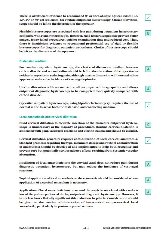
308 285
309 286
310 287
311 288
312 289
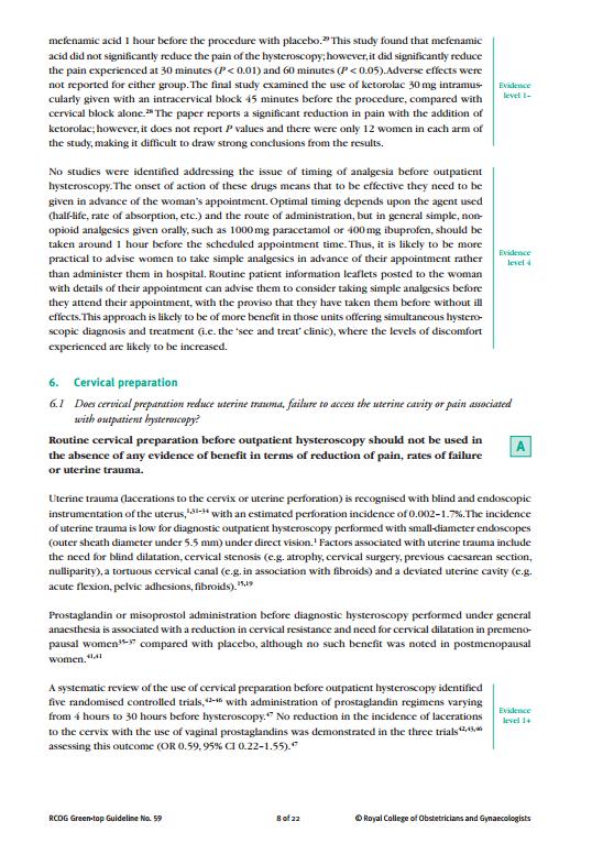
313 290
314 291
315 292
316 293
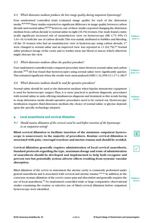
317 294
318 295
319 296
320 297

321 298

322 299
323 300
324 301
325 302
326 303
327 304
Investigating HMB- an evidence based approach
 BSGE Meeting: Contemporary management of heavy menstrual bleeding (HMB) in primary and secondary care: (7 th December 2018, RCOG) Investigating HMB- an evidence based approach T. Justin Clark MB ChB, MD(Hons),
Heavy Menstrual Bleeding. Mr Nick Nicholas MD FRCOG Grad Dip Law. Consultant Gynaecologist
 Heavy Menstrual Bleeding Mr Nick Nicholas MD FRCOG Grad Dip Law. Consultant Gynaecologist Why is HMB so important? 1:20 women aged 30-49 consult their GP with HMB Once referred to gynaecologist, surgical
Pain relief for outpatient hysteroscopy(review)
") Cochrane Database of Systematic Reviews Pain relief for outpatient hysteroscopy(review) AhmadG,SalujaS,O FlynnH,SorrentinoA,LeachD,WatsonA AhmadG,SalujaS,O FlynnH,SorrentinoA,LeachD,WatsonA. Pain relief
Cochrane Database of Systematic Reviews Pain relief for outpatient hysteroscopy(review) AhmadG,SalujaS,O FlynnH,SorrentinoA,LeachD,WatsonA AhmadG,SalujaS,O FlynnH,SorrentinoA,LeachD,WatsonA. Pain relief
Menstrual Disorders & Ambulatory Gynaecology
 Menstrual Disorders & Ambulatory Gynaecology Mr. Nagui Lewis Aziz M B, CH B, FRCOG Consultant Gynaecologist The Royal Oldham Hospital 01/09/2018 Heavy menstrual bleeding (HMB ) is a common problem responsible
Is Outpatient Hysteroscopy the New Gold Standard?
 Is Outpatient Hysteroscopy the New Gold Standard? McIlwaine K, Readman E, Ma T, Manwaring J, Ellett L, Hicks L, Porter J, Cameron M, Maher P. Mercy Hospital for Women, Melbourne, Australia Background Abnormal
Heavy menstrual bleeding (update)
") National Institute for Health and Care Excellence Final Heavy menstrual bleeding (update) A: Evidence reviews for diagnostic test accuracy in investigation for women presenting with heavy menstrual bleeding
National Institute for Health and Care Excellence Final Heavy menstrual bleeding (update) A: Evidence reviews for diagnostic test accuracy in investigation for women presenting with heavy menstrual bleeding
Heavy menstrual bleeding (update)
") National Institute for Health and Care Excellence Draft Heavy menstrual bleeding (update) Evidence reviews for diagnostic test accuracy in investigation for women presenting with heavy menstrual bleeding
National Institute for Health and Care Excellence Draft Heavy menstrual bleeding (update) Evidence reviews for diagnostic test accuracy in investigation for women presenting with heavy menstrual bleeding
INTERVENTIONAL PROCEDURES PROGRAMME
 NATIONAL INSTITUTE FOR CLINICAL EXCELLENCE INTERVENTIONAL PROCEDURES PROGRAMME Interventional procedure overview of microwave endometrial ablation Introduction This overview has been prepared to assist
Postmenopausal bleeding (PMB) guidelines for women with abnormal vaginal bleeding
 guidelines for women with abnormal vaginal bleeding") Guideline Postmenopausal bleeding (PMB) guidelines for women with abnormal vaginal bleeding 1 Scope For local use. 2 Purpose To provide evidence based care relating to several areas of the service including
Guideline Postmenopausal bleeding (PMB) guidelines for women with abnormal vaginal bleeding 1 Scope For local use. 2 Purpose To provide evidence based care relating to several areas of the service including
NATIONAL INSTITUTE FOR HEALTH AND CLINICAL EXCELLENCE. Health and social care directorate. Quality standards and indicators.
 NATIONAL INSTITUTE FOR HEALTH AND CLINICAL EXCELLENCE Health and social care directorate Quality standards and indicators Briefing paper Quality standard topic: Heavy menstrual bleeding Output: Prioritised
Abnormal uterine bleeding:
 Primary Care Women s Health Forum 16th June 2010 Abnormal uterine bleeding: The University Of Birmingham T Justin Clark MD (Hons), MRCOG Consultant Obstetrician and Gynaecologist Birmingham Women s Hospital
Your visit to the Outpatient Hysteroscopy Clinic
 Your visit to the Outpatient Hysteroscopy Clinic Department of Gynaecology Patient Information What What is the is an Outpatient outpatient Cystoscopy hysteroscopy? Clinic? An outpatient hysteroscopy means
Heavy menstrual bleeding: assessment and management
 Heavy menstrual bleeding: assessment and management NICE guideline Draft for consultation, August 0 This guideline covers assessing and treating heavy menstrual bleeding. It aims to help healthcare professionals
NICE guideline Published: 14 March 2018 nice.org.uk/guidance/ng88
 Heavy menstrual bleeding: assessment and management NICE guideline Published: 14 March 2018 nice.org.uk/guidance/ng88 NICE 2018. All rights reserved. Subject to Notice of rights (https://www.nice.org.uk/terms-and-conditions#notice-ofrights).
INTERVENTIONAL PROCEDURES PROGRAMME
 NATIONAL INSTITUTE FOR CLINICAL EXCELLENCE INTERVENTIONAL PROCEDURES PROGRAMME Interventional procedure overview of balloon thermal endometrial ablation (Cavaterm) Introduction This overview has been prepared
Out Patient Hysteroscopy Unit GUIDELINES
 Out Patient Hysteroscopy Unit GUIDELINES 1 AIMS The aim of the menstrual assessment clinic [MAC] (incorporating outpatient hysteroscopy) at Queen Charlotte s and Chelsea Hospital will be to provide a one-stop
Advanced 3D Ultrasound Incorporating Fly Thru Virtual Imaging Promotes the Concept of Ultrasound Hysteroscopy
 Advanced 3D Ultrasound Incorporating Fly Thru Virtual Imaging Promotes the Concept of Ultrasound Hysteroscopy Bill Smith Clinical Diagnostics Services, London, UK Introduction Conventional hysteroscopy
We are IntechOpen, the world s leading publisher of Open Access books Built by scientists, for scientists. International authors and editors
 We are IntechOpen, the world s leading publisher of Open Access books Built by scientists, for scientists 3,800 116,000 120M Open access books available International authors and editors Downloads Our
Citation for published version (APA): Timmermans, A. (2009). Postmenopausal bleeding : studies on the diagnostic work-up
: Timmermans, A. (2009). Postmenopausal bleeding : studies on the diagnostic work-up") UvA-DARE (Digital Academic Repository) Postmenopausal bleeding : studies on the diagnostic work-up Timmermans, A. Link to publication Citation for published version (APA): Timmermans, A. (2009). Postmenopausal
UvA-DARE (Digital Academic Repository) Postmenopausal bleeding : studies on the diagnostic work-up Timmermans, A. Link to publication Citation for published version (APA): Timmermans, A. (2009). Postmenopausal
Excessive menstrual blood loss
 Ian Chilcott Excessive menstrual blood loss >80mls - That interferes with physical, emotional, social and material quality of life 1 in 20 women aged 30 to 49 years consult their GP each year with menorrhagia
Hysteroscopy Clinic. Patient Information. Women and Children - Gynaecology
 8 Hysteroscopy Clinic Patient Information Women and Children - Gynaecology When a woman is first told that she has a gynaecological condition that requires further investigation at a specialised hospital
NATIONAL INSTITUTE FOR HEALTH AND CLINICAL EXCELLENCE GUIDANCE EXECUTIVE (GE)
") NATIONAL INSTITUTE FOR HEALTH AND CLINICAL EXCELLENCE GUIDANCE EXECUTIVE (GE) Review of TA78 fluid filled thermal balloon and microwave endometrial ablation for menstrual bleeding This guidance was issued
NATIONAL INSTITUTE FOR HEALTH AND CLINICAL EXCELLENCE GUIDANCE EXECUTIVE (GE) Review of TA78 fluid filled thermal balloon and microwave endometrial ablation for menstrual bleeding This guidance was issued
Uterine artery embolisation for treating adenomyosis
 Uterine artery embolisation for treating Issued: December 2013 guidance.nice.org.uk/ipg NICE has accredited the process used by the NICE Interventional Procedures Programme to produce interventional procedures
National inpatient diagnostic hysteroscopy survey
 Gynecol Surg (2010) 7:53 59 DOI 10.1007/s10397-009-0508-0 CLINICAL PRACTICE National inpatient diagnostic hysteroscopy survey Sherif Tawfeek & Peter Scott Received: 21 June 2009 /Accepted: 9 July 2009
PRE-ASS ESSMENT. Endometrial Ablation for Menorrhagia
 PRE-ASS ESSMENT No. 30 Feb 2004 Before decides to undertake a health technology assessment, a pre-assessment of the literature is performed. Pre-assessments are based on a limited literature search; they
Hysteroscopy. Department of Gynaecology. Patient information
 Hysteroscopy Department of Gynaecology Patient information What is is a hysteroscopy? a Hysteroscopy? A hysteroscopy is a procedure during which the inside of the uterus (womb) is examined using a thin
Outpatient hysteroscopy direct diagnostic access & new therapeutic procedures
 Outpatient hysteroscopy direct diagnostic access & new therapeutic procedures Mrs Katy Clifford Consultant Gynaecologist St Mary s Hospital Imperial College Healthcare NHS Trust Diagnostic outpatient hysteroscopy
The 6 th Scientific Meeting of the Asia Pacific Menopause Federation
 Abnormal uterine bleeding in the perimenopause Perimenopausal menstrual problems are among the most common causes for family practitioner and specialist referral. Often it is due to the hormone changes
bleeding Studies naar de diagnostiek van endom triumcarcinoom bij vrouwen met postm nopauzaal bloedverlies. Studies on the
 Studies on the diagnosis of endometria cancer in women with postmenopausal bleeding. Studies naar de diagnostiek va endometriumcarcinoom bij vrouwen m postmenopauzaal bloedverlies. Studies on the diagnosis
Role of diagnostic hysteroscopy in evaluation of abnormal uterine bleeding and its histopathological correlation
 International Journal of Reproduction, Contraception, Obstetrics and Gynecology Chaudhari KR et al. Int J Reprod Contracept Obstet Gynecol. 2014 Sep;3(3):666-670 www.ijrcog.org pissn 2320-1770 eissn 2320-1789
Endometrial Ablation for Heavy Menstrual Bleeding. Jonathan Lord Consultant gynaecologist
 Endometrial Ablation for Heavy Menstrual Bleeding Jonathan Lord Consultant gynaecologist Affiliation: Declaration of Interests NICE HMB guideline committee member Expenses & honaria: Hologic (manufacturer
A systematic review of the effect of the distension medium on pain during outpatient hysteroscopy
 A systematic review of the effect of the distension medium on pain during outpatient hysteroscopy Natalie A. M. Cooper, M.B., Ch.B., a Paul Smith, B.Sci., M.B., Ch.B., b Khalid S. Khan, M.B., B.S., M.Sc.,
Consent Advice No. XX (Joint with BSGE) Peer Review Draft Spring Morcellation for Laparoscopic Myomectomy or Hysterectomy
 Peer Review Draft Spring Morcellation for Laparoscopic Myomectomy or Hysterectomy") 1 2 3 4 5 6 7 8 9 10 11 12 13 14 15 16 17 18 19 20 21 22 23 24 25 26 27 28 29 30 31 32 33 34 35 36 37 38 39 40 41 42 43 44 45 46 47 48 49 50 51 Consent Advice No. XX (Joint with BSGE) Peer Review Draft
1 2 3 4 5 6 7 8 9 10 11 12 13 14 15 16 17 18 19 20 21 22 23 24 25 26 27 28 29 30 31 32 33 34 35 36 37 38 39 40 41 42 43 44 45 46 47 48 49 50 51 Consent Advice No. XX (Joint with BSGE) Peer Review Draft
Endometrial Cancer Biopsy of the endometrium Evaluation of women of all ages
 Endometrial Cancer Biopsy of the endometrium Evaluation of women of all ages Barbara S. Apgar, MD, MS Professor of Family Medicine University of Michigan Health System Ann Arbor, Michigan Cancer of the
Chapter 4. Hysteroscopic morcellator for removal of intrauterine polyps and myomas: a randomised controlled study among residents in training
 Chapter 4 Hysteroscopic morcellator for removal of intrauterine polyps and myomas: a randomised controlled study among residents in training Heleen van Dongen Mark Hans Emanuel Ron Wolterbeek J. Baptist
PROSPERO International prospective register of systematic reviews
 PROSPERO International prospective register of systematic reviews Closed reduction methods for acute anterior shoulder dislocation [Cochrane Protocol] Kanthan Theivendran, Raj Thakrar, Subodh Deshmukh,
Fibroid mapping. Haitham Hamoda MD FRCOG Consultant Gynaecologist, Subspecialist in Reproductive Medicine & Surgery King s College Hospital
 Fibroid mapping Haitham Hamoda MD FRCOG Consultant Gynaecologist, Subspecialist in Reproductive Medicine & Surgery King s College Hospital Fibroids Common condition >70% of women by onset of menopause.
INTERVENTIONAL PROCEDURES PROGRAMME
 NATIONAL INSTITUTE FOR CLINICAL EXCELLENCE INTERVENTIONAL PROCEDURES PROGRAMME Interventional procedure overview of laparoscopic laser myomectomy Introduction This overview has been prepared to assist
Medical Management of Fibroids Esmya. Dr Paula Briggs Consultant in Sexual and Reproductive Health
 Medical Management of Fibroids Esmya Dr Paula Briggs Consultant in Sexual and Reproductive Health Treatment options for Uterine Fibroids ESMYA Selective Uterine Artery Embolisation Fibroid ablation (hysteroscopic
Systematic reviews and meta-analyses of observational studies (MOOSE): Checklist.
: Checklist.") Systematic reviews and meta-analyses of observational studies (MOOSE): Checklist. MOOSE Checklist Infliximab reduces hospitalizations and surgery interventions in patients with inflammatory bowel disease:
Systematic reviews and meta-analyses of observational studies (MOOSE): Checklist. MOOSE Checklist Infliximab reduces hospitalizations and surgery interventions in patients with inflammatory bowel disease:
NETSCC, HTA 24 November 2009
 NETSCC, HTA 24 November 2009 Protocol 09/63/01(Clark TJ et al)1 PROJECT PROTOCOL PROJECT TITLE Cost-effectiveness of diagnostic strategies for the management of abnormal uterine bleeding (heavy menstrual
Database of Abstracts of Reviews of Effects (DARE) Produced by the Centre for Reviews and Dissemination Copyright 2017 University of York.
 Produced by the Centre for Reviews and Dissemination Copyright 2017 University of York.") A comparison of the cost-effectiveness of five strategies for the prevention of non-steroidal anti-inflammatory drug-induced gastrointestinal toxicity: a systematic review with economic modelling Brown
A comparison of the cost-effectiveness of five strategies for the prevention of non-steroidal anti-inflammatory drug-induced gastrointestinal toxicity: a systematic review with economic modelling Brown
Gynaecology Cancer Red Flags. Dr Dina Bisson Consultant Obstetrician and Gynaecologist Southmead Hospital North Bristol NHS Trust 27 April 2017
 Gynaecology Cancer Red Flags Dr Dina Bisson Consultant Obstetrician and Gynaecologist Southmead Hospital North Bristol NHS Trust 27 April 2017 Gynaecological Cancers Endometrial Cancer Ovarian Cancer Cervical
Surveillance report Published: 17 March 2016 nice.org.uk
 Surveillance report 2016 Ovarian Cancer (2011) NICE guideline CG122 Surveillance report Published: 17 March 2016 nice.org.uk NICE 2016. All rights reserved. Contents Surveillance decision... 3 Reason for
Laparoscopy-Hysteroscopy
 Laparoscopy-Hysteroscopy Patient Information Laparoscopy The laparoscope, a surgical instrument similar to a telescope, is inserted through a small incision (cut) in the belly button during laparoscopy.
Policy for Hysteroscopy Page 1 of 5. Policy for Hysteroscopy. V1.1 OPCS/ICD codes added. Version of: Version of: March 2018
 Policy for Hysteroscopy Version of: 16.03.2018 Version of: March 2018 Version Number: Changes Made: V1.1 OPCS/ICD codes added. V1 Ratified policy agreed by Healthier Lancashire and South Cumbria s Joint
Summary CHAPTER 1. Introduction
 Summary This thesis aims to evaluate the diagnostic work-up in postmenopausal women presenting with abnormal vaginal bleeding. The Society of Dutch Obstetrics and Gynaecology composed a guideline, which
A cost-utility analysis of low-dose hormone replacement therapy in postmenopausal women with an intact uterus Swift J A, Conway P, Purdie D W
 A cost-utility analysis of low-dose hormone replacement therapy in postmenopausal women with an intact uterus Swift J A, Conway P, Purdie D W Record Status This is a critical abstract of an economic evaluation
Outpatient Hysteroscopy. Patient Information
 Outpatient Hysteroscopy Patient Information Author ID: NM Leaflet Number: Gyn 062 Version: 1 Name of Leaflet: Outpatient Hysteroscopy Date Produced: January 2017 Review Date: January 2019 Introduction
EVALUATION OF AMBULATORY DIAGNOSIS OF ABNORMAL UTERINE BLEEDING SHAGAF HAJ BAKOUR
 EVALUATION OF AMBULATORY DIAGNOSIS OF ABNORMAL UTERINE BLEEDING by SHAGAF HAJ BAKOUR A thesis submitted to the Faculty of Medicine and Dentistry Of the University of Birmingham For the degree of DOCTOR
Treatment of Heavy Menstrual Bleeding: Ludkin (Nurse Hysteroscopist, Bradford Royal Infirmary) & M Rogers (Advanced
 & M Rogers (Advanced") Treatment of Heavy Menstrual Bleeding: Nov 14 th 2013- Authors: Prof Sian Jones (Gynaecologist, Bradford Royal Infirmary, H Ludkin (Nurse Hysteroscopist, Bradford Royal Infirmary) & M Rogers (Advanced
Treatment of Heavy Menstrual Bleeding: Nov 14 th 2013- Authors: Prof Sian Jones (Gynaecologist, Bradford Royal Infirmary, H Ludkin (Nurse Hysteroscopist, Bradford Royal Infirmary) & M Rogers (Advanced
Setting The setting was primary and secondary care. The economic study was carried out in the USA.
 Cost-effectiveness of pap smear screening for vaginal cancer after total hysterectomy for benign disease Fetters M D, Lieberman R W, Abrahamse P H, Sanghvi R V, Sonnad S S Record Status This is a critical
A randomised trial comparing the H Pipelle with the standard Pipelle for endometrial sampling at no-touch (vaginoscopic) hysteroscopy
 hysteroscopy") DOI: 10.1111/j.1471-0528.2008.01992.x www.blackwellpublishing.com/bjog Gynaecological surgery A randomised trial comparing the H Pipelle with the standard Pipelle for endometrial sampling at no-touch (vaginoscopic)
DOI: 10.1111/j.1471-0528.2008.01992.x www.blackwellpublishing.com/bjog Gynaecological surgery A randomised trial comparing the H Pipelle with the standard Pipelle for endometrial sampling at no-touch (vaginoscopic)
Membership of Heavy Menstrual Bleeding (update) guideline Committee
 guideline Committee") membership list Heavy Menstrual Bleeding (update) guideline Membership of Heavy Menstrual Bleeding (update) guideline The will operate as an advisory to NICE s Board, developing a guideline on Post- The
membership list Heavy Menstrual Bleeding (update) guideline Membership of Heavy Menstrual Bleeding (update) guideline The will operate as an advisory to NICE s Board, developing a guideline on Post- The
Chapter 2. Implementation of hysteroscopic surgery in The Netherlands. Heleen van Dongen Wendela Kolkman Frank Willem Jansen
 Chapter 2 Implementation of hysteroscopic surgery in The Netherlands Heleen van Dongen Wendela Kolkman Frank Willem Jansen Adapted from Eur J Obstet Gynecol Reprod Biol 07;132:232-236 Introduction Diagnostic
VirtaMed GynoS hysteroscopy Module descriptions
 VirtaMed GynoS hysteroscopy Module descriptions VirtaMed AG Rütistr. 12, 8952 Zurich Switzerland info@virtamed.com www.virtamed.com Phone: +41 44 500 9690 Table of contents Table of contents... 1 Essential
Treating cancer of the endometrium. Information for patients Gynaecology
 Treating cancer of the endometrium Information for patients Gynaecology We have written this leaflet to provide you with information following your diagnosis of cancer of the endometrium. We understand
Research Article A Structured Assessment to Decrease the Amount of Inconclusive Endometrial Biopsies in Women with Postmenopausal Bleeding
 International Surgical Oncology Volume 2016, Article ID 3039261, 5 pages http://dx.doi.org/10.1155/2016/3039261 Research Article A Structured Assessment to Decrease the Amount of Inconclusive Endometrial
PATIENT GROUP CONDEMNS NICE REVISED HEAVY MENSTRUAL BLEEDING GUIDELINES UNSAFE FOR WOMEN MANY MORE WOMEN WILL DIE AND SUFFER SERIOUS COMPLICATIONS
 PRESS RELEASE For immediate release 20 th February 2018 PATIENT GROUP CONDEMNS NICE REVISED HEAVY MENSTRUAL BLEEDING GUIDELINES UNSAFE FOR WOMEN MANY MORE WOMEN WILL DIE AND SUFFER SERIOUS COMPLICATIONS
Laparoscopy and Hysteroscopy
 AMERICAN SOCIETY FOR REPRODUCTIVE MEDICINE Laparoscopy and Hysteroscopy A Guide for Patients PATIENT INFORMATION SERIES Published by the American Society for Reproductive Medicine under the direction of
National Heavy Menstrual Bleeding Audit
 First Annual Report May 2011 Funding provided by: National Heavy Menstrual Bleeding Audit A national audit to assess patient outcomes and experiences of care for women with heavy menstrual bleeding in
STOP/START. On the Web. 12 intraoperative videos from Dr. Garcia, at
 Diagnostic hysteroscopy spies polyp previously missed on transvaginal ultrasound and dilation and curettage. STOP performing dilation and curettage for the evaluation of abnormal uterine bleeding START
Hysteroscopy. What you need to know. 139 Dumaresq Street Campbelltown Phone Fax
 Hysteroscopy What you need to know 139 Dumaresq Street Campbelltown Phone 4628 5292 Fax 4628 0349 www.nureva.com.au September 2015 HYSTEROSCOPY AND DILATATION & CURETTAGE OF THE UTERUS Introduction I have
Virtual Hysteroscopy With 3D Sonohysterography In Comparison To Office Hysteroscopy For The Diagnosis Of Endometrial Polyps
 ISPUB.COM The Internet Journal of Gynecology and Obstetrics Volume 23 Number 1 Virtual Hysteroscopy With 3D Sonohysterography In Comparison To Office Hysteroscopy For The Diagnosis Of Endometrial Polyps
Professor Christian Phillips BSc Hons BM DM FRCOG Consultant Gynaecologist and Urogynaecologist
 Professor Christian Phillips BSc Hons BM DM FRCOG Consultant Gynaecologist and Urogynaecologist Laparoscopic Hysterectomy What is a laparoscopic hysterectomy? Laparoscopic hysterectomy is an operation
Ameer Abdallah. Department of Obstetrics Gynaecology, Faculty of Medicine, Minia University, Minia Egypt
 International Journal of Research Studies in Biosciences (IJRSB) Volume 3, Issue 4, April 2015, PP 57-64 ISSN 2349-0357 (Print) & ISSN 2349-0365 (Online) www.arcjournals.org Randomized Study Comparing
Uterine sarcoma. Information for patients Gynaecology
 Uterine sarcoma Information for patients Gynaecology page 2 of 12 This leaflet is intended to offer you support and information at a very difficult and stressful time in your life. To be told that you
Management of Endometrial Hyperplasia
 Management of Endometrial Hyperplasia Green-top Guideline No. 67 RCOG/SGE Joint Guideline February 2016 Management of Endometrial Hyperplasia This is the first edition of this guideline.this is a joint
Levosert levonorgestrel 20mcg/24hour intrauterine device
 Levosert levonorgestrel 20mcg/24hour intrauterine device Verdict: Formulary inclusion: Formulary category: Restrictions: Reason for inclusion: Link to formulary: Link to medicine review summary: Levosert
Subject Index. Cavaterm, endometrial ablation complications 146, 150 contraindications 152 cost analysis compared with hysterectomy
 OOOOOOOOOOOOOOOOOOOOOOOOOOOOOO Subject Index Abnormal uterine bleeding, see also Adenomyosis, Endometrial cancer, Menorrhagia dilatation and curettage 21, 22, 25 hysteroscopy of premenopausal women anesthesia
Frequency of menses. Duration of menses 3 days to 7 days. Flow/amount of menses Average blood loss with menstruation is 60-80cc.
 Frequency of menses 24 days (0.5%) to 35 days (0.9%) Age 25, 40% are between 25 and 28 days Age 25-35, 60% are between 25 and 28 days Teens and women over 40 s cycles may be longer apart Duration of menses
Over the past year, a few gems have been
 UPDATE Abnormal uterine bleeding Howard T. Sharp, MD Dr. Sharp is Professor and Vice Chair for Clinical and Quality Activities, Department of Obstetrics and Gynecology, University of Utah Health Sciences
Is diagnostic hysteroscopy an effective tool to increase ART results?
 Is diagnostic hysteroscopy an effective tool to increase ART results? Mr. Tarek El-Toukhy, MSc MD MRCOG Consultant in Reproductive Medicine and Surgery, Guy s and St. Thomas Hospital, London Summary Technical
Surgical Interruption of Pelvic Nerve Pathways for Primary and Secondary Dysmenorrhea. Original Policy Date
 MP 4.01.10 Surgical Interruption of Pelvic Nerve Pathways for Primary and Secondary Dysmenorrhea Medical Policy Section OB/Gyn/Reproduction Issue 12:2013 Original Policy Date 12:2013 Last Review Status/Date
levonorgestrel 13.5mg intrauterine delivery system (Jaydess ) SMC No. (1036/15) Bayer
 SMC No. (1036/15) Bayer") levonorgestrel 13.5mg intrauterine delivery system (Jaydess ) SMC No. (1036/15) Bayer 6 March 2015 The Scottish Medicines Consortium (SMC) has completed its assessment of the above product and advises
levonorgestrel 13.5mg intrauterine delivery system (Jaydess ) SMC No. (1036/15) Bayer 6 March 2015 The Scottish Medicines Consortium (SMC) has completed its assessment of the above product and advises
OUTPATIENT OPERATIVE HYSTEROSCOPY
 OUTPATIENT OPERATIVE HYSTEROSCOPY Martin Farrugia MD MRCOG East Kent Hospitals NHS Trust Concepts Can you do a diagnostic hysteroscopy in a clinic? Can you do a Pipelle biopsy without local anaesthesia?
Fitting of an Intrauterine Device (IUD)
") PLEASE PRINT WHOLE FORM DOUBLE SIDED ON YELLOW PAPER Patient Information to be retained by patient affix patient label What is an IUD? An IUD is a small T-shaped plastic and copper device that is put into
PLEASE PRINT WHOLE FORM DOUBLE SIDED ON YELLOW PAPER Patient Information to be retained by patient affix patient label What is an IUD? An IUD is a small T-shaped plastic and copper device that is put into
Medical treatment for uterine fibroids
 Medical treatment for uterine fibroids Prof Mary Ann Lumsden Prof of Gynaecology and Medical Education University of Glasgow Senior Vice President RCOG Conflict of Interest Chair, Guideline development
Ambulatory endoscopic treatment of symptomatic benign endometrial polyps: a feasibility study Clark T J, Godwin J, Khan K S, Gupta J K
 Ambulatory endoscopic treatment of symptomatic benign endometrial polyps: a feasibility study Clark T J, Godwin J, Khan K S, Gupta J K Record Status This is a critical abstract of an economic evaluation
Having a hysterectomy
 Having a hysterectomy Gynaecology Oncology Information for patients Gynaecology It is expected that you will have discussed other methods of treatment for your health concern with your doctor and have
HIGH-DEFINITION HYSTEROSCOPY FOR A HIGH STANDARD OF CARE.
 HIGH-DEFINITION HYSTEROSCOPY FOR A HIGH STANDARD OF CARE. Our next-generation TruClear hysteroscopes were designed with your technique and the patient experience in mind TruClear Elite Hysteroscope Mini
Prospective multicentre randomized controlled trial to evaluate factors influencing the success rate of office diagnostic hysteroscopy
 Human Reproduction Page 1 of 6 Hum. Reprod. Advance Access published November 18, 2004 doi:10.1093/humrep/deh559 Prospective multicentre randomized controlled trial to evaluate factors influencing the
RANZCOG Advanced Training Modules
 RANZCOG Advanced Training Modules Generalist Obstetrics ATM and Generalist Gynaecology ATM The Generalist ATMs in each of Obstetrics and Gynaecology provide a framework for trainees to consolidate and
Surgical Interruption of Pelvic Nerve Pathways for Primary and Secondary Dysmenorrhea
 Page: 1 of 7 Last Review Status/Date: June 2015 for Primary and Secondary Dysmenorrhea Description Two laparoscopic surgical approaches are proposed as adjuncts to conservative surgical therapy for the
Health technology The use of gonadotrophin-releasing hormone agonists (GnRHa) in women with uterine fibroids, undergoing hysterectomy or myomectomy.
 in women with uterine fibroids, undergoing hysterectomy or myomectomy.") Cost effectiveness of pre-operative gonadotrophin releasing analogues for women with uterine fibroids undergoing hysterectomy or myomectomy Farquhar C, Brown P M, Furness S Record Status This is a critical
Cost effectiveness of pre-operative gonadotrophin releasing analogues for women with uterine fibroids undergoing hysterectomy or myomectomy Farquhar C, Brown P M, Furness S Record Status This is a critical
If you have any further questions, please speak to a doctor or nurse caring for you.
 Having a laparoscopy This leaflet aims to answer your questions about having a laparoscopy. It explains the benefits, risks and alternatives, as well as what you can expect when you come to hospital. If
Uterine artery embolization for symptomatic uterine fibroids(review)
") Cochrane Database of Systematic Reviews Uterine artery embolization for symptomatic uterine fibroids (Review) GuptaJK,SinhaA,LumsdenMA,HickeyM GuptaJK,SinhaA,LumsdenMA,HickeyM. Uterine artery embolization
Cochrane Database of Systematic Reviews Uterine artery embolization for symptomatic uterine fibroids (Review) GuptaJK,SinhaA,LumsdenMA,HickeyM GuptaJK,SinhaA,LumsdenMA,HickeyM. Uterine artery embolization
PALM-COEIN: Your AUB Counseling Guide
 PALM-COEIN: Your AUB Counseling Guide 10 million+ Treat the cause, not the symptom In the U.S, more than 10 million women between the ages of 35 and 49 are affected by AUB 1 Diagnosis Cause Structural
Health Board/Region: All-Wales
 Peer Review: Cancer Sub-site: Gynaecology Health Board/Region: All-Wales Cycle: Second Date of review: February 2018 This report describes the findings and themes observed by clinical review panels during
Alcohol interventions in secondary and further education
 National Institute for Health and Care Excellence Guideline version (Draft for Consultation) Alcohol interventions in secondary and further education NICE guideline: methods NICE guideline Methods
Pain Management for Offi ce Hysteroscopy -
 International Journal of Reproductive Medicine & Gynecology Review Article Pain Management for Offi ce Hysteroscopy - Carugno Jose* and Andrade Fausto Obstetrics and Gynecology Department, Minimally Invasive
PATIENT INFORMATION SHEET (Sheffield and Southampton Only)
") PATIENT INFORMATION SHEET (Sheffield and Southampton Only) ENDOMETRIAL SCRATCH TRIAL A Multicentre Randomised Controlled Trial of Induced Endometrial Scratch in Women Undergoing First Time in Vitro Fertilisation
PATIENT INFORMATION SHEET (Sheffield and Southampton Only) ENDOMETRIAL SCRATCH TRIAL A Multicentre Randomised Controlled Trial of Induced Endometrial Scratch in Women Undergoing First Time in Vitro Fertilisation
Application for inclusion of levonorgestrel - releasing IUD for contraception in the WHO Model List of Essential Medicines
 Application for inclusion of levonorgestrel - releasing IUD for contraception in the WHO Model List of Essential Medicines 1. Summary statement of the proposal for inclusion LNG-IUS is an effective contraceptive;
Surgical Interruption of Pelvic Nerve Pathways for Primary and Secondary Dysmenorrhea
 Surgical Interruption of Pelvic Nerve Pathways for Primary and Secondary Dysmenorrhea Policy Number: 4.01.17 Last Review: 11/2013 Origination: 11/2007 Next Review: 11/2014 Policy Blue Cross and Blue Shield
Citation for published version (APA): van Hanegem, N. (2015). The diagnostic work-up of women with postmenopausal bleeding
: van Hanegem, N. (2015). The diagnostic work-up of women with postmenopausal bleeding") UvA-DARE (Digital Academic Repository) The diagnostic work-up of women with postmenopausal bleeding van Hanegem, N. Link to publication Citation for published version (APA): van Hanegem, N. (2015). The
UvA-DARE (Digital Academic Repository) The diagnostic work-up of women with postmenopausal bleeding van Hanegem, N. Link to publication Citation for published version (APA): van Hanegem, N. (2015). The
Primary Care Gynaecology Guidelines: HEAVY REGULAR MENSTRUAL BLEEDING
 Primary Care Guidelines: HEAVY REGULAR MENSTRUAL BLEEDING
5/5/2010 FINANCIAL DISCLOSURE. Abnormal Uterine Bleeding. Is This A Problem? About me % of visits to gynecologist
 Abnormal Uterine FINANCIAL DISCLOSURE I HAVE NO FINANCIAL INTEREST IN ANY OF THE PRODUCTS MENTIONED IN MY PRESENTATION Bryan K. Rone, M.D. University of Kentucky Obstetrics and Gynecology May 5, 2010 About
Guidelines for performing gynaecological endoscopic procedures
 Guidelines for performing gynaecological endoscopic procedures This statement has been developed by the Women s Health Committee. It has been reviewed by the Endoscopic Surgery Advisory Committee (RANZCOG/AGES)
Laparoscopic Hysterectomy
 Women s Service Laparoscopic Hysterectomy Information for patients Information for Patients This leaflet is for women who have been advised to have a laparoscopic hysterectomy. It outlines the common reasons