Identification of hypothalamic sites that control puberty onset and sexual maturation. Megan Louise Greenwald-Yarnell
|
|
|
- Bertina Dawson
- 5 years ago
- Views:
Transcription
1 Identification of hypothalamic sites that control puberty onset and sexual maturation by Megan Louise Greenwald-Yarnell A dissertation submitted in partial fulfillment of the requirements for the degree of Doctor of Philosophy (Neuroscience) in the University of Michigan 2013 Doctoral Committee: Professor Martin G. Myers, Jr., Chair Professor Robert Denver Professor Michael Lehman, University of Mississippi Medical Center Professor Sue Moenter Assistant Professor David Olson Professor Audrey Seasholtz
2 Basic research is what I'm doing when I don't know what I'm doing. - Wernher von Braun
3 Megan Louise Greenwald-Yarnell 2013
4 Dedication Dedicated to my amazing husband, James. I am eternally grateful for your never-ending love, understanding and encouragement. ii
5 Acknowledgements I d like to acknowledge the support of my mentor, Martin Myers, and the entire Myers lab- past and present. The culture of cooperation and teamwork that Martin expects in his lab has made my time there truly enjoyable. I am so thankful that he welcomed me into the lab and allowed me to pursue my research interests, despite the fact that at times they were so very different from what the rest of the lab was working on. I d like to thank Dr. Christa Patterson who has been so instrumental in my success in lab and also in maintaining my sanity over the years. Thank you to Meg Allison and Amy Sutton for the evenings of pizza and drinks in lab and for always being available to talk- whether the topic is science or personal. Without the guidance of former lab members Drs. Rebecca Leshan, Gwen Louis, Scott Robertson, Eneida Villanueva and Darren Opland early on, none of this would have been possible. I d also like to thank Dr. Dave Olson, who has the unfortunate position of running the lab adjacent to Martin s and has had to suffer my outbursts and fill in for Martin when he s out of town and I desperately need to discuss some recently acquired data. To Drs. Courtney Marsh and Josh Skorupski- thank you for your help with these projects and for your willingness to provide the clinical perspective. Thank you to my classmates, the Neuroscience Graduate Program iii
6 incoming class of I never would have made it through the early days of grad school without you guys. I d also like to thank Jocelyn, Ariana and Tracy for your support over the past year, especially during Jim s recent deployment. I d also like to acknowledge the contributions of our collaborators, Drs. Suzanne Moenter and Carol Elias as well as their lab members. Additionally, I d like to thank my dissertation committee for their contributions. Your assistance with this research has been invaluable and is greatly appreciated. And finally, thank you to my family. You have all been so understanding over the past five years when science demanded all my time, energy and focus. Without your encouragement and support, I never would have had the courage or stamina to accomplish this. iv
7 Table of Contents Dedication...ii Acknowledgements... iii List of Figures... vii Chapter 1 - Introduction... 1 Chapter 2 - Genetic ablation of LepRb from Nos1-expressing neurons Methods Results Conclusions & Discussion Chapter 3 - Estrogen acts via ERα in kisspeptin/neurokinin B neurons to mediate feedback on the reproductive axis and control the onset of puberty in female mice Methods Results Conclusions & Discussion Chapter 4 Direct estrogen action on Kiss1/ERα neurons is required for negative feedback on the reproductive axis of male mice Methods Results Conclusions & Discussion Chapter 5 Summary & Conclusions v
8 The role of direct leptin action on Nos1-expressing hypothalamic neurons Regulation of the reproductive axis by estrogen action via ERα on kisspeptin neurons References vi
9 List of Figures Chapter 1 Figure 1- The hypothalamic-pituitary-gonadal (HPG) axis Chapter 2 Figure 2 - Generation of Lepr Nos1 KO mice Figure 3 - Reproductive effects of ablation of LepRb from Nos1-expressing neurons Figure 4 - Body mass is increased in both female and male Lepr Nos1 KO mice.. 50 Figure 5 - ARC Pomc gene expression is altered in Lepr Nos1 KO mice Figure 6 - Sexually dimorphic alteration in T4 levels in Lepr Nos1 KO mice Chapter 3 Figure 7- Generation of mice to study Kiss1- and Tac2-expressing neurons Figure 8 Dual-label in situ hybridization/immunohistochemistry in Tac2 eyfp and Kiss1 eyfp female mice Figure 9 Co-expression of ERα and eyfp in Tac2 eyfp and Kiss1 eyfp female mice Figure 10 - Body mass and composition of female mice lacking ERα in either Kiss1- or Tac2-expressing neurons Figure 11- Precocious but incomplete reproductive maturation in the absence of ERα from either Kiss1- or Tac2-expressing neurons Figure 12 Ovarian and uterine phenotype of adult female mice lacking ERα in Kiss1- or Tac2-expressing neurons Figure 13 - Gonadotropin and gonadal hormone levels of intact adult females. 89 Figure 14 - Gonadotropin and gonadal hormone levels of intact juvenile females Figure 15 - Effect of ovariectomy and acute estradiol treatment on LH and FSH vii
10 levels Figure 16 - Altered ARC gene expression in intact female mice lacking ERα in either Kiss1- or Tac2-expressing neurons Figure 17 Kiss1 expression in the AVPV/PeN of intact females is diminished only in ERα Kiss1 KOs Figure 18 Ovariectomy results in gene expression changes in the ARC that mimic loss of ERα from either Kiss1- or Tac2-expressing neurons.. 94 Figure 19 AVPV/PeN Kiss1 expression is diminished as a result of ovariectomy or ablation of ERα from all Kiss1-expressing neurons. 95 Figure 20 - ARC kisspeptin peptide levels are unchanged by ablation of ERα from either Kiss1- or Tac2-expressing neurons Figure 21 AVPV/PeN kisspeptin peptide levels are reduced when ERα is ablated from Kiss1-expressing neurons Chapter 4 Figure 22 - Generation of mouse models to study Kiss1- and Tac2-expressing neurons in males Figure 23 Co-expression of eyfp and ERα in the hypothalamus of male Kiss1 eyfp and Tac2 eyfp mice Figure 24 - Kisspeptin immunoreactivity in the rostral hypothalamus of Kiss1 eyfp male and female mice Figure 25 - Body mass and composition are unchanged in ERα Kiss1 KO and ERα Tac2 KO male mice Figure 26 Male reproductive organ weights are affected by ablation of ERα from either Kiss1- or Tac2-expressing neurons Figure 27 Gonadotropin and gonadal hormone levels are altered by ablation of ERα from all Kiss1- but not Tac2-expressing neurons Figure 28 - Gene expression in the rostral hypothalamus and ARC viii
11 Chapter 1 - Introduction Puberty is the period of time during which a child transitions into a sexually mature adult capable of reproduction. From a biological perspective, it is the activation of the hypothalamic-pituitary-gonadal (HPG) axis, culminating in the maturation of the brain, pituitary and gonads. Throughout puberty, there is a complex interplay between environmental and endogenous factors that regulate the HPG axis. Despite decades of research, there are still large gaps in our understanding of puberty. How is the timing of puberty onset regulated? By which hormones? What are the underlying neural circuits important for puberty onset? Where do these hormones act in the brain to modulate the timing of puberty onset? This literature review summarizes existing studies of how two important hormones- estradiol and leptin- produced in the periphery by the ovaries and white adipose tissue respectively, act in the brain to affect the timing of puberty onset and subsequent sexual maturation. 1
12 Puberty onset and sexual maturation Primates Although functional by the end of gestation, the ovaries and testes become relatively inactive for a brief period around the time of birth and stop secreting gonadal steroid hormones. Within a few days, however, they become active again and remain active until several months after birth when they become quiescent again and stop secreting significant amounts of hormones. This suppression of activity is referred to as the juvenile pause and is thought to be the result of inhibitory mechanisms in the brain and to a lesser extent at the level of the pituitary gland, a small endocrine organ located below the base of the brain. The regulation of gonadal activity throughout childhood is ultimately controlled by the brain, and more specifically by an area of the brain called the hypothalamus, which functions to link the neuroendocrine system to peripheral organ systems via the pituitary gland. Feedback loops exist between the hypothalamus, pituitary gland and the gonads, which together are called the HPG axis (Figure 1). During the juvenile pause, neurons in the brain that produce gonadotropin-releasing hormone (GnRH) are relatively inactive and produce very little GnRH. Since pulsatile GnRH secretion is required for the production and release of specific hormones from the pituitary, low levels of the pituitary-derived gonadotropins luteinizing hormone (LH) and follicle-stimulating hormone (FSH) are characteristic of the period of time prior to puberty onset 1,2. Before the discorvery of FSH, GnRH was often termed luteinizing hormone releasing hormone (LHRH). 2
13 As an organism matures and approaches puberty, GnRH neurons become more active, resulting in increased secretion of GnRH and causing the gonadotropes in the anterior pituitary to also become more active. While young prepubertal boys and girls do not exhibit pulsatile LH release, nocturnal LH pulses are readily detected in older prepubertal children; the increased nocturnal LH levels are characteristic of human puberty onset 1,2. During puberty, both LH and FSH increase in overall levels, pulse frequency and amplitude 1 5. The increased pulsatile release of the gonadotropins from the anterior pituitary triggers effects in the gonads, including the production of gonadal hormones, termed androgens or estrogens. In females, LH acts on the theca cells of the ovaries, resulting in the production of androgenic precursors of estradiol, a potent estrogen. In males, LH acts on the Leydig cells of the testes, leading to production of testosterone, a potent androgen. FSH acts on granulosa cells in the ovaries and Sertoli cells in the testes to stimulate gametogenesis and gonadal growth. Additionally, in females FSH acts within the granulosa cells to promote the aromatization of thecal androgens to estradiol. With continued stimulation by the gonadotropins LH and FSH, the peripubertal gonads grow and secrete sex hormones at steadily increasing rates. During puberty, the effects of the rising hormone levels can be seen in a variety of tissues in a sex-specific manner. In females, estrogens are responsible for growth of the breasts and maturation of the female genitalia, while circulating androgens control the growth of pubic and axillary hair. In males, androgens control not only the development of genitalia and growth of 3
14 body hair, but also deepen the voice by enlarging the larynx and laryngeal muscles. The development of these secondary sexual characteristics does not occur at precisely the same chronological age in all individuals, but the sequence of changes is characteristic for each sex and generally the same across individuals. Two mechanisms for the control of puberty onset It has been hypothesized that the suppression of GnRH release prior to puberty onset is the result of two different mechanisms (which may not be equally important or prominent across species). The first mechanism relies on the fact that prior to puberty, the hypothalamus and the pituitary gland are extraordinarily sensitive to the negative feedback effect of gonadal steroid hormones, which inhibit GnRH and LH release and keep the HPG axis quiescent. If this system becomes less sensitive to gonadal hormone feedback, gonadotropin secretion can escape from negative feedback, allowing gonadal hormone levels to rise as puberty progresses. This mechanism is often referred to as the gonadostat hypothesis, and has been attributed to the endocrinologist Melvin Grumbach, although his initial description of the phenomenon was gonadal steroid-dependent LHRH increase 6. The second mechanism is independent of gonadal hormone feedback and instead postulates that there are neural mechanisms that restrain the activity of GnRH neurons prior to puberty, resulting in low gonadotropin and gonadal hormone levels in prepubertal organisms. This is referred to as the central drive hypothesis. It s possible that 4
15 in addition to active restraint of the GnRH neuron there may also be a lack of stimulatory inputs to GnRH neurons during this time. Under this hypothesis, puberty begins when the inputs to GnRH neurons change and allow more GnRH to be secreted. The primary mechanism of puberty initiation seems to vary between species and sometimes even between sexes of the same species. In most species, however, a combination of both mechanisms is likely responsible for the initiation of puberty. It has been proposed that the hormone independent mechanism may provide coarse control over pubertal timing, whereas the hormone dependent mechanism is responsible for fine control. In addition to the two systems described above, there are also numerous other signals that must be integrated to permit the pubertal increase in GnRH secretion. Leptin, a fatderived hormone and an indicator of the amount of energy stored in the periphery, is one such permissive signal and will be described later in this review. In humans, the suppression of GnRH secretion appears to be mostly independent of gonadal hormone feedback. Even though agonadal infants have elevated levels of LH and FSH during the first few years of life, they still exhibit the same qualitative pattern of quiescence that is observed in gonad-intact children in the years preceding puberty onset; levels of gonadotropins decrease during the juvenile pause compared to levels at birth 7,8. Since these individuals lack gonads, gonadal steroid hormone feedback can not be responsible for this juvenile decrease. Around the age at which puberty would normally occur, gonadotropin 5
16 levels rise in these individuals, despite the fact that they lack gonads and thus gonadal hormones. This suggests a significant contribution of the gonadal hormone independent mechanism in the restraint and subsequent activation of the GnRH pulse generator in humans. Rodents Researchers are limited in the manipulations that can be done in humans to study the regulation of puberty onset and sexual maturation. Although they have more freedom in non-human primates, the size, cost and relatively long prepubertal juvenile period of non-human primates have led to the development and use of other animal models in which to study puberty and reproductive maturation. Mice, rats, and sheep have all been popular models for researchers studying the HPG axis. Rats are an attractive animal model for studying puberty and reproduction due to their rapid growth and sexual maturation as well as easily detectable external signs of sexual maturity. During the first half of the 20 th century, studies in rats provided insight into the neuroendocrinology of puberty, including the identification of substances released from the ovaries and the pituitary that could accelerate the maturation of juvenile female rats or their gonads (reviewed elsewhere 9 ). In female rodents, the most common measures of sexual maturation are age at vaginal opening and then the presence of cornified epithelial cells in a vaginal smear. Vaginal opening is an estrogen-dependent phenomenon and precedes the first appearance of cornified epithelial cells in a 6
17 vaginal smear, which indicates that circulating estradiol was recently at levels capable of causing ovulation. In male rodents, the separation of the foreskin of the penis from the glans, called balanopreputial separation, is generally used as a measure of puberty onset 10. Although rats have proved useful in the field of reproductive biology over the last century, the advent of modern molecular biology techniques has led to an increase in the use of laboratory mice. The laboratory mouse is not without limitations as a model of HPG axis regulation, but advances in mouse genetics have allowed researchers to investigate reproductive biology in ways that are impossible in other organisms. Before the 1990s, the knockout mice available to researchers were spontaneous knockouts; that is, mice with naturally occurring alterations in specific genes that render the resulting gene product nonfunctional. With the introduction of gene-targeting technology and transgenic techniques, researchers can now manipulate genes for which spontaneous mutations had never been discovered. Additionally, in mice, the conditional deletion of receptors or proteins from specific cell types or specific areas of the body (termed conditional knockouts) has even further revolutionized the field of reproductive biology. Estrogen and its receptors Investigations into the gonadal hormone dependent mechanism of puberty onset regulation have focused mainly on estrogen action in females. Two nuclear estrogen receptors exist, encoded by two separate genes. In mice, the 7
18 genes Esr1 and Esr2 encode estrogen receptor α (ERα) and estrogen receptor β (ERβ) respectively; both receptors are class I members of the superfamily of nuclear hormone receptors and they share significant sequence homology. Each receptor is composed of 6 functional domains, including a DNA-binding domain and a domain capable of both ligand binding and activation of gene transcription. The mechanism of action is the same for both receptors: when the receptor binds to its ligand, it forms a dimer and migrates from the cell s cytosol into the nucleus where it affects transcription of target genes. Although their sequence is similar, for the most part the two estrogen receptors are expressed in different areas of the body, or in different cell types within the same tissue. Detailed descriptions of the distribution of the two receptors have been previously reported 11,12. After the Esr1 and Esr2 genes were cloned (Esr1 in 1986 and Esr2 in 1996), researchers disrupted the normal gene sequence by inserting a copy of the neomycin-resistance gene (neo) into one exon of each gene The insertion of neo prevents normal expression of the functional receptor throughout the entire body, creating global knockout mice. Since the initial reports of the αerko (lacking ERα globally) and βerko (lacking ERβ globally) mice, numerous studies have investigated the effects of loss of estrogen action on specific reproductive tissues, as well as on overall reproductive maturation and fertility. It is important to note that more than one knockout mouse line exists for each receptor, and slight differences in the resulting phenotypes have been reported. While the loss of either receptor is not lethal, both αerko and βerko 8
19 mice have reproductive impairments Female αerko mice have normal ovaries during the neonatal period and prior to puberty, and mature oocytes can be generated in young animals with a pharmacologic hormonal manipulation, but the success of this manipulation is dependent on which αerko mouse line is used 14, As adults, αerko females have enlarged cystic ovaries that lack corpora lutea, the structure that develops from a recently ruptured ovarian follicle 14 16,19. The presence of corpora lutea in the ovaries indicates that ovulation has recently occurred and that the mouse is fertile. In αerko mice, folliculogenesis proceeds through the early stages as primary and secondary follicles are visible, but they never fully mature and rupture 14 16,19. Circulating levels of both LH and estradiol are profoundly increased in αerko females 11, The elevated levels of LH may be the direct cause of some aspects of the αerko phenotype as mice that have targeted transgenic overexpression of LH are also anovulatory and share the ovarian phenotype of αerko females 22,23. The uterus of αerko females is significantly smaller than a wild-type uterus and is unresponsive to the elevated circulating estrogen levels characteristic of these mice ; in a wildtype female mouse, elevated estrogen levels cause uterine enlargement and fluid accumulation. The mammary glands of αerko mice are rudimentary and are also unresponsive to estrogenic compounds 15,24. Female βerko mice also have impaired overall fertility, but the effect of the mutation on the female reproductive axis is quite different from that observed in αerko females 13. The uterus and the vagina of βerko females are normal and undergo the expected cyclic changes associated with estrogen exposure 13. 9
20 At a gross level, the ovaries of βerko females are not different from those of wild-type females. Histological analysis reveals an increased number of early atretic (degenerating) follicles and very few corpora lutea, indicating subfertility and incomplete folliculogenesis 13. Not surprisingly, given the reduced number of corpora lutea, βerko females produce fewer litters and less pups per litter than wild-type females 13. In contrast with the reported phenotype of the αerko females, estradiol levels are unaffected, LH levels are only slightly elevated, and mammary glands are normal in females lacking ERβ globally 13,20,21. Elevated estradiol levels in αerko females but unaffected levels in βerko females indicate a requirement of ERα, but not ERβ, in estrogen negative feedback. While much information has been gleaned from these whole-animal knockouts of ERα and ERβ, the development of new genetic technologies has allowed for more site-specific investigation into the differential roles of these two receptors. As noted previously, ERα and ERβ are expressed in many different tissues of the body. For example, ERα mrna can be found in the pituitary, ovary, uterus, oviduct and mammary gland, as well as in multiple locations within the brain 25. The receptors have distinct physiological roles in different tissues and the following section of this review will focus exclusively on the central role of ERα in estrogen feedback and the use of site-specific ERα deletions to better elucidate its function in the brain. Estrogen feedback control of LH and FSH As described earlier, gonad-derived steroid hormones play an important 10
21 role in the regulation of GnRH and gonadotropin secretion. These hormones have a predominantly inhibitory role (termed negative feedback), but in postpubertal females estrogen also has a stimulatory function (positive feedback), but only during a specific time of the rodent estrous cycle (similar to the human menstrual cycle). The feedback regulation translates into differential LH secretion patterns: tonic release (in males and females) and a surge-like secretion (only in females prior to ovulation). Estrogens have an essential role in these feedback loops, but the exact site of estrogen action and the neurons responsible for the feedback regulation have remained elusive. Since a preovulatory LH surge can be generated in βerko but not αerko females and estrogen negative feedback is lost in αerko but not βerko females 19,20,26, research has focused on locating and characterizing the ERα-expressing neurons responsible for estrogen feedback. To do this, researchers have looked for ways to selectively ablate ERα from increasingly more specific sets of cells. With specific deletion studies, one can determine the physiological function and necessity of ERα in circumscribed subsets of cells, either in the central nervous system (CNS) or in the periphery. This type of research has been made possible through the use of cre recombinase, an enzyme that in vivo can be used to catalyze site-specific DNA recombination between DNA sequence repeats called loxp sites. For instance, when the expression of cre recombinase is driven by the endogenous CamKIIα promoter (which is highly expressed in forebrain neurons), the recombinase will excise any DNA between loxp sites in forebrain neurons. This mouse line 11
22 (CamKIIα-cre mice) was bred to a second mouse line that has two loxp sites flanking the third exon of Esr1, the murine gene that encodes ERα 26. This breeding scheme results in the excision of the coding sequence for the DNA binding domain of ERα from forebrain neurons and leads to production of nonfunctional ERα only in those cells in the conditional knockout mice. Ablation of functional ERα from all CamKIIα-expressing neurons results in infertility 26 ; knockout female mice exhibited abnormalities in their reproductive organs, including grossly enlarged and fluid-filled uteri, and their ovaries lacked corpora lutea, suggesting abnormally high estrogen levels and a failure to ovulate. Persistently high estradiol levels are indicative of impaired estrogen negative feedback. Additionally, an LH surge could not be generated in the knockout animals using a well-established surge-induction paradigm. At the time of the LH surge in a wild-type female mouse, the product of the immediate early gene c-fos can be readily detected in GnRH neurons and also in neurons located in the anteroventral periventricular nucleus (AVPV; an area of the brain thought to play a significant role in the generation of the LH surge), indicating recent activation of these neurons. Although there were no changes in the number or distribution of GnRH-positive neurons in the knockouts, there was a lack of c-fos immunoreactivity in GnRH neurons after the surge induction treatment 26. C-Fos immunoreactivity was also absent from the AVPV. Together, this suggests that estrogen feedback (both positive and negative) relies on estrogen action via ERα-expressing forebrain neurons. Since GnRH neurons don t seem to produce functional ERα 27, there must be neurons located 12
23 upstream of them that do express ERα and are capable of transmitting information about changing estrogen levels to GnRH neurons. Recent research indicates that those neurons may be hypothalamic kisspeptin neurons. Kisspeptin Evidence from numerous studies points to a connection between the neuropeptide kisspeptin (product of the gene Kiss1) and reproductive function. In 2003, two independent studies illustrated this point, describing absent puberty, infertility and hypogonadotropic hypogonadism in humans with loss-of-function mutations in the kisspeptin receptor GPR54, product of the gene Kiss1r 28,29. Subsequent reports of loss of functional kisspeptin itself have found a similar phenotype 30. The reproductive phenotype described in human patients has been reproduced in mice lacking functional GPR54 or kisspeptin, indicating a common function of kisspeptin across species 31. In contrast, activating Kiss1 and Kiss1r mutations in humans lead to the opposite reproductive phenotypeprecocious puberty 32,33. In addition to its role in regulating puberty onset, kisspeptin/gpr54 signaling is also essential for the preovulatory LH surge and blockade of GPR54 signaling with a kisspeptin antagonist suppresses LH pulses in sheep Within the hypothalamus, there are two distinct populations of kisspeptinsynthesizing neurons. The larger population of kisspeptin neurons, located in the arcuate nucleus (ARC), expresses ERα and two additional neuropeptides, neurokinin B (product of the gene Tac2) and dynorphin A (product of the gene 13
24 Pdyn). These neurons have been termed KNDy neurons, reflecting the expression of these three peptides (kisspeptin, neurokinin B and dynorphin A) in this population of kisspeptin neurons in both rodents and sheep The kisspeptin neurons in the second hypothalamic population do not express Tac2 or Pdyn, and are found predominantly in the anteroventral periventricular nucleus (AVPV) but extend caudally along the third ventricle into the periventricular nucleus (PeN). The majority of AVPV/PeN kisspeptin neurons also express ERα, and are activated around the time of the preovulatory LH surge 41. There is evidence that both populations of kisspeptin neurons synapse on GnRH neurons, but the ARC kisspeptin neurons also communicate extensively with each other within the ARC 40,42. What further distinguishes these two populations and points to their potential role in estrogen feedback is the effect of changing estrogen levels on Kiss1 expression in the two populations. In a gonad-intact adult female, Kiss1 mrna levels are quite low in the ARC while levels are much more robust in the AVPV/PeN 43. If the ovaries are removed, thus significantly lowering circulating estrogen levels, Kiss1 mrna levels rise in the ARC 43, indicating an inhibition of Kiss1 by estrogen in intact females. If the ovariectomized female undergoes estrogen replacement, Kiss1 mrna levels drop in the ARC 43,44. The opposite effect is observed in the AVPV/PeN: Kiss1 mrna levels drop significantly after ovariectomy but return to normal levels if the ovariectomized female is treated with estradiol 43,44. This differential gene expression response of ARC and AVPV/PeN kisspeptin neurons to gonadectomy is also evident in male mice 45. These 14
25 effects of changing estrogen levels on Kiss1 mrna are absent in female αerko mice, suggesting a critical role for ERα in this phenomenon 43. Interestingly, the overall effect is still present in male mice lacking ERα, perhaps suggesting that in males the androgen receptor (AR) is capable of effecting the same changes in Kiss1 expression as ERα is able to in females. Based on their response to changing estradiol levels and the fact that the AVPV/PeN kisspeptin neurons are active around the time of the LH surge, many researchers have hypothesized that estrogen action on kisspeptin neurons is required for estrogen feedback and more specifically that the AVPV/PeN kisspeptin neurons are responsible for estrogen positive feedback and the ARC KNDy neurons are responsible for estrogen negative feedback. To test whether estrogen action via ERα in kisspeptin neurons is truly necessary for estrogen feedback, Mayer and colleagues generated a Kiss1 cellspecific ERα knockout mouse (termed KERKO mice) 46. Without ERα in kisspeptin-producing cells, puberty onset is dramatically advanced, indicating that estrogen signaling in kisspeptin neurons acts as a pubertal brake. Since female KERKO mice don t ovulate or achieve estrous cyclicity, the researchers concluded that ERα in kisspeptin neurons is required for complete sexual maturation. Although LH levels in the knockouts are significantly higher than wild-type controls at early ages, this difference dissipates as KERKO females enter adulthood. The number of kisspeptin-immunoreactive cell bodies is diminished in the AVPV/PeN, but Kiss1 mrna levels in the ARC are significantly increased, providing further support for the idea that estrogen action through ERα 15
26 stimulates AVPV/PeN Kiss1 expression while inhibiting it in the ARC. A second group of researchers performed a similar deletion study and found very comparable results 44. Furthermore, they also describe elevated estradiol levels in the knockout females and the expected increase in uterine weight resulting from high estrogen levels. While these two studies point to a critical role for estrogen action via ERα in kisspeptin neurons in the regulation of the timing of puberty onset and subsequent sexual maturation, they do little to clarify the differential role of the two kisspeptin populations. Taking a different approach, Rance and colleagues capitalized on the fact that ARC KNDy neurons (but not AVPV/PeN kisspeptin neurons) express Tacr3, the gene that encodes the neurokinin B receptor; they ablated all neurons that express NK3r and observed reduced negative feedback in their female mice 47. The ablation did not, however, result in a complete loss of negative feedback, likely due to other hypothalamic circuits or unaffected negative feedback at the level of the pituitary. While this is the best evidence to date that ARC KNDy neurons are required for estrogen negative feedback, it doesn t definitively implicate estrogen action via ERα as necessary in these neurons. In order to test the hypothesis that estrogen action via ERα in ARC KNDy neurons is absolutely required for estrogen negative feedback, a method for eliminating ERα from these neurons (while leaving AVPV/PeN kisspeptin neurons unaltered) must be developed. Capitalizing on the fact that Tac2 is expressed in ARC KNDy neurons but not in AVPV/PeN kisspeptin neurons, I 16
27 investigated the necessity of estrogen action via ERα in ARC KNDy neurons by performing a genetic ablation of ERα from all Tac2-expressing cells and comparing the resulting phenotype to that of female mice lacking ERα in all Kiss1 neurons (discussed in detail in Chapter 3). The reproductive phenotype observed in male αerko mice (reduced testes weight, reduced sperm count, reduced fertility) suggests a role for estrogen action via ERα in the control of the male reproductive axis as well 14. Similar to females, male mice have ERα-expressing KNDy neurons in the ARC and Kiss1 expression within these cells is negatively regulated by estradiol treatment 48. The post-gonadectomy rise in LH can be attenuated by estrogen treatment, indicating a role for estrogen in negative feedback in males. The requirement of estrogen action via ERα in kisspeptin neurons in male has been investigated previously 46. Although the reported assessment of the male phenotype is extraordinarily brief, they found no effect on basal LH levels and no changes in testes weight in male KERKO mice. We sought to more completely characterize male mice lacking ERα in all kisspeptin neurons as well as compare their phenotype to that of males lacking ERα only in the ARC kisspeptin (KNDy) neurons. I employed the same genetic deletion strategy described above to investigate the effect of deletion of ERα from Kiss1- or Tac2- expressing neurons in the male mouse (discussed in detail in Chapter 4). While gonad-derived hormones clearly play an important role in the 17
28 regulation of the HPG axis, they are not the only hormones that regulate reproduction and puberty onset; other circulating hormones also function to modulate the HPG axis. Although estrogen is a well-known stimulatory signal for puberty onset in females, other signals are considered to be permissive and function to fine-tune the timing of puberty onset and sexual maturation. These signals do not dramatically advance puberty onset like estrogen does, but they are still necessary regulatory components of the reproductive system. Identification of these permissive signals and their neural substrates will be necessary if researchers are to fully understand the regulation of mammalian puberty onset and sexual maturation. The link between energy balance and reproduction During times of undernutrition or food deprivation, all organisms must halt energy-intensive but non-critical activities and direct all their energy toward survival until food is readily available again. These energy-intensive but noncritical physiological activities include growth, immune function and reproduction. The regulatory mechanisms underlying these activities must be able to respond to changing levels of metabolic cues. One such metabolic cue is leptin, a hormone secreted by white adipose tissue in proportion to the amount of fat stored in the body. Thus, circulating leptin levels generally reflect the amount of energy that an organism has stored in the form of fat. Leptin acts in the brain to mediate not only energy balance and food consumption, but also a variety of neuroendocrine processes, including reproduction. Without leptin or its receptor, 18
29 energy balance is severely disrupted, resulting in profound obesity and diabetes. Additionally, immune function, growth and fertility are negatively impacted. The reported reproductive impairment of humans and mice that have mutations in the gene encoding either leptin or its receptor has led to the hypothesis that the hormone leptin and its receptor are critical for normal reproductive function. Mice A naturally-occurring single gene mutation causing profound obesity and hyperphagia in mice was first described in the literature in 1950 by researchers at what would eventually become the Jackson Laboratory 49. The gene product and the site of its synthesis would remain unknown until the mid-1990s. The mutated gene causing the obesity, Lep, was eventually mapped and cloned, and its gene product was named leptin 50,51. Mice lacking either leptin (called ob/ob mice) or its receptor (db/db mice) are profoundly obese, hyperphagic, hyperglycemic, cold-intolerant and have impaired immune function. Treating ob/ob mice with leptin rapidly and significantly reduces their food intake and body weight, confirming that the hyperphagia and obesity are a direct result of the lack of circulating leptin 51,52. In addition to the impairments described above, homozygous ob/ob mice are also infertile, although heterozygote animals have unaffected fertility 49. The reproductive failure in male mice lacking leptin has been attributed to insufficient production of LH and testosterone, resulting in increased FSH levels, and reduced testes weight 53,54. The sterility in male ob/ob mice can be reversed with 19
30 leptin treatment or food restriction- thwarting the ob/ob s natural predilection for over-eating 52,55,56. While the degree of infertility of the ob/ob mouse is somewhat strain-dependent, the fact that it can be reversed with leptin treatment indicates a critical role for leptin in the regulation of male reproductive function. Similar to their male counterparts, the sterility of female ob/ob mice can be corrected with chronic leptin treatment, but can not, however, be reversed with food restriction 55,57,58. Uterine weight, ovarian weight, total follicle number and serum LH are all significantly increased in ob/ob adult females after chronic leptin treatment 55. The reproductive axis of ob/ob females responds positively to a crude GnRH extract made from pituitary stalk/median eminence, evidence that the pituitary gland is functional but lacks appropriate stimulation by secretions from the hypothalamus 59. Ovulation is only possible in ob/ob females after gonadotropin treatment, providing further evidence that the reproductive impairment in ob/ob females is due to a central deficit in leptin action and not an impairment in either the pituitary or gonads 54,60,61. Together, these data reveal an important role for leptin in the central regulation of the female reproductive axis in mice. As illustrated above, adult mice with congenital deficits in leptin signaling have severe reproductive impairments. Additionally, leptin has an effect on prepubertal animals; leptin treatment slightly accelerates puberty onset in wild-type female mice 62,63. Similar to leptin treatment, transgenic leptin overexpression also leads to slightly advanced puberty onset in females 64. While increasing circulating levels of leptin in prepubertal animals either genetically or 20
31 pharmacologically does accelerate puberty onset, the advancement is very small and may suggest that in mice, sufficient leptin levels are one of the final permissive signals needed for puberty onset. The magnitude of advancement of puberty in these two cases is incomparable to the advancement observed when, for example, estrogen feedback to the hypothalamus is disrupted 44,46. Hence, leptin is an important permissive signal for puberty onset, but should not be classified as a stimulatory signal. Humans Additional evidence of leptin s regulation of the reproductive axis has been gleaned from rare occurrences of LEP or LEPR (the human genes for leptin and its receptor) mutations in the human population. Although initial mutational screenings of large groups of obese individuals failed to identify any individuals with mutations in the genes for leptin or the leptin receptor 65,66, there are a scant number of documented occurrences of patients with mutations in these genes. The first report described two young cousins from a consanguineous family who were homozygous for a single nucleotide deletion in LEP, resulting in severe obesity and hyperphagia 67. In these young patients, chronic leptin treatment reduced energy intake during a test meal (indicating reduced hyperphagia) and produced a remarkable reduction in fat mass 68,69. The young age of these affected individuals initially prevented researchers from drawing any conclusions as to leptin s role in human puberty or fertility, but continued observation and treatment did eventually inform researchers of leptin s role in human reproductive 21
32 axis function. To determine the effect of leptin on the onset of puberty, three pre- or peripubertal individuals underwent chronic leptin replacement 69. Chronic treatment of the two prepubertal children previously described did not lead to elevation of basal LH and FSH levels and sex steroid levels above the prepubertal range. In contrast, chronic leptin treatment of the third peripubertal individual resulted in a gradual increase in gonadotropin and estradiol levels, and eventually she developed secondary sexual characteristics and began menstruating regularly. In an additional peripubertal individual with a leptin mutation, leptin treatment beginning at age 14 years and 9 months led to a rapid rise in basal and stimulated LH and FSH from prepubertal to pubertal levels 70. After chronic leptin treatment, her uterine volume and estradiol levels increased substantially. Taken together, data from these four leptin-deficient patients (two prepubertal and two peripubertal) reveal that in humans, leptin treatment does not stimulate precocious puberty in prepubertal individuals but instead acts in a permissive manner; leptin allows puberty to commence at an appropriate time and only when all other signals that affect pubertal timing are present. Consistent with the notion that leptin is a permissive but necessary signal that allows puberty to commence, adults with either leptin or leptin receptor mutations not only exhibit profound obesity but also hypogonadotropic hypogonadism with severely delayed or absent puberty 71,72. In one case, three sisters in a consanguineous Algerian family all possessed the same single base substitution in exon 16 of LEPR, the leptin receptor gene 71. None of the affected 22
33 sisters had entered puberty despite the fact that their ages were between 13 and 19 years. They had no mammary gland development, sparse pubic hair and no axillary hair- all secondary sex characteristics that develop during puberty- and they were amenorrhoeic. Along with their low estradiol and LH levels, all three fit the criteria for central hypogonadism. This absence of sexual maturation due to leptin deficiency provides further evidence of the necessity of leptin action for normal reproductive function in human females. Leptin signaling is also necessary for human male puberty commencement and sexual maturation. The report of an adult male (age 22) with a LEP mutation describes hypogonadism in addition to obesity and hyperinsulinemia; he never entered into puberty and lacked normal secondary sexual characteristics such as pubic and axillary hair 72. Treatment of this affected individual with either human chorionic gonadotropin or GnRH significantly increased circulating levels of testosterone and FSH and LH, respectively, leading to a correction of his central hypogonadism. Additional studies have described other affected adults with hypogonadism and delayed/absent puberty who lack secondary sexual characteristics as a result of leptin receptor mutations, although due to the nature of the mutation (affecting the receptor instead of leptin production), leptin administration would be ineffective as a treatment for the hypogonadism 69,71,73. Due to their increased fat mass, these individuals already have elevated leptin levels, but without a functional leptin receptor, the circulating leptin is ineffective at regulating either energy balance or reproduction. 23
34 In agreement with findings from animal models of impaired leptin action, most evidence indicates that in both male and female humans, leptin is necessary for puberty commencement and full sexual maturation. However, a report of pregnancy in a woman with a known LEPR mutation has recently called into question the necessity of leptin signaling for human fertility 74. In both mice and humans, the threshold for the amount of leptin needed for normal reproductive capacity and puberty onset seems relatively low compared to other physiological functions that require leptin signaling. Despite higher fat mass and decreased circulating leptin, mice that are heterozygous for the ob mutation (ob/+ mice) are fertile 75. This suggests that while one wild-type copy of the gene encoding leptin is not sufficient to properly regulate energy balance, it is able to support normal function of the reproductive axis. An additional model of decreased leptin action lends support to the previous report of spared fertility in mice with low leptin levels; when ob/ob mice were bred to a mouse strain that expresses a weak human leptin transgene, the offspring were fertile despite the fact that their leptin levels were approximately half that of wildtype mice 76. Some but not all other neuroendocrine effects were also corrected in the transgenic ob/ob animals, supporting the hypothesis that not all physiologic functions that require leptin signaling require the same degree of normal leptin action. In humans, an individual with a heterozygous LEP or LEPR mutation is clearly fertile since the homozygous affected individuals described above have parents who are unaffected heterozygous carriers of the mutation. Insights into leptin s role in the regulation of the reproductive axis also 24
35 come from patients with congenital generalized lipodystrophy, a condition characterized by a lack of adipose tissue and thus extremely low leptin levels. Women with congenital generalized lipodystrophy are infertile; and suffer from reproductive symptoms that include hyperandrogenism, oligomenorrhea or amenorrhea, and polycystic ovaries 77,78. Severe insulin resistance is also a hallmark of congenital generalized lipodystrophy and is likely to be the main cause of the excessive ovarian androgen production, whereas the lack of leptin may be responsible for the reduced pulsatile gonadotropin release. Leptin administration is a common treatment for affected individuals and corrects not only the severe insulin resistance and resulting hyperandrogenism, but also improves menstrual cyclicity 77,78. Chronic leptin treatment has been shown to be sufficient to restore fertility and support pregnancy in a lipodystrophic female patient 79. An additional opportunity to study leptin s effect on fertility comes from situations where leptin levels fall acutely. A laboratory-controlled starvation experiment in adult males illustrated the ability of leptin replacement to strongly regulate the HPG axis; leptin treatment was able to fully restore LH pulsatility and testosterone levels, despite a 72-hour fast that resulted in a dramatic decrease in circulating leptin 80. In females, amenorrhea due to negative energy balance can also be corrected with leptin treatment, as can the reduced LH levels, ovarian volume and estradiol levels associated with the reduced leptin levels resulting from negative energy flux
36 Leptin receptor signaling The gene encoding the leptin receptor was cloned and identified shortly after leptin, its only known ligand, was cloned and identified 82. Several leptin receptor isoforms exist, all derived from alternative splicing of the Lepr mrna. Five isoforms have been identified in the mouse, differing at the C terminus but identical in their ligand-binding and membrane-spanning domains. Only the isoform called Ob-rb or LepRb has a significant intracellular domain (approximately 300 amino acids); this intracellular domain contains all the protein motifs capable of activating the Jak-STAT signal transduction pathway. Transgenic expression of LepRb in the brains of mice lacking all five LepR isoforms corrects most of the db/db phenotype 83, confirming the importance of this isoform and its central action. Leptin binding to its receptor activates the LepRb-associated Jak2 tyrosine kinase and promotes its autophosphorylation. This in turn phosphorylates three intracellular LepRb tyrosine residues: Tyr 985, Tyr 1077 and Tyr Although the Jak2 tyrosine kinase activity is necessary for LepRb signaling, it is not sufficient to mediate most leptin action as LepRb mutant mice that can bind and activate Jak2 but lack all three tyrosine residues and other intracellular LepRb motifs (termed LepRb Δ65 mice) are virtually indistinguishable from db/db mice 84. Each tyrosine phosphorylation site recruits specific SH2 domain-containing effector proteins. Phosphorylation of Tyr 985 leads to recruitment of SHP2 and SOCS3. During leptin signaling in cultured cells, SHP2 participates in ERK activation 85,86. In vitro, SOCS3 functions to attenuate LepRb signaling Phosphorylation of 26
37 Tyr 985 does not appear to play a significant role in LepRb s control of metabolism and neuroendocrine physiology as mice homozygous for a Tyr Leu mutation at 985 (named l/l mice) are lean, fertile and are more sensitive to leptin than controls 88. They show no neuroendocrine impairment and actually exhibit protection from high-fat diet-induced obesity. Both male and female l/l mice are fertile. Thus LepRb signaling via phosphorylation of Tyr 985 is not necessary for normal fertility. Phosphorylation of Tyr 1077 leads to the activation of the latent transcription factor signal transducer and activator of transcription-5 (STAT5). Mice homozygous for a Tyr Phe mutation at Tyr 1077 (named f/f mice), exhibit only a very mild metabolic phenotype consisting mainly of increased fat mass and an increase in food consumption when maintained on a high-fat diet 89, suggesting that phosphorylation Tyr 1077 plays only a minor role in leptin s control of metabolism. f/f mice do, however, have slightly impaired reproductive function, with females exhibiting long intervals between estrus cycles. Phosphorylation of Tyr 1138 leads to the activation of the latent transcription factor STAT3. Mice homozygous for a Tyr Ser mutation at Tyr1138 (named s/s mice) exhibit hyperphagia and obesity similar to that of db/db animals, but unlike db/db mice, they are fertile, have increased body length compared to controls and have only slightly elevated glucose levels 90. In addition to the hyperphagia and obesity, s/s mice share other aspects of the db/db phenotype: they have difficulty maintaining body temperature in response to acute cold exposure (although not to the same degree as db/db animals), equivalently decreased 27
38 expression of uncoupling protein-1 (UCP1) in brown adipose tissue (BAT), and a similar repression of thyroid axis function 91. Thus, leptin-induced phosphorylation of Tyr 1138 is indispensable for many physiological functions, but not overall fertility. In summary, it appears that the three identified tyrosine residues that are found on the intracellular domain of the leptin receptor are each responsible for different aspects of the physiology of leptin action. Mice completely devoid of leptin signaling are obese, hyperphagic, infertile, and exhibit a wide array of neuroendocrine deficits. Based on the phenotype of the l/l mice, it appears that signaling via Tyr 985 does not play a large role in either metabolic or neuroendocrine control by leptin. Based on the phenotype of the f/f mice, signaling via Tyr 1077 plays a role in leptin s control of the reproductive axis, but only a very minor role in leptin s control of metabolism. And finally, based on the observed phenotype of the s/s mice, signaling via Tyr 1138 seems to be integral to the obesity and hyperphagia phenotype of db/db mice, as well as many of the their neuroendocrine impairments such as decreased thyroid axis function, but not their infertility. Central leptin action The major effects of leptin action on metabolism and reproduction appear to be mediated by the central nervous system Neurons expressing the long form of the leptin receptor, LepRb, are found throughout the brain , within discrete hypothalamic nuclei and also in circumscribed areas of the midbrain and 28
39 brainstem. In the hypothalamus, there are a large number of LepRb-expressing neurons in the arcuate nucleus (ARC), dorsomedial hypothalamus (DMH), lateral hypothalamus (LHA), ventromedial hypothalamus (VMH), posterior hypothalamus (PH) and the ventral premammillary nucleus (PMv). Although some brain areas that contain LepRb-expressing neurons are known to be involved in regulation of the reproductive axis, GnRH neurons do not express LepRb Therefore, there must be LepRb-expressing neurons located upstream of GnRH neurons, the final output to the neuroendocrine reproductive axis. Since ARC Kiss1 expression is reduced in ob/ob mice and leptin treatment ameliorates this deficit, some have postulated that direct leptin action on ARC kisspeptin neurons may be the mechanism by which leptin regulates the HPG axis 105. There is some debate over the extent to which kisspeptin neurons express LepRb 103,105, but regardless, a genetic deletion of LepRb from all Kiss1-expressing neurons did not alter puberty onset or fertility 106. This indicates that direct leptin action through LepRb on kisspeptin neurons is not the main mechanism for leptin s effect on puberty regulation or adult fertility. Using a mouse model that allows us to visualize neurons that are in synaptic contact with GnRH neurons, we determined that LepRb-expressing neurons in the PMv and the striohypothalamic nucleus (StHy) contact GnRH neurons 103. Although the LepRb-expressing neurons in the StHY have not been previously reported to be involved in the control of reproduction, the PMv is known to play an important role in the control of the reproductive axis. Neurons 29
40 in the PMv project to both GnRH and kisspeptin neurons and lesions to the PMv disrupt estrous cyclicity Lesions to the PMv also alter the changes in Kiss1 and Gnrh1 expression that are normally part of the proestrus estrus transition in female rodents 111. Although these studies point to an important role for the PMv in modulating the reproductive axis, they don t necessarily implicate leptin action in the PMv as a required component of this regulation. As many of the LepRbexpressing neurons in the PMv also express neuronal nitric oxide synthase (nnos, product of the Nos1 gene), we performed a Nos1-specific ablation of LepRb 112. Excision of Lepr exon 17 by cre recombinase causes a frameshift in the last exon producing a premature stop codon and a null leptin receptor only in cells that express the gene Nos1. We investigated the metabolic and neuroendocrine effects of this Nos1 cell-specific ablation of LepRb (discussed in detail in Chapter 2). Our results suggest that while leptin action in Nos1- expressing neurons is not necessary for overall female fertility, it does play an important role in energy balance and pubertal maturation. Future studies will be needed to determine whether there is a population of LepRb-expressing neurons that is absolutely crucial for sexual maturation and if it does exist, where in the brain those neurons reside. It is possible, however, that no such circumscribed population exists and that instead, leptin action in a distributed network of neurons is responsible for the metabolic control of puberty onset and fertility in mice. 30
41 Puberty and sexual maturation are highly regulated and incredibly important aspects of mammalian physiology. It is imperative that an organism not begin to reproduce until all levels of the HPG axis (the hypothalamus, the pituitary and the gonads) are fully mature and the organism has sufficient energy stores to support gametogenesis and sexual reproduction. Estrogen and leptin are two crucial hormonal signals that function within the hypothalamus to regulate the timing of puberty onset and also the completion of puberty. A better understanding of where these signals act and what exactly their function is within specific subsets of hypothalamic neurons is necessary if we are to implement better treatments for disorders such as precocious or delayed puberty, or other diseases with symptoms that include impaired pubertal onset or maturation. 31
 and luteinizing hormone (LH).")
42 Figure 1- The hypothalamic-pituitary-gonadal (HPG) axis Gonadotropin-releasing hormone (GnRH) is released in a pulsatile manner from GnRH neurons residing in the hypothalamus into the hypophyseal portal system where it can reach the gonadotropes in the anterior pituitary. The pituitary gonadotropes produce the gonadotropins follicle-stimulating hormone (FSH) and luteinizing hormone (LH). The effects of the gonadotropins on the gonads depend on the organism s sex and the pattern of release of the gonadotropins from the pituitary. Gonadal hormones (as well as many other signals) feed back to the hypothalamus and the pituitary gland to modulate the axis. 32
43 Chapter 2 - Genetic ablation of LepRb from Nos1-expressing neurons * The fat-derived hormone leptin controls a diverse array of physiological processes. Loss-of-function mutations in the gene for either leptin or its receptor (product of the gene Lepr) reveal that leptin action is necessary for proper regulation of feeding, energy balance and a variety of other neuroendocrine functions Conditional knockout mice have allowed researchers to begin to determine the importance and necessity of individual subsets of Leprexpressing neurons. Genetic ablation of Lepr from all hypothalamic neurons, for example, leads to increased body mass, adiposity and food intake, confirming the importance of hypothalamic leptin action in the regulation of energy balance 116. More restricted deletions have uncovered subsets of Lepr-expressing neurons that are responsible for satiety 117,118 and Lepr-expressing neurons that play a role in dopamine-mediated behaviors While the neurons responsible for some aspects of leptin action have been identified, the neuronal * The work described here has been previously published 112. The targeting construct used to generate the Nos1 IRES-cre mice was the work of Yusong Gong. Lepr Nos1 KO mice were first generated and studied by Dr. Rebecca Leshan. Dr. Leshan also performed the validation of the Nos1 IRES-cre mouse line. 33
44 populations that control some well-established functions of leptin action remain elusive. Global Lepr mutants (db/db mice) or Lep mutants (ob/ob mice) of both sexes have severe reproductive impairments. The obesity that results from the loss of leptin action is not the cause of the infertility as reducing obesity with food restriction does not reverse infertility in ob/ob female mice 57. In db/db mice, infertility can be completely rescued with transgenic expression of the long isoform of the leptin receptor (LepRb) exclusively in neurons 97. While it s clear that the reproductive problems in ob/ob and db/db mice are due to a central deficit in leptin action, the location of the LepRb-expressing neurons that modulate the reproductive axis has not yet been determined. LepRb-expressing neurons are found in many hypothalamic nuclei that have a known role in regulating the HPG axis. While much attention is paid to LepRb neurons in the arcuate nucleus (ARC), selective expression of LepRb in the ARC of leptin receptor null mice does not rescue their reproductive function 123, indicating that other brain areas mediate the effects of leptin action on reproduction. Re-expression of LepRb in the ventral premammillary nucleus (PMv), however, improves several aspects of reproductive function 106. There is a large population of LepRb-expressing neurons in the PMv, a hypothalamic nucleus reciprocally connected with brain regions that are central to reproduction including the preoptic nucleus (POA) and the anteroventral periventricular nucleus (AVPV) 107,108, Lesions to the PMv disrupt the estrous cycle and prevent the activation of the HPG axis that occurs at the time of the preovulatory 34
45 LH surge 109. Additionally, neurotoxic lesions to the PMv of female ob/ob mice prevented leptin from stimulating LH secretion, indicating that direct leptin action in the PMv is an important modulator of the reproductive axis 106. Based on these data implicating PMv LepRb neurons as an integral component in leptin s regulation of the reproductive axis, we have developed a genetic approach to test the necessity of direct leptin action in the PMv. Using a unique genetic marker of PMv LepRb neurons (including a small number of LepRb neurons elsewhere in the hypothalamus), we have ablated LepRb from a subset of hypothalamic neurons and investigated the effects of the deletion on reproduction, energy balance and neuroendocrine function. Our results suggest that direct leptin action on Nos1-expressing neurons is necessary for normal pubertal maturation and energy balance, but plays only a minor role in neuroendocrine function. 35
46 Methods Animals All animals were bred in our colony in the Unit for Laboratory Animal Medicine at the University of Michigan. All animals and procedures used were in accordance with the guidelines and approval of the University Committee on the Care and Use of Animals. The generation of the Nos1 IRES-cre mice has been previously reported 112. Briefly, an IRES-cre cassette was inserted downstream of the STOP codon of the Nos1 gene. Lepr flox/flox mice 127, which were initially provided by Dr. Streamson Chua and subsequently propagated in our colony, were crossed with Nos1 IRES-cre mice to produce Nos1-specific Lepr knockout animals 112. Cre expression during early development in some animals led to recombination globally and resulted in a Δ (or null) Lepr allele. Cre-mediated excision of exon 17 causes a frameshift in subsequent exons, resulting in obesity that is indistinguishable from the phenotype of db/db mice. Nos1 IRES-cre/+ ;Lepr Δ/+ or Nos1 IRES-cre/+ ;Lepr flox/+ mice were bred to Lepr flox/flox mice in order to produce experimental (Nos1 IREScre/+ ;Lepr Δ/flox Lepr Nos1 KO ) and control (Lepr Δ/flox and Nos1 IRES-cre/+ ;Lepr Δ/+ Control, Nos1 IRES-cre/+ ;Lepr Δ/Δ LeprKO ) animals. Genotyping was performed as described previously 112. Phenotypic studies Beginning at weaning (PND 21), mice were housed individually. Female mice were monitored daily for vaginal opening and then vaginal cytology was 36
47 assessed daily to determine estrous cyclicity. Body mass was measured and recorded prior to dissection when mice were 7-8 weeks old. After rapid decapitation without anesthesia, blood was collected and allowed to clot at room temperature for 30 to 90 minutes. Serum was isolated by centrifugation at 2,000xg for 15 minutes and stored at -20 C. An additional cohort of female mice was generated to assess overall fertility. 7-8 week old females were housed with naïve C57BL/6J males purchased from Jackson Laboratory. For each female mouse, latency to produce pups was recorded; females were deemed infertile if no pups were born within 90 days of mating. Brain microdissection and analysis by RT-qPCR During the week preceding dissection, mice were briefly handled each day. All dissections took place between 13:00 and 16:00. At the time of dissection, mice were quickly decapitated and the brain was removed from the skull. The brain was placed in a rodent coronal brain matrix (1 mm divisions). The ARC and an area of the rostral hypothalamus that includes the AVPV/PeN were dissected and immediately frozen in separate tubes on dry ice. RNA was extracted from microdissected tissue using Trizol (Invitrogen) and then converted to cdna using SuperScript First-Strand Synthesis system for reverse transcriptase PCR. cdna was analyzed in triplicate by quantitative realtime-pcr on an Applied Biosystems StepOnePlus Real-Time PCR System for Gapdh (endogenous control) and each of the following: Pomc, Agrp, Npy, 37
48 Socs3 and Kiss1. All Taqman assays were acquired from Applied Biosystems (Foster City, CA). Hormone analysis Serum thyroxine levels were analyzed in duplicate by the Michigan Diabetes Research and Training Center (MDRTC) Chemistry Laboratory using radioimmunoassay (TKT41, Siemens). Serum corticosterone levels were analyzed in duplicate using an enzyme immunoassay kit (K014-H1, Arbor Assays). Absorbance was measured using a Tecan Infinite F200 plate reader. Image collection, data analysis and statistics Gene expression levels were calculated using the 2 -ΔΔCT method, normalized to expression in control animals. Corticosterone levels were determined by carrying out four parameter logistic curve (4PLC) fitting using Magellan Data Analysis Software (Tecan). One-way ANOVA with Bonferroni post-hoc analysis was used to test for significant differences between genotypes. Vaginal opening, first estrus and parturition datasets were analyzed using a Mantel-Cox (logrank) test. Differences were deemed significant if p<.05. Data are presented as mean±sem. 38
49 Results Generation of mice lacking LepRb in all Nos1-expressing neurons In order to determine the physiological role of PMv LepRb-expressing neurons, we generated mice in which cre recombinase was inserted into the Nos1 locus by homologous recombination (Figure 2 a). The restriction of cre expression to Nos1-expressing neurons has been previously confirmed 112. The co-expression of cre (Nos1) and LepRb was determined and revealed that nearly all PMv LepRb-expressing neurons are Nos1-positive. Nos1/LepRb neurons are also found in the ARC and in the dorsomedial nucleus of the hypothalamus (DMH) 112. Nos1 IRES-cre mice were bred to Lepr-flox mice to eventually generate Nos1 cell-specific LepRb knockout animals (Figure 2 b). Intermittent expression of cre during early development occasionally led to early recombination (producing what we refer to as a Δ Lepr allele) around the LoxP sites in the modified Lepr allele, precluding the use of Nos1 IRES-cre/+ ;Lepr flox/flox mice in the following studies. Instead, we compared experimental (Nos1 IRES-Cre/+ ;Lepr Δ/flox Lepr Nos1 KO ) and control (Lepr Δ/flox and Nos1 IRES-Cre/+ ;Lepr Δ/+ Control ) animals as well as global Lepr knockouts (Nos1 Cre Lepr Δ/Δ LeprKO ) in some analyses. Ablation of LepRb from Nos1 neurons results in a reproductive phenotype in female mice Since the PMv has previously been implicated in the regulation of the 39
50 reproductive axis 106,107, , we investigated several reproductive parameters in our female mice (Figure 3); age at vaginal opening, age at first vaginal estrus, and the latency to produce pups were all determined. While we did not observe any differences in the age at vaginal opening when comparing Lepr Nos1 KO females to their littermate controls (Figure 3 a), there was a significant effect of the deletion on the age at which females had their first estrus (Figure 3 b). Despite the observed delay in reaching reproductive maturity, overall fertility was not impaired and Lepr Nos1 KO females were capable of producing pups (Figure 3 c). Reports suggest that the neuropeptide kisspeptin may be integral in relaying energy balance information to the HPG axis in a variety of species 103, Since Kiss1 expression is regulated by both acute and chronic alterations in leptin levels 129,130, we investigated whether Kiss1 levels were altered in our Lepr Nos1 KO mice. In the ARC, Kiss1 levels were completely unchanged by ablation of LepRb from Nos1-expressing neurons (Figure 3 d). We found that AVPV/PeN Kiss1 levels were significantly reduced in the global LeprKOs compared to controls, but there was no significant alteration in Lepr Nos1 KOs (Figure 3 e). While the delay in first estrus suggests a slight deficit in pubertal maturation, the spared overall fertility indicates that either leptin action in Nos1-expressing neurons is not necessary for fertility or that other neural circuits are able to compensate for the loss of direct leptin action on Nos1 neurons. 40
51 Deletion of LepRb from Nos1 neurons results in impaired energy balance in both male and female mice Lepr-expressing neurons in many different areas of the brain play a role in the regulation of energy balance. Although there are no prior studies implicating the PMv in energy balance regulation, Nos1/LepRb neurons are found in more areas than just the PMv, leading us to investigate other physiological parameters in the Lepr Nos1 KO animals. We found that female Lepr Nos1 KO mice are heavier than their littermate controls, but not as heavy as the global LeprKOs (Figure 4 a). Compared to their littermate controls, the male Lepr Nos1 KOs are also obese, but more closely resemble global LeprKOs (although they are still significantly lighter) (Figure 4 b). To determine whether the classical regulatory neuropeptides produced in the ARC were affected by the deletion of LepRb from Nos1 neurons, we next examined mrna levels of Npy, Agrp, Pomc and Socs3 in the ARC of control, Lepr Nos1 KO and LeprKO mice (Figure 5). In agreement with previous reports 90,97,131, global loss of leptin signaling resulted in increased expression of Npy and Agrp, but reduced expression of Pomc in the ARC (Figure 5 a-c). Compared to controls, Lepr NOS1 KOs had unchanged Npy and Agrp expression, but significantly reduced Pomc, such that they were not different compared to LeprKOs. Expression of Socs3, a negative regulator of LepRb signaling is significantly decreased in global LeprKOs, but not significantly altered by ablation of LepRb from Nos1-expressing neurons (Figure 5 d). The obesity (and its severity) observed in Lepr Nos1 KO mice was surprising 41
52 since the majority of Nos1/LepRb neurons are found in the PMv, an area without a known role in energy balance. Deletion of LepRb from GABAergic neurons causes profound obesity, but deletion from glutamatergic neurons has only a very minor effect on body mass 132 ; since the PMv is a mostly glutamatergic nucleus, it is likely that extra-pmv GABAergic Nos1/LepRb neurons are responsible for the obesity observed in our Lepr Nos1 KO mice. The neuroendocrine axis is affected in male but not female Nos1-specific Lepr knockouts Hypothalamic-pituitary-adrenal (HPA) axis over-activation and elevated serum corticosterone levels are known effects of either leptin or leptin receptor deficiency The Lepr-expressing neurons responsible for this effect have not previously been reported, so we assessed HPA axis activity in our mice lacking LepRb in Nos1-expressing neurons. We found that in females, corticosterone levels were not significantly altered in either Lepr Nos1 KO or global LeprKOs compared to controls (Figure 6 a). This is in contrast with previous work that has found elevated corticosterone levels in female db/db mice 133. We did, however, find corticosterone levels to be significantly elevated in male global LeprKOs, but not Lepr Nos1 KOs (Figure 6 b). Thus, direct leptin action on Nos1 neurons is not required for proper regulation of the HPA axis in either females or males. The hypothalamic-pituitary-thyroid (HPT) axis is also affected by deficient leptin signaling; decreased thyroxine (T4) levels have been reported previously in 42
53 mice lacking either leptin or its receptor 91,137. Since the neuronal population responsible for this effect has not yet been determined, we investigated whether serum T4 levels were significantly altered as a result of ablation of LepRb from Nos1-expressing neurons. As expected, we observed significantly lower serum T4 levels in LeprKO mice of both sexes compared to littermate controls (Figure 6 c, d). T4 levels in female Lepr Nos1 KO mice were not, however, significantly changed compared to their littermate controls (Figure 6 c). In male Lepr Nos1 KOs, T4 levels were significantly lower than those of controls, but not as low as what we observed in the global LeprKOs (Figure 6 d). This sexual dimorphism in thyroxine deficiency suggests either a sex difference in the neural regulation of thyroid function, or that the greater obesity of male Lepr Nos1 KOs has a secondary effect on the thyroid gland. 43
54 Conclusions & Discussion When energy stores are limited, organisms must prioritize physiological functions. Immune function, growth and reproduction are often suspended while all energy is shifted to food acquisition and consumption. The suspension of reproductive axis function has been described and studied for many decades. The hypothesis that a certain body mass is necessary for normal functioning of the reproductive axis has largely been supported by research in both humans and rodents since its introduction in the 1970s 138. For example, despite their substantial energy stores, db/db mice lack the necessary receptor isoform (LepRb) to inform the central nervous system of the amount of energy stored in the periphery. As a result, they exhibit deficits in many of the systems described above- immune function, growth and reproduction. We set out to identify the neuronal population which functions to transmit information about energy stores to the hypothalamic output neurons of the reproductive axis- the GnRH neurons. The leptin receptor isoform LepRb is found throughout the hypothalamus, in several nuclei that are known to regulate the reproductive axis. We chose to investigate the necessity of direct leptin action on ventral premammillary nucleus (PMv) neurons since numerous studies have identified the PMv as an important component of the neural circuitry connecting energy balance to reproduction 106, Because Nos1 is expressed in most PMv LepRb neurons, we developed and utilized a Nos1 IRES-cre mouse to selectively ablate LepRb from all Nos1-expressing neurons. 44
55 Female mice lacking LepRb in all Nos1 neurons (Lepr Nos1 KO mice) exhibited a mild impairment in reproductive maturation; although age at vaginal opening (puberty onset) wasn t affected by our genetic manipulation, Lepr Nos1 KO females were older than their littermate controls when they first went into estrus (completion of puberty). While this indicates a delay in reaching reproductive maturity, adult knockouts are fertile as evidenced by their ability to successfully produce live litters. Thus, direct leptin action in Nos1 neurons appears to be unnecessary for fertility, but does modulate sexual maturation of female mice. Given the importance of reproduction to all organisms, it is not surprising that there are redundant neuronal pathways or perhaps even pathways that are plastic enough to compensate for this loss of direct leptin action on Nos1- expressing neurons. The mechanism by which Nos1/LepRb neurons modulate the reproductive axis does not appear to involve changes in expression of the gene that encodes the neuropeptide kisspeptin; although decreased Kiss1 expression has been postulated to contribute to the reproductive impairments associated with leptindeficient states 105, we found ARC Kiss1 levels to be normal in both Lepr Nos1 KO females and mice lacking LepRb globally (LeprKO mice). Kisspeptin neurons are also found in the AVPV/PeN, but again, we found unchanged Kiss1 levels in that area of Lepr Nos1 KO females. Thus, it appears that Nos1/LepRb neurons may modulate the GnRH neuron directly and don t rely on kisspeptin neurons as an interneuron or that the mechanism doesn t involve changes in Kiss1 gene expression. 45
56 While the reproductive phenotype in mice lacking LepRb in Nos1- expressing neurons was not as striking as we had expected, these mice did exhibit a dramatic metabolic phenotype. Both male and female Lepr Nos1 KO mice are profoundly obese, although not quite as obese as mice lacking LepRb globally. As the PMv has no known role in energy balance regulation (despite the number of LepRb neurons found there), we believe that non-pmv Nos1/LepRb neurons are responsible for this metabolic phenotype. Additionally, the loss of LepRb from glutamatergic neurons does not significantly alter energy balance and the PMv is a mostly glutamatergic nucleus 132. As such, GABAergic Nos1/LepRb neurons are likely critical regulators of energy balance and should be the focus of future investigations into hypothalamic leptin action. Leptin deficiency causes significant and well-characterized gene expression changes in the ARC. We assessed what effect, if any, deletion of LepRb from Nos1-expressing neurons had on the expression of ARC Agrp, Pomc, Npy and Socs3. While Agrp, Npy and Socs3 expression was unchanged in Lepr Nos1 KO mice compared to littermate controls, we found a significant decrease in Pomc expression in Lepr Nos1 KO mice. Pomc/LepRb neurons do not express Nos1, so LepRb has not been genetically ablated from them; the change in Pomc expression is not an effect of the loss of direct leptin action on ARC Pomc neurons. The change in expression, then, is due to either the effect of high circulating leptin in these animals 112, or is an effect of altered neuronal inputs to the Pomc neuron as a result of the deletion. Other hypothalamic-pituitary axes in addition to the HPG axis are affected 46
57 by leptin deficiency, so we assessed whether our deletion had any significant effects on them as well. We found no effect of our deletion on circulating corticosterone levels, indicating a normally functioning hypothalamic-pituitaryadrenal (HPA) axis in both sexes. While global LeprKO males exhibited the expected increase in corticosterone as a result of deficient leptin action ,139, we did not see the same effect in females, indicating a possible sexual dimorphism in the effect of leptin deficiency on the HPA axis. Activity of the hypothalamic-pituitary-thyroid (HPT) axis is suppressed in states of leptin deficiency 134,139, so we measured thyroxine levels in Lepr Nos1 KO mice of both sexes. LeprKO male and female mice have diminished thyroxine levels, but we saw an effect of our conditional deletion only in males. This likely reflects a sexual dimorphism in leptin s regulation of the HPT axis, or is an effect of the greater obesity in male Lepr Nos1 KO mice. Using a conditional deletion of LepRb from all Nos1-expressing neurons, we have uncovered a requirement of leptin action on Nos1 neurons for proper energy balance regulation but not for fertility. Although in males they contribute modestly to neuroendocrine function, their main function appears to be control of energy balance. As the distribution of Nos1/LepRb neurons expands into multiple hypothalamic nuclei, future studies should focus on further dissection of this neuronal population and characterization of individual subpopulations of Nos1/LepRb neurons within the hypothalamus. 47
58 Figure 2 - Generation of Lepr Nos1 KO mice (a) An IRES-driven cre coding sequence was inserted in the final Nos1 exon between the stop codon and the pa site. A neomycin cassette has previously been removed from the Nos1 IRES-cre mice. (b) Nos1 IRES-cre mice were mated with Lepr flox/flox mice 127 to eventually generate Lepr Nos1 KO animals and littermate controls. ATG, start codon; IRES, internal ribosome entry site; pa, polyadenylation. 48
59 Figure 3 - Reproductive effects of ablation of LepRb from Nos1-expressing neurons Percentage of female Lepr Nos1 KO and littermate control mice that have undergone (a) vaginal opening (n = 18-24) and (b) first vaginal estrus (n = 17-23). Mantel-Cox (logrank) test: **, p<.01. (c) Percentage of adult female Lepr Nos1 KO and littermate control mice that have produced a live litter of pups (n = 9-12). Relative Kiss1 expression in the (d) ARC (n = 9-13) and (e) AVPV/PeN (n = 10-15) of adult female controls, Lepr Nos1 KOs and global LeprKOs. One-way ANOVA, Bonferroni post-hoc: **, p<.01. VO, vaginal opening. 49
 adult littermate control, Lepr Nos1 KO and global LeprKO mice. One-way ANOVA, Bonferroni posthoc: *, p<.05; **, p<.01; ***, p<.")
60 Figure 4 - Body mass is increased in both female and male Lepr Nos1 KO mice Body mass of (a) female (n = 9-18) and (b) male (n = 12-25) adult littermate control, Lepr Nos1 KO and global LeprKO mice. One-way ANOVA, Bonferroni posthoc: *, p<.05; **, p<.01; ***, p<
61 Figure 5 - ARC Pomc gene expression is altered in Lepr Nos1 KO mice Relative expression levels of (a) Npy (n = 14-25), (b) Agrp (n = 14-25), (c) Pomc (n = 15-25) and (d) Socs3 (n = 15-24) in the ARC of littermate control, Lepr Nos1 KO and global LeprKO mice. One-way ANOVA, Bonferroni post-hoc: *, p<.05; **, p<.01; ***, p<
62 Figure 6 - Sexually dimorphic alteration in T4 levels in Lepr Nos1 KO mice Serum corticosterone levels in (a) female (n = 8-14) and (b) male (n = 6-12) littermate control, Lepr Nos1 KO and global LeprKO mice. Serum T4 levels in adult (c) females (n = 8-16) and (d) males (n = 6-12). One-way ANOVA, Bonferroni post-hoc: *, p<.05; **, p<.01; ***, p<.001. T4, thyroxine. 52
63 Chapter 3 - Estrogen acts via ERα in kisspeptin/neurokinin B neurons to mediate feedback on the reproductive axis and control the onset of puberty in female mice Neurons producing the neuropeptide kisspeptin mediate important aspects of the neural control of the reproductive axis. Humans with Kiss1 or Kiss1r (the kisspeptin receptor) mutations present with hypothalamic infertility 28,29, prompting a multitude of experiments in rodents as well as other mammals investigating the precise physiological role of kisspeptin neurons. Experiments in rodents illustrate a critical role for kisspeptin and its receptor in puberty regulation and fertility; the importance of kisspeptin neurons in the anteroventral periventricular nucleus/periventricular nucleus (AVPV/PeN) in estrogen positive feedback and generation of the pre-ovulatory luteinizing hormone (LH) surge has been previously reported 41,140. Kisspeptin-producing neurons are also found in the hypothalamic arcuate nucleus (ARC) but the function of these kisspeptin neurons is less clear. Although some have concluded from mrna experiments that ARC kisspeptin neurons control estrogen negative feedback 43, In these studies, Dr. Courtney Marsh assisted with collecting the data presented in Figure 10- Figure 12 and performed some of the quantification of data presented in Figure 9. Drs. Carol Elias and Roberta Cravo generated the data presented in Figure 8. 53
64 electrophysiological assessments of this population find no evidence of their role in negative feedback 141. Ablation of ARC kisspeptin neurons does, however, result in altered feedback on the reproductive axis, providing some evidence that they may play a role in estrogen negative feedback 47. Both populations of kisspeptin neurons express estrogen receptor alpha (ERα), the receptor necessary for both positive and negative estrogen feedback 40,43. Manipulation of the kisspeptin neuron s response (or ability to respond) to circulating estrogen will reveal their role in estrogen feedback. Not surprisingly, genetic ablation of ERα from all Kiss1-expressing neurons in the mouse results in a dramatic reproductive phenotype; Kiss1 cell-specific ERα knockout females have precocious vaginal opening but never reach reproductive maturity as they don t have estrous cycles and are anovulatory 46. One limitation of this model, however, is that ERα was ablated from both populations of kisspeptin neurons and doesn t allow for specific delineation of the function of each population individually. Kiss1 gene expression is regulated by estrogen differently in the two neuronal populations; AVPV/PeN Kiss1 expression is positively regulated by estrogen while ARC Kiss1 expression is negatively regulated by estrogen treatment 43. The populations differ not only in their response to circulating estrogen and their location in the brain, but also in the other neuropeptides that they produce. ARC kisspeptin neurons also produce neurokinin B (NKB), a product of the gene Tac2, and dynorphin A, an endogenous opioid peptide produced by the gene Pdyn. These neurons have been nicknamed KNDy 54
65 neurons to reflect the coexpression of these three peptides and distinguish them from the kisspeptin neurons residing in the AVPV/PeN which do not produce NKB or dynorphin A. In the present study, we capitalized on the fact that ARC kisspeptin neurons express the gene Tac2 and AVPV/PeN kisspeptin neurons do not. We generated a mouse that expresses cre recombinase under the control of the endogenous Tac2 promoter and used this mouse line to ablate ERα from the ARC KNDy neurons without disturbing estrogen action in the AVPV/PeN kisspeptin neurons. We hypothesized that ERα in ARC KNDy neurons is required for restraint of the reproductive axis prior to puberty onset and also for normal estrogen negative feedback in adult female mice. Indeed, our results suggest that direct estrogen action on ARC KNDy neurons via ERα is required for 1) restraint of the reproductive axis prior to puberty and 2) for proper estrogen negative feedback in adult female mice. 55
66 Methods Animals All animals were bred in our colony in the Unit for Laboratory Animal Medicine at the University of Michigan in accordance with the guidelines and approval of the University Committee on the Care and Use of Animals. The Esr1-flox mouse line has been previously described 142. Genotyping primers for this mouse line are as follows: For, 5 - aaggctgcaaggctttcttt 3 ; Rev, 5 gaaattcttagccacagcttc 3. Early recombination around the loxp sites of this allele did occasionally occur and could be detected in a tail biopsy using a different forward primer: 5 gaatccaccagctgctgtag 3 and the reverse primer described above. Generation of Kiss1 IRES-cre and Tac2 IRES-cre mice 5.6 kb of genomic DNA containing portions of exon 2 and the 3 UTR of the murine Kiss1 gene was amplified by PCR from R1 ES cells and cloned into a plasmid containing a FRT-NEO-FRT-IRES-CRE cassette such that the cassette deleted a small sequence located 3 to the Kiss1 STOP codon but 5 to the polyadenylation site. The targeting construct was linearized using the NotI restriction enzyme and electroporated into R1 mouse embryonic stem cells at the University of Michigan Transgenic Animal Model Core. Neomycin-resistant clones were analyzed by quantitative real-time PCR 143 and further confirmed by Southern blotting using the restriction enzyme HindIII and an external 580 bp 56
67 probe. Correctly-targeted ES cells were injected into C57Bl/6J blastocysts to generate chimeras. Male chimeras were then bred to C57Bl/6J females and pups were genotyped to confirm insertion of IRES-cre into the Kiss1 locus. Primers used for genotyping are as follows: WT for, 5 ccaaggcagggagcttcta 3 ; common rev, 5 gagggtgtgggcatatgagt 3 ; Cre for, 5 acctggcctggtctggacac - 3. To remove the FRT-flanked NEO cassette, mice were bred to a Flp recombinase deleter mouse line (Jax ) and excision was confirmed by PCR. 5.2 kb of genomic DNA containing portions of exon 6 and the 3 UTR of the Tac2 gene was amplified by PCR from R1 ES cells and cloned into the same FRT-NEO-FRT-IRES-CRE containing plasmid as described above. Electroporation occurred as described above and resistant clones were analyzed by quantitative real-time PCR and then Southern blotting using the restriction enzyme EcoRV and an external 560 bp probe. The targeting construct was linearized with NotI. Chimeras were generated and bred as described above, and pups were genotyped for insertion of IRES-cre into the Tac2 locus. Primers used for genotyping: WT for, 5 cccccagctttggcatcctc 3 ; common rev, 5 gcgaatgacagaccatctctcc 3 ; Cre for, 5 - acctggcctggtctggacac 3 (same as what is used for Kiss1 IRES-cre genotyping). Removal of the FRT-flanked NEO cassette was the same as described above. Mouse model validation Kiss1 IRES-cre/+ and Tac2 IRES-cre/+ male mice were bred to homozygous cre- 57
68 inducible eyfp reporter females (Jax ) to generate Kiss1 eyfp and Tac2 eyfp animals respectively, allowing visualization and quantification of creexpressing cells as eyfp expression is restricted to cells that express cre recombinase. Dual-label immunohistochemistry/in situ hybridization Dual-label immunohistochemistry/in situ hybridization (IHC/ISH) was performed as previously described 144 on brains from adult female Kiss1 eyfp and Tac2 eyfp mice. eyfp was detected using a chicken anti-gfp primary antibody (Abcam, 1:5000) and riboprobes were generated using the following primers: Kiss1 F: 5 - AATTAACCCTCACTAAAGGGAGACTGTGTCGCCACCTATGG - 3'; Kiss1 R: 5 - TAATACGACTCACTATAGGGAGATCTAGAAGCTCCCTGCCTTG - 3'; Tac2 F: 5 - AATTAACCCTCACTAAAGGGAGAGCTGTCCTCGCCCTCAGCT TGGCTTGGA - 3'; Tac2 R: 5 - TAATACGACTCACTATAGGGAGATTTGAGGA TGCCAAAGCTGGGGGTG - 3' Perfusion and immunohistochemistry Blood was drawn from the heart of female mice (age 21 days or 9-10 weeks) after receiving an overdose of sodium pentobarbital. They were then perfused transcardially with phosphate-buffered saline (PBS, ph 7.4) followed by 10% formalin. Blood was allowed to clot for 30 minutes at room temperature and then centrifuged for 15 minutes at 2,000 x g and the serum was removed and stored at -20 C. The brain was removed and postfixed in 10% formalin for
69 hours and then dehydrated in 30% sucrose in PBS until the time of sectioning. Brains were cut in 30 µm coronal sections on a sliding microtome, collected in four representative series, and stored at -20 C in cryoprotectant. For kisspeptin immunostaining, free-floating brain sections were pretreated in 1% H 2 O 2 in PBS, then blocked in 3% normal donkey serum (NDS)/3% Triton X-100 in PBS and then incubated with primary antibody (rabbit anti-kisspeptin, Millipore, 1:2000) overnight. Sections were thoroughly washed with PBS to remove the cryoprotectant, incubated in biotinylated donkey-antirabbit (Jackson ImmunoResearch, 1:200) for 2 hours, followed by avidin-biotincomplex labeling (Vectastain Elite kit, Vector Laboratories). Signals were developed with diaminobenzidine resulting in a brown precipitate. For eyfp and ERα immunostaining, sections were blocked as described above and then incubated in primary antibodies (chicken anti-gfp, Abcam, 1:1000; rabbit anti-erα, Santa Cruz, 1:1000) overnight. Brain sections were washed and then incubated with Alexa Fluor-conjugated secondary antibodies (goat anti-chicken 488 and donkey anti-rabbit 568, Invitrogen, 1:250). Sections were mounted onto gelatin-coated slides and coverslipped with ProLong Antifade mounting medium (Invitrogen). Longitudinal study Cre-positive males (either Kiss1 IRES-cre/+ or Tac2 IRES-cre/+ ) were bred to ERα flox/flox females. The resulting Kiss1 IRES-cre/+ /ERα flox/+ or Tac2 IRES-cre/+ /ERα flox/+ males and ERα flox/+ females were bred to each other to generate Kiss1- or Tac2-59
70 specific ERα knockouts (Kiss1 IRES-cre/+ /ERα flox/flox, ERα Kiss1 KO or Tac2 IREScre/+ /ERα flox/flox, ERα Tac2 KO ) and their littermate controls ERα flox/flox and Kiss1 IREScre/+ or Tac2 IRES-cre/+. Females were identified using toe-clipping and checked daily for vaginal opening beginning at PND10. Mice were individually housed beginning at PND21 and body weight was recorded weekly. After vaginal opening, estrous cycle phase was determined daily based on vaginal cytology 145. At 8 weeks of age, body composition was determined by a NMR-based Minispec LF90II (Bruker Optics) analyzer. 9-week-old mice were dissected (some controls in diestrus and some in estrus, based on vaginal cytology). Blood was collected after an overdose of sodium pentobarbital was given and allowed to clot for 30 minutes at room temperature. It was then centrifuged for 15 minutes at 2,000 x g and the serum was collected and stored at -20 C. Using a mouse brain matrix, the ARC and a triangle of tissue containing the AVPV/PeN as well as the preoptic area were dissected and individually snap-frozen on dry ice. Brain tissue was stored at -80 for later RNA extraction. Ovaries and uterus were removed, weighed, and fixed overnight in Z-fix (Anatech) and then transferred to 70% ethanol for storage. Ovaries were sectioned and stained with hematoxylin and eosin stain (H&E) by the University of Michigan Comprehensive Cancer Center Tissue Core. The total number of follicles or corpora lutea (both ovaries, one representative section per ovary) were counted by an individual blind to the genotype. RNA was extracted from microdissected brain tissue using TRIzol 60
71 (Invitrogen) and then converted to cdna using the iscript cdna Synthesis Kit (Bio-Rad). Gene expression was analyzed in triplicate by quantitative RT-PCR using Gapdh (endogenous control), Kiss1, Pdyn, Tac2 and Gnrh1 Taqman assays (Invitrogen). Relative mrna expression values were calculated by the 2 - ΔΔCt method, with normalization of each sample s ΔCt value to the average ΔCt of the intact control samples. Ovariectomy and estradiol treatment Nine week old female mice (ERα Kiss1 KO, ERα Tac2 KO and their littermate controls) were ovariectomized ( OVX ) under isofluorane anesthesia. Blood was collected in a heparinized capillary tube from the tail vein prior to ovariectomy. Estradiol treatment was based on a previously established negative feedback induction paradigm 146 ; after a 12-day recovery from OVX, blood was collected again from the tail vein and mice received a sub-cutaneous injection of β- Estradiol 3-benzoate (1μg/20g body mass, Sigma Aldrich) in sesame oil. 3 hours later, blood was collected again. All blood samples were centrifuged and plasma was frozen at -20 C for later analysis. Seven days after estradiol treatment, all mice were dissected and the brains were microdissected as previously described. Microscopy and image analysis Microscopic images were obtained using an Olympus BX-51 microscope with a DP30BW camera (Olympus) or a Nikon Eclipse 90i microscope with a DS- 61
72 Fi1 color camera (Nikon) (for color images [ovaries histology, dual IHC/ISH]). Images from fluorescent labeling experiments were pseudo-colored and merged using Adobe Photoshop. Hormone analysis Serum or plasma was analyzed by the University of Virginia Center for Research in Reproduction Ligand Assay and Analysis Core or the University of Michigan Chemistry Core. Estradiol levels were determined by ELISA (Calbiotech) while LH and FSH levels were assessed using the Millipore MILLIPLEX MAP rat pituitary panel. Statistics Student s t-test was used when only two groups were compared. Oneway ANOVA with Bonferroni post-hoc analysis was used when comparing three or more groups. Repeated measures two-way ANOVA was used to analyze body weight and serum gonadotropin levels in the ovariectomy experiment. Data were analyzed and graphs were generated using either OriginPro 8 or GraphPad Prism software. Differences were deemed significant if p <.05. Data are presented as mean±sem. 62
73 Results Generation and validation of Tac2 and Kiss1 cell-specific mouse lines To study the effect of the loss of ERα from ARC KNDy neurons, we first used gene targeting in embryonic stem cells to develop a knock-in mouse line that expresses cre recombinase under the control of the endogenous murine Tac2 promoter (Figure 7 a). An internal ribosome entry site (IRES) followed by cre recombinase cdna was inserted downstream of the Tac2 STOP codon. In Tac2 IRES-cre mice, transcription of the modified Tac2 allele yields a bicistronic mrna from which neurokinin B and cre recombinase are independently translated. Tac2 IRES-cre mice were first crossed to a cre-activated fluorescent reporter mouse line (ROSA26-EYFP mice), generating Tac2 eyfp mice and allowing us to determine sites of cre expression (Figure 7 b). To specifically ablate ERα from Tac2-expressing neurons, we used a mouse strain in which exon 3 of the Esr1 gene, which encodes the ERα binding domain, is flanked by loxp sites (Esr1-flox mice 142, Figure 7 c). Breeding this mouse line to the Tac2 IRES-cre line described above generated ERα Tac2 KO animals. We also developed a Kiss1 knock-in IRES-cre line (Figure 7 d) in order to compare the effect of ERα ablation from just a subset of kisspeptin neurons (using the Tac2 IRES-cre mice) to the effect of loss of ERα from all kisspeptin neurons (using the Kiss1 IRES-cre mice). Kiss1 IRES-cre mice were also crossed to the ROSA26-EYFP line, generating Kiss1 eyfp mice, and to the Esr1-flox line, generating ERα Kiss1 KO animals. 63
74 To verify that cre expression was restricted to Kiss1- and Tac2-expressing cells in the two novel mouse lines, we performed dual-label in situ hybridization/immunohistochemistry on hypothalamic sections from Kiss1 eyfp and Tac2 eyfp female mice. In the ARC of a Tac2 eyfp female mouse, most eyfp cells express Tac2 mrna (Figure 8 a) and also Kiss1 mrna (Figure 8 b). Although we observed strong Kiss1 hybridization lining the third ventricle in the AVPV of a female Tac2 eyfp female, we found no eyfp cells in that area (Figure 8 c), confirming that AVPV/PeN kisspeptin neurons do not express Tac2. There was a high degree of co-localization between hybridized Kiss1 probe and eyfp immunoreactivity in the ARC of a Kiss1 eyfp mouse (Figure 8 d) confirming that cre expression was limited to kisspeptin neurons. Using Kiss1 eyfp female mice, we determined that in the ARC, nearly all eyfp cells express ERα (93±0.8%; n=3) and in the AVPV/PeN, ~65% (66.2±7.2%; n=3) of eyfp cells express ERα (Figure 9 a-b). In Tac2 eyfp female mice, nearly all ARC eyfp cells express ERα (96.6±0.3%; n=3) but we detected no eyfp/erα cells in the AVPV/PeN (Figure 9 c-d). While we found scattered eyfp/erα cells elsewhere in the brain of Tac2 eyfp mice, they were few in number and the ARC eyfp/erα cells represent 88% of all eyfp/erα cells. Generation and initial characterization of ERα Kiss1 KO and ERα Tac2 KO females To generate Kiss1-specific ERα knockout mice, we first crossed Kiss1 IREScre heterozygotes to Esr1-flox homozygotes (ERα flox/flox ) and then crossed the resultant Kiss1 IRES-cre/+ /ERα flox/+ males to ERα flox/+ females. The F2 progeny 64
75 consisted of Kiss1 IRES-Cre/+ /ERα flox/flox ( ERα Kiss1 KO ) mice, ERα flox/flox mice (without the modified Kiss1 allele) and Kiss1 IRES-Cre/+ mice (without a modified Esr1 allele). The latter two genotypes were used as littermate controls to the ERα Kiss1 KO animals in all studies. When appropriate, Kiss1 IRES-cre/+ and ERα flox/flox female mice were combined into one larger control group. The same strategy was used to generate Tac2-specific ERα knockout mice, eventually generating littermate Tac2 IRES-Cre/+ /ERα flox/flox ( ERα Tac2 KO ) mice, ERα flox/flox mice (without the modified Tac2 allele) and Tac2 IRES-Cre/+ mice (without a modified Esr1 allele). The body mass of ERα Kiss1 KO females was not significantly altered compared to littermate controls at early ages, but at 8 weeks of age ERα Kiss1 KO females were very slightly but significantly heavier than control littermates (Figure 10 a). A similar effect was observed in ERα Tac2 KO females; they were slightly but significantly heavier than their littermate controls at 5, 6, 7, and 8 weeks of age (Figure 10 c). Body composition analysis of 8-week-old females revealed that both ERα Kiss1 KO and ERα flox/flox mice both had lower body fat percentage than Kiss1 IRES-cre/+ mice and also that ERα Kiss1 KO females have increased fluid percentage compared to both littermate control genotypes (Figure 10 b). The same effect was observed in ERα Tac2 KO mice and their littermate controls; Tac2 IRES-cre/+ mice have significantly higher fat percentage compared to both ERα flox/flox and ERα Tac2 KO animals and we found increased fluid percentage in ERα Tac2 KO females compared to both littermate control genotypes (Figure 10 d). 65
76 Puberty onset and reproductive phenotype of ERα Kiss1 KO and ERα Tac2 KO females Vaginal opening is an external signal of puberty onset in rodents and can be significantly accelerated by a variety of different manipulations, including treatment with exogenous kisspeptin or estrogenic compounds We found vaginal opening to be significantly and profoundly advanced to a similar extent in mice lacking ERα in either Kiss1 (Figure 11 a) or Tac2 (Figure 11 f) neurons compared with their littermate controls. Vaginal cytology revealed a lack of estrous cyclicity in ERα Kiss1 KOs; they spent significantly less time in diestrus or metestrus and significantly more time in estrus than their littermate controls (Figure 11 b & e). While Kiss1 IRES-cre/+ (Figure 11 c) and ERα flox/flox (Figure 11 d) showed all stages of estrous cyclicity, ERα Kiss1 KO females exhibited constant vaginal cornification for the entire extent of the study (Figure 11 e). A similar lack of estrous cyclicity was also observed in ERα Tac2 KO females (Figure 11 g & j); they exhibited persistent vaginal cornification while Tac2 IRES-cre/+ (Figure 11 h) and ERα flox/flox (Figure 11 i) mice showed all stages of estrous cyclicity. In addition to their impaired estrous cyclicity, both ERα Kiss1 KO and ERα Tac2 KO females exhibit the same striking abnormalities in their reproductive organs (Figure 12). The uterus of ERα Kiss1 KO animals was significantly enlarged and filled with fluid (Figure 12 a). Additionally, in these animals we observed an increase in the total number of follicles within the ovary but a stark reduction in the number of corpora lutea (Figure 12 b). Histological analysis of the ovary from a representative control adult female (Figure 12 c) revealed several corpora 66
77 lutea while the representative ovary from an age-matched ERα Kiss1 KO female (Figure 12 d) contained a hemorrhagic cyst and no corpora lutea. The same effect was observed in the ERα Tac2 KO females. We found enlarged, fluid-filled uteri in the knockouts (Figure 12 e), an increase in the total number of follicles and a nearly complete lack of corpora lutea (Figure 12 f). Histological analysis of representative ovary sections from a control adult female (Figure 12 g) revealed a typical-looking ovary with several corpora lutea while a section from an ERα Tac2 KO animal (Figure 12 h) contained an increased number of follicles, several hemorrhagic cysts, and no corpora lutea. Ablation of ERα from either Kiss1- or Tac2-expressing neurons results in accelerated puberty onset but incomplete sexual maturation. Their ovarian and uterine histology suggest a hyperstimulation of the HPG axis, possibly due to reduced negative feedback on gonadotropin secretion. Gonadotropins and gonadal hormones To assess whether deletion of ERα from either Kiss1 or Tac2 neurons altered feedback on gonadotropin and gonadal hormone levels, we analyzed serum from adult (9-week-old) (Figure 13) female knockouts and their littermate controls. Due to the fact that both ERα Kiss1 KO and ERα Tac2 KO adult females showed persistent vaginal cornification, a group of littermate controls were dissected in estrus and analyzed alongside the knockouts and the controls dissected in diestrus. We found LH levels to be slightly higher in ERα Kiss1 KO adult females only when compared to estrus controls but not when compared to 67
78 diestrus controls (Figure 13 a). FSH levels were completely unchanged (Figure 13 b). Estradiol levels in ERα Kiss1 KO adult females were approximately three times higher than levels in either control group (Figure 13 c). A similar but slightly more profound effect was observed in ERα Tac2 KO females. When gonadotropin levels were assessed in adult females, we found a significant elevation in serum LH in the ERα Tac2 KOs compared to both control groups (Figure 13 d). As we saw in the ERα Kiss1 KOs, FSH levels were not significantly changed between ERα Tac2 KOs and the two control groups (Figure 13 e). The estradiol levels of the ERα Tac2 KO females were, however, significantly elevated (Figure 13 f), and to the same extent as what we observed in the ERα Kiss1 KO females. While the increase in LH levels in the ERα Tac2 KO adults was significant, the small magnitude of the difference led us to investigate the hormonal milieu in animals around the time of puberty; it is possible that developmental compensation may explain the relatively small elevation in LH levels in our adult ERα Tac2 KOs and the surprisingly normal levels in ERα Kiss1 KOs. When gonadotropin levels were assessed in juvenile animals, we saw a profound elevation in the ERα Kiss1 KO females serum LH concentration compared to littermate controls (Figure 14 a), but no effect on FSH (Figure 14 b). Not surprisingly, given their precocious vaginal opening, juvenile ERα Kiss1 KO females also had significantly higher estradiol levels than their littermate controls (Figure 14 c). We observed the same effect on serum LH (Figure 14 d) and estradiol (Figure 14 f) in the ERα Tac2 KO females, but unlike ERα Kiss1 KO juveniles, they 68
79 also had significantly elevated FSH levels (Figure 14 e). While these data suggest reduced negative feedback on the reproductive neuroendocrine system, we also wanted to assess the effect of ovariectomy and acute estradiol treatment in the knockout females (Figure 15). Since ovariectomy is a complete removal of estrogen feedback, the extent of the disrupted negative feedback in our knockout females could be determined by comparing their gonadotropin levels to those of ovariectomized females. As a result of ovariectomy, both LH (Figure 15 a) and FSH (Figure 15 b) levels rose in both ERα Kiss1 KOs and their littermate controls, although the elevation of FSH was significantly blunted in the conditional knockouts. Acute estradiol treatment was effective in reducing LH levels equally well in both ERα Kiss1 KOs and their littermate controls, but FSH levels remained lower than those of controls. The same effect was observed in ERα Tac2 KOs; LH (Figure 15 c) and FSH (Figure 15 d) rose in both the knockouts and controls after ovariectomy, but the effect on FSH was blunted in ERα Tac2 KOs compared to controls. Acute estradiol treatment reduced LH levels similarly in both ERα Tac2 KOs and controls, although FSH levels remained lower than littermate controls. Thus, despite our observations of reduced estrogen negative feedback in both ERα Kiss1 KO and ERα Tac2 KO intact females, ovariectomy revealed that neither knockout had completely lost negative feedback on gonadotropin secretion. Both knockout mouse lines were also still sensitive to the negative feedback effects of acute estradiol treatment. 69
80 Hypothalamic gene expression ERα belongs to a class of receptors that when bound to their ligands act as transcription factors, translocating to the nucleus and regulating gene expression. In both the ARC and the AVPV/PeN, expression of Kiss1 is affected by fluctuating estrogen levels 43,44. To determine how gene expression was affected by ablation of ERα from either Kiss1- or Tac2-expressing neurons, we extracted RNA from microdissected tissue (ARC and rostral hypothalamus), converted it to cdna and then determined relative gene expression levels. We found that ablation of ERα from kisspeptin neurons significantly altered the expression of the genes encoding the three neuropeptides found in ARC KNDy neurons, all of which are known to be estrogen-responsive (Figure 16). Relative expression of Kiss1 (Figure 16 a), Tac2 (Figure 16 b) and Pdyn (Figure 16 c) were all significantly higher in the ARC of ERα Kiss1 KO adult females compared to littermate controls dissected in either diestrus or estrus. The same effect was observed when ERα was ablated from all Tac2- expressing neurons; relative expression of Kiss1 (Figure 16 d), Tac2 (Figure 16 e) and Pdyn (Figure 16 f) in the ARC were all also significantly elevated in ERα Tac2 KO adult females compared to controls. The relatively small magnitude of the difference in Tac2 levels between ERα Tac2 KOs and controls appears to be due to an effect of the insertion of the IRES-cre sequence into the Tac2 gene as expression was diminished in Tac2 IRES-cre/+ controls relative to ERα flox/flox controls (data not shown). Since the changes in ARC gene expression are the same between ERα Kiss1 KOs and ERα Tac2 KOs, we believe this effect to be due to loss of 70
81 ERα from ARC KNDy neurons since this is common to both knockout lines studied here. Gene expression in the rostral hypothalamus, where the AVPV/PeN kisspeptin neurons and GnRH neurons are found, was altered by ablation of ERα from all Kiss1-expressing neurons but not by ablation of ERα from Tac2- expressing neurons (Figure 17). In stark contrast to what we observed in the ARC, relative expression of AVPV/PeN Kiss1 in ERα Kiss1 KO females was substantially diminished compared to controls (Figure 17 a). Despite all indications that the reproductive axis in these mice is overactive, we found no changes in Gnrh1 gene expression (Figure 17 b). Unlike what we observed in ERα Kiss1 KO mice, ERα Tac2 KO females exhibited no changes in Kiss1 expression in the AVPV/PeN (Figure 17 c). Again we saw no effect of ablation of ERα from Tac2 neurons on Gnrh1 expression (Figure 17 d). Since we saw a change in AVPV/PeN Kiss1 expression in the ERα Kiss1 KOs but not in the ERα Tac2 KOs, we believe this effect to be due to the ablation of ERα from AVPV/PeN Kiss1-expressing neurons. While the ablation of ERα from either Kiss1- or Tac2-expressing neurons prevents direct estrogen action, we wanted to assess whether the gene expression changes we observed in intact ERα Kiss1 KO and ERα Tac2 KO were identical to the changes that would occur as a result of ovariectomy, a complete loss of estrogen negative feedback. To do this, we compared the expression of hypothalamic Kiss1, Tac2, Pdyn and Gnrh1 in intact controls to levels in ovariectomized controls, intact knockouts and ovariectomized knockouts. 71
82 We found that in the ARC, Kiss1 expression rose dramatically as a result of ovariectomy, to levels that were indistinguishable from those of intact and ovariectomized ERα Kiss1 KO females (Figure 18 a). Although we previously were able to detect a significant increase in ARC Tac2 in the ERα Kiss1 KO mice (Figure 16 b), we found no significant changes in Tac2 when comparing intact controls to ovariectomized controls, intact knockouts or ovariectomized knockouts due to problems specifically with the Tac2 assay (Figure 18 b). ARC Pdyn expression was elevated in ovariectomized control animals to the same extent as intact ERα Kiss1 KOs (Figure 18 c). Of the genes that we analyzed, none were significantly different between ovariectomized controls and intact ERα Kiss1 KOs, suggesting that the alterations in ARC gene expression resulting from ovariectomy are due to loss of direct estrogen action on Kiss1-expressing neurons. ARC Kiss1 was elevated in ovariectomized controls to the same extent as what we observed in both intact and ovariectomized ERα Tac2 KO females (Figure 18 d). Again, even though we had previously detected a significant increase in ARC Tac2 expression in ERα Tac2 KO females compared to littermate controls (Figure 16 e), a problem with the assay prevented us from finding any possible differences when comparing intact controls to ovariectomized controls, intact knockouts or ovariectomized knockouts (Figure 18 e). There was a significant increase in Pdyn expression as a result of ovariectomy, but no difference between ovariectomized controls and intact ERα Tac2 KOs and no additive effect of ovariectomy on Pdyn levels in the knockouts (Figure 18 f). Furthermore, the fact 72
83 that expression of all three of these genes are altered similarly between ovariectomized controls and intact ERα Tac2 KOs suggests that the increases in ARC gene expression resulting from ovariectomy are due to loss of direct estrogen action from not just Kiss1-expressing neurons but more specifically from the loss of ERα from ARC KNDy neurons specifically. We next wanted to determine the effect of ovariectomy on gene expression in the rostral hypothalamus of control and Kiss1- or Tac2-specific ERα knockouts (Figure 19). As we found previously, AVPV/PeN Kiss1 expression was severely diminished in ERα Kiss1 KO females, but we did not observe a significant reduction in controls as a result of ovariectomy as has been previously reported 43 (Figure 19 a). Ovariectomy did, however, reduce AVPV/PeN Kiss1 levels in ERα Kiss1 KO females even further to undetectable levels. Gnrh1 expression was not affected by ovariectomy or by ablation of ERα from all Kiss1 neurons (Figure 19 b). The substantial reduction in AVPV/PeN Kiss1 as a result of either ovariectomy or ERα deletion confirms the loss of positive regulation by direct estrogen action on AVPV/PeN kisspeptin neurons. We detected a profound and significant decrease in Kiss1 expression as a result of ovariectomy when we analyzed the littermate controls of the ERα Tac2 KOs, but as we found before, ablation of ERα from Tac2 neurons does not significantly affect AVPV/PeN Kiss1 expression (Figure 19 c). Our analysis revealed that Gnrh1 expression was unaffected by ovariectomy or by the ablation of ERα from Tac2 neurons (Figure 19 d). The reduction in AVPV/PeN Kiss1 levels in ERα Kiss1 KO but not ERα Tac2 KO mice provides even more evidence that 73
84 1) our deletion is specific for ARC kisspeptin neurons and doesn t directly affect AVPV/PeN kisspeptin neurons, and 2) the severe reproductive phenotype of ERα Tac2 KO females is a result of affected ARC kisspeptin neuron function. Analysis of kisspeptin peptide levels in the ARC and AVPV/PeN The dramatic changes in hypothalamic Kiss1 gene expression that we discovered in the conditional ERα knockouts prompted us to investigate whether these changes led to significant alterations in kisspeptin peptide as well. In young (21-day-old) animals, we found that ablation of ERα from all kisspeptin neurons had no effect on ARC kisspeptin peptide immunoreactivity (Figure 20 a & b). Despite the fact that Kiss1 mrna levels are dramatically increased in ERα Kiss1 KO adult females, we found no changes in ARC kisspeptin peptide staining in adults (~ 9 weeks old) (Figure 20 e & f). Similarly, in both young and adult ERα Tac2 KOs, we observed no changes in ARC kisspeptin staining (Figure 20 c & d, g & h). In the AVPV/PeN, we saw the expected changes in kisspeptin peptide staining based on previously published data and our own gene expression findings 46 (Figure 21). In young ERα Kiss1 KOs and their littermate controls, we saw very little kisspeptin immunoreactivity (Figure 21 a & b), but kisspeptin staining was significantly diminished in adult ERα Kiss1 KO females compared to littermate controls, in agreement with our gene expression data (Figure 21 e & f). In young ERα Tac2 KO females, however, we saw a slight increase in AVPV/PeN kisspeptin immunoreactivity compared to their littermate controls 74
85 (Figure 21 c & d). In contrast with the diminished amount of kisspeptin in the AVPV/PeN of ERα Kiss1 KO adult females, there was no effect of ERα ablation from Tac2 neurons on adult AVPV/PeN kisspeptin staining (Figure 21 g & h). In summary, while we saw no effect of either deletion on the amount of ARC kisspeptin peptide immunoreactivity, AVPV/PeN kisspeptin immunoreactivity was diminished in ERα Kiss1 KO adults and increased in young ERα Tac2 KOs. 75
86 Conclusions & Discussion While substantial progress has been made in understanding how the female HPG axis functions, there are still many unanswered questions. Central estrogen action via ERα is required for normal reproductive function in female rodents; without it, female mice exhibit reproductive organ anomalies and impaired estrogen feedback, resulting in anovulation and infertility 26. Although the GnRH neurons are the final output of the hypothalamus, regulating the reproductive axis, they don t express detectable levels of ERα. Thus, there must be ERα-expressing neurons that reside upstream of the GnRH neurons that are critical for normal regulation of the female reproductive axis. The location and identity of those neurons are one of those remaining unanswered questions in the field of reproductive biology. We used a genetic approach to test the necessity of direct estrogen action via ERα in either all Kiss1-expressing neurons or within a subset of kisspeptin neurons (the ARC KNDy neurons). Ablation of ERα from all Kiss1 neurons or from only the ARC KNDy neurons resulted in the same dramatic acceleration of puberty onset in female mice, in agreement with previous reports that ablated ERα from all kisspeptin neurons 44,46. As adults, both conditional knockout genotypes exhibited a complete lack of estrous cyclicity and reduced ovarian function; we found few corpora lutea and an increased total number of follicles in addition to a number of hemorrhagic cysts. Although the ERα Kiss1 KO and ERα Tac2 KO females entered puberty earlier than littermate controls, their lack of 76
87 cyclicity or ovulation suggests a failure to reach sexual maturity. The nearly identical phenotype suggests that the defect in sexual maturation is due to the loss of ERα from the ARC KNDy neurons since this is common to both knockout lines. When we investigated the hormonal milieu in these mice around the time of puberty onset, we found that both ERα Kiss1 KO and ERα Tac2 KO juvenile females had elevated LH and estradiol levels. While the increased estradiol explains the early vaginal opening, elevated LH levels in the face of elevated circulating estradiol indicates impaired estrogen negative feedback in both knockout mouse lines. Serum analysis was performed in adult females as well; again, we found elevated LH levels in ERα Tac2 KOs, but the LH levels in adult ERα Kiss1 KOs were now unchanged compare to littermate controls. Since estradiol levels were profoundly elevated in both adult ERα Kiss1 KO and adult ERα Tac2 KO female mice, the unchanged and elevated LH levels, respectively, suggest a reduction in estrogen negative feedback. Ovariectomy results in a complete loss of estrogen negative feedback and gonadotropin levels rise quickly and dramatically. Even though LH levels were significantly elevated in ERα Tac2 KO adult females, they were not as high as what we observed in ovariectomized females and in fact LH levels in these animals rose even further upon ovariectomy. After ovariectomy, acute administration of exogenous estradiol suppressed gonadotropin levels in both conditional knockouts. This implies additional redundant or compensatory negative feedback systems which can partially restrain LH levels in an intact animal and 77
88 upon acute estradiol treatment after ovariectomy, but are not able to properly restrain puberty onset or allow normal estrous cyclicity. Thus, while estrogen action via ERα in ARC KNDy neurons may not be the only estrogen negative feedback system in the mouse, its loss can not be completely compensated for by other systems under normal physiological conditions. mrna levels of several genes within the hypothalamus of the mouse are sensitive to estrogen feedback. Ablation of ERα from either all kisspeptin neurons or only ARC kisspeptin neurons resulted in a complete loss of negative feedback on ARC Kiss1 and Pdyn gene expression, mimicking the levels found in ovariectomized animals. While we could detect a significant increase in ARC Tac2 expression in both ERα Kiss1 KO and ERα Tac2 KO mice compared to littermate controls, technical difficulties with the gene expression assay prevented us from determining whether those levels were comparable to what is observed in an ovariectomized mouse. These findings indicate that while additional negative feedback systems may exist for the restraint of LH levels, loss of ERα from ARC KNDy neurons results in a complete loss of regulation of Kiss1 and Pdyn and possibly Tac2 as well. In the rostral hypothalamus, AVPV/PeN kisspeptin neurons reside in close proximity to GnRH neurons. From the same microdissected tissue sample, we were able to assess expression levels of both Kiss1 and Gnrh1. While Kiss1 expression in the AVPV/PeN was not affected by loss of ERα from ARC KNDy neurons, it was significantly decreased as a result of ablation of ERα from all kisspeptin neurons. This reduction mimics what we observed in ovariectomized 78
89 mice, indicating the necessity of direct estrogen action via ERα on AVPV/PeN kisspeptin neurons for proper maintenance of Kiss1 expression in those neurons. While we saw dramatic increases in adult ARC Kiss1 expression in both conditional knockout mouse lines, this effect was not apparent at the peptide level when we performed immunohistochemistry. This may be due to limitations in the ability to quantify IHC staining, so future studies should address whether ARC kisspeptin neurons produce and release more kisspeptin as a result of ERα ablation. In the AVPV/PeN we observed the expected decrease in kisspeptin in ERα Kiss1 KO but not ERα Tac2 KO females. While there appeared to be a slight increase in kisspeptin immunoreactivity in the AVPV/PeN of juvenile ERα Tac2 KOs, IHC is likely not the best way to quantify an increase in peptide levels and future studies are needed to confirm this finding. Regardless, this potential increase in kisspeptin peptide is not likely to be the cause of the precocious vaginal opening or elevated LH and estradiol in juvenile animals since these effects were common to both knockout lines and we didn t see the same peptide change in juvenile ERα Kiss1 KO females. Interestingly, the phenotype that we have described here in the ERα Kiss1 KO and ERα Tac2 KO female mice is quite similar to that described by Singh and colleagues when they performed a selective deletion of ERα from the pituitary 150. They found fewer corpora lutea and more atretic follicles and cysts in the ovary, as well as elevated LH and estradiol levels. They similarly found no change in Gnrh1 expression despite an obvious reduction in estrogen negative feedback. Interestingly, while this selective deletion of ERα from the pituitary 79
90 resulted in decreased estrogen negative feedback, a different group performing a similar genetic deletion found no evidence of impaired estrogen feedback in their mice 151. While we are the first to perform a genetic ablation of ERα selectively from ARC KNDy neurons, others have investigated the overall necessity of those neurons 47. Surprisingly, mice lacking ARC KNDy neurons don t exhibit elevated basal LH levels, implying intact estrogen negative feedback. Taking into account our data which illustrates a reduction in negative feedback when ARC KNDy neurons lose ERα, this implies that the increase in LH as a result of decreased estrogen negative feedback requires ARC KNDy neurons. The precise manner by which the unrestrained KNDy neurons affect GnRH output to increase LH levels was not investigated here but should be the focus of future work. Here, we show for the first time a clear reduction in estrogen negative feedback after loss of direct estrogen action via ERα in ARC KNDy neurons. This is in contrast with only a slight reduction in negative feedback on gonadotropin secretion in mice lacking ERα in all kisspeptin neurons. Despite the differences in the magnitude of feedback reduction between the two conditional knockout mouse lines, the overall reproductive phenotype is nearly identical. The regulation of puberty onset and sexual maturation is extremely sensitive to alterations in gonadal steroid feedback, and ERα in ARC KNDy neurons is a critical aspect of this system. 80
91 Figure 7- Generation of mice to study Kiss1- and Tac2-expressing neurons (a) Tac2 IRES-cre mice carry an IRES-cre cassette in the 3 UTR of the endogenous murine Tac2 gene. (b) Tac2 eyfp mice were generated by crossing the Tac2 IREScre mouse to a commercially available ROSA26 fluorescent reporter strain (Jax ), which after recombination around the loxp sites results in eyfp production in all cre-expressing cells and thus fluorescent labeling of Tac2- expressing cells. (c) Tac2-specific ERα knockout animals ( ERα Tac2 KO ) were generated by crossing the Tac2 IRES-cre mice to Esr1-flox mice which have loxp sites flanking exon 3 which contains the sequence for the DNA binding domain of the receptor 142. Cre-mediated recombination around the loxp sites results in a Tac2 cell-specific ERα null mouse. (d) Kiss1 IRES-cre mice carry the same IREScre cassette in the 3 UTR of the murine Kiss1 gene. They were used to generate Kiss1 eyfp and ERα Kiss1 KO mice. IRES, internal ribosome entry site; pa, polyadenylation site; frt, flippase recognition target. 81
 and (b) Kiss1 mrna (black dots). (c) In the AVPV/PeN of a female Tac2 eyfp mouse, there are no eyfp-ir neurons (brown) co-localizing with the Kiss1 mrna (black dots) along the third ventricle.")
92 Figure 8 Dual-label in situ hybridization/immunohistochemistry in Tac2 eyfp and Kiss1 eyfp female mice (a) Most eyfp-ir (brown) neurons in the ARC of a female Tac2 eyfp mouse express Tac2 mrna (black dots) and (b) Kiss1 mrna (black dots). (c) In the AVPV/PeN of a female Tac2 eyfp mouse, there are no eyfp-ir neurons (brown) co-localizing with the Kiss1 mrna (black dots) along the third ventricle. (d) In the ARC of a Kiss1 eyfp mouse, most eyfp-ir (brown) neurons express Kiss1 mrna. Scale bars in (a,b,d) are 100 μm. Scale bar in (c) is 200 μm. 3V, third ventricle; ME, median eminence. 82
 ARC and (d) PeN of a Tac2 eyfp mouse. Neurons expressing both eyfp and ERα appear white.")
93 Figure 9 Co-expression of ERα and eyfp in Tac2 eyfp and Kiss1 eyfp female mice eyfp and ERα immunoreactivity (pseudocolored green and purple respectively) in the (a) ARC and the (b) PeN of a Kiss1 eyfp mouse. eyfp and ERα immunoreactivity in the (c) ARC and (d) PeN of a Tac2 eyfp mouse. Neurons expressing both eyfp and ERα appear white. ARC, Arcuate nucleus; PeN, periventricular nucleus; 3V, third ventricle; ME, median eminence. 83
94 84
95 85 Figure 10 - Body mass and composition of female mice lacking ERα in either Kiss1- or Tac2-expressing neurons (a) Body mass of littermate controls (Kiss1 IRES-cre/+ and ERα flox/flox ; Control ; filled squares) and Kiss1-specific ERα knockout ( ERα Kiss1 KO ; open circles) mice from 3-9 weeks of age (n = 6-16). (b) Body composition of 8-week-old control and ERα Kiss1 KO mice (n = 10 each group). (c) Body mass of littermate controls (Tac2 IRES-Cre/+ and ERα flox/flox ; Control ; filled squares) and Tac2-specific ERα knockout ( ERα Tac2 KO ; open circles) mice from 3-9 weeks of age (n = 10-18). (d) Body composition of 8-week-old control and ERα Tac2 KO mice (n = 8-9). (a & c) Repeated measures ANOVA, Bonferroni s multiple comparisons test: *, p<.05; **, p<.01. (b & d) One-way ANOVA, Bonferroni post-hoc: *, p<.05; **, p<.01; ***, p<.001.
96 86
97 Figure 11- Precocious but incomplete reproductive maturation in the absence of ERα from either Kiss1- or Tac2-expressing neurons (a) Average age at vaginal opening and first vaginal estrus of littermate controls and ERα Kiss1 KO females (n = 9-10). (b) Percentage of time spent in diestrus/metestrus and estrus from the time of vaginal opening until 8 weeks of age (n = 5-8). Representative vaginal cytology of (c) Kiss1 IRES-Cre/+, (d) ERα flox/flox and (e) ERα Kiss1 KO females. (f) Average age at vaginal opening and first vaginal estrus of littermate controls and ERα Tac2 KO females (n = 7-11). (g) Percentage of time spent in diestrus/metestrus and estrus (based on vaginal cytology) from the time of vaginal opening until 8 weeks of age (n = 3-6). Representative vaginal cytology of 2 individual (h) Tac2 IRES-Cre/+, (i) ERα flox/flox and (j) ERα Tac2 KO females, beginning at the time of vaginal opening. One-way ANOVA, Bonferroni post-hoc: *, p<.05; **, p<.01; ***, p<.001. VO, vaginal opening; VE, first vaginal estrus; D/M, diestrus/metestrus; E, estrus; C, cornified epithelials; N, nucleated epithelials; L, leukocytes; ND, not determined. 87
![Figure 12 Ovarian and uterine phenotype of adult female mice lacking ERα in Kiss1- or Tac2-expressing neurons (a) Uterine mass [normalized to body mass (g/g)] of 9-week-old control (Kiss1 IRES-Cre/+](/docs-images/83/88392128/images/98-0.jpg "and ERα flox/flox females dissected in diestrus/metestrus), estrus control (littermate controls dissected in estrus) and ERα Kiss1 KO females (n = 7-18).")
98 Figure 12 Ovarian and uterine phenotype of adult female mice lacking ERα in Kiss1- or Tac2-expressing neurons (a) Uterine mass [normalized to body mass (g/g)] of 9-week-old control (Kiss1 IRES-Cre/+ and ERα flox/flox females dissected in diestrus/metestrus), estrus control (littermate controls dissected in estrus) and ERα Kiss1 KO females (n = 7-18). (b) Number of follicles and corpora lutea per two representative sections from each mouse (n = 7-18). Representative H&E-stained ovary sections from (c) a littermate control and (d) an ERα Kiss1 KO. Yellow asterisks designate corpora lutea. (e) Uterine mass of control, estrus control and ERα Tac2 KO females (n = 8-16). (f) Quantification of the number of follicles and number of corpora lutea per two representative ovary sections from each mouse (n = 11-14). Representative H&E-stained ovary sections from (g) a littermate control and (h) an ERα Tac2 KO. One-way ANOVA, Bonferroni post-hoc: ***, p<.001. Student s t test: *, p<.05; **, p<.01; ***, p<
99 Figure 13 - Gonadotropin and gonadal hormone levels of intact adult females Serum (a) LH (n = 7-29), (b) FSH (n = 7-30) and (c) estradiol (7-18) levels of 9- week-old control [littermate controls (Kiss1 IRES-Cre/+ and ERα flox/flox ) in diestrus at the time of blood collection], estrus control (littermate controls in estrus) and ERα Kiss1 KO females. (d) LH (n = 6-36), (e) FSH (n = 6-35) and (f) estradiol (n = 5-18) levels of 9-week-old control and ERα Tac2 KO females. One-way ANOVA, Bonferroni post-hoc: *, p<.05; **, p<.01; ***, p<.001. LH, luteinizing hormone; FSH, follicle-stimulating hormone. 89
 and ERα Kiss1 KO females. (d) LH (n = 8-10), (e) FSH (n = 9-10) and (f) estradiol (n = 8-10) levels of 3-week-old control and ERα Tac2 KO females. Student s t test: *, p<.")
100 Figure 14 - Gonadotropin and gonadal hormone levels of intact juvenile females Serum (a) LH (n = 9-10), (b) FSH (n = 9-10) and (c) estradiol (n = 9-10) levels of 3-week-old control (Kiss1 IRES-Cre/+ and ERα flox/flox ) and ERα Kiss1 KO females. (d) LH (n = 8-10), (e) FSH (n = 9-10) and (f) estradiol (n = 8-10) levels of 3-week-old control and ERα Tac2 KO females. Student s t test: *, p<.05; **, p<.01; **, p<.01; ***, p<.001. LH, luteinizing hormone; FSH, follicle-stimulating hormone. 90
101 Figure 15 - Effect of ovariectomy and acute estradiol treatment on LH and FSH levels Effect of OVX and subsequent acute estradiol treatment on serum (a) LH and (b) FSH levels in ERα Kiss1 KO adult females (open circles) and their littermate controls (filled circles) (n = 8-10). Effect of the same treatment on serum (c) LH and (d) FSH levels of ERα Tac2 KO adult females (open circles) and their littermate controls (filled circles) (n = 8-10). Repeated measures ANOVA, Bonferroni s multiple comparisons test: *, p<.05; ***, p<.001. LH, luteinizing hormone; FSH, follicle-stimulating hormone; OVX, ovariectomy. 91
102 Figure 16 - Altered ARC gene expression in intact female mice lacking ERα in either Kiss1- or Tac2-expressing neurons Relative expression of (a) Kiss1, (b) Tac2 and (c) Pdyn in the ARC of adult female littermate controls [in either diestrus/metestrus ( Control ) or estrus ( Estrus control ) at the time of tissue collection] and ERα Kiss1 KOs (n = 7-19). Relative expression of (d) Kiss1, (e) Tac2, and (f) Pdyn in the ARC of adult ERα Tac2 KO females and littermate controls (n = 6-19). Expression levels in estrus controls and KO animals are normalized to control (diestrus/metestrus) levels. One-way ANOVA, Bonferroni post-hoc: **, p<.01; ***, p<.001. ARC, arcuate nucleus. 92
103 Figure 17 Kiss1 expression in the AVPV/PeN of intact females is diminished only in ERα Kiss1 KOs Relative expression of (a) Kiss1 and (b) Gnrh1 in the rostral hypothalamus of control [littermate controls in either diestrus/metestrus ( Control ) or in estrus ( Estrus control ) at the time of tissue collection] and ERα Kiss1 KO adult females (n = 8-17). (c) Kiss1 and (d) Gnrh1 expression in the rostral hypothalamus of control and ERα Tac2 KO adult females (n = 8-19). Expression levels in estrus controls and KO animals are normalized to control (diestrus/metestrus) levels. One-way ANOVA, Bonferroni post-hoc: *, p<.05. AVPV/PeN, anteroventral periventricular nucleus/periventricular nucleus. 93
, and (c) Pdyn (n = 7-8) in the ARC of adult female ERα Kiss1 KO and littermate controls (Kiss1 IRES-Cre/+ and ERα flox/flox ), either intact or ovariectomized.")
104 Figure 18 Ovariectomy results in gene expression changes in the ARC that mimic loss of ERα from either Kiss1- or Tac2-expressing neurons Relative expression of (a) Kiss1 (n = 6-8), (b) Tac2 (n = 4 per group), and (c) Pdyn (n = 7-8) in the ARC of adult female ERα Kiss1 KO and littermate controls (Kiss1 IRES-Cre/+ and ERα flox/flox ), either intact or ovariectomized. Relative expression of (d) Kiss1 (n = 7-8), (e) Tac2 (n = 1-4), and (f) Pdyn (n = 6-8) in the ARC of adult female ERα Tac2 KO and littermate controls, either intact or ovariectomized. One-way ANOVA, Bonferroni post-hoc: *, p<.05; **, p<.01; ***, p<.001. OVX, ovariectomized; ARC, Arcuate nucleus. 94
105 Figure 19 AVPV/PeN Kiss1 expression is diminished as a result of ovariectomy or ablation of ERα from all Kiss1-expressing neurons Relative expression of (a) Kiss1 (n = 5-7) and (b) Gnrh1 (n = 8 per group) in the rostral hypothalamus of adult female ERα Kiss1 KO and littermate controls (Kiss1 IRES-Cre/+ and ERα flox/flox ), either intact or ovariectomized. Relative expression of (c) Kiss1 (n = 5-7) and (d) Gnrh1 (n = 7-8) in the rostral hypothalamus of adult female ERα Tac2 KO and littermate controls, either intact or ovariectomized. One-way ANOVA, Bonferroni post-hoc: *, p<.05; **, p<.01. AVPV/PeN, anteroventral periventricular/periventricular nucleus; OVX, ovariectomized; ND, not determined; NS, not significant. 95
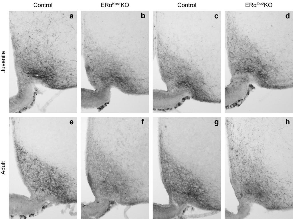
106 96
107 97 Figure 20 - ARC kisspeptin peptide levels are unchanged by ablation of ERα from either Kiss1- or Tac2- expressing neurons Kisspeptin immunoreactivity in the ARC of a juvenile (21-day-old) (a) littermate control and a (b) ERα Kiss1 KO female. Kisspeptin immunoreactivity in the ARC of a juvenile (c) control and a (d) ERα Tac2 KO female. Kisspeptin immunoreactivity in the ARC of an adult ( 9 weeks old) (e) control and a (f) ERα Kiss1 KO female. Kisspeptin immunoreactivity in the ARC of an adult (g) control and a (h) ERα Tac2 KO female. ARC, Arcuate nucleus.
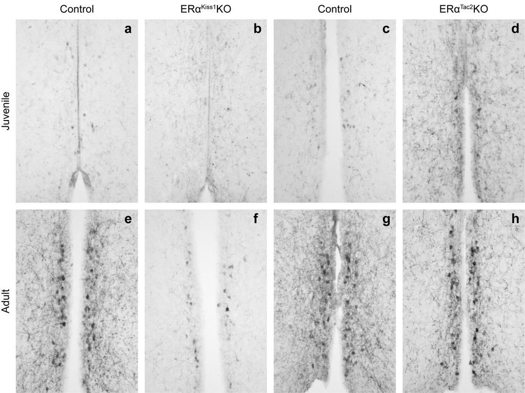
108 98
109 99 Figure 21 AVPV/PeN kisspeptin peptide levels are reduced when ERα is ablated from Kiss1-expressing neurons Kisspeptin immunoreactivity in the AVPV of a juvenile (21-day-old) (a) control and a (b) ERα Kiss1 KO female. Kisspeptin immunoreactivity in the AVPV of a juvenile (c) control and a (d) ERα Tac2 KO female. Kisspeptin immunoreactivity in the AVPV of an adult ( 9 weeks old) (e) control and a (f) ERα Kiss1 KO female. Kisspeptin immunoreactivity in the AVPV of an adult (g) control and a (h) ERα Tac2 KO female. AVPV/PeN, anteroventral periventricular/periventricular nucleus.
110 Chapter 4 Direct estrogen action on Kiss1/ERα neurons is required for negative feedback on the reproductive axis of male mice In males, the testes exert negative feedback effects on the hypothalamopituitary system, modulating the release of gonadotropin-releasing hormone (GnRH) from the hypothalamus and the subsequent release of the gonadotropins luteinizing hormone (LH) and follicle-stimulating hormone (FSH) from the anterior pituitary (Figure 1). This effect is demonstrated by experiments in which removal of the testes (and thus circulating testosterone) results in substantial increases in LH and FSH levels; the increase in gonadotropin levels can be attenuated by administration of testosterone As testosterone can act either directly through the androgen receptor (AR) or be aromatized to estradiol and then act through either isoform of the estrogen receptor (ERα or ERβ), many studies have sought to determine which effects of testosterone require action via AR and which require one of the estrogen receptor isoforms. The suppression of LH levels by testosterone treatment in a castrated In these studies, Dr. Courtney Marsh performed the quantification of the colocalization of ERα and eyfp in both mouse lines as well as assisting with the dissections at the end of the longitudinal study. Luhong Wang performed the gene expression analysis. 100
111 male is actually an estradiol-dependent phenomenon since it doesn t occur if dihydrotestosterone (DHT), a non-aromatizable androgen, is administered instead of testosterone 152. The elevated LH levels in a castrated male can be suppressed using estradiol treatment in wild-type, but not in mice lacking ERα globally (αerko mice) 48,152, implicating ERα in gonadal hormone negative feedback in the male. As fertility is not affected in male mice lacking ERβ globally, ERα appears to be the estrogen receptor isoform that plays a critical role in the male reproductive axis 13. It s generally accepted that GnRH neurons themselves don t possess functional ERα, so the identity of the ERα-expressing neurons in the hypothalamus that relay sex steroid signals to the GnRH neurons has not yet been elucidated. It has been proposed that neurons in the arcuate nucleus (ARC) that synthesize the neuropeptide kisspeptin may be the ERα-expressing neurons responsible for negative feedback regulation of GnRH neurons in male mice 45. This hypothesis is based solely on the regulation of ARC Kiss1 mrna levels by testosterone; while castration leads to increased Kiss1 expression, treatment with testosterone after the castration significantly reduces Kiss1 levels 45. While DHT treatment also reduces ARC Kiss1 levels, the small magnitude of this reduction suggests that the majority of the negative regulation of ARC Kiss1 expression requires an estrogen receptor. As is the case in females, the majority of ARC kisspeptin neurons in the male produce two other neuropeptides: neurokinin B (product of the gene Tac2) and dynorphin A (product of the gene Pdyn) 48. As a result, these neurons have been termed KNDy neurons. 101
112 There is a second population of hypothalamic kisspeptin neurons in the anteroventral periventricular/periventricular nucleus (AVPV/PeN) which does not express either Tac2 of Pdyn. Changing testosterone levels have the opposite effect on Kiss1 mrna levels in the AVPV/PeN; castration reduces Kiss1 levels while testosterone or estrogen- but not DHT- treatment normalizes them 45. The physiological relevance of this differential regulation of Kiss1 expression in males is unknown at this time, although some have suggested that the ARC kisspeptin neurons may be involved in testosterone-mediated negative feedback control of gonadotropin secretion, whereas the AVPV/PeN kisspeptin neurons are simply a remnant of the estrogen positive feedback system that is present only in females for the purpose of generating the preovulatory LH surge 45. In order to test whether ERα in ARC kisspeptin neurons is required for proper regulation of gonadotropin secretion and overall function of the male reproductive axis, we performed a conditional genetic deletion of ERα from all Kiss1-expressing neurons and compared the resulting phenotype to that of mice lacking ERα only in ARC KNDy neurons. Our results do not support the hypothesis that ERα in ARC KNDy neurons functions as part of the gonadal negative feedback system. Instead, it appears that a population of non-tac2- expressing kisspeptin/erα neurons is responsible for estrogen negative feedback in the male. 102
113 Methods Animals All animals were bred in our colony in the Unit for Laboratory Animal Medicine at the University of Michigan. All animals and procedures used were in accordance with the guidelines and approval of the University Committee on the Care and Use of Animals. Generation of Kiss1 IRES-cre and Tac2 IRES-cre mice The generation of mouse lines containing IRES-cre coding sequence knocked in to the 3 untranslated regions of the endogenous mouse Kiss1 and Tac2 alleles has been described previously (see Chapter 3). Mouse model validation Kiss1 IRES-cre/+ and Tac2 IRES-cre/+ male mice were bred to homozygous creinducible eyfp reporter females (Jax ) to generate Kiss1 eyfp and Tac2 eyfp animals respectively, allowing visualization and quantification of creexpressing cells as eyfp expression is restricted to cells that express cre recombinase. Perfusion and immunohistochemistry Adult and juvenile (3-week-old) mice received an overdose of sodium pento-barbital and were then perfused transcardially with phosphate-buffered saline (PBS, ph 7.4) followed by 10% formalin. Testes and seminal vesicles 103
114 (from adult animals only) were isolated and weighed. The brain was removed and post-fixed in 10% formalin for 2-4 hours and then dehydrated in 30% sucrose in PBS until the time of sectioning. Brains were cut in 30 µm coronal sections on a sliding microtome, collected in four representative series, and either stored at - 20 C in cryoprotectant. For eyfp, kisspeptin and ERα immunostaining, sections were washed, blocked in normal donkey serum and then incubated in primary antibodies (chicken anti-gfp, Abcam, 1:1000; rabbit anti-erα, Santa Cruz, 1:1000; rabbit anti-kisspeptin, Millipore, 1:2000) overnight. Brain sections were then thoroughly washed and incubated with Alexa Fluor-conjugated secondary antibodies (goat anti-chicken 488 and donkey anti-rabbit 568, Invitrogen, 1:250) for two hours. Sections were mounted onto gelatin-coated slides and coverslipped with ProLong Antifade mounting medium (Invitrogen). Longitudinal study Cre-positive males (either Kiss1 IRES-cre/+ or Tac2 IRES-cre/+ ) were bred to ERα flox/flox females. The resulting Kiss1 IRES-cre/+ / ERα flox/+ or Tac2 IRES-cre/+ / ERα flox/+ males and ERα flox/+ females were bred to each other to generate Kiss1- or Tac2- specific ERα knockouts (Kiss1 IRES-cre/+ / ERα flox/flox, ERα Kiss1 KO or Tac2 IRES-cre/+ / ERα flox/flox, ERα Tac2 KO ) and their littermate controls ERα flox/flox and Kiss1 IRES-cre/+ or Tac2 IRES-cre/+. Genotyping was performed as previously described for the females (Chapter 3). Male mice were individually housed beginning at PND21 and body weight 104
115 was recorded weekly. Body composition at 8 weeks of age was determined by a NMR-based Minispec LF90II (Bruker Optics) analyzer. At 9 weeks of age, mice were dissected between 13:00 and 16:00. Blood was collected after an overdose of sodium pentobarbital was given and allowed to clot for 30 minutes at room temperature. It was then centrifuged for 15 minutes at 2,000 x g and the serum was collected and stored at -20 C. Using a mouse brain matrix, the ARC and a block of tissue containing the AVPV/PeN as well as the preoptic area were dissected and individually snap-frozen on dry ice. Brain tissue was stored at -80 for later RNA extraction. RNA was extracted from microdissected brain tissue using TRIzol (Invitrogen) and then converted to cdna with the iscript cdna Synthesis Kit (Bio-Rad). cdna was analyzed in triplicate by quantitative realtime-pcr using Gapdh (endogenous control), Kiss1, pdyn, Tac2 and GnRH Taqman assays (Invitrogen). Relative expression levels were calculated using the 2 -ΔΔCt method, with normalization of each sample s ΔCt value to the average ΔCt of the control samples. Microscopy and image analysis Microscopic images were obtained using an Olympus BX-51 microscope with a DP30BW camera (Olympus). Dual-channel images were pseudo-colored and merged using Adobe Photoshop. Hormone analysis 105
116 Serum was analyzed by the Ligand Assay and Analysis Core of the University of Virginia Center for Research in Reproduction or the University of Michigan Chemistry Core. Testosterone levels were determined by RIA (catalog # TKTT2, Siemens) while LH and FSH levels were determined using the Millipore MILLIPLEX MAP rat pituitary panel. Statistics Student s t-test was used when only two groups were compared. Oneway ANOVA with Bonferroni post-hoc analysis was used when comparing three or more groups. Repeated measures two-way ANOVA was used to analyze longitudinal body weight. Data was analyzed and graphs were generated using either OriginPro 8 or GraphPad Prism software. Differences were deemed significant if p <.05. Data are presented as mean±sem. 106
117 Results Generation of mouse models to study Kiss1- and Tac2-expressing neurons in the male mouse brain To study the physiological role of hypothalamic ERα in the male mouse, we utilized the same mouse models described previously (Chapter 3). Briefly, the coding sequence for an IRES-cre cassette was knocked in to the endogenous Tac2 (Figure 22 a) and Kiss1 (Figure 22 d) loci. This results in the production of cre recombinase specifically in cells that express either of those genes. To visualize the pattern of cre expression, Kiss1 IRES-cre and Tac2 IRES-cre mice were bred to commercially available cre-mediated fluorescent reporter mice (Figure 22 b). Excision of the transcriptional blocker located between the loxp sites allows production of the fluorescent protein eyfp in all cre-expressing cells. The two cre-expressing mouse lines were also crossed to a mouse line that contains loxp sites flanking the third exon of the Esr1 gene (Figure 22 c). Cremediated recombination around these sites results in excision of the coding sequence for the DNA binding domain of ERα, resulting in ablation of ERα in all cre-expressing neurons. We assessed the pattern of cre expression in both Tac2 eyfp and Kiss1 eyfp male mice (Figure 23). As expected, we saw eyfp immunoreactivity in the ARC (Figure 23 a) and PeN (Figure 23 b) in a Kiss1 eyfp male brain. In this mouse line, 96±0.2% (N = 5) of eyfp cells in the ARC and 20±2.8% (N = 5) of eyfp cells in the AVPV/PeN co-express ERα. In agreement with the known 107
118 expression of Tac2, the expression of eyfp in a Tac2 eyfp male brain was more extensive than what we observed in Kiss1 eyfp mice, but the colocalization of eyfp and ERα was mostly restricted to the ARC (Figure 23 c), where 95±0.5% (N = 3) of Tac2 cells also express ERα. We did not identify any eyfp/erα cells in the AVPV/PeN of the Tac2 eyfp male brain (Figure 23 d). In the adult Kiss1 eyfp brain, not all eyfp cells contained detectable kisspeptin peptide, although the presence of eyfp suggests that expression of Kiss1 is transient in these cells. Although staining in the ARC of a Kiss1 eyfp male reveals an abundance of kisspeptin-immunoreactive fibers (data not shown), staining in the vicinity of the AVPV/PeN revealed no kisspeptin-ir cells and few fibers but several eyfp-ir cells (Figure 24 a). The absence of kisspeptin immunoreactivity in males is not because of technical issues because we can readily detect kisspeptin cells and fiber in the AVPV/PeN of female Kiss1 eyfp mice using the same experimental method (Figure 24 b). We also detect some eyfp cells in the female that aren t currently producing detectable levels of kisspeptin peptide. These findings reflect the fact that there are neurons in the vicinity of the AVPV/PeN that only transiently express Kiss1, but may express ERα and thus still play a role in the regulation of the reproductive axis. Generation of Kiss1- and Tac2-specific ERα knockout animals To generate Kiss1-specific ERα knockout mice, we first crossed Kiss1 IREScre heterozygotes to Esr1-flox homozygotes (ERα flox/flox mice) and then crossed the resultant Kiss1 IRES-cre/+ /ERα flox/+ males to ERα flox/+ females. The F2 progeny 108
119 consisted of Kiss1 IRES-Cre/+ /ERα flox/flox ( ERα Kiss1 KO ) mice, ERα flox/flox mice (without the modified Kiss1 allele) and Kiss1 IRES-Cre/+ mice (without a modified Esr1 allele). The latter two genotypes were used as littermate controls to the ERα Kiss1 KO animals in all studies. When appropriate, the two groups of control genotypes were combined into one larger group. The same breeding strategy was used to generate Tac2-specific ERα knockout mice, eventually generating littermate Tac2 IRES-Cre/+ /ERα flox/flox ( ERα Tac2 KO ) mice, ERα flox/flox mice (without the modified Tac2 allele) and Tac2 IRES-Cre/+ mice (without a modified Esr1 allele). Body mass and body composition Global ERα knockout male mice have slightly decreased body mass but increased white adipose tissue mass 155,156. We assessed body mass in our male mice from the time of weaning (3 weeks) until the time of dissection and body composition was determined in adult animals prior to dissection (Figure 25). We found absolutely no effect of ablation of ERα from Kiss1-expressing cells on body mass (Figure 25 a) or adult body composition (Figure 25 b). Similarly, ablation of ERα from all Tac2-expressing cells had no effect on body mass (Figure 25 c) or adult body composition (Figure 25 d). Thus estrogen action through ERα in Kiss1 or Tac2 cells is not necessary for the proper regulation of body mass or body composition in males. Reproductive organ weights The effect of global deletion of ERα on reproductive organ weights in 109
120 males has yielded mixed results 155,157,158. We found testes weight to be unchanged in adult ERα Kiss1 KO males compared to their littermate controls (Figure 26 a). The seminal vesicles (accessory sex glands that produce the fluid that becomes the copulatory plug) are enlarged as a result of ablation of ERα from Kiss1 neurons (Figure 26 b). This effect on seminal vesicle weight is not seen in global ERα knockouts 152,158. Since we had observed early puberty onset in females lacking ERα in either Kiss1 or Tac2 neurons (Figure 11), we measured testes weight of 3 week old male mice as an indicator of sexual maturation. We found that the testes of young ERα Kiss1 KO males weighed more than those of their littermate controls (Figure 26 c). While adult male ERα Tac2 KO mice had unaffected testes (Figure 26 d) and seminal vesicle (Figure 26 e) weights, we saw increased testes weight in juvenile ERα Tac2 KOs compared to their littermate controls (Figure 26 f). These significant changes in juvenile testes weight indicate a potential acceleration in sexual maturation as a result of selective ERα ablation. Gonadotropin and gonadal steroid hormone levels Alterations in gonadotropin and gonadal hormone levels have been reported in male mice with decreased estrogen action, but the findings are inconsistent 152,158. In adult ERα Kiss1 KO males, we found significantly elevated LH levels, decreased FSH levels and elevated testosterone levels (Figure 27 a- c). In contrast, we found no significant changes in any of these hormones in ERα Tac2 KO adults (Figure 27 d-f), suggesting the existence of a population of 110
121 Kiss1/ERα neurons which are critical for estrogen negative feedback on gonadotropin secretion but are separate from ARC Kiss1/Tac2 neurons. Gene expression Expression of certain reproduction-related genes in the ARC and in the rostral hypothalamus are sensitive to gonadal steroid levels in the male mouse 45,48,152. Since testosterone can act not only directly through the androgen receptor but also be aromatized to estrogen and act through ERα, we assessed relative expression levels of Kiss1, Tac2 and Pdyn in the ARC and Kiss1 and Gnrh1 in the rostral hypothalamus in our mice. In mice lacking ERα in all Kiss1- expressing neurons, we found no effect on expression of Kiss1 or Gnrh1 in the rostral hypothalamus (Figure 28 a) or on expression of Kiss1, Tac2 or Pdyn in the ARC (Figure 28 b). Although we observed a significant increase in AVPV/PeN Kiss1 expression in ERα Tac2 KO males compared to littermate controls, the magnitude of this increase is very small (Figure 28 c). Similar to what we found in the ERα Kiss1 KO males, we observed no effect on Gnrh1 expression (Figure 28 c) in ERα Tac2 KO males, or any of the three genes (Kiss1, Tac2, Pdyn) we investigated in the ARC (Figure 28 d). Thus, ablation of ERα from either all Kiss1-expressing neurons or just from Tac2-expressing neurons has no virtually no detectable effect on the expression of genes in the hypothalamus that are known to be important modulators of GnRH neuron function. 111
122 Conclusions & Discussion Studies in a multitude of species have shown that the central actions of estradiol, the aromatization product of testosterone, play a critical role in the regulation of pulsatile gonadotropin release and thus the entire male reproductive axis. Yet the identity and location of the estrogen receptor-expressing neuronal population required for this effect has not yet been uncovered. As it doesn t appear that GnRH neurons themselves are responsible for this effect of estradiol on the system, we hypothesized that neurons producing the neuropeptide kisspeptin could be the critical population. To address the putative contribution of direct estrogen action on kisspeptin neurons to the regulation of the reproductive axis, we used cre-loxp technology to genetically ablate ERα from either all Kiss1 neurons or from only the ARC kisspeptin neurons which coexpress Tac2. We saw almost no effect of deletion of ERα from only ARC KNDy neurons in males. While juvenile (3-week-old) ERα Tac2 KO males had slightly elevated testes weight, this difference was absent in adults. Although this may indicate accelerated puberty onset, the age at balano-preputial separation was not assessed in these studies and should be addressed in the future. The adult ERα Tac2 KO males exhibited no significant hormonal changes and only a very slight (but significant) increase in Kiss1 expression in the AVPV/PeN. In contrast with the underwhelming phenotype of the ERα Tac2 KO males, ERα Kiss1 KO males do show signs of impaired negative feedback on the 112
123 reproductive system. Adult ERα Kiss1 KO males have elevated LH and testosterone levels, indicating reduced restraint on the anterior pituitary. This was surprising since Mayer and colleagues previously found no changes in LH in their adult knockouts 46. This discrepancy may be due to subtle differences in cre expression between the two different Kiss1 IRES-cre lines used or slightly different study conditions. In agreement with previous studies of αerko males, we did not observe any changes in adult testes weight but their seminal vesicles were significantly heavier than their littermate controls. The ERα Kiss1 KOs elevated testosterone levels may be the cause of this effect since seminal vesicle weight is sensitive to circulating testosterone 152. When we investigated the effect of conditional ERα deletion on hypothalamic gene expression we found a small but significant increase in AVPV/PeN Kiss1 expression in ERα Tac2 KO males. This is likely due to the slightly (but not significantly [p =.08]) elevated testosterone levels in these mice, which stimulates Kiss1 expression in this area in an aromatase-dependent manner 45. There were no significant alterations in any of the other genes we analyzed in the hypothalamus of ERα Kiss1 KO males. If the loss of estrogen action through ERα on kisspeptin neurons resulted in a complete loss of negative feedback, we would expect effects on gene expression that mimic the effect of castration. This result was unlikely in our mouse models since negative feedback also occurs through the androgen receptor which we have not manipulated here. While others have reported that there are more Tac2-expressing neurons 113
124 in the male ARC than there are Tac2/Kiss1 neurons 48, we didn t observe any evidence of this in our comparison of Tac2 eyfp and Kiss1 eyfp mice. The neurons affected by our deletion of ERα from all Kiss1 neurons include all those affected by the Tac2-specific deletion with the addition of any non-tac2-expressing Kiss1/ERα neurons. We believe that the phenotypic differences between our two conditional ERα knockouts are a result of the non-tac2-expressing Kiss1/ERα cells in the AVPV/PeN that expressed Kiss1 at some point during development, but no longer produce detectable levels of the peptide. More work is needed to characterize these neurons and better determine their function in the regulation of the male reproductive system. Additionally, the development of an inducible Kiss1 IRES-cre mouse line would allow researchers to further investigate this system without the potential confound of the neurons that only transiently express Kiss1 at some point during development. Using mouse models that allow genetic manipulation of either all Kiss1- expressing or only ARC Kiss1/Tac2 neurons, we have uncovered a critical role of direct estrogen action on non-tac2-expressing Kiss1/ERα neurons in negative feedback on the male reproductive system. Further study of these neurons is needed in order to determine the mechanism by which they affect GnRH output from the hypothalamus and thus regulate the functioning of the male reproductive axis. 114
125 Figure 22 - Generation of mouse models to study Kiss1- and Tac2- expressing neurons in males (a) Tac2 IRES-cre mice carry an IRES-cre cassette in the 3 UTR of the endogenous murine Tac2 gene. (b) Tac2 eyfp mice were generated by crossing the Tac2 IREScre mouse to a commercially available ROSA26 fluorescent reporter strain (Jax ), which after recombination around the loxp sites results in eyfp production in all cre-expressing cells and thus fluorescent labeling of Tac2- expressing cells. (c) Tac2-specific ERα knockout animals ( ERα Tac2 KO ) were generated by crossing the Tac2 IRES-cre mice to Esr1-flox mice which have loxp sites flanking exon 3 which contains the sequence for the DNA binding domain of the receptor 142. Cre-mediated recombination around the loxp sites results in a Tac2 cell-specific ERα null mouse. (d) Kiss1 IRES-cre mice carry the same IREScre cassette in the 3 UTR of the murine Kiss1 gene. They were used to generate Kiss1 eyfp and ERα Kiss1 KO mice. IRES, internal ribosome entry site; pa, polyadenylation site; frt, flippase recognition target. 115
 PeN of a male Kiss1 eyfp mouse. eyfp and ERα immunoreactivity in the (c) ARC and (d) PeN of a male Tac2 eyfp mouse.")
126 Figure 23 Co-expression of eyfp and ERα in the hypothalamus of male Kiss1 eyfp and Tac2 eyfp mice eyfp and ERα immunoreactivity (pseudocolored green and purple respectively) in the (a) ARC and the (b) PeN of a male Kiss1 eyfp mouse. eyfp and ERα immunoreactivity in the (c) ARC and (d) PeN of a male Tac2 eyfp mouse. Neurons expressing both eyfp and ERα appear white and some are indicated with a white arrow. 3V, third ventricle; ME, median eminence. Scale bar = 200 μm. 116
 and a female (b) Kiss1 eyfp adult mouse. Neurons expressing both eyfp and kisspeptin appear white and some are denoted with a white arrow. 3V, third ventricle.")
127 117 Figure 24 - Kisspeptin immunoreactivity in the rostral hypothalamus of Kiss1 eyfp male and female mice eyfp (pseudo-colored green) and kisspeptin (pseudo-colored purple) immunoreactivity in the vicinity of the AVPV/PeN of a male (a) and a female (b) Kiss1 eyfp adult mouse. Neurons expressing both eyfp and kisspeptin appear white and some are denoted with a white arrow. 3V, third ventricle. Scale bar = 200 μm.
128 118 Figure 25 - Body mass and composition are unchanged in ERα Kiss1 KO and ERα Tac2 KO male mice (a) Body mass of littermate controls (Kiss1 IRES-Cre/+ and ERα flox/flox ; Control ; filled squares) and Kiss1-specific ERα knockout ( ERα Kiss1 KO ; open circles) male mice from 3-9 weeks of age (n = 10-11). (b) Body composition of 8-week-old control and ERα Kiss1 KO mice (n = 10-11). (c) Body mass of littermate controls (Tac2 IRES-Cre/+ and ERα flox/flox ; Control ; filled squares) and Tac2-specific ERα knockout ( ERα Tac2 KO ; open circles) mice from 3-9 weeks of age (n = 8-20). (d) Body composition of 8-week-old control and ERα Tac2 KO mice (n = 8-9).
 (n = 10-14). (c) Testes weight of juvenile (3-week-old) ERα Kiss1 KO and littermate controls (n = 8-13).")
129 Figure 26 Male reproductive organ weights are affected by ablation of ERα from either Kiss1- or Tac2-expressing neurons (a) Testes and (b) seminal vesicle weights of adult ( 9 weeks old) ERα Kiss1 KO and littermate controls (Kiss1 IRES-Cre/+ and ERα flox/flox ) (n = 10-14). (c) Testes weight of juvenile (3-week-old) ERα Kiss1 KO and littermate controls (n = 8-13). (d) Testes and (e) seminal vesicle weights of adult ( 9 weeks old) ERα Tac2 KO and littermate controls (n = 8-12). (f) Testes weight of juvenile (3-week-old) ERα Tac2 KO and littermate controls (n = 7-8). All tissue weights are normalized to body weight (g/g). Student s t test: **, p<.01; ***, p<
 levels in adult ERα Kiss1 KO and littermate control (Kiss1 IRES-Cre/+ and ERα flox/flox ) males.")
130 Figure 27 Gonadotropin and gonadal hormone levels are altered by ablation of ERα from all Kiss1- but not Tac2-expressing neurons Serum (a) LH (n = 11-17), (b) FSH (n = 10-20) and (c) testosterone (n = 11-15) levels in adult ERα Kiss1 KO and littermate control (Kiss1 IRES-Cre/+ and ERα flox/flox ) males. Serum (d) LH (n = 9-19), (e) FSH (n = 10-19) and (f) testosterone (n = 10-18) levels in adult ERα Tac2 KO and littermate control males. Student s t test: *, p<.05; **, p<.01. LH, luteinizing hormone; FSH, follicle-stimulating hormone. 120
 Kiss1, Tac2 and Pdyn expression in the ARC of ERα Kiss1 KO and littermate control (Tac2 IRES-Cre/+ and ERα flox/flox ) males (n = 10-20).")
131 Figure 28 - Gene expression in the rostral hypothalamus and ARC (a) Kiss1 and Gnrh1 expression in the rostral hypothalamus of ERα Kiss1 KO and littermate control males (n = 11-20). (b) Kiss1, Tac2 and Pdyn expression in the ARC of ERα Kiss1 KO and littermate control (Tac2 IRES-Cre/+ and ERα flox/flox ) males (n = 10-20). (c) Relative expression of Kiss1 and GnRH in the rostral hypothalamus of ERα Tac2 KO and littermate control males (n = 9-19). (d) Kiss1, Tac2 and Pdyn expression in the ARC of ERα Tac2 KO and littermate control males (n = 8-19). Expression levels in KO animals are normalized to Control levels. Student s t test: **, p<.01. ARC, arcuate nucleus. 121
Reproductive cyclicity 19. Introduction. Page 1. repro and its story lines. Male repro: a simpler way of control
 Reproductive cyclicity 19 Male repro: a simpler way of control Menstrual cycles: ovary / uterine anatomy and cell types, follicular phase, ovulation, luteal phase, cyclicity Race events: removal of P4
Reproduction. Introduction
 Reproduction The goal of these lectures is to discuss basic physiology associated with the control of reproduction (from sexual diferentiation to adult reproductive function). 26 The sections for this
Reproductive physiology
 Reproductive physiology Sex hormones: Androgens Estrogens Gestagens Learning objectives 86 (also 90) Sex Genetic sex Gonadal sex Phenotypic sex XY - XX chromosomes testes - ovaries external features Tha
Menstrual Cycle. Last example of how a circle works. Course Outline. Topic #! Topic lecture! Silverthorn! Membranes (pre-requisite material)!!
!!") The goal of these lectures is to discuss how control system is formed and operates. For this, basic physiology associated with the control the menstrual cycle will be used. The sections for this lecture
The goal of these lectures is to discuss how control system is formed and operates. For this, basic physiology associated with the control the menstrual cycle will be used. The sections for this lecture
Hormonal Control of Human Reproduction
 Hormonal Control of Human Reproduction Bởi: OpenStaxCollege The human male and female reproductive cycles are controlled by the interaction of hormones from the hypothalamus and anterior pituitary with
Peripubertal, leptin-deficient ob/ob female mice were used in an investigation of
 ESSICK-BROOKSHIRE, ELIZABETH ANN, M.S. The Effects of Peripherally Administered 17-β Estradiol and BIBP3226, a NPY Y1 Receptor Antagonist, on Food Intake, Body Mass, Reproductive Development and Behavior
CASE 41. What is the pathophysiologic cause of her amenorrhea? Which cells in the ovary secrete estrogen?
 CASE 41 A 19-year-old woman presents to her gynecologist with complaints of not having had a period for 6 months. She reports having normal periods since menarche at age 12. She denies sexual activity,
Reproductive Hormones
 Reproductive Hormones Male gonads: testes produce male sex cells! sperm Female gonads: ovaries produce female sex cells! ovum The union of male and female sex cells during fertilization produces a zygote
Reproductive FSH. Analyte Information
 Reproductive FSH Analyte Information 1 Follicle-stimulating hormone Introduction Follicle-stimulating hormone (FSH, also known as follitropin) is a glycoprotein hormone secreted by the anterior pituitary
Animal and Veterinary Science Department University of Idaho. REGULATION OF REPRODUCTION AVS 222 (Instructor: Dr. Amin Ahmadzadeh) Chapter 5
 Chapter 5") Animal and Veterinary Science Department University of Idaho REGULATION OF REPRODUCTION AVS 222 (Instructor: Dr. Amin Ahmadzadeh) Chapter 5 I. DEFINITIONS A. Endocrine Gland B. Hormone Chemical messenger
Animal and Veterinary Science Department University of Idaho REGULATION OF REPRODUCTION AVS 222 (Instructor: Dr. Amin Ahmadzadeh) Chapter 5 I. DEFINITIONS A. Endocrine Gland B. Hormone Chemical messenger
BIOL212 Biochemistry of Disease. Metabolic Disorders - Obesity
 BIOL212 Biochemistry of Disease Metabolic Disorders - Obesity Obesity Approx. 23% of adults are obese in the U.K. The number of obese children has tripled in 20 years. 10% of six year olds are obese, rising
SISTEMA REPRODUCTOR (LA IDEA FIJA) Copyright 2004 Pearson Education, Inc., publishing as Benjamin Cummings
 Copyright 2004 Pearson Education, Inc., publishing as Benjamin Cummings") SISTEMA REPRODUCTOR (LA IDEA FIJA) How male and female reproductive systems differentiate The reproductive organs and how they work How gametes are produced and fertilized Pregnancy, stages of development,
SISTEMA REPRODUCTOR (LA IDEA FIJA) How male and female reproductive systems differentiate The reproductive organs and how they work How gametes are produced and fertilized Pregnancy, stages of development,
Molecular and neural mechanisms of leptin action: Focus on Reproduction
 Molecular and neural mechanisms of leptin action: Focus on Reproduction Martin G. Myers, Jr., M.D., Ph.D. and collaborators Division of Metabolism, Endocrinology and Diabetes Department of Internal Medicine
WEIGHT GAIN DURING MENOPAUSE EMERGING RESEARCH
 MENOPAUSE WHEN DOES IT OCCUR? The cessation of the menstrual cycle for one year. WEIGHT GAIN DURING MENOPAUSE EMERGING RESEARCH Jan Schroeder, Ph.D. Chair of The Department of Kinesiology California State
Chapter 14 Reproduction Review Assignment
 Date: Mark: _/45 Chapter 14 Reproduction Review Assignment Multiple Choice Identify the choice that best completes the statement or answers the question. 1. Use the diagram above to answer the next question.
REPRODUCCIÓN. La idea fija. Copyright 2004 Pearson Education, Inc., publishing as Benjamin Cummings
 REPRODUCCIÓN La idea fija How male and female reproductive systems differentiate The reproductive organs and how they work How gametes are produced and fertilized Pregnancy, stages of development, birth
Hormones of brain-testicular axis
 (Hormone Function) Hormones of brain-testicular axis anterior pituitary drives changes during puberty controlled by GnRH from hypothalamus begins to secrete FSH, LH LH targets interstitial endocrinocytes
SCHOOL OF MEDICINE AND HEALTH SCIENCES DIVISION OF BASIC MEDICAL SCIENCES DISCIPLINE OF BIOCHEMISTRY & MOLECULAR BIOLOGY
 1 SCHOOL OF MEDICINE AND HEALTH SCIENCES DIVISION OF BASIC MEDICAL SCIENCES DISCIPLINE OF BIOCHEMISTRY & MOLECULAR BIOLOGY PBL SEMINAR: SEX HORMONES PART 1 An Overview What are steroid hormones? Steroid
PedsCases Podcast Scripts
 PedsCases Podcast Scripts This is a text version of a podcast from Pedscases.com on Puberty and Pubertal Disorders Part 2: Precocious Puberty. These podcasts are designed to give medical students an overview
9.4 Regulating the Reproductive System
 9.4 Regulating the Reproductive System The Reproductive System to unite a single reproductive cell from a female with a single reproductive cell from a male Both male and female reproductive systems include
FLASH CARDS. Kalat s Book Chapter 11 Alphabetical
 FLASH CARDS www.biologicalpsych.com Kalat s Book Chapter 11 Alphabetical alpha-fetoprotein alpha-fetoprotein Alpha-Fetal Protein (AFP) or alpha-1- fetoprotein. During a prenatal sensitive period, estradiol
MULTIPLE CHOICE: match the term(s) or description with the appropriate letter of the structure.
 or description with the appropriate letter of the structure.") Chapter 27 Exam Due NLT Thursday, July 31, 2015 Name MULTIPLE CHOICE: match the term(s) or description with the appropriate letter of the structure. Figure 27.1 Using Figure 27.1, match the following:
Chapter 27 Exam Due NLT Thursday, July 31, 2015 Name MULTIPLE CHOICE: match the term(s) or description with the appropriate letter of the structure. Figure 27.1 Using Figure 27.1, match the following:
Reproductive physiology. About this Chapter. Case introduction. The brain directs reproduction 2010/6/29. The Male Reproductive System
 Section Ⅻ Reproductive physiology Ming-jie Wang E-Mail: mjwang@shmu.edu.cn About this Chapter The reproductive organs and how they work the major endocrine functions of sexual glands actions of sex hormones
Chapter 27 The Reproductive System. MDufilho
 Chapter 27 The Reproductive System 1 Figure 27.19 Events of oogenesis. Before birth Meiotic events 2n Oogonium (stem cell) Mitosis Follicle development in ovary Follicle cells Oocyte 2n Primary oocyte
Hypothalamus & Pituitary Gland
 Hypothalamus & Pituitary Gland Hypothalamus and Pituitary Gland The hypothalamus and pituitary gland form a unit that exerts control over the function of several endocrine glands (thyroid, adrenals, and
KISSPEPTIN AND GNIH CONTROL OF GNRH IN FEMALE MAMMALS
 KISSPEPTIN AND GNIH CONTROL OF GNRH IN FEMALE MAMMALS M.J. Zamiri Department of Animal Science, College of Agriculture, Shiraz University, Shiraz, Iran mjzamiri@gmail.com Introduction Since the discovery
REPRODUCTIVE ENDOCRINOLOGY OF THE MALE
 Reproductive Biotechnologies Andrology I REPRODUCTIVE ENDOCRINOLOGY OF THE MALE Prof. Alberto Contri REPRODUCTIVE ENDOCRINOLOGY OF THE MALE SPERMATOGENESIS AND REPRODUCTIVE BEHAVIOR RELATED TO THE ACTIVITY
Leptin part 2. Mary ET Boyle
 Leptin part 2 Mary ET Boyle Leptin Feedback: leptin levels drop during starvation, when fat depots are depleted to support the organism s basic energy needs, leptin levels rise during refeeding where fat
Action of reproductive hormones through the life span 9/22/99
 Action of reproductive hormones through the life span Do reproductive hormones affect the life span? One hypothesis about the rate of aging asserts that there is selective pressure for either high rate
10.7 The Reproductive Hormones
 10.7 The Reproductive Hormones December 10, 2013. Website survey?? QUESTION: Who is more complicated: men or women? The Female Reproductive System ovaries: produce gametes (eggs) produce estrogen (steroid
Female Reproductive System. Lesson 10
 Female Reproductive System Lesson 10 Learning Goals 1. What are the five hormones involved in the female reproductive system? 2. Understand the four phases of the menstrual cycle. Human Reproductive System
Reproductive Health and Pituitary Disease
 Reproductive Health and Pituitary Disease Janet F. McLaren, MD Assistant Professor Division of Reproductive Endocrinology and Infertility Department of Obstetrics and Gynecology jmclaren@uabmc.edu Objectives
1 The cell. Checkup Chapter 5. (pp ) 1. Look at the following illustration.
 1. Look at the following illustration.") Checkup Chapter 5 1 The cell (pp. 126 131) 1. Look at the following illustration. a) What three cell components are visible under a light (optical) microscope? Cell membrane Cytoplasm Nucleus b) What is
Checkup Chapter 5 1 The cell (pp. 126 131) 1. Look at the following illustration. a) What three cell components are visible under a light (optical) microscope? Cell membrane Cytoplasm Nucleus b) What is
Reproductive System. Testes. Accessory reproductive organs. gametogenesis hormones. Reproductive tract & Glands
 Reproductive System Testes gametogenesis hormones Accessory reproductive organs Reproductive tract & Glands transport gametes provide nourishment for gametes Hormonal regulation in men Hypothalamus - puberty
REPRODUCTION & GENETICS. Hormones
 REPRODUCTION & GENETICS Hormones http://www.youtube.com/watch?v=np0wfu_mgzo Objectives 2 Define what hormones are; Compare and contrast the male and female hormones; Explain what each hormone in the mail
Why is my body not changing? Conflicts of interest. Overview 11/9/2015. None
 Why is my body not changing? Murthy Korada Pediatrician, Pediatric Endocrinologist Ridge Meadows Hospital Surrey Memorial Hospital None Conflicts of interest Overview Overview of normal pubertal timing
Reproductive Endocrinology. Isabel Hwang Department of Physiology Faculty of Medicine University of Hong Kong Hong Kong May2007
 Reproductive Endocrinology Isabel Hwang Department of Physiology Faculty of Medicine University of Hong Kong Hong Kong May2007 isabelss@hkucc.hku.hk A 3-hormone chain of command controls reproduction with
The reproductive lifespan
 The reproductive lifespan Reproductive potential Ovarian cycles Pregnancy Lactation Male Female Puberty Menopause Age Menstruation is an external indicator of ovarian events controlled by the hypothalamicpituitary
Biology of Reproduction- Zool 346 Exam 2
 Biology of Reproduction- Zool 346 Exam 2 ANSWER ALL THE QUESTIONS ON THE ANSWER SHEET. THE ANSWER ON THE ANSWER SHEET IS YOUR OFFICIAL ANSWER. Some critical words are boldfaced. This exam is 7 pages long.
Ch 11: Endocrine System
 Ch 11: Endocrine System SLOs Describe the chemical nature of hormones and define the terms proand prepro-hormone. Explain mechanism of action of steroid and thyroid hormones Create chart to distinguish
Kisspeptin and other neuropeptides. New opportunities for reproductive endocrinology Nobel Laureates. Richard A Anderson
 Kisspeptin and other neuropeptides Higher centres Timing of puberty Stress New opportunities for reproductive endocrinology E2 +ve E2 -ve Richard A Anderson Obstetrics and Gynaecology University of Edinburgh
Unit 15 ~ Learning Guide
 Unit 15 ~ Learning Guide Name: INSTRUCTIONS Complete the following notes and questions as you work through the related lessons. You are required to have this package completed BEFORE you write your unit
LH and FSH. Women. Men. Increased LH. Decreased LH. By Ronald Steriti, ND, PhD 2011
 LH and FSH By Ronald Steriti, ND, PhD 2011 Luteinizing hormone (LH) and follicle-stimulating hormone (FSH) are gonadotropins that stimulate the gonads - the testes in males, and the ovaries in females.
Female Reproductive System. Justin D. Vidal
 Female Reproductive System Justin D. Vidal If you cannot identify the tissue, then it is probably part of the female reproductive system! Introduction The female reproductive system is constantly changing,
Ayman Mesleh & Leen Alnemrawi. Bayan Abusheikha. Faisal
 24 Ayman Mesleh & Leen Alnemrawi Bayan Abusheikha Faisal We were talking last time about receptors for lipid soluble hormones.the general mechanism of receptors for lipid soluble hormones: 1. Receptors
Puberty and Pubertal Disorders Part 3: Delayed Puberty
 PedsCases Podcast Scripts This is a text version of a podcast from Pedscases.com on Puberty and Pubertal Disorders Part 3: Delayed Puberty These podcasts are designed to give medical students an overview
Study Guide Answer Key Reproductive System
 Biology 12 Human Biology Textbook: BC Biology 12 Study Guide Answer Key Reproductive System 1. Distinguish between a gamete and a gonad using specific examples from the male and female systems. Gonads
Ying Wan Master of Neuroscience
 The refinement of the pulsatile Luteinizing Hormone (LH) profile following pubertal maturation: a potential mechanism for enhanced recruitment of ovarian follicles for improved fertility Ying Wan Master
MULTIPLE CHOICE. Choose the one alternative that best completes the statement or answers the question.
 MULTIPLE CHOICE. Choose the one alternative that best completes the statement or answers the question. 1) Which of the following hormones controls the release of anterior pituitary gonadotropins? A) LH
Hormones. Prof. Dr. Volker Haucke Institut für Chemie-Biochemie Takustrasse 6
 Hormones Prof. Dr. Volker Haucke Institut für Chemie-Biochemie Takustrasse 6 Tel. 030-8385-6920 (Sekret.) 030-8385-6922 (direkt) e-mail: vhaucke@chemie.fu-berlin.de http://userpage.chemie.fu-berlin.de/biochemie/aghaucke/teaching.html
BIOLOGY - CLUTCH CH.45 - ENDOCRINE SYSTEM.
 !! www.clutchprep.com Chemical signals allow cells to communicate with each other Pheromones chemical signals released to the environment to communicate with other organisms Autocrine signaling self-signaling,
Functions of male Reproductive System: produce gametes deliver gametes protect and support gametes
 Functions of male Reproductive System: produce gametes deliver gametes protect and support gametes Spermatogenesis occurs in the testes after puberty. From the testes they are deposited into the epididymas
Testes (male gonads) -Produce sperm -Produce sex hormones -Found in a sac called the scrotum -Suspended outside of the body cavity for temperature
 -Produce sperm -Produce sex hormones -Found in a sac called the scrotum -Suspended outside of the body cavity for temperature") REPRODUCTION Testes (male gonads) -Produce sperm -Produce sex hormones -Found in a sac called the scrotum -Suspended outside of the body cavity for temperature reduction -Testes wall made of fibrous connective
REPRODUCTION Testes (male gonads) -Produce sperm -Produce sex hormones -Found in a sac called the scrotum -Suspended outside of the body cavity for temperature reduction -Testes wall made of fibrous connective
Reproductive System (Hormone Function) Physiology Department Medical School, University of Sumatera Utara
 Physiology Department Medical School, University of Sumatera Utara") Reproductive System (Hormone Function) Physiology Department Medical School, University of Sumatera Utara 1 Endocrine Control: Three Levels of Integration Hormones of the hypothalamic-anterior pituitary
Reproductive System (Hormone Function) Physiology Department Medical School, University of Sumatera Utara 1 Endocrine Control: Three Levels of Integration Hormones of the hypothalamic-anterior pituitary
TESTOSTERONE DEFINITION
 DEFINITION A hormone that is a hydroxyl steroid ketone (C19H28O2) produced especially by the testes or made synthetically and that is responsible for inducing and maintaining male secondary sex characteristics.
Chapter 22 The Reproductive System (I)
") Chapter 22 The Reproductive System (I) An Overview of Reproductive Physiology o The Male Reproductive System o The Female Reproductive System 22.1 Reproductive System Overview Reproductive system = all
Chapter 22 The Reproductive System (I) An Overview of Reproductive Physiology o The Male Reproductive System o The Female Reproductive System 22.1 Reproductive System Overview Reproductive system = all
Leptin levels and menstrual function in HIV-infected women in rural India
 Leptin levels and menstrual function in HIV-infected women in rural India Annie Phoebe. K¹, Mini Jacob.S¹, Hemalatha.R², Sivakumar M.R¹ ¹Department of Experimental Medicine, The Tamil Nadu Dr. MGR Medical
The reproductive system
 The reproductive system THE OVARIAN CYCLE HORMONAL REGULATION OF OOGENSIS AND OVULATION hypothalamic-pituitary-ovary axis Overview of the structures of the endocrine system Principal functions of the
Module J ENDOCRINE SYSTEM. Learning Outcome
 Module J ENDOCRINE SYSTEM Topic from HAPS Guidelines General functions of the endocrine system Chemical classification of hormones & mechanism of hormone actions at receptors. Control of hormone secretion
HCG (human chorionic gonadotropin); Novarel Pregnyl (chorionic gonadotropin); Ovidrel (choriogonadotropin alfa)
; Novarel Pregnyl (chorionic gonadotropin); Ovidrel (choriogonadotropin alfa)") Federal Employee Program 1310 G Street, N.W. Washington, D.C. 20005 202.942.1000 Fax 202.942.1125 5.08.09 Subject: HCG Page: 1 of 5 Last Review Date: June 19, 2015 HCG Powder, Novarel, Pregnyl, Ovidrel
Federal Employee Program 1310 G Street, N.W. Washington, D.C. 20005 202.942.1000 Fax 202.942.1125 5.08.09 Subject: HCG Page: 1 of 5 Last Review Date: June 19, 2015 HCG Powder, Novarel, Pregnyl, Ovidrel
Leptin Intro/Signaling. ATeamP: Angelo, Anthony, Charlie, Gabby, Joseph
 Leptin Intro/Signaling ATeamP: Angelo, Anthony, Charlie, Gabby, Joseph Overview Intro to Leptin Definition & Sources Physiology Bound vs. Free Receptors Signaling JAK/STAT MAPK PI3K ACC Experimental findings
Female Reproductive Physiology. Dr Raelia Lew CREI, FRANZCOG, PhD, MMed, MBBS Fertility Specialist, Melbourne IVF
 Female Reproductive Physiology Dr Raelia Lew CREI, FRANZCOG, PhD, MMed, MBBS Fertility Specialist, Melbourne IVF REFERENCE Lew, R, Natural History of ovarian function including assessment of ovarian reserve
The Role of Kisspeptin and KNDy Cells in the Reproductive Neuroendocrine System
 Western University Scholarship@Western Electronic Thesis and Dissertation Repository August 2013 The Role of Kisspeptin and KNDy Cells in the Reproductive Neuroendocrine System Christina M. Merkley The
Growth Hormone, Somatostatin, and Prolactin 1 & 2 Mohammed Y. Kalimi, Ph.D.
 Growth Hormone, Somatostatin, and Prolactin 1 & 2 Mohammed Y. Kalimi, Ph.D. I. Growth Hormone (somatotropin): Growth hormone (GH) is a 191 amino acid single chain polypeptide (MW 22,000 daltons). Growth
Chapter 14 The Reproductive System
 Biology 12 Name: Reproductive System Per: Date: Chapter 14 The Reproductive System Complete using BC Biology 12, page 436-467 14. 1 Male Reproductive System pages 440-443 1. Distinguish between gametes
I. Endocrine System & Hormones Figure 1: Human Endocrine System
 I. Endocrine System & Hormones Figure 1: Human Endocrine System Endocrine System: a) Endocrine glands are ductless since they lack specific vessels for the transport of hormones throughout the body. Instead,
Model Answer. M.Sc. Zoology (First Semester) Examination Paper LZT 103 (Endocrinology)
 Examination Paper LZT 103 (Endocrinology)") Model Answer M.Sc. Zoology (First Semester) Examination-2013 Paper LZT 103 (Endocrinology) Section A 1. (i) d (ii) b (iii) b (iv) c (v) c (vi) a (vii) c (viii) a (ix) d (x) b Section B Q.2 Answer Hormonal
Model Answer M.Sc. Zoology (First Semester) Examination-2013 Paper LZT 103 (Endocrinology) Section A 1. (i) d (ii) b (iii) b (iv) c (v) c (vi) a (vii) c (viii) a (ix) d (x) b Section B Q.2 Answer Hormonal
Fukushima-ku, Osaka. Synopsis. and LH release by investigating the effects of exogenous estrogen on the progesteroneinduced
 Further Studies on the Causal Relationship between the Secretion of Estrogen and the Release of Luteinizing Hormone in the Rat FUMIHIKO KOBAYASHI, KATSUMI HARA AND TAMOTSU MIYAKE Shionogi Research Laboratory,
Generating kisspeptin cell lines to investigate their role in reproduction
 Generating kisspeptin cell lines to investigate their role in reproduction Dakota C. Jacobs 1 Jadwiga M. Giebultowicz 2, and Patrick E. Chappell 3 1 Bioresource Research, 2 Department of Integrative Biology,
Sexual Development. 6 Stages of Development
 6 Sexual Development 6 Stages of Development Development passes through distinct stages, the first of which is fertilization, when one sperm enters one ovum. To enter an ovum, a sperm must undergo the
Physiology of Male Reproductive System
 Physiology of Male Reproductive System the anterior pituitary gland serves as the primary control of reproductive function at puberty Ant Pituitary secretes FSH & large amounts of LH (ICSH) FSH & LH cause
The menstrual Cycle. François Pralong
 The menstrual Cycle François Pralong Services d Endocrinologie, Diabétologie et Métabolisme, Hôpitaux Universitaires de Genève et Lausanne Centre des Maladies CardioVasculaires et Métaboliques, Lausanne
The Role of Leptin in Anorexia Nervosa: Clinical Implications THE INSULINATORS IRIS ULLOA, SUNDUS MOHAMED, KATIE NINH, AMANDA BUSTAMANTE
 The Role of Leptin in Anorexia Nervosa: Clinical Implications THE INSULINATORS IRIS ULLOA, SUNDUS MOHAMED, KATIE NINH, AMANDA BUSTAMANTE AN LEPTIN RELATED FINDINGS + PATHWAY Introduction What is leptin?
GONADAL FUNCTION: An Overview
 GONADAL FUNCTION: An Overview University of PNG School of Medicine & Health Sciences Division of Basic Medical Sciences Clinical Biochemistry BMLS III & BDS IV VJ Temple 1 What are the Steroid hormones?
Human Physiology 6.6- Hormones, Homeostasis, and Reproduction
 Human Physiology 6.6- Hormones, Homeostasis, and Reproduction Essential idea: Hormones are used when signals need to be widely distributed. Application: William Harvey s investigation of sexual reproduction
Web Activity: Simulation Structures of the Female Reproductive System
 differentiate. The epididymis is a coiled tube found along the outer edge of the testis where the sperm mature. 3. Testosterone is a male sex hormone produced in the interstitial cells of the testes. It
Animal Reproduction Chapter 46. Fission. Budding. Parthenogenesis. Fragmentation 11/27/2017
 Animal Reproduction Chapter 46 Both asexual and sexual reproduction occur in the animal kingdom Sexual reproduction is the creation of an offspring by fusion of a male gamete (sperm) and female gamete
HORMONES & REPRODUCTION OUTLINE
 1 HORMONES & REPRODUCTION Dr. Steinmetz OUTLINE 2 The Endocrine System Sexual Reproduction Hormonal Role in Sexual Differentiation Gender Differences and Gender Identity Characterizing Complex Behaviors
Chapter 28: REPRODUCTIVE SYSTEM: MALE
 Chapter 28: REPRODUCTIVE SYSTEM: MALE I. FUNCTIONAL ANATOMY (Fig. 28.1) A. Testes: glands which produce male gametes, as well as glands producing testosterone 2. Seminiferous tubules (Fig.28.3; 28.5) a.
Biology of Reproduction-Biol 326
 Biology of Reproduction-Biol 326 READ ALL INSTRUCTIONS CAREFULLY. ANSWER ALL THE QUESTIONS ON THE ANSWER SHEET. THE ANSWER ON THE ANSWER SHEET IS YOUR OFFICIAL ANSWER REGARDLESS OF WHAT YOU MARK ON THE
The menstrual cycle. François Pralong
 The menstrual cycle François Pralong Services d Endocrinologie, Diabétologie et Métabolisme, Hôpitaux Universitaires de Genève et Lausanne Centre des Maladies CardioVasculaires et Métaboliques, Lausanne
Major endocrine glands and their hormones
 Chapter 18 Major endocrine glands and their hormones Endocrine glands Pituitary gland Has two major parts Anterior lobe called the adenohypophysis is epithelial in origin Posterior lobe called the neurohypophysis
2.0 Synopsis. Lupron Depot M Clinical Study Report R&D/09/093. (For National Authority Use Only) to Part of Dossier: Name of Study Drug:
 to Part of Dossier: Name of Study Drug:") 2.0 Synopsis Abbott Laboratories Individual Study Table Referring to Part of Dossier: Name of Study Drug: Volume: Abbott-43818 (ABT-818) leuprolide acetate for depot suspension (Lupron Depot ) Name of
2.0 Synopsis Abbott Laboratories Individual Study Table Referring to Part of Dossier: Name of Study Drug: Volume: Abbott-43818 (ABT-818) leuprolide acetate for depot suspension (Lupron Depot ) Name of
Endocrine System Hormones (Ch. 45)
") Endocrine System Hormones (Ch. 45) Regulation Why are hormones needed? chemical messages from one body part to another communication needed to coordinate whole body daily homeostasis & regulation of large
Endocrine System Hormones (Ch. 45) Regulation Why are hormones needed? chemical messages from one body part to another communication needed to coordinate whole body daily homeostasis & regulation of large
NROSCI/BIOSC 1070 and MSNBIO 2070 September 11, 2017 Control Mechanisms 2: Endocrine Control
 NROSCI/BIOSC 1070 and MSNBIO 2070 September 11, 2017 Control Mechanisms 2: Endocrine Control Hormones are chemical messengers that are secreted into the blood by endocrine cells or specialized neurons.
Laura Stewart, MD, FRCPC Clinical Associate Professor Division of Pediatric Endocrinology University of British Columbia
 Precocious Puberty Laura Stewart, MD, FRCPC Clinical Associate Professor Division of Pediatric Endocrinology University of British Columbia Faculty Disclosure Faculty: Laura Stewart No relationships with
Section 39 1 The Endocrine System (pages )
") Chapter 39 Endocrine and Reproductive Systems 11 What is the function of the parathyroid glands? Section 39 1 The Endocrine System (pages 997 1002) Key Concepts What is the function of the endocrine system?
Chapter 39 Endocrine and Reproductive Systems 11 What is the function of the parathyroid glands? Section 39 1 The Endocrine System (pages 997 1002) Key Concepts What is the function of the endocrine system?
Leptin: Amenorrhea, Reproduction, Anorexia. Hazel Leung, Ahrad Nathan, Seja Saddy, Judy Tang
 Leptin: Amenorrhea, Reproduction, Anorexia Hazel Leung, Ahrad Nathan, Seja Saddy, Judy Tang Introduction: What is leptin? Adipocyte-derived hormone (WAT) Receptor roles Class I cytokine receptor superfamily
Refer to the figure below, a diagram of a renal tubule, to answer the following questions.
 1. The digestion and utilization of which nutrient creates the greatest need for osmoregulation by the kidneys? a. protein b. starch c. fat d. oil e. cellulose 2. Which of the following is true of urea?
1. During the follicular phase of the ovarian cycle, the hypothalamus releases GnRH.
 1. During the follicular phase of the ovarian cycle, the hypothalamus releases GnRH. 2. This causes the anterior pituitary to secrete small quantities of FSH and LH. 3. At this time, the follicles in the
Section 39 1 The Endocrine System (pages )
") Chapter 39 Endocrine and Reproductive Systems Section 39 1 The Endocrine System (pages 997 1002) Key Concepts What is the function of the endocrine system? How does the endocrine system maintain homeostasis?
Chapter 39 Endocrine and Reproductive Systems Section 39 1 The Endocrine System (pages 997 1002) Key Concepts What is the function of the endocrine system? How does the endocrine system maintain homeostasis?
Outline. Male Reproductive System Testes and Sperm Hormonal Regulation
 Outline Male Reproductive System Testes and Sperm Hormonal Regulation Female Reproductive System Genital Tract Hormonal Levels Uterine Cycle Fertilization and Pregnancy Control of Reproduction Infertility
Endocrine System Notes
 Endocrine System Notes is the tendency to maintain a stable internal environment. - parts of the body that secrete hormones directly into the body. - parts of the body that make secretions which travel
ROLE OF HORMONAL ASSAY IN DIAGNOSING PCOD DR GAANA SREENIVAS (JSS,MYSURU)
") ROLE OF HORMONAL ASSAY IN DIAGNOSING PCOD DR GAANA SREENIVAS (JSS,MYSURU) In 1935, Stein and Leventhal described 7 women with bilateral enlarged PCO, amenorrhea or irregular menses, infertility and masculinizing
ROLE OF HORMONAL ASSAY IN DIAGNOSING PCOD DR GAANA SREENIVAS (JSS,MYSURU) In 1935, Stein and Leventhal described 7 women with bilateral enlarged PCO, amenorrhea or irregular menses, infertility and masculinizing
Reproduction and Development. Female Reproductive System
 Reproduction and Development Female Reproductive System Outcomes 5. Identify the structures in the human female reproductive system and describe their functions. Ovaries, Fallopian tubes, Uterus, Endometrium,
Why Reproduce? In order to ensure the continuation of the species and the continuation of life in general by producing offspring
 HUMAN REPRODUCTION Why Reproduce? In order to ensure the continuation of the species and the continuation of life in general by producing offspring Asexual vs Sexual Reproduction Remember: Asexual reproduction:
Growing up. W Worksheet 3.1: Percentile growth charts
 3.1 W WWorksheet 3.1: Percentile growth charts Growing up From the day we are conceived, each and every one of us follows a life cycle. The human life cycle can be divided into stages such as baby, child,
Lecture 3. Reproductive Endocrinology. (Sep 30, 2008)
") Lecture 3 Reproductive Endocrinology (Sep 30, 2008) Major achievements that speed up the development: 1. The anterior pituitary controls the function of the gonads. 2. Gonads produce steroid hormones that
Lecture 3 Reproductive Endocrinology (Sep 30, 2008) Major achievements that speed up the development: 1. The anterior pituitary controls the function of the gonads. 2. Gonads produce steroid hormones that
clinical outcome and hormone profiles before and after laparoscopic electroincision of the ovaries in women with polycystic ovary syndrome
 & clinical outcome and hormone profiles before and after laparoscopic electroincision of the ovaries in women with polycystic ovary syndrome Zulfo Godinjak¹*, Ranka Javorić² 1 Gynecology and Obstetrics
DAX1, testes development role 7, 8 DFFRY, spermatogenesis role 49 DMRT genes, male sex differentiation role 15
 Subject Index N-Acetylcysteine, sperm quality effects 71 Ambiguous genitalia, origins 1, 2 Anti-Müllerian hormone function 13 receptors 13 Sertoli cell secretion 10, 38 Apoptosis assays in testes 73, 74