REPRODUCTIVE TRACT OF ANIMALS FED A DIET DEFICIENT IN VITAMIN A ALCOHOL BUT CON TAINING VITAMIN A ACID. I. THE MALE RAT
|
|
|
- Barry Rose
- 6 years ago
- Views:
Transcription
1 HISTOLOGY OF THE LESIONS PRODUCED IN THE REPRODUCTIVE TRACT OF ANIMALS FED A DIET DEFICIENT IN VITAMIN A ALCOHOL BUT CON TAINING VITAMIN A ACID. I. THE MALE RAT J. McC. HOWELL, J. N. THOMPSON and G. A. J. PITT Departments of Veterinary Pathology and Biochemistry, University of Liverpool {Received 20th August 1962) Summary. Male rats maintained on a diet in which the vitamin A alcohol had been replaced by vitamin A acid developed lesions in the reproductive tract. The testicular changes were a sloughing of the cells of the germinal epithelium followed an by obliteration of the lumen of the tubule by Sertoli cells. Testicular regeneration was produced by the administration ofvitamin A alcohol. The lesions are comparable to those of vitamin A deficiency as described by Mason (1933) but are un complicated by the secondary manifestations of vitamin A deficiency. The present paper also describes for the first time lesions in the testes of healthy growing prepubertal rats fed the vitamin A acid diet. INTRODUCTION Vitamin A acid, the carboxylic acid corresponding to the primary alcohol, vitamin A, was thought to replace vitamin A alcohol in all its known functions except that it could not provide vitamin A aldehyde for the prosthetic group of the visual pigments. Thus Dowling & Wald (1960) a using diet deficient in vitamin A alcohol, but supplemented with vitamin A acid, found that rats grew well and appeared normal, except that they were blind. It has been suggested that vitamin A acid is the form of the vitamin active systemically, i.e. concerned with growth and general tissue maintenance (Moore, 1953; Dowling & Wald, 1960). However, Thompson, Howell & Pitt (in preparation) have shown that rats maintained on a diet deficient in vitamin A alcohol, but containing vitamin A acid, showed normal mating behaviour but failed to reproduce. Lesions were present in the reproductive tract of male and female rats. The purpose of this communication is to give a more detailed report of the histological changes in the male reproductive tract, and to discuss these changes in relation to those of orthodox vitamin A deficiency as described by Mason (1933). 159
2 160 J. McC. Howell, J. N. Thompson and G. A. J. Pitt MATERIALS AND METHODS ANIMALS The animals used in our experiments were hooded rats from the closed colony maintained in the Biochemistry Department of the University of Liverpool. They could be divided into two types according to their vitamin A reserves at weaning {see Table 1). Type 1 rats were born of stock females which had been maintained on a diet containing a generous amount of vitamin A (Diet No. 4, British Extracting Co. Bromborough). The Type 1 rats were fed from weaning the basic diet (see Table 1 parentage and dietary regime of the rats used in this work Dam Diet Vitamin A stores at weaning Type 1 rat Stock rat Basic diet from weaning Present Control to Type 1 Stock rat Basic diet plus vitamin A alcohol given in oil by a weekly dose Type 2 rat Type 1 rat given doses of 0-5 to 2-0 Mg of vitamin A acetate in oil by mouth every day during preg nancy Control to Type 2 Type 1 rat given doses of 0-5 to 2-0 v-g of vitamin A acetate in oil by mouth every day during preg nancy mouth as of 140 or 200 ug ofvitamin A acetate OR the basal diet without vitamin A acid Basic diet available from birth Basic diet plus vitamin A alcohol given in oil by mouth as a weekly dose of 140 or 200 Mg of vitamin A acetate Present Absent Absent below), i.e. a diet containing vitamin A acid but not vitamin A alcohol. They had stores of vitamin A alcohol in the liver which lasted from 4 to 5 weeks after weaning. The control rats were fed the basic diet with modifications as indicated in Table 1 and in the text. Type 2 rats were born of Type 1 females which were maintained on the basic the minimal amount of vitamin A alcohol diet and given during pregnancy (given as the acetate) required for the successful delivery of living young (Thompson, Howell & Pitt, in preparation). The administration of vitamin A alcohol ceased as soon as the young were born. At weaning the Type 2 rats were continued on the basic diet. Any stores of vitamin A alcohol in the Type 2 rats were exhausted before these animals were weaned. Control rats, born under this regimen, were fed the basic diet with modifications as indicated in Table 1 and in the text.

3 Vitamin A alcohol deficiency in the rat 161 DIET The basic diet (Thompson, Howell & Pitt, in preparation) consisted of: sucrose 65%, casein 18%, yeast 8%, edible ground-nut oil 5%, and minerals 4 %. Supplements were added containing adequate amounts of all known vitamins, with the exception of vitamin A alcohol. In the supplements vitamin A acid was substituted for vitamin A alcohol. The vitamin A acid was mixed into the food as the methyl ester, the dose being 10 pg/g of diet. A new supply of diet was mixed at least once a week and usually more frequently. Certain animals received higher levels of vitamin A acid given as 1 mg of vitamin A acid methyl ester by mouth every day in 0 1 ml of ground-nut oil or mixed with the diet at a level of up to 0-5 mg/g of diet. The basic diet contained 0-15 g //-oc-tocopheryl acetate per kg of diet. EXPERIMENTAL TECHNIQUES In our regeneration experiments, Type 1 rats were divided into pairs matched for body weight. Under ether anaesthesia the right testis of these rats was re moved and fixed in Bouin's fluid. The animals were retained on the basic diet but one of the pair also received vitamin A alcohol as a dietary supplement, i.e. in addition to vitamin A acid. The vitamin A was given by mouth in oil as a weekly dose of 200 µg of vitamin A acetate. All rats were killed by chloroform inhalation. Testes and seminal vesicles were removed and weighed prior to fixation. Material for histological examina tion was fixed in Bouin's fluid as soon as possible after death. The tissue was embedded in paraffin wax and sections were cut at 4 µ and stained with haematoxylin and eosin; selected sections were also stained by the periodic acid-schiff technique. Where appropriate, sections were cut on the freezing microtome and stained with Sudan IV or by the pas technique. Type 1 rats RESULTS HISTOLOGY OF THE TESTIS AND EPIDIDYMIS The rats were weaned on to the basic diet when they were 3 to 4 weeks old. accumulated a store of vitamin A alcohol. At this time they had already Such rats, given the basic diet from which the vitamin A acid was omitted, showed the classical symptoms of vitamin A deficiency (failure to maintain a gain in weight, xerophthalmia) after 4 to 5 weeks. The first lesion in the male reproductive tract of the rats given the basic diet was seen after 5 weeks. This first lesion was an increase in the number of At 5 weeks cells, probably spermatids, in the ductus epididymis (PL 1, Fig. 1). the testes weighed less than those of an animal given this diet supplemented with vitamin A alcohol, but little difference could be seen in the histological structure. As the length of time on the basic diet increased, the number of sloughed cells in the ductus epididymis became more numerous, being replaced by degenerate cells, which finally disappeared leaving only eosinophilic material.

4 162 J. McC. Howell, J. N. Thompson and G. A. J. Pitt The number of spermatozoa in the ductus epididymis decreased and they were absent from the ductus epididymis of rats killed when 20 weeks old. They were, however, seen to persist in the proximal part of the vas deferens and a few were found in this site in an animal killed when 11 months old. The first lesion to be seen in the testis was a loss of spermatids. This was apparent after about 6 to 8 weeks on the basic diet. This lesion occurred at about the same time in animals completely deficient in vitamin A, i.e. animals fed the basic diet without the vitamin A acid. This early lesion was followed by a patchy loss of spermatocytes from the seminiferous tubules. At this time some tubules contained healthy spermatocytes together with a few cells having dark structureless nuclei and homogeneous darkly eosinophilic cytoplasm. These were thought to be degenerating spermatocytes. Although many spermatocytes had disappeared from the testes of rats examined after a period of 20 weeks on the diet, isolated spermatocytes persisted and were seen in the testes of one animal killed when 1 year old (PL 1, Fig. 2). During the time that the spermatocytes were disappearing, vacuolation was seen in the cytoplasm of the Sertoli and spermatogonia cell layers (PL 1, Fig. 3). An examination of frozen sections revealed that these vacuoles con tained neither sudanophilic nor pas positive material. After the disappearance of the spermatocytes and spermatogonia, the lumen of the tubule became obliterated by a mass of Sertoli cells (PL 1, Fig. 4). These lesions were patchily distributed throughout the degenerating testis. Thus in any one section of the testis the following changes could be seen: tubules completely filled with Sertoli cells, and tubules with a lumen, lined by Sertoli cells and spermatogonia. Many of the latter showed vacuolation and some contained spermatocytes (PL 1, Fig. 5). Type 2 rats These rats were given the basic diet as soon as they could eat. They were born with negligible stores of vitamin A which would not last until weaning. Thus they were deficient in vitamin A alcohol from the start of the experiment. Type 2 rats fed on the basic diet never formed spermatids (PL 1, Fig. 6). A lumen was formed in the seminiferous tubule, only to be closed later by Sertoli cells. Healthy dividing spermatocytes and degenerating spermatocytes were seen in the same tubule (PL 2, Fig. 7) and in the early stages spermatocytes were numerous (PL 1, Fig. 6). They persisted in reduced numbers for a con siderable period of time and some spermatocytes were still present in the tubules of a rat killed when 231 days old. Vacuolation was seen in the cytoplasm of Ser toli cells. Loss of spermatocytes and spermatogonia was followed by what would seem to be a proliferation of Sertoli cells to fill the seminiferous tubules. This was seen in animals only 61 days old (PL 2, Fig. 8) but filling of the tubule was still only to be seen in isolated tubules in the testes of rats aged 80 days. The majority of tubules were filled when the rats were 130 days old, but even in a rat killed when 231 days old this lesion was not present in all the tubules (PL 2, Fig. 9). Varying stages in the development of the lesion could be seen in any one section of the testis. The ductus epididymis of these rats at no time contained spermatozoa
.")
5 Vitamin A alcohol deficiency in the rat 163 (PL 2, Fig. 10). In a few rats one or two degenerating cells were observed but the usual picture was of a ductus that was empty or which contained only eosinophilic material (PL 2, Fig. 11). REGENERATION OF THE TUBULAR EPITHELIUM These experiments were performed on Type 1 rats. The right testis was re moved after 2 to 4 months on the basic diet. This was done in order to observe regeneration from different stages in the development of the lesion. All stages in the development of the lesion were seen, ranging from a testis with a normal histological structure but with an increase in the number of cells in the epi didymis, to a testis having almost all its tubules filled with Sertoli cells. The period allowed for regeneration varied from 49 days to 99 days. There was only slight evidence of regeneration in the left testis examined after 49 days, but definite evidence of regeneration was seen after longer periods on the basic diet supplemented with vitamin A alcohol (PL 2, Fig. 12 and PL 3, Fig. 13). Sections prepared from the testis removed at operation from one such animal, shown in PL 2, Fig. 12, contained no spermatids, but after 69 days regeneration spermatids were present in about half the tubules of the remaining testis and there were some immature spermatozoa (Leblond & Cleremont, 1952). Our results indicated that regeneration could be produced in severely degenerate testes if spermatocytes or spermatogonia were present in the tubules. In all cases the testes of the animals maintained on the basic diet alone showed a more severe degree of degeneration at the end of the experiment than did the testis that had previously been removed. PREVENTION OF DEVELOPMENT OF THE LESION Type 1 rats, whose diet was supplemented with vitamin A alcohol at or up 3 weeks after weaning, did to not show degeneration of the germinal epithelium. Normal testicular development was seen even in the Type 2 rats on such a regimen. High dietary levels of vitamin A acid, even those sufficient to produce signs of hypervitaminosis A (Thompson & Pitt, 1960) in no way altered the develop ment of the lesion. Animals on high levels of vitamin A acid who also received a supplement of vitamin A alcohol had normal testes. MACROSCOPIC APPEARANCE OF THE TESTIS In Type 1 rats the first macroscopic sign of testicular abnormality was a change in colour. There was a change from the normal white to a slightly pink appear ance. This was followed by a reduction in size and weight; the degenerate testes had a greyish pink translucent appearance and they were markedly oedematous. Even when the synthetic diet was supplemented with a high level of vitamin A acid, the testes weighed less than those of similar rats whose diet was supplemented with vitamin A alcohol (Thompson, Howell & Pitt, in preparation). The testes of the Type 2 rats were small and slightly pink but of a relatively firm consistency (PL 3, Fig. 14), and they were not markedly oedematous.

6 164 J. McC. Howell, J. TV. Thompson and G. A. J. Pitt ACCESSORY SEXUAL ORGANS and Type 2 rats fed on the basic diet were The seminal vesicles of Type 1 smaller and weighed less than those of equivalent animals receiving this diet supplemented with vitamin A alcohol. In the regeneration experiments the rats whose diet had been supplemented with vitamin A alcohol had larger seminal vesicles at the end of the experiment than those rats which had been maintained on the basic diet alone (Thompson, Howell & Pitt, in preparation). No marked histological changes were seen in either the prostatic acini or the seminal vesicles. Foci of squamous metaplasia and keratinization were not seen in any of the rats given the basic diet. Such lesions were frequently present in the prostate and seminal vesicles of rats fed this diet without vitamin A acid. DISCUSSION Dowling & Wald (1960) concluded that "... the only function vitamin A may perform directly in the rat is to supply the prosthetic group of its visual pig ments. All other functions growth, general tissue maintenance are served equally well by vitamin A acid". Our results have indicated that this statement must be modified (Thompson, Howell & Pitt, in preparation). Rats main tained on such a diet, the basic diet, show severe degenerative changes in the testes. These changes are similar to those described by Mason (1933) in rats deficient in vitamin A, although Mason's results can only be compared with those obtained in our Type 1 rats. Mason observed the first changes in the reproductive organs at about the time xerophthalmia developed, and he illustrates this lesion in a rat that had been on the deficient diet for about 50 days. This lesion was essentially a sloughing of the cells of the germinal epithelium. The first cells to be sloughed were the spermatids. At this time many cells were seen in the ductus epididy mis. He noted: "This sloughing process was often observed in sections of the ductuli efferentes and ductus epididymis before any other pathological change could be noted in the tubules themselves". He further stated: "Often, small numbers of deeply staining pycnotic cells were observed throughout the epithelium of the seminiferous tubules". These were in all probability the cells that we consider to be degenerating spermatocytes. In the advanced stages of vitamin A deficiency few germ cells were left. Mason recorded a reduction in tubule size throughout the course of the development of the lesion, but he did not mention vacuole formation although vacuole-like structures can be seen in Figs. 11 and 51 of his paper. Neither does Mason report that the lesion of vitamin A deficiency had a patchy distribution throughout the testes, but some evidence of a patchy distribution of the lesions can be seen in Figs. 8, 11, 26, 27 and 51 of his paper. These are essentially the same lesions observed in our animals fed on a diet deficient in vitamin A alcohol but supplemented with vitamin A acid. They are the characteristic lesions of vitamin A deficiency as observed in the testes. However, Mason did not report Sertoli cells filling the seminiferous tubules, and this lesion was not seen in any of our control animals which were given at

7 Vitamin A alcohol deficiency in the rat 165 weaning a diet completely deficient in vitamin A, i.e. the basic diet without vitamin A acid. The proliferation of Sertoli cells was first seen in Type 1 rats fed from weaning the basic diet, when they were 11 weeks old, and the lesion was seen in one or two tubules only. However, Beaver (1961) working on vitamin A deficiency in germ-free rats does show Sertoli cells filling the semi niferous tubules {seefig. 25 of his paper) ; three of the germ-free rats in his experi ment lived for 109 days and the average survival time in eight animals allowed to die was 95 days. One would expect vitamin A deficient rats kept under normal laboratory conditions to die when about 9 weeks old. It is thought that our results only differ from those of Mason in that our rats lived longer and did not suffer from inanition. Had Mason's deficient rats lived longer, presumably they too would have shown tubules full of Sertoli cells. The results of our experiments in Type 1 2 rats indicate that the and Type basic lesion of vitamin A deficiency in the testes is to be found at a stage in spermatocytogenesis before the meiotic division of the spermatocytes to form spermatids. Spermatids were never formed in our Type 2 rats. Degenerating spermatocytes were seen for a considerable period of time and during this time healthy and even dividing spermatocytes might be seen in the same tubule. Eventually all the spermatocytes and probably all the spermatogonia disappear leaving a tubule filled with Sertoli cells. The mechanism for these changes remains obscure. In the Type 2 rats we have demonstrated for the first time, the importance of vitamin A in the development and maturation of the germinal epithelium of the testes. Mason considered that one of the main differences between testicular damage due to vitamin A deficiency and that due to vitamin E deficiency was that regeneration could be produced in the former but not in the latter. Striking regeneration was produced in our rats by the administration ofvitamin A alcohol. Evidence has been cited by Mason (1939) that "variable degrees of gross atrophy and the occasional appearance of foci of epithelial keratinization, have been observed in the epididymis, vas deferens, seminal vesicle, prostate and preputial glands of the A deficient rat". He went on to say: "That (the atrophy) of the prostate and seminal vesicle appears to be more closely related to the body growth and nutritive state of the animal than to the degree of A de ficiency." This cannot relate to our findings with vitamin A acid for in our experiments Type 1 rats had a body weight equal to that of the control rats on the same diet but supplemented with vitamin A alcohol, but the seminal vesicles of the former were smaller. It is interesting to note that in the re generation experiment the rats receiving the vitamin A alcohol postoperatively seminal vesicles than the controls maintained on vitamin A acid had bigger (Thompson, Howell & Pitt, in preparation). These results indicate that vitamin A alcohol has an effect on the seminal and nutritive state vesicles which is independent of its effect on the growth of the animal. Fiske (1941) has shown that pituitary, seminal vesicle and testes weights of

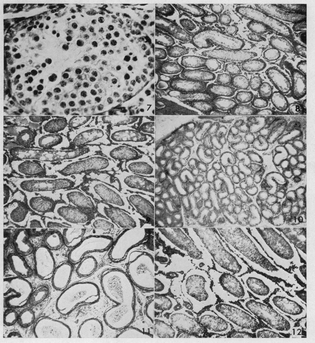
8 166 J. McC. Howell, J. jv. Thompson and G. A. J. Pitt in rats rats kept in the dark were smaller than the weights of these organs kept under normal laboratory conditions. However, there was little histolo gical difference between the testes of these two groups. In the Type 1 rats the testes were markedly degenerate and the seminal vesicles were reduced in size before one would have expected the rats to be blind (Dowling & Wald, 1960). Regeneration of the retina may have in fluenced the seminal vesicle weights of the rats receiving vitamin A alcohol in our regeneration experiment. One would expect a much earlier development of the retinal lesion in the Type 2 rats. If this is so, blindness could be a contributory factor in the develop ment of the lesions in the reproductive tract of these animals. This aspect of the problem is now being investigated. ACKNOWLEDGMENTS We wish to thank Professor D. L. Hughes and Professor R. A. Morton, f.r.s. for their encouragement and advice, Miss M. W. Royston, a.i.m.l.t. and Mr J. Camp for technical assistance, Mr G. Weston, f.i.m.l.t. for the photographs, and Dr O. Isler of F. Hoffmann-La Roche & Co, Basel for supplies of vitamin A acid. This work was supported in part by a grant from the Agricultural Re search Council. REFERENCES Beaver, D. L. (1961) Vitamin A deficiency in the germ-free rat. Amer. J. Path. 38, 335. Dowling, J. E. & Wald, G. (1960) The biological function of vitamin A acid. Proc. nat Acad. Sci., Wash. 46, 587. Fiske, V. M. (1941) Effect of light on sexual maturation, oestrous cycles, and anterior pituitary of the rat. Endocrinology, 29, 187. Leblond, C. P. & Cleremont, Y. (1952) Spermiogenesis of rat, mouse, hamster and guinea-pig as revealed by the 'periodic acid-fuchsin sulfurous acid' technique. Amer. J. Anat. 90, 167. Mason, K. E. (1933) Differences in testes injury and repair after vitamin A deficiency, vitamin E deficiency and inanition. Amer. J. Anat. 52, 153. Mason, K. E. (1939) Relation of the vitamins to the sex glands. Sex and Internal Secretions, 2nd edn. Eds. E. Allen, C. H. Danforth and E. A. Doisy. Williams & Wilkins, Baltimore. Moore, T. (1953) Vitamin A in the normal individual. Symposium on Nutrition. Ed. R. M. Herriott. Johns Hopkins Press, Baltimore. Thompson, J. N. & Pitt, G. A. J. (1960) Vitamin A acid and hypervitaminosis A. Nature, Lond. 188, 672. EXPLANATION OF PLATES PLATE 1 Fig. 1. Spermatozoa and sloughed cells in the ductus epididymis. Type 1 rat fed on the basic diet for 6 weeks, i.e. a diet containing vitamin A acid but not vitamin A alcohol. The histological structure of the testis was normal. Haematoxylin and eosin Fig. 2. Isolated spermatocytes present in the seminiferous tubule. One-year-old-Type 1 rat. Haematoxylin and eosin Fig. 3. Vacuolation in the cytoplasm of the cells lining the tubules. Type 1 rat fed the basic diet for 11 weeks. Haematoxylin and eosin Fig. 4. Tubules filled with Sertoli cells. Eosinophilic oedema fluid surrounds these tubules. Type 1 rat fed the basic diet for 15 weeks. Haematoxylin and eosin. X 189. Fig. 5. Some tubules are filled with Sertoli cells, others have a lumen. Vacuolation and spermatocytes can be seen in the latter. Type 1 rat fed the basic diet for 15 weeks. Haematoxylin and eosin Fig. 6. Seminiferous tubule containing numerous spermatocytes but no spermatids. Fifty-six-day-old Type 2 rat. Periodic acid Schiff reagent. x375.


9 PLATE 1 (Facing p. 166)

10 PLATE 2


11 PLATE 3 (Facing p. 167)

12 Vitamin A alcohol deficiency in the rat PLATE 2 Fig. 7. Tubule from the testis shown in Fig. 6. Healthy spermatocytes and degenerating cells are present. Periodic acid-schiff reagent, 500. Fig. 8. The tubule in the centre of the field is filled with Sertoli cells. Sixty-one-day-old Type 2 rat. Haematoxylin and eosin Fig. 9. Testes. Some of the tubules still have a lumen. Two hundred and thirty-oneday-old Type 2 rat. Haematoxylin and eosin Fig. 10. The ductus epididymis is empty. Fifty-six-day-old Type 2 rat (see Fig. 6). Haematoxylin and eosin Fig. 11. The ductus epididymis is either empty or contains eosinophilic material. Two hundred and thirty-one-day-old Type 2 rat (see Fig. 9). Haematoxylin and eosin. X52-5. Fig. 12. Right testis showing severe degeneration. Tubule in the centre of the field con tains spermatocytes. Eighteen-week-old Type 1 rat. This testis was removed surgically during a regeneration experiment. The animal had been fed on the basic diet. Haema toxylin and eosin. 70. PLATE 3 Fig. 13. Left testis removed 69 days after the surgical removal of the right testis which is shown in Fig. 12. Postoperatively the rat had been fed on the basic diet supplemented with a weekly dose of vitamin A alcohol, given in oil, by mouth as 200 Mg of vitamin A acetate. Spermatids are present. Many of these are immature spermatozoa. Twentyeight-weeks-old Type 1 rat. Haematoxylin and eosin. X 70. Fig. 14. Testes and seminal vesicles of a 67-day-old Type 2 rat and of a 67-day-old control animal. The control animal was born on the same regimen as the Type 2 rat but from 40 days old it had been fed the basic diet supplemented with a weekly dose of vitamin A alcohol. The testes and seminal vesicles of this animal, shown at the top of the figure, are larger than those of the Type 2 rat.
THE EFFECT OF OESTRIN ON THE TESTIS OF THE ADULT MOUSE
 389 THE EFFECT OF OESTRIN ON THE TESTIS OF THE ADULT MOUSE BY MARJORIE ALLANSON. (Harold Row Research Scholar, King's College, London.) (Received 5th March, 1931.) (With One Plate.) I. INTRODUCTION. THE
THE EFFECTS OF LIGATION OF CAUDA EPIDIDYMIDIS ON THE DOG TESTIS
 Copyright 1974 The American Fertility Society FERTILITY AND STERILITY Vol. 25, No.3, March, 1974 Printed in U.S.A. THE EFFECTS OF LIGATION OF CAUDA EPIDIDYMIDIS ON THE DOG TESTIS A. M. VARE, M.B.B.S.,
Morphogenesis of the residual body of the mouse testis
 93 Morphogenesis of the residual body of the mouse testis By CASIMIR F. FIRLIT and JOSEPH R. DAVIS (From the Department of Pharmacology and Therapeutics, Stritch School of Medicine, and Graduate School,
Histology of Male Reproductive system (1)
") Histology of Male Reproductive system (1) Prof. Dr. Malak A. Al-yawer Learning Objectives At the end of this lecture, the medical student will be able to: State the organization of the testis Define seminiferous
Histology of Male Reproductive system (1) Prof. Dr. Malak A. Al-yawer Learning Objectives At the end of this lecture, the medical student will be able to: State the organization of the testis Define seminiferous
MALE REPRODUCTIVE SYSTEM
 MALE REPRODUCTIVE SYSTEM The male reproductive system consists of primary sex organs (testes) and secondary or accessory sex organs. The secondary organs consist of a series of genital ducts (ductules
IN normal male fowls, four developmental stages of spermatogenetic activity
 Development of the Testis Tubule in the Fowl By GAMAL A. R. KAMAR (From the Animal Production Department, Faculty of Agriculture, Cairo University, Giza, Egypt) With three plates (figs. 1-3) SUMMARY Three
THE EFFECTS OF REPEATED INJECTIONS OF CHORIONIC GONADOTROPIN ON THE TESTES OF THE LEOPARD FROG (RANA PIPIENS SCHREBER)
") THE EFFECTS OF REPEATED INJECTIONS OF CHORIONIC GONADOTROPIN ON THE TESTES OF THE LEOPARD FROG (RANA PIPIENS SCHREBER) ROBERT P. McCOURT Department of Zoology and Entomology, The Ohio State University,
THE EFFECTS OF REPEATED INJECTIONS OF CHORIONIC GONADOTROPIN ON THE TESTES OF THE LEOPARD FROG (RANA PIPIENS SCHREBER) ROBERT P. McCOURT Department of Zoology and Entomology, The Ohio State University,
LABORATORY EXERCISES FOR MALE REPRODUCTIVE SYSTEM
 LABORATORY EXERCISES FOR MALE REPRODUCTIVE SYSTEM Slide #101 (1096). Testis, rat. sustentacular ( Sertoli ) cells Nuclei of Sustentacular cells Leydig cells Spermatogonia Spermatocytes Spermatids pale
Male Reproductive System
 Male Reproductive System organs that function in: gamete and hormone production not all in abdominal cavity paired testicles = controlled by LH & FSH duct systems accessory glands Testis: Gross Histology
HISTOLOGIC CHANGES IN THE SEMINIFEROUS TUBULES AFTER VASECTOMY
 FERTILItY AND STI!RILITY Copyright 1974 The American Fertility Society Vol. 25, No.8, August 1974 PTillted in U.S.AI HISTOLOGIC CHANGES IN THE SEMINIFEROUS TUBULES AFTER VASECTOMY FLETCHER C. DERRICK,
MALE REPRODUCTIVE SYSTEM
 1 MALE REPRODUCTIVE SYSTEM SCPA 602 Anatomical Basis for Pathological Study Updated: 20.09.2018 Lect. Nisamanee Charoenchon, PhD nisamanee.cha@mahidol.ac.th Department of Pathobiology, Mahidol University
5 15/3/2012. Malik Al-Momani
 5 15/3/2012 Malik Al-Momani بسم هللا الرحمن الرحيم Spermatogenesis Note : Please refer to slides so see photos. Quick Revision : - Testis is divided by septum into testicular lobules, inside the lobules
Basic histology 5/4/2015
 Male reproductive system The male reproductive system is composed of the testes, genital ducts (the adjoining epididymis, and the vas deferens, a accessory sex glands (the seminal vesicles, the prostrate
Effects of Ablation of the Submaxillary Gland in Guinea Pigs IV. Cause of deterioration of the tubules in the testes
 1961 475 Effects of Ablation of the Submaxillary Gland in Guinea Pigs IV. Cause of deterioration of the tubules in the testes Kazuo Suzuki Received August 1, 1960 Shakujii Institute, Tokyo Medical College,
Identification of the spermatogenic stages in living seminiferous tubules of man
 Identification of the spermatogenic stages in living seminiferous tubules of man V. Nikkanen, K.-O. S\l=o"\derstr\l=o"\m and M. Parvinen Department of Obstetrics and Gynecology, Turku University Central
Male Reproductive System
 Male Reproductive System Constitution of male reproductive system Genital gland ----testis Genital ducts epididymis / ductus deferens / urinary duct Accessory sex glands Penis prostate gland Seminal vesicle
612.6I7.5:612.6I6.I. different, but most of them appear to be satisfactory from a qualitative
 442 612.6I7.5:612.6I6.I SIZE CHANGES IN THE SEMINAL VESICLES OF THE MOUSE DURING DEVELOPMENT AND AFTER CASTRATION. BY RUTH DEANESLY AND A. S. PARKES'. (From the National Institute for Medical Research,
Title. Author(s)KANAGAWA, Hiroshi; ISHIKAWA, Tsune; KAWATA, Keiichir. CitationJapanese Journal of Veterinary Research, 13(1): Issue Date
KANAGAWA, Hiroshi; ISHIKAWA, Tsune; KAWATA, Keiichir. CitationJapanese Journal of Veterinary Research, 13(1): Issue Date") Title A CASE OF CANINE TESTICULAR SERTOLI CELL TUMOR Author(s)KANAGAWA, Hiroshi; ISHIKAWA, Tsune; KAWATA, Keiichir CitationJapanese Journal of Veterinary Research, 13(1): 11-1 Issue Date 1965-03 DOI 10.14943/jjvr.13.1.11
Title A CASE OF CANINE TESTICULAR SERTOLI CELL TUMOR Author(s)KANAGAWA, Hiroshi; ISHIKAWA, Tsune; KAWATA, Keiichir CitationJapanese Journal of Veterinary Research, 13(1): 11-1 Issue Date 1965-03 DOI 10.14943/jjvr.13.1.11
The Use of Rabbits in Male Reproductive Toxicology
 Environmental Health Perspectives Vol. 77, pp. 5-9, 1988 The Use of Rabbits in Male Reproductive Toxicology by Daniel Morton* The rabbit is the smallest and least expensive laboratory animal in which serial
Adapted from Preg. & Part., Senger
 MALE ENDOCRINOLOGY AND SPERMATOGENESIS (Chapter 10) AVS 222 (Instructor: Dr. Amin Ahmadzadeh) I. MALE ENDOCRINOLOGY (Figure10-1 to 10-3) A. Glands and their respective hormones 1) Hypothalamic hormone:
THE VITAMINS AND SEX HORMONES CONCERNED IN REPRODUCTION
 THE VITAMINS AND SEX HORMONES CONCERNED IN REPRODUCTION D. ROY McCULLAGH, Ph.D. The vitamins, the hormones, and the enzymes constitute three important groups of substances of special interest to the biological
Male Reproduction Organs. 1. Testes 2. Epididymis 3. Vas deferens 4. Urethra 5. Penis 6. Prostate 7. Seminal vesicles 8. Bulbourethral glands
 Outline Terminology Human Reproduction Biol 105 Lecture Packet 21 Chapter 17 I. Male Reproduction A. Reproductive organs B. Sperm development II. Female Reproduction A. Reproductive organs B. Egg development
Medical School Histology Basics Male Reproductive System. VIBS 289 lab
 Medical School Histology Basics Male Reproductive System VIBS 289 lab Larry Johnson Texas A&M University OBJECTIVE To conduct a histologic examination of the testis (which produce spermatozoa), excretory
Efferent Ducts and Epididymis
 increase) the secretion of each of the androgen regulated proteins. Regulation of spermatogenesis is therefore an extremely complex cascade of cell-cell interactions with the Leydig cells supporting germ
fibroproliferation, and mononuclear cell infiltration. An autoallergic aetiology (Received 13th December 1972)
") EARLY HISTOLOGICAL CHANGES IN EXPERIMENTAL CONTRALATERAL EPIDIDYMO\x=req-\ ORCHITIS IN THE RABBIT E. ZAPPI and S. SHULMAN Department of Microbiology, Mew York Medical College, New York, N.Y. 10029, U.S.A.
EARLY HISTOLOGICAL CHANGES IN EXPERIMENTAL CONTRALATERAL EPIDIDYMO\x=req-\ ORCHITIS IN THE RABBIT E. ZAPPI and S. SHULMAN Department of Microbiology, Mew York Medical College, New York, N.Y. 10029, U.S.A.
The Male Reproductive System
 The Male Reproductive System YONG-MEI CHEN ( 陈咏梅 ) Dept. of Anatomy, Histology & Embryology Peking Union Medical College Tel:69156461 E-mail address: pumc_he@126.com Content Spermatogenesis Spermiogenesis
Pathology of Male Reproductive System 1
 Pathology of Male Reproductive System 1 Professor dr Ali Hassan Altimimi Professor of Pathology& Histology MSc, PHD, MD(UK) MALE REPRODUCTIVE SYSTEM The internal male genitalia consist of the testes with
Variability in Weight and Histological Appearance of the Prostate of Beagle Dogs Used in Toxicology Studies
 Toxicologic Pathology, 36: 917-925, 2008 Copyright 2008 by Society of Toxicologic Pathology ISSN: 0192-6233 print / 1533-1601 online DOI: 10.1177/0192623308324958 Variability in Weight and Histological
Congenital mesonephric defects in male infants
 J. clin. Path. (1969), 22, 725-730 Congenital mesonephric defects in male infants with mucoviscidosis J. R. OLSON AND D. K. WEAVER From the Department ofpathology, The University of Michigan Medical Center,
abruptly retrogress and remain quiescent throughout the winter. The insecti
 HISTOCHEMICAL STUDY OF THE SEMINAL VESICLE SECRETION OF THE INDIAN HEDGEHOG WITH PARTICULAR REFERENCE TO THE CRYSTALLOID BODIES PUSHPAMALA P. BIDWAI and S. R. BAWA Department of Biophysics, Panjab University,
CHRONIC RENAL DISEASE IN RATS FOLLOWING A TEMPORARY
 40 CHRONIC RENAL DISEASE IN RATS FOLLOWING A TEMPORARY DEFICIENCY OF POTASSIUM P. FOURMAN, R. A. McCANCE AND R. A. PARKER From the Departments of Experimental Medicine and Pathology, University of Cambridge
relatively unpredictable environmental factors. PATTERNS OF CHANGE IN THE REPRODUCTIVE ORGANS OF THE MALE POCKET GOPHER, GEOMYS PINETIS
 PATTERNS OF CHANGE IN THE REPRODUCTIVE ORGANS OF THE MALE POCKET GOPHER, GEOMYS PINETIS KATHERINE CARTER EWEL Department of Zoology, University offlorida, Gainesville, Florida {Received 6th April 1971,
THE EFFECT OF UNILATERAL CASTRATION ON THE REMAINING TESTIS OF THE MOUSE
 402 THE EFFECT OF UNILATERAL CASTRATION ON THE REMAINING TESTIS OF THE MOUSE BY I. W. ROWLANDS. (From the Department of Zoology, University College of North Wales, Bangor.) (Received 14th April, 1934.)
Chapter 26: Reproductive Systems. Male 11/29/2015. Male reproductive system is composed of... BIO 218 Fall Gonads (testes)
") Chapter 26: Reproductive Systems BIO 218 Fall 2015 Male Male reproductive system is composed of... Gonads (testes) Duct system (epididymis, ductus deferens, ejaculatory ducts, urethra) Accessory sex glands
Chapter 26: Reproductive Systems BIO 218 Fall 2015 Male Male reproductive system is composed of... Gonads (testes) Duct system (epididymis, ductus deferens, ejaculatory ducts, urethra) Accessory sex glands
The Reproductive System
 Essentials of Human Anatomy & Physiology Elaine N. Marieb Seventh Edition Chapter 16 The Reproductive System Slides 16.1 16.20 Lecture Slides in PowerPoint by Jerry L. Cook The Reproductive System Gonads
androgen on the seminal vesicles it had neither a blocking effect on the penile
 MORPHOLOGICAL AND BEHAVIOURAL EFFECTS OF AN 'ANTIANDROGEN' IN MALE RATS F. A. BEACH and W. H. WESTBROOK Department of Psychology, University of California, Berkeley, California 94720, U.S.A. (Received
- production of two types of gametes -- fused at fertilization to form zygote
 Male reproductive system I. Sexual reproduction -- overview - production of two types of gametes -- fused at fertilization to form zygote - promotes genetic variety among members of a species -- each offspring
The Reproductive System
 16 PART A The Reproductive System PowerPoint Lecture Slide Presentation by Jerry L. Cook, Sam Houston University ESSENTIALS OF HUMAN ANATOMY & PHYSIOLOGY EIGHTH EDITION ELAINE N. MARIEB The Reproductive
To General Embryology Dr: Azza Zaki
 Introduction To General Embryology The Human Development is a continuous process that begins when an ovum from a female is fertilized by a sperm from a male. Cell division, growth and differentiation transform
Cyclical Changes in the Distribution of the Testis Lipids of a Seasonal Mammal (Talpa europaea) By B. LOFTS
 By B. LOFTS") 199 Cyclical Changes in the Distribution of the Testis Lipids of a Seasonal Mammal (Talpa europaea) By B. LOFTS (From the Department of Zoology and Comparative Anatomy, St. Bartholomew's Medical College,
199 Cyclical Changes in the Distribution of the Testis Lipids of a Seasonal Mammal (Talpa europaea) By B. LOFTS (From the Department of Zoology and Comparative Anatomy, St. Bartholomew's Medical College,
Reproductive Tract Pathology in Hyperkeratosis
 Reproductive Tract Pathology in Hyperkeratosis of Cattle and Sheep Kenneth McEntee, D.V.M., and Peter Olafson, D.V.M. HYPERKERATOSIS (X disease) of cattle was first observed in May, 1941, in New York State.
ESUR SCROTAL AND PENILE IMAGING WORKING GROUP MULTIMODALITY IMAGING APPROACH TO SCROTAL AND PENILE PATHOLOGIES 2ND ESUR TEACHING COURSE
 ESUR SCROTAL AND PENILE IMAGING WORKING GROUP MULTIMODALITY IMAGING APPROACH TO SCROTAL AND PENILE PATHOLOGIES 2ND ESUR TEACHING COURSE NORMAL ANATOMY OF THE SCROTUM MICHAEL NOMIKOS M.D. F.E.B.U. UROLOGICAL
18 Urinary system. 19 Male reproductive system. Female reproductive system. Blok 11: Genital and Urinary Tract Diseases
 Blok 11: Genital and Urinary Tract Diseases 18 Urinary System 19 Male Genital System 20 Female Genital System 18 Urinary system You should be able to: 1. Describe the structures and associated functions
Male Anatomy. testes, genetically determined in mammals - testis releases hormones that then control the development of secondary sex characteristics
 Male Anatomy Male Anatomy Primary Organ testes, genetically determined in mammals - testis releases hormones that then control the development of secondary sex characteristics 1) Secondary Organs internal
The Male Reproductive System
 The Male Reproductive System The male reproductive system Testes Genital ducts Accessory sex glands: seminal vesicles prostate bulbourethral glands External genitalia: penis Structure of the Testis Tunica
Primary sex organs (gonads): testes and ovaries. Accessory reproductive organs: ducts, glands, and external genitalia
: testes and ovaries. Accessory reproductive organs: ducts, glands, and external genitalia") Male Reproductive System Primary sex organs (gonads): testes and ovaries Produce sex cells (gametes) Secrete steroid sex hormones Androgens (males) Estrogens and progesterone (females) Accessory reproductive
Male Reproductive System Primary sex organs (gonads): testes and ovaries Produce sex cells (gametes) Secrete steroid sex hormones Androgens (males) Estrogens and progesterone (females) Accessory reproductive
SISTEMA REPRODUCTOR (LA IDEA FIJA) Copyright 2004 Pearson Education, Inc., publishing as Benjamin Cummings
 Copyright 2004 Pearson Education, Inc., publishing as Benjamin Cummings") SISTEMA REPRODUCTOR (LA IDEA FIJA) How male and female reproductive systems differentiate The reproductive organs and how they work How gametes are produced and fertilized Pregnancy, stages of development,
SISTEMA REPRODUCTOR (LA IDEA FIJA) How male and female reproductive systems differentiate The reproductive organs and how they work How gametes are produced and fertilized Pregnancy, stages of development,
Male reproduction. Cross section of Human Testis ผศ.ดร.พญ.ส ว ฒณ ค ปต ว ฒ ภาคว ชาสร รว ทยา คณะแพทยศาสตร ศ ร ราชพยาบาล 1. Aims
 Aims Male reproduction Male reproductive structure Spermatogenesis ส ว ฒณ ค ปต ว ฒ ห อง 216 โทร: 7578 Hypothalamo-pituitary-testicular axis Male sex hormone action Male reproductive structure Male reproductive
T H1s PRESENTATION concerns the extent to which specific dietary deficiencies
 The Relation of Dietary Deficiencies to Male Fertility Landrum B. Shettles, M.D. T H1s PRESENTATION concerns the extent to which specific dietary deficiencies may affect the male reproductive potential.
describe the parts and function of semen and the glands that contribute to it
 You need to be able to: describe spermatogenesis (How is sperm made?) describe the anatomy of a sperm describe the parts and function of semen and the glands that contribute to it How is sperm made? Spermatogenesis
Chapter 22 The Reproductive System (I)
") Chapter 22 The Reproductive System (I) An Overview of Reproductive Physiology o The Male Reproductive System o The Female Reproductive System 22.1 Reproductive System Overview Reproductive system = all
Chapter 22 The Reproductive System (I) An Overview of Reproductive Physiology o The Male Reproductive System o The Female Reproductive System 22.1 Reproductive System Overview Reproductive system = all
THE HOLOCRINE CELLS of the epithelium of the rat epididymis and vas
 Holocrine Cells of the Human Epididymis JAN MARTAN, Ph.D.,"' PAULL. RISLEY, Ph.D., and ZDENEK HRUBAN, M.D., Ph.D. t THE HOLOCRINE CELLS of the epithelium of the rat epididymis and vas deferens undergo
Male Reproductive Physiology
 Male Reproductive Physiology Overview Anatomy Function Endocrine and spermatogenesis Testis epididymus,vas deferens,seminal vesicles and prostate Hypothalamic pituitary testicular axis Hormones of the
Cycle of the Seminiferous Epithelium of the Guinea Pig
 Cycle of the Seminiferous Epithelium of the Guinea Pig A Method for Identification of the Stages Yves Clermont, Ph.D. IN THE GUINEA PIG, the cells of the seminiferous epithelium are arranged in definite
Chapter 36 Active Reading Guide Reproduction and Development
 Name: AP Biology Mr. Croft Chapter 36 Active Reading Guide Reproduction and Development Section 1 1. Distinguish between sexual reproduction and asexual reproduction. 2. Which form of reproduction: a.
ADVERSE EFFECTS OF VASECTOMY: SPERM GRANULOMA OF EPIDIDYMIDES V. P. DIXIT
 ADVERSE EFFECTS OF VASECTOMY: SPERM GRANULOMA OF EPIDIDYMIDES V. P. DIXIT Reproduct ion Physiology Section, Department of Zoology, University of Rajasthan, Jaipur-302004 Summary: Rats and mice were vasectomized
Cytological Studies on Human Spermatogenic and Sustentacular (Sertoli) Cells
 Cells") Cytological Studies on Human Spermatogenic and Sustentacular (Sertoli) Cells By Setsuko Ogata Department of Anatomy, Tokyo Women's Medical College, Shinjuku, Tokyo, Japan (Director : Prof. Dr. Kura Kubota)
Cytological Studies on Human Spermatogenic and Sustentacular (Sertoli) Cells By Setsuko Ogata Department of Anatomy, Tokyo Women's Medical College, Shinjuku, Tokyo, Japan (Director : Prof. Dr. Kura Kubota)
Testes (male gonads) -Produce sperm -Produce sex hormones -Found in a sac called the scrotum -Suspended outside of the body cavity for temperature
 -Produce sperm -Produce sex hormones -Found in a sac called the scrotum -Suspended outside of the body cavity for temperature") REPRODUCTION Testes (male gonads) -Produce sperm -Produce sex hormones -Found in a sac called the scrotum -Suspended outside of the body cavity for temperature reduction -Testes wall made of fibrous connective
REPRODUCTION Testes (male gonads) -Produce sperm -Produce sex hormones -Found in a sac called the scrotum -Suspended outside of the body cavity for temperature reduction -Testes wall made of fibrous connective
IN a previous publication (Hewitt, 1954) a description was given of the
 a description was given of the") i 9 9 Further Observations on the Histochemistry of Fat Absorption in the Small Intestine of the Rat By W. HEWITT, M.B., B.S. (From the Department of Anatomy, St. Thomas' Hospital Medical School, London,
i 9 9 Further Observations on the Histochemistry of Fat Absorption in the Small Intestine of the Rat By W. HEWITT, M.B., B.S. (From the Department of Anatomy, St. Thomas' Hospital Medical School, London,
The Reproductive System
 PowerPoint Lecture Slide Presentation by Patty Bostwick-Taylor, Florence-Darlington Technical College The Reproductive System 16PART A The Reproductive System Gonads primary sex organs Testes in males
Spermatogenesis in Man
 Spermatogenesis in Man I. Nuclear Morphology During Spermatogenesis in Man BRUNETTO CHIARELLI, PH.D., ARTHUR FALEK, PH.D., KAREN J. BACK, B.S., and C. THOMAS COWART, M.D. THE SEQUENCE of transformations
12/3/12. Managing Bull Development to Optimize Fertility Rearing bulls for fertility
 Managing Bull Development to Optimize Fertility Rearing bulls for fertility Effect of post weaning nutrition (after normal calf hood nutrition) - testis size - age at puberty - semen quality Effect of
SEASONAL CHANGES IN HISTOLOGYOF THE THYROID GLAND CALOTIS VERSICOLOR
 SEASONAL CHANGES IN HISTOLOGYOF THE THYROID GLAND CALOTIS VERSICOLOR M. D. Kulkarni And A. H. Shinde Department of Zoology Yashwantrao Chavan Arts & Science College, Mangrulpir Dist. Washim. (Received
Physiologic Anatomy of the Male Sexual Organs
 Reproductive and Hormonal Functions of the Male The reproductive functions of the male can be divided into three major subdivisions: (1) spermatogenesis, which means simply the formation of sperm; (2)
Evaluation of antifertility efficacy of Carica papaya seed extract through histological indices in Male Albino Rats
 International Journal of Chemical and Pharmaceutical Sciences 2013, Sep., Vol. 4 (3) ISSN: 0976-9390 IJCPS Evaluation of antifertility efficacy of Carica papaya seed extract through histological indices
Chapter 28: REPRODUCTIVE SYSTEM: MALE
 Chapter 28: REPRODUCTIVE SYSTEM: MALE I. FUNCTIONAL ANATOMY (Fig. 28.1) A. Testes: glands which produce male gametes, as well as glands producing testosterone 2. Seminiferous tubules (Fig.28.3; 28.5) a.
EFFECT OF DIETARY LINOLEIC AND LINOLENIC ACIDS ON TESTICULAR DEVELOPMENT IN THE RAT
 Quarterly Journal of Experimental Physiology (1983), 68, 221-231 221 Printed in Great Britain EFFECT OF DIETARY LINOLEIC AND LINOLENIC ACIDS ON TESTICULAR DEVELOPMENT IN THE RAT W. M. F. LEAT, CHRISTINE
Potassium Dichromate Impact on Male Reproductive Integrity Biomarker in Rat. Two Generation Study
 Potassium Dichromate Impact on Male Reproductive Integrity Biomarker in Rat. Two Generation Study Jelena Rankov, Alexandra Trif, Diana Brezovan, Florin Muselin Faculty of Veterinary Medicine 300645, Timisoara,
The spermatogenesis CHARACTERISTICS OF THE SPERMATOZOON 26/04/2017. Reproductive Biotechnologies Andrology I. Prof. Alberto Contri
 Reproductive Biotechnologies Andrology I The spermatogenesis Prof. Alberto Contri CHARACTERISTICS OF THE SPERMATOZOON 1) Aploid cell with high condensed DNA 2) Forward motility - flagellum 3) Enzymes for
(6, 7, 8, 9). cycle, a result in agreement with the experiments of PAPANICOLAOU and
. cycle, a result in agreement with the experiments of PAPANICOLAOU and") THE NATURE OF THE ANCESTROUS CONDITION RESULTING FROM VITAMIN B DEFICIENCY. By A. S. PARKES, Beit Memorial Research Fellow. From the Department of Physiology and Biochemistry, University College, London.
THE NATURE OF THE ANCESTROUS CONDITION RESULTING FROM VITAMIN B DEFICIENCY. By A. S. PARKES, Beit Memorial Research Fellow. From the Department of Physiology and Biochemistry, University College, London.
STRUCTURE AND FUNCTION OF THE MALE REPRODUCTIVE SYSTEM
 Unit 7A STRUCTURE AND FUNCTION OF THE MALE REPRODUCTIVE SYSTEM LEARNING OBJECTIVES 1. Learn the structures of the male reproductive system. 2. Learn the functions of the male reproductive system. 3. Learn
Sperm production. Sperm production. Meiosis. Mitosis. The cells of Leydig in testes secrete
 Sperm production Ductus deferens Epididymis The cells of Leydig in testes secrete Seminiferous testosterone (T) tubules T secreted at puberty produces 2 o sex characteristics, spermatogenesis, & maintain
Sperm production. Sperm production. Controlling sperm production. Meiosis. Mitosis. The cells of Leydig in testes secrete
 Ductus deferens Sperm production Epididymis The cells of Leydig in testes secrete Seminiferous testosterone (T) tubules T secreted at puberty produces 2 o sex characteristics, spermatogenesis, & maintain
Spermatogenesis Following Experimental Testicular Ischemia
 Spermatogenesis Following Experimental Testicular Ischemia Frank Hinman, Jr, MD, and Gilbert I Smith, MD REGENERATION of the spermatogenic elements of the testis after depression by testosterone and by
REPRODUCCIÓN. La idea fija. Copyright 2004 Pearson Education, Inc., publishing as Benjamin Cummings
 REPRODUCCIÓN La idea fija How male and female reproductive systems differentiate The reproductive organs and how they work How gametes are produced and fertilized Pregnancy, stages of development, birth
11. SEXUAL DIFFERENTIATION. Germinal cells, gonocytes. Indifferent stage INDIFFERENT STAGE
 11. SEXUAL DIFFERENTIATION INDIFFERENT STAGE Early in pregnancy, (within 10-15 % of the pregnancy s expected length) a genital ridge is formed in the sides of the embryonic tissue, ventral to the mesonephros
GROWTH AND OBSERVATIONS OF CHINESE HAMSTER SEMINIFEROUS EPITHELIUM IN VITRO
 J. Cell Sci. 6, 19S-205 (1970) Printed in Great Britain GROWTH AND OBSERVATIONS OF CHINESE HAMSTER SEMINIFEROUS EPITHELIUM IN VITRO D. J. ELLINGSON AND K. T. S. YAO U.S. Department of Health, Education
川北医学院讲稿. Under low power note the testis is enclosed by a strong fibrous. layer of serous epithelium. These fibrous tissue
 川北医学院讲稿 Experiment 5: Male and Female Reproductive System Hello, everybody, class is begin,keep quiet, please. And this is the last experimental class. Today we will learn 5 slices and review all structures
Reproductive Endocrinology. Isabel Hwang Department of Physiology Faculty of Medicine University of Hong Kong Hong Kong May2007
 Reproductive Endocrinology Isabel Hwang Department of Physiology Faculty of Medicine University of Hong Kong Hong Kong May2007 isabelss@hkucc.hku.hk A 3-hormone chain of command controls reproduction with
Mohammad Sha ban. Basheq Jehad. Hamzah Nakhleh
 11 Mohammad Sha ban Basheq Jehad Hamzah Nakhleh Physiology of the reproductive system In physiology, we are concerned with the mechanisms in which the system functions, and how the system responds to different
Reproductive System Purpose General Structures Male Structures Functions Female Anatomy Structures Functions Clinical Applications
 The Reproductive System: Male, Ch 23 Outline of class lecture After studying the male reproductive system you should be able to: 1. Define the purpose of reproduction and identify the general organs of
DEVELOPMENT OF THE RECTUM-TESTIS TEMPERATURE DIFFERENCE IN THE POST-NATAL RAT
 DEVELOPMENT OF THE RECTUM-TESTIS TEMPERATURE DIFFERENCE IN THE POST-NATAL RAT MARTTI KORMANO Department of Anatomy, University of Helsinki, Siltavuorenpenger, Helsinki, and Research Laboratory of the Sauna-Seura,
A COMPARATIVE STUDY OF GERM CELL KINETICS IN THE TESTES OF CHILDREN WITH UNILATERAL CRYPTORCHIDISM: A PRELIMINARY REPORT*
 FERTILITY AND STERILITY Copyright 1970 by the Williams & Wilkins Co. Vol. 21, No. 11, November 1970 Printed in U.S.A. A COMPARATIVE STUDY OF GERM CELL KINETICS IN THE TESTES OF CHILDREN WITH UNILATERAL
COMMUNICATIONS PHOTOCOAGULATION OF THE RETINA* OPHTHALMOSCOPIC AND HISTOLOGICAL FINDINGS. photocoagulation of the rabbit's retina.
 Brit. J. Ophthal. (1963) 47, 577. COMMUNICATIONS PHOTOCOAGULATION OF THE RETINA* OPHTHALMOSCOPIC AND HISTOLOGICAL FINDINGS BY A. LAVYEL Haifa, Israel SINCE the introduction of the photocoagulator by Meyer-Schwickerath
Chapter 46 ~ Animal Reproduction
 Chapter 46 ~ Animal Reproduction Overview Asexual (one parent) fission (parent separation) budding (corals) fragmentation & regeneration (inverts) parthenogenesis Sexual (fusion of haploid gametes) gametes
 MOCK TEST I (HUMAN REPRODUCTION) 1. In human transfer of sperms into female genital tract is called as 1) Fertilization 2) Implantation 3) Insemination 4) Gestation 2. In male human scrotum maintains the
Reproductive Effects of Feeding Gossypol and Vitamin E to Bulls
 Reproductive Effects of Feeding Gossypol and Vitamin E to Bulls J. Velasquez-Pereira P.J. Chenoweth L.R. McDowell C.A. Risco C.A. Staples D. Prichard F.G. Martin M.C. Calhoun S.N. Williams N.S. Wilkinson
Chapter 14 Reproduction Review Assignment
 Date: Mark: _/45 Chapter 14 Reproduction Review Assignment Multiple Choice Identify the choice that best completes the statement or answers the question. 1. Use the diagram above to answer the next question.
Physiology of Male Reproductive System
 Physiology of Male Reproductive System the anterior pituitary gland serves as the primary control of reproductive function at puberty Ant Pituitary secretes FSH & large amounts of LH (ICSH) FSH & LH cause
Embryology 3. Spermatogenesis:
 Embryology 3 Spermatogenesis: The 2 testis in males are each divided into lobes and lobules by connective tissue septa forming 250 lobule and in each lobule there are 1 to 4 seminefrous tubule ( so almost
ROLE OF SOME ANTIOXIDANTS ON MERCURY CHLORIDE INDUCED SPERMATOGENESIS IN SWISS ALBINO MICE DURING PRE PUBERTAL PHASE OF LIFE
 Indian J.Sci.Res.1(2) : 19-25, 2010 ROLE OF SOME ANTIOXIDANTS ON MERCURY CHLORIDE INDUCED SPERMATOGENESIS IN SWISS ALBINO MICE DURING PRE PUBERTAL PHASE OF LIFE a1 DUGESH NANDINI SHARMA AND LATA BHATTACHARYA
BIOL 2402 Reproductive Systems
 Collin College Dr. Chris Doumen BIOL 2402 Reproductive Systems 1 Reproductive System Most systems between males and females in the human body are similar in structure. The exception of course are the organs
on 0 1nurl 3 n.u.-o.~.2529
 ,,.,11,., 1n~t1'111u, on 0 1nurl 3 n.u.-o.~.2529 FAILURE OF INDUCING SPERMATOGENESIS WITH FSH, LH AND TESTOSTERONE IN VITAMIN A DEFICIENT RAT Vichitra Leardkamolkarn, Reon Somana, Sasitorn Rojananuengnit
epididymis, respectively. The occurrence and disposition of the droplet
 MORPHOLOGICAL CHANGES IN RABBIT SPERMATOZOA DURING PASSAGE THROUGH THE EPIDIDYMIS J. M. BEDFORD Department of Physiology, Royal Veterinary College, London, N. W. 1 {Received 28th August 1962) Summary.
Basic Reproduction & Genetics. Steve Pritchard UNL Extension Educator Boone-Nance Counties
 Basic Reproduction & Genetics Steve Pritchard UNL Extension Educator Boone-Nance Counties Hormonal Regulation of the Estrous Cycle Several hormones regulate the estrous cycle Changes in the concentrations
Histology of Male Reproductive System
 Histology of Male Reproductive System Lecture Objectives Describe the histological features of the male reproductive system Male Reproductive System The male structures of reproduction include the: testes,
Action of Testosterone, Dihydrotestosterone and 5a Androstane 3tr, 17(3 Diol on the Spermatogenesis of Immature Rats
 BIOLOGY OF REPRODUCTION 14, 332-338 (1976) Action of Testosterone, Dihydrotestosterone and 5a Androstane 3tr, 17(3 Diol on the Spermatogenesis of Immature Rats H. E. CHEMES1, E. PODESTA and M. A. RIVAROLA
(Received 6th October 1972)
") EFFECT OF TESTOSTERONE POLYDIMETHYL- SILOXANE IMPLANTS UPON SPERM PRODUCTION, LIBIDO AND ACCESSORY SEX ORGAN FUNCTION IN RABBITS L. L. EWING, L. G. STRATTON and C. DESJARDINS Department of Physiological
EFFECT OF TESTOSTERONE POLYDIMETHYL- SILOXANE IMPLANTS UPON SPERM PRODUCTION, LIBIDO AND ACCESSORY SEX ORGAN FUNCTION IN RABBITS L. L. EWING, L. G. STRATTON and C. DESJARDINS Department of Physiological
hypophysectomized rat. Marenzi & Gerschman [1934] studied six of the University and Royal Infirmary, Glasgow (Received 13 December 1937)
![hypophysectomized rat. Marenzi & Gerschman [1934] studied six of the University and Royal Infirmary, Glasgow (Received 13 December 1937)](/thumbs/96/129025719.jpg "hypophysectomized rat. Marenzi & Gerschman [1934] studied six of the University and Royal Infirmary, Glasgow (Received 13 December 1937)") 124 J. Physiol. (I938) 92, I24-130 6i2.492.5:6I2.I26 THE EFFECT OF HYPOPHYSECTOMY ON THE BLOOD CALCIUM AND PHOSPHORUS OF THE RAT BY A. B. ANDERSON AND E. G. OASTLER From the Biochemical Laboratory, Department
124 J. Physiol. (I938) 92, I24-130 6i2.492.5:6I2.I26 THE EFFECT OF HYPOPHYSECTOMY ON THE BLOOD CALCIUM AND PHOSPHORUS OF THE RAT BY A. B. ANDERSON AND E. G. OASTLER From the Biochemical Laboratory, Department
Male Reproductive System. Dr Maan Al-Abbasi PhD, MSc, MBChB, MD
 Male Reproductive System Dr Maan Al-Abbasi PhD, MSc, MBChB, MD Learning Objectives 1. Describe the General Anatomy of the Male Reproductive System 2. Identify the structures that are related to the prostate.