UNIT 05: ASSESSMENT OF THE ABDOMEN, ANUS & RECTUM
|
|
|
- Dylan Baldric Paul
- 6 years ago
- Views:
Transcription
 Instructor New Life College of Nursing September 26, 2015 Elsevier items and derived items 2008, 2004, 2000, 1996, 1992 by Saunders, an imprint of Elsevier")
1 In The Name of God (A PROJECT OF NEW LIFE COLLEGE OF NURSING KARACHI) UNIT 05: ASSESSMENT OF THE ABDOMEN, ANUS & RECTUM Shahzad Bashir RN, BScN, DCHN, MScN (Std. DUHS) Instructor New Life College of Nursing September 26, 2015 Elsevier items and derived items 2008, 2004, 2000, 1996, 1992 by Saunders, an imprint of Elsevier Inc.
2 Objectives. By the end of the unit, learner will be able to: Discuss the pertinent health history questions necessary to perform the assessment of abdomen, anus & rectum. Describe the specific assessment to be made during the physical examination of the abdomen. Practice assessment skills of abdomen. Discuss component of a rectal examination. Documents finding. List the changes in abdomen that are characteristics of aging process. Slide 21-2
3 General Considerations The patient should have an empty bladder. The patient should be lying supine on the exam table and appropriately draped. The examination room must be quiet to perform adequate auscultation and percussion. Watch the patient's face for signs of discomfort during the examination 3
4 General Considerations Disorders in the chest will often manifest with abdominal symptoms. It is always wise to examine the chest when evaluating an abdominal complaint. Consider the inguinal/rectal examination in males. Consider the pelvic/rectal examination in females. 4
5 Structure and Function Subjective Data Health History Questions Objective Data The Physical Exam Abnormal Findings Slide 21-5
6 Structure and Function Surface landmarks Borders of abdominal cavity Abdominal muscles Internal anatomy (viscera) Solid viscera Liver Pancreas Spleen Adrenal glands Kidneys Ovaries Uterus Slide 21-6

7 Slide 21-7

8 Slide 21-8
9 Structure and Function (cont.) Internal anatomy (viscera) (cont.) Hollow viscera Stomach Gallbladder Small intestine Colon Bladder Slide 21-9

10 Internal Anatomy Slide 21-10
11 Deep Internal Anatomy Pat Thomas, Slide 21-11
12 Structure and Function (cont.) Abdominal wall divided into four quadrants Right upper (RUQ) Left upper (LUQ) Right lower (RLQ) Left lower (LLQ) Slide 21-12

13 Slide 21-13
14 Location! Location! Location! RIGHT UPPER QUADRANT(RUQ) Liver Gallbladder Duodenum (small intestine) Head of Pancreas Right kidney and adrenal Hepatic flexure of colon Part of ascending and transverse colon 14
15 Location! Location! Location! LEFT UPPER QUADRANT(LUQ) Left lobe of liver Stomach Spleen Left kidney and adrenal Body of pancreas Splenic flexure of colon Parts of transverse and descending colon 15

16 Location! Location! Location! RIGHT LOWER QUADRANT(RLQ) Cecum Appendix Section of the ascending colon Right ovary Right Fallopian tube Right ureter Right spermatic cord Part of uterus (If enlarged) 16
17 Location! Location! Location! LEFT LOWER QUADRANT (LUQ) Sigmoid Colon Part of descending colon Left Ovary Left fallopian tube Left ureter Left spermatic cord Part of uterus(if enlarged) 17
18 Subjective Data Health History Questions Appetite Dysphagia Food intolerance Abdominal pain Nausea/vomiting, Regurgitation Bowel habits Abdominal history Medications Nutritional assessment Slide 21-18
19 Subjective Data Health History Questions Heartburn Rectal bleeding Hemorrhoids Previous surgery Weight gain or loss Slide 21-19
20 Objective Data The Physical Exam Preparation Lighting and draping Measures to enhance abdominal wall relaxation Equipment needed Stethoscope Small centimeter ruler Skin-marking pen Alcohol swab Slide 21-20
21 PREPARATION Equipment - stethoscope, marking pen, ruler, paper for documentation Patient lie on back, pillow under head, knees slightly flexed Empty bladder Short fingernails Proper light Privacy maintain e.g. side screen Slide 21-21
22 SEQUENCE OF ASSESSMENT INSPECTION AUSCULTATION PERCUSSION PALPATION 22
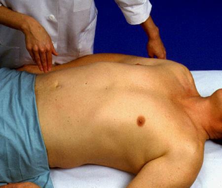
23 Objective Data The Physical Exam (cont.) Inspect the abdomen: Contour Symmetry Umbilicus Skin (Pigmentation, Lesions, Striae (elevated/depressed), Turgor Pulsation or movement Hair distribution Demeanor Slide 21-23

24 Contour Slide 21-24

25 INSPECT ABDOMINAL CONTOUR 25
26 Objective Data The Physical Exam (cont.) Auscultate the abdomen: Bowel sounds 5-35/min Increased, decreased, absent bowel sounds. Borborygymus Silent abdomen Vascular sounds (bruits) Percuss the abdomen: General tympany Liver span Usual technique Scratch test Splenic dullness Costovertebral angle tenderness Special procedures Fluid wave Shifting dullness Slide 21-26
27 AUSCULTATION Active bowel sounds 5-35/min Hypoactive 4/min or less( K+, Paralytic Ileus, Chronic use of Laxative) Hyperactive 35 or more /min (Dysentery, Diarrhea, Early sign of Intestinal Obstruction). Bruits (blowing sound) Aorta Renal Iliac Friction rub (Obstruction two layers of organs rubbing each other). Slide 21-27
28 GUT SOUNDS Use the diaphragm of your stethoscope to listen to gut sounds Normal gut sounds are gurgling, 5 to 35 per minute Borborygmi (Rumbling sounds caused by gas moving through the intestines (stomach "growling ) are loud, easily audible sounds. They are normal, too. High pitched, Tinkling (raindrops in a barrel) sounds are a sign of early intestinal obstruction Succussion splash, A loud sound like splashing water, is often heard without a stethoscope as the patient moves from side to side. It occurs when the abdomen is filled with air or fluid and indicates delayed gastric emptying from an obstruction or gastric dilatation. 28
29 GUT SOUNDS Decreased sounds: (none for a minute) are a sign of decreased gut activity. Gut sounds may be markedly decreased after abdominal surgery; abdominal infection (peritonitis) or injury. Absent Sounds : (no sounds for 5 minutes) are a bad sign. They can be caused by longer-lasting intestinal obstruction, intestinal perforation or intestinal (mesenteric) ischemia or infarction. 29
30 BRUITS SOUNDS (A sound, especially an abnormal one. A bruit may be heard over an artery or vascular channel, reflecting turbulence of flow) OR VENOUS HUMS. Use the bell of your stethoscope to listen for bruits: Aortic bruits: Are heard in the epigastrium. They may be a sign of abdominal aortic (a sac formed by localized dilatation of the wall of an artery, a vein) Slide 21-30
, which is a potentially treatable cause of hypertension.")
31 BRUITS SOUNDS Renal artery bruits: Are in each upper quadrant. They may be a sign of renal artery stenosis (A narrowing), which is a potentially treatable cause of hypertension. Iliac/femoral bruits: Are in the lower quadrants. They may be a sign of peripheral atherosclerosis Slide 21-31

32 AUSCULTATION FOR BRUITS 32
33 PERCUSSION To determine the size of solid organs and presence of masses, fluid and gas Tympanic sound Percuss in all four quadrants Percuss for liver Percuss for spleen Percuss bladder if indicated 33
34 THE TECHNIQUE FOR PERCUSSION There are two basic sounds which can be elicited: 1. Tympanitic (drum-like) sounds produced by percussing over air filled structures. 2. Dull sounds that occur when a solid structure (e.g. liver) or fluid (E.g. Ascites) lies beneath the region being examined. Special note should be made if percussion produces pain, which may occur if there is underlying inflammation, as in peritonitis. This would certainly be supported by other historical and exam findings. 34

35 Abdominal Percussion Pattern Slide 21-35
36 PERCUSSION Percussing the body gives one of three notes: 1. Tympany is found in most of the abdomen, caused by air in the gut. It has a higher pitch than the lung. 2. Resonance is found in normal lung. It is lower pitched and hollow. 3. Dullness is a flat sound, without echoes. The liver and spleen, and fluid in the peritoneum (ascites), give a dull note. 36

37 Cont... Slide 21-37

38 PERCUSS THE LIVER 38
39 PERCUSSION OF THE SPLEEN When significantly enlarged, percussion in the left upper quadrant will produce a dull tone. Splenomegaly suggested by percussion should then be verified by palpation Percuss in left anterior axillary line, just above lowest rib Ask your patient to take a deep breath and percuss again. Dullness with full inspiration may be a sign of enlarged spleen. Slide 21-39

40 PERCUSSION OF SPLEEN Slide 21-40

41 PERCUSSION OF SPLEEN Slide 21-41
42 PERCUSSION If dullness in flank (on side) - check for shifting dullness If indicated check for fluid wave 42
43 SHIFTING DULLNESS With the patient supine, begin percussion at the level of the umbilicus and proceed down laterally. In the presence of ascites, you will reach a point where the sound changes from tympanitic to dull. This is the intestine-fluid interface and should be roughly equidistant(central) from the umbilicus on the right and left sides as the fluid. Mark this point on both the right and left sides of the abdomen and then have the patient roll into a lateral decubitus position (i.e. onto either their right or left sides). 43

")
44 SHIFTING DULLNESS (REAL PATIENT) 44
45 Objective Data The Physical Exam (cont.) Palpate the liver: Measures to enhance muscle relaxation Light palpation Deep palpation Bimanual palpation Normally palpable structures Liver Spleen Kidneys Aorta Usual technique Hooking technique Slide 21-45
46 PALPATION Light palpation to evaluate general condition, Four quadrants, 1-3cm. Special organs. Nature of any distention, and abnormalities and painfulness. E.g. Inguinal nodes, Hernia. Deep palpation 4-5cm, both hand technique or one hand, to detect any organ enlargement, abdominal masses or swellings Palpate for liver and spleen Rebound tenderness (Inflammation of appendix/ Peritoneal inflammation 46


47 PALPATION OF LIVER Bimanual technique for liver palpation Hooking technique for liver palpation 47

48 Normal Liver Span Slide 21-48

49 Slide 21-49

50 PALPATION OF SPLEEN 50

51 REBOUND TENDERNESS/ Blumberg s sign 51

52 ABDOMINAL AORTIC ANEURYSM THE EXAM METHOD: THE PATIENT S ABDOMEN SHOULD BE RELAXED WITH THE KNEES FLEXED. THE EXAMINER UMBILICUS FOR THE AORTIC PULSATION. PLACE BOTH HANDS ON THE ABDOMEN WITH THE INDEX FINGER ON EITHER SIDE OF THE PULSATING AORTA. ESTIMATE THE WIDTH ( NL <2.5CM IN WIDTH). 52
53 ON BACK CHECK FOR RENAL BRUITS COSTOVERTEBRAL ANGLE TENDERNESS 53

54 Slide 21-54

55 BLUNT PERCUSION OF KIDNEY 55

56 POSTERIOR VIEW: LOCATION OF THE KIDNEYS 56
57 Important Sign s ROVSING S SIGN Also know as indirect tenderness. the sign is positive when pressure applied to the left lower quadrant results in right lower quadrant pain. RECTAL TENDERNESS Patients with appendicitis involving/ the pelvis may have rectal tenderness on examination. 57

58 MC BURNEY S POINT TENDERNESS A point 11/3-2 inches on the abdomen that is on the line connecting the umbilicus with the ASIS (or anterior superior iliac spine). This is associated with the bottom margin of the appendix. The point is one third of the distance when measured from ASIS to umbilicus. In 1889 charles mcburney stated that all patients with appendicitis had maximal pain at this point. Slide 21-58
59 Special Procedures for Advanced Practice Rebound tenderness (Blumberg s sign) Inspiratory arrest (Murphy s sign) Iliopsoas muscle test Obturator test Slide 21-59

60 Normally Palpable Structures Slide 21-60

61 Sample Charting Slide 21-61

62 Sample Charting (cont.) Slide 21-62
63 Abnormal Findings Abdominal Distention Obesity Air or gas Ascites Ovarian cyst Pregnancy Feces Tumor Slide 21-63
64 Abnormal Findings Abnormalities on Inspection Umbilical hernia Epigastric hernia Incisional hernia Slide 21-64
65 Abnormal Findings Abnormal Bowel Sounds Succussion splash Hypoactive bowel sounds Hyperactive bowel sounds Slide 21-65
66 Abnormal Findings Abnormalities on Palpation of Enlarged Organs Enlarged liver (Hepatomegaly) Enlarged nodular liver Enlarged gallbladder (Cholecystitis, Cholithiasis) Enlarged spleen (Splenomegaly) Enlarged kidney (Pylonephritis, Polycystic Kidney) Aortic aneurysm Slide 21-66
67 EXAMINATION OF THE ANUS AND RECTUM This information is sometimes included with the abdominal assessment and at times with assessment of the male and female genitalia. 67
68 GENERAL PRINCIPLES Anal canal is the final segment of digestive system. It measures from 2.5 cm to 4 cm long. It is lined with skin that contains no hair or sebaceous glands but does contain many somatic sensory nerves, making it very sensitive to touch. Within the anus are the two sphincters that normally hold the anal canal closed except when passing gas and feces. 68
69 Examination of anus & Rectum. History: Bowel habits(changes). Character of stools(blood). Rectal Pain C/O, Constipation, Diarrhoea Hemorrhoids Screening, (PR Proctoscopy) Use of Laxatives or medications Prostate problems Slide 21-69
70 RECTAL EXAMINATION Assist patient into position: Male left lateral, or standing upper body resting on a table. Female Lithotomy Then I. Inspection II. Palpation: Males Females Slide 21-70
71 RECTAL EXAMINATION Rectal Examination in Men. Inspect the perianal areas. Palpate the anal canal, rectum, and prostate. If the patient cannot stand, examine the genitalia before doing the rectal examination. Genital and Rectal Examination in Women. Examine the external genitalia, vagina, and cervix. Obtain a Pap smear (a sample of secretions and superficial cells of the uterine cervix and uterus; examined with a microscope to detect any abnormal cells). Palpate the uterus and adnexa (ovaries). Do a rectovaginal and rectal examination. 71
72 Examination of anus & Rectum. Inspection: Position- Side lying is preferred or lithotomy if genitalia exam in female or standing with upper body resting on a table for men. Inspect perianal tissue/ Sacrococygeal area by retracting buttocks. Look for skin characteristics, Lumps, lesions, hemorrhoids, ulcers, Rashes, Redness, inflammation, pigmentation. Ask client to bear down prolapse of rectum or hemorrhoids. Slide 21-72
73 Examination of anus & Rectum. Palpation: Surrounding tissue for lumps and tenderness. Per rectal examination, anal sphincter, tone, grasp, laxity. Rectal wall, irregularity, tenderness nodular, lesions. Prostate gland, round, heart shaped, 2.5-4cm, firm & non tender, palpable on anterior rectal wall. Observe fecal matter on gloved finger for color (blood) & consistency. Slide 21-73
74 HEMORRHOIDS With increased venous (portal) pressure, vein can enlarge. This is a hemorrhoid or a varicosity External hemorrhoids occur below the anorectal junction Itch and bleed with defecation Painful and swollen with thrombosis Resolve and leave flabby(loose) skin top around Anal opening. 74
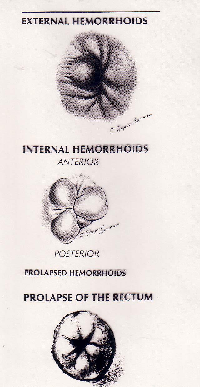
75 HEMORRHOIDS 75
76 continued Internal hemorrhoids originate above anorectal junction Covered with mucosa May appear as red mass with pressure (valsalva in heart abnormalities) 76

77 THE NORMAL PROSTATE 77

78 CANCER OF THE RECTUM 78

79 FISTULA VERSUS FISSURE 79

80 Slide 21-80

81 DRAPING THE ABDOMEN 81

82 VARIOUS CAUSES OF ABDOMINAL DISTENSION Obese abdomen 82

83 VARIOUS CAUSES OF ABDOMINAL DISTENSION Hepatomegaly 83

84 VARIOUS CAUSES OF ABDOMINAL DISTENSION Ascites 84

85 VARIOUS CAUSES OF ABDOMINAL DISTENSION Markedly enlarged gall bladder (labelled "GB") 85

86 VARIOUS CAUSES OF ABDOMINAL DISTENSION Umbilical Hernia 86

87 VARIOUS CAUSES OF ABDOMINAL DISTENSION Umbilical hernia 87

88 CAPUT MEDUSAE (Dilated cutaneous veins around the umbilicus, seen mainly in the newborn and in patients with cirrhosis.) 88

89 GYNECOMASTIA Abnormal Enlarged breast of male 89


90 Adult Stethoscope: Diaphragm and Bell Incorporated Into Single Side. Adult Stethoscope Auscultation Combination Adult & Pediatric Stethoscope Newborn Stethoscope 90

91 EXAMINING FOR A FLUID WAVE: 91

92 Tenderness over the liver suggests inflammation, as in hepatitis, or congestion, as in heart failure. 92
93 GI Variations Due to Age Aging- should not affect GI function unless associated with a disease process. Decreased: salivation, sense of taste, gastric acid secretion, esophageal emptying, liver size, bacterial flora. Increased: constipation! 2/26/
94 GI Variations with pregnancy Decrease in gastric motility High incidence of N, V (r/t pregnancy hormones) and heartburn or acid reflux Bowel sounds diminished r/t enlarged uterus displacing intestines Linea nigra- increased pigmentation of abd midline Striae Gravidarum 2/26/
95 References 1. Anderson, K. (1996). Mosby s Medical, Nursing and Allied Health Dictionary, ed. 4. St. Louis: C.V. Mosby. 2. Barkauskas,V.H., Stoltenberg-Allen, K., Baumann, L.C., and Darling- Fisher. (1998). Health and Physical Assessment, ed. 2. St. Louis: Mosby Year Book. 3. Bates, B., Bickley, L.S., and Hoekelman, R.A. (1995).Physical Examination and History Taking, ed. 6. Philadelphia: Lippincott-Raven. 4. Becker, K.L., and Stevens, S.A. (1988). Performing in-depth abdominal assessment. Nursing88 18(6): Zator Estes, M.E. (1998). Health Assessment and Physical Examination. Albany,N.Y.: Delmar Publishers. 95
Abdominal Examination Benchmarks
 Abdominal Examination Benchmarks Preparation and Positioning: Stand on the right side of the patient. The patient should be supine and double draped so only the abdomen is exposed o To relax the abdominal
BIOE221. Session 6. Abdominal Examination. Bioscience Department. Endeavour College of Natural Health endeavour.edu.au
 BIOE221 Session 6 Abdominal Examination Bioscience Department Examination of the Abdomen Session objectives Understand the organs / structures that are present in the abdominal cavity Understand the importance
Module Seven. Application of Health Assessment NUR 225. Physical examination of gastrointestinal and urinary system. King Saud University
 King Saud University Collage of Nursing Medical Surgical Nursing depart Application of Health Assessment NUR 225 Module Seven Physical examination of gastrointestinal and urinary system Learning Outcomes:
Abdominal Assessment
 Abdominal Assessment Mary Marian, MS,RD,CSO University of AZ, Tucson, AZ Neha Parekh, MS,RD,LD,CNSC Cleveland Clinic, Cleveland, OH Objectives: 1. Outline the steps in performing an abdominal examination.
BATES VISUAL GUIDE TO PHYSICAL EXAMINATION. Vol. 13: Abdomen
 BATES VISUAL GUIDE TO PHYSICAL EXAMINATION Vol. 13: Abdomen So tell me a little bit about the abdominal pain that you ve been experiencing. Your learning objectives for mastering the abdomen examination
Application of Health Assessment NUR 225. Module Seven. Physical examination of gastrointestinal and urinary system
 King Saud University Collage of Nursing Medical Surgical Nursing depart Application of Health Assessment NUR 225 Module Seven Physical examination of gastrointestinal and urinary system 1 Gastrointestinal
Gastro system. Examination
 Gastro system Examination 1. INSPECTION: Skin lesions- scars Blood vessels: ABDOMEN Nine regions Inf vena cava Obstruction shows veins in flanks and emptying from distal to proximal SVC Portal vein Obstruction
Abdominal Exam. Winter Quarter Adapted from previous years by Amanda Kocoloski, OMS IV
 Abdominal Exam Winter Quarter 2010 Adapted from previous years by Amanda Kocoloski, OMS IV Agenda ó History ó Anatomy ó Physical ó Prac4ce cases ó 2 gastrointes4nal complaints ó Work on incorpora4ng GI
PHYSICAL DIAGNOISS OF THE ABDOMEN Eve Bargmann, M.D.
 PHYSICAL DIAGNOISS OF THE ABDOMEN Eve Bargmann, M.D. CASE 1: A 48-year-old woman with abdominal cramping, vomiting, and no bowel movement for 2 days. Does this patient have intestinal obstruction (blockage
Abdominal Examination
 Abdominal Examination Dr AbdulQader Said Murshed Consultant General, G.I., & Laparoscoic Surgeon FRCS Glasg, FRCSI, Jordanian Board Wednesday, 27/6/2018 Preparation The environment: warmth, privacy, good
NEO 111 Melanie Jorgenson, RN, BSN
 NEO 111 Melanie Jorgenson, RN, BSN Inspection: performing deliberate, purposeful observations in a systematic manner Palpation: using the sense of touch Percussion: striking one object against another
Chapter 24 - Abdominal_Emergencies
 Introduction to Emergency Medical Care 1 OBJECTIVES 24.1 Define key terms introduced in this chapter. 13, 15, 18, 20 22 24.2 Describe the location, structure, and function of the organs in the abdominal
Abdominal System Examination Subtitle Calibri Regular 14pt. Presenter/s Name Calibri Bold 14pt Title Calibri Regular 14pt Date 00/00/00
 Abdominal System Examination Subtitle Calibri Regular 14pt Presenter/s Name Calibri Bold 14pt Title Calibri Regular 14pt Date 00/00/00 Resources http://www.medicine.tcd.ie/clinical-skills/ Approaching
STRUCTURAL BASIS OF MEDICAL PRACTICE EXAMINATION 3. October 16, 2015
 STRUCTURAL BASIS OF MEDICAL PRACTICE EXAMINATION 3 October 16, 2015 PART l. Answer in the space provided. (12 pts) 1. Identify the structures. (2 pts) A. B. A B C. D. C D 2. Identify the structures. (2
Gastrointestinal & Genitourinary Emergencies. Lesson Goal. Learning Objectives 9/10/2012
 Gastrointestinal & Genitourinary Emergencies Lesson Goal Recognize, assess & provide care to patients with abdominal cavity injuries Learning Objectives Discuss different causes of nontraumatic abdominal
Anatomy of the Large Intestine
 Large intestine Anatomy of the Large Intestine 2 Large Intestine Extends from ileocecal valve to anus Length = 1.5-2.5m = 5 feet Regions Cecum = 2.5-3 inch Appendix= 3-5 inch Colon Ascending= 5 inch Transverse=
Muscle spasm Diminished bowel sounds Nausea/vomiting
 3 4 5 6 7 8 9 0 Chapter 8: Abdomen and Genitalia Injuries Abdominal Injuries Abdomen is major body cavity extending from to pelvis. Contains organs that make up digestive, urinary, and genitourinary systems.
Lab Monitor Images Dissection of the Abdominal Vasculature + Lower Digestive System
 Lab Monitor Images Dissection of the Abdominal Vasculature + Lower Digestive System Stomach & Duodenum Frontal (AP) View Nasogastric tube 2 1 3 4 Stomach Pylorus Duodenum 1 Duodenum 2 Duodenum 3 Duodenum
STRUCTURAL BASIS OF MEDICAL PRACTICE EXAMINATION 3. October 17, 2014
 STRUCTURAL BASIS OF MEDICAL PRACTICE EXAMINATION 3 October 17, 2014 PART l. Answer in the space provided. (12 pts) 1. Identify the structures. (2 pts) A. B. A B C. D. C D 2. Identify the structures. (2
Introduction to Evidence Based Medicine:
 Introduction to Evidence Based Medicine: General Abdominal Examination o Inspection Cullen s sign and Grey Turner s sign are indications of intraperitoneal or retroperitoneal hemorrhage. Traditionally,
Biology Human Anatomy Abdominal and Pelvic Cavities
 Biology 351 - Human Anatomy Abdominal and Pelvic Cavities Please place your name and I.D. number on the back of the last page of this exam. You must answer all questions on this exam. Because statistics
SMALL GROUP SESSION 18B. January 20 th or January 22 nd
 SMALL GROUP SESSION 18B January 20 th or January 22 nd Abdominal Pain Case and Abdominal Examination Workshop Suggested Readings: Complete the abdominal exam module on the POM-1 web-site Optional: http://medicine.ucsd.edu/clinicalmed/abdomen.htm
Preview from Notesale.co.uk Page 1 of 34
 Abdominal viscera and digestive tract Digestive tract Abdominal viscera comprise majority of the alimentary system o Terminal oesophagus, stomach, pancreas, spleen, liver, gallbladder, kidneys, suprarenal
Gastrointestinal Examination
 1. General inspection (end of bed) Patient: - Jaundice? - General well being - Attitude of patient - Mental state - encephalopathy Gastrointestinal Examination Around the bed - Specific diet e.g. diabetic,
The Human Body: An Overview of Anatomy. Anatomy. Physiology. Anatomy - Study of internal and external body structures
 C H A P T E R 1 The Human Body: An Orientation An Overview of Anatomy Anatomy The study of the structure of the human body Physiology The study of body function Anatomy - Study of internal and external
Nursing Principles & Skills II. Bowel Sounds Constipation Fecal Impaction
 Nursing Principles & Skills II Bowel Sounds Constipation Fecal Impaction Bowel Sounds Definitionthe noise or sounds made by the peristaltic waves of the intestinal muscle contracting and relaxing Bowel
Biology Human Anatomy Abdominal and Pelvic Cavities
 Biology 351 - Human Anatomy Abdominal and Pelvic Cavities You must answer all questions on this exam. Because statistics demonstrate that, on average, between 2-5 questions on every 100-point exam are
physical therapy assessment of abdominal pain
 physical therapy assessment of abdominal pain abdominal landmarks anatomy in the RUQ anatomy in the LUQ anatomy of the RLQ anatomy of the LLQ problem based history (OLDCART) abdominal pain nausea and vomiting
SUBJECTS 2nd year, 1st semester I. 1. Primitive gut - limits, derivatives 2. Foregut -limits, evolution, derivatives 3. Midgut -limits, evolution,
 SUBJECTS 2nd year, 1st semester I. 1. Primitive gut - limits, derivatives 2. Foregut -limits, evolution, derivatives 3. Midgut -limits, evolution, derivatives 4. Hindgut- limits, evolution, derivatives
Intro to Functional P.E. Dicken Weatherby, N.D..
 4 Quadrants of Functional Diagnosis Functional & Nutritional P.E. Inspection The Basics of P.E. Palpation Percussion Auscultation Inspection Teaching your eyes to see. General appearance State of their
PARAMEDIC RESOURCE MANUAL
 .. ONTARIO BASE HOSPITAL GROUP PARAMEDIC RESOURCE MANUAL THE ABDOMEN SECTION EIGHT Version 1.1 2010 Update PARAMEDIC RESOURCE MANUAL OJECTIVES: THE ABDOMEN The objectives indicate what you should know,
Body Regions Review. Anatomical Position. Anatomical Planes. Supine versus Prone 9/9/2009
 Body Regions Review The fundamental divisions of the human body Christine Sparks Anatomy / Physiology I Sept. 9, 2009 Anatomical Position Universal terms are used to describe the body accurately and result
Exploring Anatomy: the Human Abdomen
 Exploring Anatomy: the Human Abdomen PERITONEUM AND PERITONEAL CAVITY PERITONEUM The peritoneum is a thin serous membrane that lines the abdominal cavity and covers, in variable amounts, the viscera within
Causes of abdominal pain Doctors in the ED spend lots of time and money diagnosing abdominal pain. They still often do not know the exact cause
 1 2 3 What's Going On in There? EMS and Abdominal Pain Kevin McFarlane BSN,RN,CEN,CPEN,EMT Southwest Emergency Education and Consulting What is going on in there Acute Abdomen Sudden onset of pain within
Appendix 5. EFSUMB Newsletter. Gastroenterological Ultrasound
 EFSUMB Newsletter 87 Examinations should encompass the full range of pathological conditions listed below A log book listing the types of examinations undertaken should be kept Training should usually
Plain abdomen The standard films are supine & erect AP views (alternative to erect, lateral decubitus film is used in ill patients).
.") Plain abdomen The standard films are supine & erect AP views (alternative to erect, lateral decubitus film is used in ill patients). The stomach can be readily identified by its location, gastric rugae
Plain abdomen The standard films are supine & erect AP views (alternative to erect, lateral decubitus film is used in ill patients). The stomach can be readily identified by its location, gastric rugae
Compliance Department ELEMENTS OF GENITOURINARY EXAMINATION 11/2010
 Compliance Department ELEMENTS OF GENITOURINARY EXAMINATION 11/2010 Elements of Examination Constitutional Measurement of any three of the following seven vital signs: 1) sitting or standing blood pressure,
Chapter 32 Gastroenterology General Pathophysiology General Risk Factors for GI emergencies: Excessive Consumption Excessive Smoking Increased
 1 2 3 4 5 6 7 Chapter 32 Gastroenterology General Pathophysiology General Risk Factors for GI emergencies: Excessive Consumption Excessive Smoking Increased Ingestion of Caustic Substances Poor Bowel Habits
Peritoneum: Def. : It is a thin serous membrane that lines the walls of the abdominal and pelvic cavities and clothes the viscera.
 Peritoneum: Def. : It is a thin serous membrane that lines the walls of the abdominal and pelvic cavities and clothes the viscera. Layers of the peritoneum: 1. Outer Layer ( Parietal Peritoneum) : lines
BY DR NOMAN ULLAH WAZIR
 BY DR NOMAN ULLAH WAZIR The stomach (from ancient Greek word stomachos, stoma means mouth) is a muscular, hollow and the most dilated part of the GIT. It starts from the point where esophagus ends. It
Components of a Health Assessment Health history Review of Systems Physical assessment head-to-toe sequence, system sequence
 1 2 3 4 5 6 7 Introduction to Health Assessment Taylor Chapter 25 Purposes of the Health Assessment Establish the nurse patient relationship Gather data about the patient s general health status Identify
Basic Body Structure
 Basic Body Structure The Cell All life consists of microscopic living structures called cells. They perform various functions throughout the body. All cells are similar in structure, but not identical.
GASTROINTESTINAL SYSTEM
 GASTROINTESTINAL SYSTEM Topographic Anatomy of the Abdomen Surface Landmarks Xiphoid process T9/T10 Inferior costal margin L2/L3 Iliac Crest L4 level ASIS L5/S1 level Pubic symphysis level of greater trochanter
Abdominal Ultrasound
 Abdominal Ultrasound Imaging Control Buttons Depth The organ imaged should take up 3/4 of the screen Frequency = Penetration Use high frequencies (harmonics) for fluid filled and superficial structures
CHAPTER 2 Terms Pertaining to the Body as a Whole
 CHAPTER 2 Terms Pertaining to the Body as a Whole OBJECTIVES 1. Define terms that apply to the structural organization of the body. 2. Identify the body cavities and the organs contained within the cavities.
Accessory Glands of Digestive System
 Accessory Glands of Digestive System The liver The liver is soft and pliable and occupies the upper part of the abdominal cavity just beneath the diaphragm. The greater part of the liver is situated under
HEALTH ASSESSMENT. Afnan Tunsi BSN, RN, MSc.
 HEALTH ASSESSMENT Afnan Tunsi BSN, RN, MSc. Learning Outcomes 2 After completion of this lecture, the student will be able to: Describe suggested sequencing to conduct a thorax and lungs physical health
Guidelines, Policies and Statements D5 Statement on Abdominal Scanning
 Guidelines, Policies and Statements D5 Statement on Abdominal Scanning Disclaimer and Copyright The ASUM Standards of Practice Board have made every effort to ensure that this Guideline/Policy/Statement
F A M N O P R S ! D !
 A B C D E F A M N O P Q G H I J R S T U V 595 W http://www.encognitive.com/images/digestive-system-2.jpg K L M A N B C O P D E F G D Q R H S I J K http://apbrwww5.apsu.edu/thompsonj/anatomy%20&%20physiology/2020/2020%20exam%20reviews/exam%203/colon%20diagram.jpgd
GI & Renal Professional Skills (Based on the doctor s info)
") GI & Renal Professional Skills (Based on the doctor s info) 1. Greet patient, ask for permission, ensure privacy, expose the patient s abdomen (from nipple/xiphisternal joint to pubic symphysis/mid- thigh)
GI & Renal Professional Skills (Based on the doctor s info) 1. Greet patient, ask for permission, ensure privacy, expose the patient s abdomen (from nipple/xiphisternal joint to pubic symphysis/mid- thigh)
Abdomen and Genitalia Injuries. Chapter 28
 Abdomen and Genitalia Injuries Chapter 28 Hollow Organs in the Abdominal Cavity Signs of Peritonitis Abdominal pain Tenderness Muscle spasm Diminished bowel sounds Nausea/vomiting Distention Solid Organs
ABDOMEN - GI. Duodenum
 TALA SALEH ABDOMEN - GI Duodenum - Notice the shape of the duodenum, it looks like capital G shape tube which extends from the pyloroduodenal junction to the duodenojejunal junction. - It is 10 inches
Human Anatomy Key Points Unit 1/ Study Guide
 Human Anatomy Key Points Unit 1/ Study Guide I. Anatomy and Physiology a. Anatomy 1. Means cutting apart (dissection) 2. Study of the body and the relationships of its parts to each other. 3. Dissection
Jhia Anjela D. Rivera 1 1. BS Biology, Department of Biology, College of Science, Polytechnic University of the Philippines
 DIGESTIVE SYSTEM Jhia Anjela D. Rivera 1 1 BS Biology, Department of Biology, College of Science, Polytechnic University of the Philippines DIGESTIVE SYSTEM Consists of the digestive tract (gastrointestinal
This lab activity is aligned with Visible Body s Human Anatomy Atlas app. Learn more at visiblebody.com/professors
 1 This lab activity is aligned with Visible Body s Human Anatomy Atlas app. Learn more at visiblebody.com/professors 2 A. Digestive System Overview To Start: Go to the Views menu and scroll down to the
Unit # 10 B Assessment of Ears
 In The Name of God (A PROJECT OF NEW LIFE HEALTH CARE SOCIETY KARACHI) Unit # 10 B Assessment of Ears Shahzad Bashir RN, BScN, DCHN, MScN (Std. DUHS) Instructor New Life College of Nursing Updated, January
To describe the liver. To list main structures in porta hepatis.
 GI anatomy Lecture: 6 د. عصام طارق Objectives: To describe the liver. To list main structures in porta hepatis. To define portal system & portosystemic anastomosis. To list parts of biliary system. To
The abdominal Esophagus, Stomach and the Duodenum. Prof. Oluwadiya KS
 The abdominal Esophagus, Stomach and the Duodenum Prof. Oluwadiya KS www.oluwadiya.com Viscera of the abdomen Abdominal esophagus: Terminal part of the esophagus The stomach Intestines: Small and Large
Anatomy: Know Your Abdomen
 Anatomy: Know Your Abdomen Glossary Abdomen - part of the body below the thorax (chest cavity); separated by the diaphragm. Anterior - towards the front of the body. For example, the umbilicus is anterior
- Digestion occurs during periods of low activity - Produces more energy than it uses. - Mucosa
 Introduction Digestive System Chapter 29 Provides processes to break down molecules into a state easily used by cells - A disassembly line: Starts at the mouth and ends at the anus Digestive functions
OVARIES URETER FALLOPIAN TUBES BLADDER UROGENITAL OPENINGS (BOTH SEXES) PENIS VAGINA UTERUS
 PENIS VAGINA UTERUS") URETER OVARIES FALLOPIAN TUBES BLADDER UROGENITAL OPENINGS (BOTH SEXES) PENIS VAGINA UTERUS REPRODUCTIVE PRODUCE FEMALE HORMONES EXCRETORY FROM KIDNEY TO BLADDER EXCRETORY STORES URINE REPRODUCTIVE TRANSPORTS
URETER OVARIES FALLOPIAN TUBES BLADDER UROGENITAL OPENINGS (BOTH SEXES) PENIS VAGINA UTERUS REPRODUCTIVE PRODUCE FEMALE HORMONES EXCRETORY FROM KIDNEY TO BLADDER EXCRETORY STORES URINE REPRODUCTIVE TRANSPORTS
Midgut. Over its entire length the midgut is supplied by the superior mesenteric artery
 Gi Embryology 3 Midgut the midgut is suspended from the dorsal abdominal wall by a short mesentery and communicates with the yolk sac by way of the vitelline duct or yolk stalk Over its entire length the
Abdominal radiology 腹部放射線學
 Abdominal radiology 腹部放射線學 台北醫學大學 - 市立萬芳醫院 留偉順 laowilson@hotmail.com The Normal Abdominal Series Chest Supine abdomen Erect abdomen Left lateral decubitus abdomen Learning objectives Understanding normal
SAEMS ABDOMINAL PAIN STANDING ORDER Self-Learning Module
 SAEMS ABDOMINAL PAIN STANDING ORDER Self-Learning Module Dawn Daniels Tucson Medical Center January 2016 PURPOSE This SAEMS Standing Order Training Module has been developed to serve as a template for
Pancreas & Biliary System. Dr. Vohra & Dr. Jamila
 Pancreas & Biliary System Dr. Vohra & Dr. Jamila 1 Objectives At the end of the lecture, the student should be able to describe the: Location, surface anatomy, parts, relations & peritoneal reflection
ASSESSING THE PLAIN ABDOMINAL RADIOGRAPH M A A M E F O S U A A M P O F O
 ASSESSING THE PLAIN ABDOMINAL RADIOGRAPH M A A M E F O S U A A M P O F O Introduction The abdomen (less formally called the belly, stomach, is that part of the body between the thorax (chest) and pelvis,
Biology 218 Human Anatomy. Adapted from Martini Human Anatomy 7th ed. Chapter 1 Foundations: An Introduction to Anatomy
 Adapted from Martini Human Anatomy 7th ed. Chapter 1 Foundations: An Introduction to Anatomy Introduction Anatomy The study of external structures The study of internal structures The study of the relationship
Safe Answers For The American Board of Surgery Certifying Exam & Recertifying Exam
 Safe Answers For The American Board of Surgery Certifying Exam & Recertifying Exam By Sarmad Aji, MD., FACS. A comprehensive review of the most commonly asked questions on the American Board of Surgery
Chapter 18 - Gastrointestinal & Urologic Emergencies
 1 2 3 4 5 6 7 8 Chapter 18 Gastrointestinal and Urologic National EMS Education Standard Competencies (1 of 4) Medicine Applies fundamental knowledge to provide basic emergency care and transportation
Percussion These 4 techniques are the foundation of the physical exam. Respiration Blood pressure Body
 1 Chapter 11: Physical Exam Techniques 2 Introduction Although patient assessment formally starts with the, the physical examination actually begins when you first set eyes on your patient. The purpose
د. عصام طارق. Objectives:
 GI anatomy Lecture: 5 د. عصام طارق Objectives: To describe anatomy of stomach, duodenum & pancreas. To list their main relations. To define their blood & nerve supply. To list their lymph drainage. To
Nasogastric tube. Stomach. Pylorus. Duodenum 1. Duodenum 2. Duodenum 3. Duodenum 4
 Esophagus Barium Swallow Stomach and Duodenum 4 year old Upper GI Nasogastric tube Stomach and Duodenum 4 year old Upper GI Nasogastric tube Stomach Pylorus Duodenum 1 Duodenum 2 Duodenum 3 Duodenum 4
Duodenum retroperitoneal
 Duodenum retroperitoneal C shaped Initial region out of stomach into small intestine RETROperitoneal viscus Superior 1 st part duodenal cap ; moves upwards and backwards to lie on the R crura medial to
FY 2016 MCRCEDP Approved ICD-10 Code List
 Approved List C18.0 Malignant neoplasm of cecum C18.1 Malignant neoplasm of appendix C18.2 Malignant neoplasm of ascending colon C18.3 Malignant neoplasm of hepatic flexure C18.4 Malignant neoplasm of
1 Right & left Hepatic ducts Gastric Impression of spleen
 Pancreatic Model 1 Right & left Hepatic ducts 14 Gastric Impression of spleen 2 Common hepatic duct 15 Renal Impression of spleen 3 Cystic Duct 16 Colic Impression of spleen 4 Common Bile Duct 17 Splenic
CT abdomen and pelvis
 CT abdomen and pelvis General indications: Assessment of vague abdominal symptoms (pain, colics,distenstion,...) Varifecation of a lesion discovered by other diagnostic modalities as US, barium,ivp, Staging
Swelling. Size: measure exact size in cm using a tape measure (measure longitudinal and transverse axis and if possible the depth)
") Swelling Inspection Site: exact anatomic position Number: single or multiple Shape: spherical, oval, kidney-shaped or irregular Size: measure exact size in cm using a tape measure (measure longitudinal
Swelling Inspection Site: exact anatomic position Number: single or multiple Shape: spherical, oval, kidney-shaped or irregular Size: measure exact size in cm using a tape measure (measure longitudinal
The Peripheral Vascular System
 The Peripheral Vascular System Anatomy and Physiology Arteries Arteries contain 3 concentric layers of tissue: - the intima - the media - the adventitia The intima The endothelium of the intima has metabolic
- Digestion occurs during periods of low activity - Produces more energy than it uses. 3 Copyright 2016 by Elsevier Inc. All rights reserved.
 Introduction Digestive System Chapter 29 Provides processes to break down molecules into a state easily used by cells - A disassembly line: Starts at the mouth and ends at the anus Digestive functions
GI Tract Lynn Ta Jennifer Zhang July 6, 2006 GI TRACT. 1) Other Names: Gastrointestinal tract Digestive tract Alimentary tract
 Other Names: Gastrointestinal tract Digestive tract Alimentary tract") GI Tract Lynn Ta Jennifer Zhang July 6, 2006 GI TRACT 1) Other Names: Gastrointestinal tract Digestive tract Alimentary tract 2) Definition/Location: Digestion and absorption are the primary functions
GI Tract Lynn Ta Jennifer Zhang July 6, 2006 GI TRACT 1) Other Names: Gastrointestinal tract Digestive tract Alimentary tract 2) Definition/Location: Digestion and absorption are the primary functions
Development of pancreas and Small Intestine. ANATOMY DEPARTMENT DR.SANAA AL-AlSHAARAWY DR.ESSAM Eldin Salama
 Development of pancreas and Small Intestine ANATOMY DEPARTMENT DR.SANAA AL-AlSHAARAWY DR.ESSAM Eldin Salama OBJECTIVES At the end of the lecture, the students should be able to : Describe the development
General description: Detailed description:
 18. Course Description: General description: This course focuses on Gastro Intestinal system to understand structures, biochemical aspects, physiological functions, pathological disorders, microbial, parasitic
Percussion, auscultation. Dr. Szathmári Miklós Semmelweis University First Department of Medicine 23. Sept. 2013
 Percussion, auscultation Dr. Szathmári Miklós Semmelweis University First Department of Medicine 23. Sept. 2013 The physical principles of the percussion Percussion sets the body surface (chest wall) and
FHS Appendicitis US Protocol
 FHS Appendicitis US Protocol Reviewed By: Shireen Khan, MD; Sarah Farley, MD; Anna Ellermeier, MD Last Reviewed: May 2018 Contact: (866) 761-4200 **NOTE for all examinations: 1. If documenting possible
Figure Care of the Patient with a Gastrointestinal Disorder. Location of digestive organs.
 Care of the Patient with a Gastrointestinal Disorder 1 Slide 1 Slide 2 Figure 45-1 (From Thibodeau, G.A., Patton, K.T. [1987]. Anatomy and physiology. St. Louis: Mosby.) Location of digestive organs. Slide
My Patient Has Abdominal Pain PoCUS of the Biliary Tract and the Urinary Tract
 My Patient Has Abdominal Pain PoCUS of the Biliary Tract and the Urinary Tract Objectives PoCUS for Biliary Disease PoCUS for Renal Colic PoCUS for Urinary Retention Biliary Disease A patient presents
Development of the Digestive System. W.S. O The University of Hong Kong
 Development of the Digestive System W.S. O The University of Hong Kong Plan for the GI system Then GI system in the abdomen first develops as a tube suspended by dorsal and ventral mesenteries. Blood
The Digestive System or tract extends from the mouth to the anus.
 The Digestive System or tract extends from the mouth to the anus. FUNCTION The Digestive System breaks down and absorbs food materials e.g. amino acids, glucose DEFINITIONS: Ingestion: Ingestion is the
Inferior Pelvic Border
 Pelvis + Perineum Pelvic Cavity Enclosed by bony, ligamentous and muscular wall Contains the urinary bladder, ureters, pelvic genital organs, rectum, blood vessels, lymphatics and nerves Pelvic inlet (superior
BATES VISUAL GUIDE TO PHYSICAL EXAMINATION. Vol. 11: Peripheral Vascular System
 BATES VISUAL GUIDE TO PHYSICAL EXAMINATION Vol. 11: Peripheral Vascular System Hello, Mrs. Roth, welcome to our clinic. Thank you. Your learning objectives for mastering the examination of the Peripheral
BLOCK IV: OFFICIAL BODY PARTS LIST FOR ANTERIOR ABDOMINAL WALL AND ABDOMINAL CONTENTS
 BLOCK IV: OFFICIAL BODY PARTS LIST FOR ANTERIOR ABDOMINAL WALL AND ABDOMINAL CONTENTS External oblique muscle Muscular portion Aponeurotic portion Superficial inguinal ring Lateral (inferior) crus Medial
#8 - Ventral Body Cavity Organs
 #8 - Objectives: Use a cat dissection to study the organs of the ventral body cavity; Use virtual human dissection software and a human model to observe the organs of the Respiratory, Digestive, Urinary,
SYLLABUS. Exam: Abdomen and Thorax Lecture DIAG units, 33 hours (3 hours lecture)
") SYLLABUS Name of Course: Length of Course: Course Description: Prerequisites: Corequisite: Course Offered by: Required Text: Recommended Text: Reference Texts: Materials: Method of Instruction: Exam: Abdomen
SYLLABUS Name of Course: Length of Course: Course Description: Prerequisites: Corequisite: Course Offered by: Required Text: Recommended Text: Reference Texts: Materials: Method of Instruction: Exam: Abdomen
In the name ofgod. Abdomen 3. Dr. Zahiri
 In the name ofgod Abdomen 3 Dr. Zahiri Peritoneum Peritoneum It is the serous membrane(a type of loose connective tissue and is covered by mesothelium) that lines the abdominal cavity. Extensions of the
Fetal Pig Visual Dissection Guide
 Fetal Pig Visual Dissection Guide WARD470156-776 Orientation Cranial Anterior Sagittal plane Frontal plane Ventral Dorsal Transverse plane Caudal Posterior 1 Incisions 1 Gender Key Male Female Both 4 3
The posterior abdominal wall. Prof. Oluwadiya KS
 The posterior abdominal wall Prof. Oluwadiya KS www.oluwadiya.sitesled.com Posterior Abdominal Wall Lumbar vertebrae and discs. Muscles opsoas, quadratus lumborum, iliacus, transverse, abdominal wall
The peritoneum. Prof. Oluwadiya KS, MBBS, FMCS(Orthop) Website:
 Website:") The peritoneum Prof. Oluwadiya KS, MBBS, FMCS(Orthop) Website: http://oluwadiya.com The peritoneum Serous membrane that lines the abdominopelvic cavity and invests the viscera The largest serous membrane
The peritoneum Prof. Oluwadiya KS, MBBS, FMCS(Orthop) Website: http://oluwadiya.com The peritoneum Serous membrane that lines the abdominopelvic cavity and invests the viscera The largest serous membrane
The Digestive System. Chapter
 The Digestive System Chapter 15.1 Functions: mechanical and chemical breakdown of food *absorption of nutrients Consists of alimentary canal and accessory organs Wall of the Alimentary Canal 15.2 Characteristics
3 Circulatory Pathways
 40 Chapter 3 Circulatory Pathways Systemic Arteries -Arteries carry blood away from the heart to the various organs of the body. -The aorta is the longest artery in the body; it branches to give rise to
HBA 531 THE BODY. Trunk Examination September 30, What is the effect of the parasympathetic nervous system on: (2.5)
") HBA 531 THE BODY Trunk Examination September 30, 2013 Name: 1. What is the effect of the parasympathetic nervous system on: (2.5) a) Heart rate b) Male reproductive function c) Pylorus d) Internal anal
HBA 531 THE BODY Trunk Examination September 30, 2013 Name: 1. What is the effect of the parasympathetic nervous system on: (2.5) a) Heart rate b) Male reproductive function c) Pylorus d) Internal anal