CT abdomen and pelvis
|
|
|
- Tyrone Freeman
- 6 years ago
- Views:
Transcription
1 CT abdomen and pelvis General indications: Assessment of vague abdominal symptoms (pain, colics,distenstion,...) Varifecation of a lesion discovered by other diagnostic modalities as US, barium,ivp, Staging of extraabdominal malignancies (breast cancer, bronchogenic ca.,...) Assessment of patients with lymphoma Assessment of abdominal masses Specific indications: Assessment of certian intraabdominal organ (liver, spleen, pancreas, kidneys, suprarenal, aorta, IVC, pelvic organs, ) Examples: Diagnosis and staging of intraabdominal malignancies Blunt or penetrating abdominal trauma Diagnosis of intraabdoiminal vascular lesions (aortic aneurysm, IVC thrombosis,...) (CT angiography) Diagnosis of pelvic lesions: inflammatory or neoplastic Technique of examination Patient s preparation: For adequate examination of the abdomen and pelvis by CT, sometimes we need to opacity the gastrointestinal tract by an orally administrated contrast material [gastrographin 38%].. The contrast material is diluted by water to a concentration of 2-4% and taken orally by the patient at intervals to opacity the Gl tract from the stomach to the anus. The amount of contrast material and the intervals of intake are subject to great variations Oral contrast is not usually given in the diagnosis of some emergency cases such as acute abdominal trauma, acute renal colic,... It is also not given for dehydrated children Oral contrast may be given for patients suspected to have acute viscus perforation acute intestinal obstrection Patients indicated for IV contrast injection are injected with 4-5 ampules (20 ml each) as a bolus just before the start of examination
2 Almost all patients examined by CT for abdominal or pelvic lesions are indicated for contrast injection except some limited circumstances [e.g acute renal colic,..] Fasting for 4-6 hours prior to contrast injection is necessary. NB Non contrast scans should be obtained before contrast Injection when examining specific organs such as the liver, kidney and urinary bladder. CT sections are usually obtained at 10mm intervals for general indications and most of the specific indications. 5mm sections are obtained when assessing small organs such as the pancreas, suprarenal glands and urinary bladder or pelvic organs in general. Using the multidetector CT [MDCT] facilities we may obtain reconstructed images in the sagittal and coronal planes which are very helpful in the diagnosis specially in cases of: Diaphragmatic lesions Thoracic lesions extending into the abdomen and vice versa Renal and suprarenal masses Vascular lesions specially the aortic and iliac vessels Pelviabdominal masses Abdominopelvic lymphadenopathy Peritoneal and mesenteric pathology MDCT angiography is also helpful in the diagnosis of vascular lesions such as : Aortic lesions: arteriosclerosis,aneurysms, dissection,.. Renal arteries: renal artery stenosis, donors of renal transplantation Mesenteric vessels: abdominal angina, vascular occlusions, aneurysms,.. CT portography for portal venous abnormalities: portal hypertension, esophageal variccs, portal vein thrombosis,..
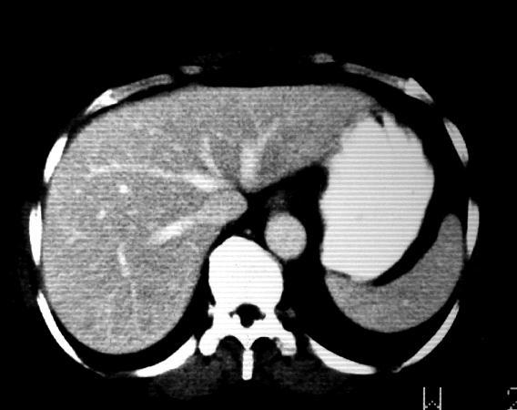
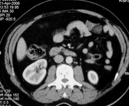
3 Triphasic study of the liver Is now the accurate way to detect and diagnose hepatic lesions Using the MDCT and automatic pumb injector for contrast injection [to avoid human error] The patient lies supine on the CT machine Frontal scanogram is performed Precontrast scans for the hepatic area are obtained [from the diaphragm to the iliac crest] Start contrast injection by the pumb, the rate of injection is 2-4 ml/ sec. The injected contrast- material will reach the hepatic artery in about 20 sec. from the start time of injection then we scan the liver [arterial phase] The contrast material will reach the portal vein after about 50 sec. from the start of injection then we scan te liver again [portal phase] Then we wait for about 3 minutes and scan the liver again [delayed phase] The triphasic study is now completed - Arterial phase - Portal phase (venous phase) - Delayed phase Normal Anatomy Liver [Rt hypochondrium] Average hepatic size 15 cm cranio candal diameter i.e 15 CT sections provided the section thickeness is 1 cm. Allow ± 1 section for respiration. The use of MDCT can avoid this misregestration as the patient can hold his breath during the scanning period for each phase The normal density of the liver is homogenous with no focal lesions. Normal intra hepatic bile ducts are not usually seen on CT scans Hepatic CT density is almost similar to that of the spleen after contrast injection Hepatic vessels appear as tubular enhancing structures after contrast injection Hepatic veins are seen in the higher sections entring the IVC while lying in its fossa on the viseral surface of the liver
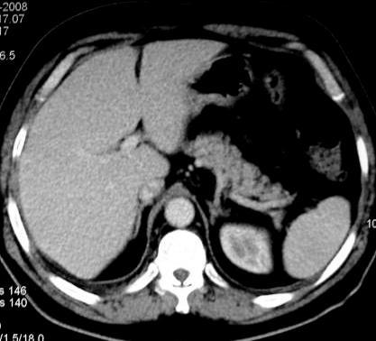
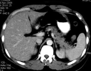
4 Hepatic artery branches are not seen on regular CT scan but can be seen in the arterial phase of the triphasic study The rest of vascular structures seen through out the liver parenchyma are portal venous radicles The porta hepatis represents the hilum of the liver. It contains fat, lymph nodes, CBD, hepatic artery and portal vein. All these structures are not usually seen on regular CT scan except for fat and portal vein. Liver segments: Left lobe: Medial segment (MS), lateral segment (LS) Rt lobe : Anteriorsegment (AS), posterior segment (PS) Caudate lobe is present between the porta hepatis and IVC Spleen [left hypochondriam] Average size is 12 cm cranio caudal diamiter More than 12 cm= splenomegaly... less than 12 cm = normal spleen Density is homogenous.. no focal lesion Pancreas [Retro peritonium] Lies obliquely in the upper abdomen, head lower than body and tail Density is homogenous less than that of the liver and spleen Pancreatic size: head (3cm), body (2.5cm), tail(2cm)in normal adults. Atrophic changes of the pancreas are normally seen in old age where the pancreas is small in size and shows fatty infiltration of its parenchyma The normal common pancreatic duct is not seen on regular CT Splenic vein lies along the posterior border of the pancreas. The splenic vein joins the SMV to form the portal vein in the upper CT sections. The normal anatomic landmark of the pancreatic head is the SMA and SMV The pancreatic head lies within the concavity of the doudenal loop, if the loop is not adequately opacified by oral grastrographin, it will not be distinguished from the pancreatic head impression of enlarged head of pancreas.
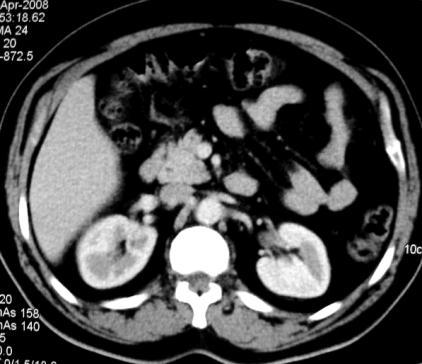
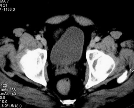
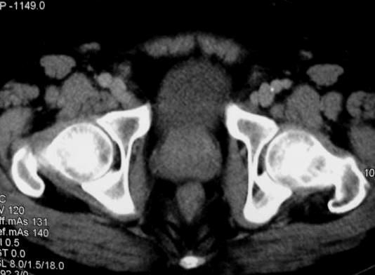
5 Suprarenal glands[ Retroperitonium] Best seen in obease patients with large amount of retro peritoneal fat It is sometimes difficult to identify the suprarenal glands in children and thin patients by CT The suprarenal gland lies at the upper pole of each kidney The left suprarenal gland is easily identified compared to the Rt one, because the Rt suprarenal gland is closely related to the Rt hepatic lobe The suprarenal gland has an inverted Y configuration with main stem, medial limb and lateral limb Kidneys [Retro peritonium] CT evaluation of the kidneys should include pre and post contrast scans Renal size is appeoximately 12 cm x 6cm x 3cm [length, width, thickness] The renal hilum contains fat, renal pelvis, artery and vein. The left renal vein is a constant finding. It crosses the midline in front or behind (retroaortic) the aorta to join the IVC The renal outline is smooth and regular, apart from fetal tobulations (normal variant) The Gerotta fascia surrounds the kidney and separates the perirenal from the pararenal spaces. The fascia itself is not usually seen on CT unless thickened Urinary bladder [pelvis] CT evaluation of urinary bladder lesions should include pre and post contrast scans Urinary bladder usually shows contrast fluid level [urine is above the contrast material] Posterior to the urinary bladder the seminal vesicles and rectum are seen in males, the uterine cervix and rectum are seen in the females The ovaries are better assessed by ultrasound or MRI The ischiorectal fossa is a triangular shaped fat containing area on either side of the anal canal
6 Abdominal muscles: The psoas muscle lies on either side of the vertebral bodies In the pelvis the psoas blends with the iliacus muscle which lies on the inner aspect of the iliac bone to form the iliopsoas muscle The rectus abdominis muscles form the anterior abdominal wall on either side of the midline
 14- seminal vesicles (male) 15- Rectum 16- Ureter 17- psoas muscle 18- Rectus abdomins muscle 19 -Gluteus maximus muscle 20- Intestine LS = lateral segment MS =")
7 1- Spleen 2- Liver 3- Stomach 4-Hepatic veins 5 -Suprarenal glands 6- Upper pole of the kidney 7- Tail of pancreas 8- Body of pancreas 9- Head of pancreas 10- kidney 11- Gall bladder 12- urinary bladder 13- uterine cervix (female) 14- seminal vesicles (male) 15- Rectum 16- Ureter 17- psoas muscle 18- Rectus abdomins muscle 19 -Gluteus maximus muscle 20- Intestine LS = lateral segment MS = medial segment AS = anterior segment PS = posterior segment A = aorta V = IVC SV = splenic vein SMA = superior mesentric artery SMV = superior mesentric vein
8
9
10
1 Right & left Hepatic ducts Gastric Impression of spleen
 Pancreatic Model 1 Right & left Hepatic ducts 14 Gastric Impression of spleen 2 Common hepatic duct 15 Renal Impression of spleen 3 Cystic Duct 16 Colic Impression of spleen 4 Common Bile Duct 17 Splenic
Lab Monitor Images Dissection of the Abdominal Vasculature + Lower Digestive System
 Lab Monitor Images Dissection of the Abdominal Vasculature + Lower Digestive System Stomach & Duodenum Frontal (AP) View Nasogastric tube 2 1 3 4 Stomach Pylorus Duodenum 1 Duodenum 2 Duodenum 3 Duodenum
Duodenum retroperitoneal
 Duodenum retroperitoneal C shaped Initial region out of stomach into small intestine RETROperitoneal viscus Superior 1 st part duodenal cap ; moves upwards and backwards to lie on the R crura medial to
Anatomy of the renal system. Professor Nawfal K. Al-Hadithi
 Anatomy of the renal system Professor Nawfal K. Al-Hadithi Objectives To describe the posterior abdominal wall To identify the main anatomical landmarks of the kidneys & ureters To describe the suprarenal
Pancreas & Biliary System. Dr. Vohra & Dr. Jamila
 Pancreas & Biliary System Dr. Vohra & Dr. Jamila 1 Objectives At the end of the lecture, the student should be able to describe the: Location, surface anatomy, parts, relations & peritoneal reflection
Abdomen and Pelvis CT (1) By the end of the lecture students should be able to:
 By the end of the lecture students should be able to:") RAD 451 Abdomen and Pelvis CT (1) By the end of the lecture students should be able to: State the common indications for Abdomen and pelvis CT exams Identify possible contra indications for Abdomen and
RAD 451 Abdomen and Pelvis CT (1) By the end of the lecture students should be able to: State the common indications for Abdomen and pelvis CT exams Identify possible contra indications for Abdomen and
The Spleen. Dr Fahad Ullah
 The Spleen BY Dr Fahad Ullah Spleen The spleen is an largest lymphoid organ shaped like a shoe that lies relative to the 9th and 11th ribs and is located in the left hypochondrium. Thus, the spleen is
Appendix 5. EFSUMB Newsletter. Gastroenterological Ultrasound
 EFSUMB Newsletter 87 Examinations should encompass the full range of pathological conditions listed below A log book listing the types of examinations undertaken should be kept Training should usually
د. عصام طارق. Objectives:
 GI anatomy Lecture: 5 د. عصام طارق Objectives: To describe anatomy of stomach, duodenum & pancreas. To list their main relations. To define their blood & nerve supply. To list their lymph drainage. To
My Patient Has Abdominal Pain PoCUS of the Biliary Tract and the Urinary Tract
 My Patient Has Abdominal Pain PoCUS of the Biliary Tract and the Urinary Tract Objectives PoCUS for Biliary Disease PoCUS for Renal Colic PoCUS for Urinary Retention Biliary Disease A patient presents
The posterior abdominal wall. Prof. Oluwadiya KS
 The posterior abdominal wall Prof. Oluwadiya KS www.oluwadiya.sitesled.com Posterior Abdominal Wall Lumbar vertebrae and discs. Muscles opsoas, quadratus lumborum, iliacus, transverse, abdominal wall
Peritoneum: Def. : It is a thin serous membrane that lines the walls of the abdominal and pelvic cavities and clothes the viscera.
 Peritoneum: Def. : It is a thin serous membrane that lines the walls of the abdominal and pelvic cavities and clothes the viscera. Layers of the peritoneum: 1. Outer Layer ( Parietal Peritoneum) : lines
GI module Lecture: 9 د. عصام طارق. Objectives:
 GI module Lecture: 9 د. عصام طارق Objectives: To list structures forming posterior abdominal wall. To follow aorta & its main branches. To describe IVC & its main tributaries. To list nerves of posterior
The abdominal Esophagus, Stomach and the Duodenum. Prof. Oluwadiya KS
 The abdominal Esophagus, Stomach and the Duodenum Prof. Oluwadiya KS www.oluwadiya.com Viscera of the abdomen Abdominal esophagus: Terminal part of the esophagus The stomach Intestines: Small and Large
Anatomy of the Large Intestine
 Large intestine Anatomy of the Large Intestine 2 Large Intestine Extends from ileocecal valve to anus Length = 1.5-2.5m = 5 feet Regions Cecum = 2.5-3 inch Appendix= 3-5 inch Colon Ascending= 5 inch Transverse=
MDCT signs differentiating retroperitoneal and intraperitoneal lesions- diagnostic pearls
 MDCT signs differentiating retroperitoneal and intraperitoneal lesions- diagnostic pearls Poster No.: C-0987 Congress: ECR 2015 Type: Educational Exhibit Authors: D. V. Bhargavi, R. Avantsa, P. Kala; Bangalore/IN
Accessory Glands of Digestive System
 Accessory Glands of Digestive System The liver The liver is soft and pliable and occupies the upper part of the abdominal cavity just beneath the diaphragm. The greater part of the liver is situated under
Guidelines, Policies and Statements D5 Statement on Abdominal Scanning
 Guidelines, Policies and Statements D5 Statement on Abdominal Scanning Disclaimer and Copyright The ASUM Standards of Practice Board have made every effort to ensure that this Guideline/Policy/Statement
ASSESSING THE PLAIN ABDOMINAL RADIOGRAPH M A A M E F O S U A A M P O F O
 ASSESSING THE PLAIN ABDOMINAL RADIOGRAPH M A A M E F O S U A A M P O F O Introduction The abdomen (less formally called the belly, stomach, is that part of the body between the thorax (chest) and pelvis,
Lab 9 Abdomen MUSCLES
 Lab 9 Abdomen MUSCLES External abdominal oblique continuous with the external intercostal muscle; its fibers point in a caudal direction as it moves anteriorly until it inserts on the linea alba via its
A Frame of Reference for Anatomical Study. Anatomy and Physiology Mr. Knowles Chapter 1 Liberty Senior High School
 A Frame of Reference for Anatomical Study Anatomy and Physiology Mr. Knowles Chapter 1 Liberty Senior High School Anatomical Terms of Direction and Position Created for communicating the direction and
Surface Anatomy. Location Shape Weight Role of Five Surfaces Borders Fissures Lobes Peritoneal Lig
 The Liver Functions Bile production and secretion Detoxification Storage of glycogen Protein synthesis Production of heparin and bile pigments Erythropoiesis (in fetus) Surface Anatomy Location Shape Weight
Lecture 02 Anatomy of the LIVER
 Lecture 02 Anatomy of the LIVER BY Dr Farooq Khan Aurakzai Dated: 02.01.2018 Introduction to Liver Largest gland in the body. 2 nd largest organ of the body. Weight approximately 1500 gm, and is roughly
Gross Anatomy of the Urinary System
 Gross Anatomy of the Urinary System Lecture Objectives Overview of the urinary system. Describe the external and internal anatomical structure of the kidney. Describe the anatomical structure of the ureter
Q129. Which of the following is NOT true about lymph node?
 Q129. Which of the following is NOT true about lymph node? (1). Normal lymph node is not seen in the ultrasound image (2). It is general that high frequency probe is used due to normal lymph node is located
THE ABDOMEN SUPRARENAL GLANDS KIDNEY URETERS URINARY BLADDER
 THE ABDOMEN SUPRARENAL GLANDS KIDNEY URETERS URINARY BLADDER THE SUPRARENAL GLANDS The suprarenal (adrenal) glands lie immediately superior and slightly anterior to the upper pole of either kidney. Golden
Abdomen and Retroperitoneum Ultrasound Protocols
 Abdomen and Retroperitoneum Ultrasound Protocols Reviewed By: Anna Ellermeier, MD Last Reviewed: March 2018 Contact: (866) 761-4200, Option 1 **NOTE for all examinations: 1. If documenting possible flow
The Kidneys. (L., ren; Gk, nephros; hence the adjectives renal and nephric) & Suprarenal (Adrenal) Glands. Dr Maan Al-Abbasi PhD, MBChB
 & Suprarenal (Adrenal) Glands. Dr Maan Al-Abbasi PhD, MBChB") The Kidneys (L., ren; Gk, nephros; hence the adjectives renal and nephric) & Suprarenal (Adrenal) Glands Dr Maan Al-Abbasi PhD, MBChB Functions of Urinary System Regulate electrolytes (K+, Na+, etc) Regulate
The Kidneys (L., ren; Gk, nephros; hence the adjectives renal and nephric) & Suprarenal (Adrenal) Glands Dr Maan Al-Abbasi PhD, MBChB Functions of Urinary System Regulate electrolytes (K+, Na+, etc) Regulate
UNIVERSITY DEVELOPMENT CENTER. Course Specification 2015/2016 For the Anatomy (first year) Medicine Anatomy and Embryology Department 29/12/2015
 Medicine Anatomy and Embryology Department 29/12/2015") Course Specification 2015/2016 For the Anatomy (first year) Faculty : Department : Medicine Anatomy and Embryology Department Course Specification: Programme (s) on which the course is given : M.B.B.Ch
Course Specification 2015/2016 For the Anatomy (first year) Faculty : Department : Medicine Anatomy and Embryology Department Course Specification: Programme (s) on which the course is given : M.B.B.Ch
To describe the liver. To list main structures in porta hepatis.
 GI anatomy Lecture: 6 د. عصام طارق Objectives: To describe the liver. To list main structures in porta hepatis. To define portal system & portosystemic anastomosis. To list parts of biliary system. To
CT Chest. Verification of an opacity seen on the straight chest X ray
 CT Chest Indications: To assess equivocal plain x-ray findings Staging of lung neoplasm Merastatic workup of extra thoraces malignancies Diagnosis of diffuse lung diseases with HRCT Assessment of bronchietasis
Imaging in gastric cancer
 Imaging in gastric cancer Gastric cancer remains a deadly disease because of late diagnosis. Adenocarcinoma represents 90% of malignant tumors. Diagnosis is based on endoscopic examination with biopsies.
Anatomy: Know Your Abdomen
 Anatomy: Know Your Abdomen Glossary Abdomen - part of the body below the thorax (chest cavity); separated by the diaphragm. Anterior - towards the front of the body. For example, the umbilicus is anterior
Anatomy. Contents Brain (Questions)
") Anatomy 12 Contents 12.1 Brain (Questions).................................................... 683 12.2 Head and Neck (Questions)............................................. 685 12.3 Thorax (Questions)...................................................
Anatomy 12 Contents 12.1 Brain (Questions).................................................... 683 12.2 Head and Neck (Questions)............................................. 685 12.3 Thorax (Questions)...................................................
4/9/2018 OBJECTIVES PANCREAOTO BILIARY ULTRASOUND: BEYOND CHOLECYSTITIS
 PANCREAOTO BILIARY ULTRASOUND: BEYOND CHOLECYSTITIS Jean Yves Sewah Kaiser Permanente West Los Angeles 1 OBJECTIVES Discuss the role of ultrasound in the evaluation of the gallbladder, biliary tree and
Introduction to Anatomical Terms. Packet #3
 Introduction to Anatomical Terms Packet #3 Directional Terms Directional terms describe the positions of structures relative to other structures or locations in the body. Introduction Superior vs. Inferior
Abdominal Ultrasound
 Abdominal Ultrasound Imaging Control Buttons Depth The organ imaged should take up 3/4 of the screen Frequency = Penetration Use high frequencies (harmonics) for fluid filled and superficial structures
ANATOMY OF PELVICAYCEAL SYSTEM -DR. RAHUL BEVARA
 1 ANATOMY OF PELVICAYCEAL SYSTEM -DR. RAHUL BEVARA 2 KIDNEY:ANATOMY OVERVIEW Kidneys are retroperitoneal, in posterior abdominal region, extending from T12 L3 Bean-shaped Right kidney is lower than left
Basic Abdominal Sonography
 24S Basic Abdominal Sonography Procedural Overview JOHN FATCHETT II, RDMS is provided. Patient preparation (i.e., fasting) scanning techniques, spleen, transducer. evaluation of abdominal anatomy in the
Pancreas and Biliary System
 Pancreas and Biliary System Please view our Editing File before studying this lecture to check for any changes. Color Code Important Doctors Notes Notes/Extra explanation Objectives At the end of the lecture,
Biology Human Anatomy Abdominal and Pelvic Cavities
 Biology 351 - Human Anatomy Abdominal and Pelvic Cavities Please place your name and I.D. number on the back of the last page of this exam. You must answer all questions on this exam. Because statistics
Vascular CT Protocols
 Vascular CT Protocols V 1D: Chest and abdominal CT angiogram (aortic dissection protocol) V 1T: Chest CT angiogram (aortic trauma protocol) V 2: Abdominal and pelvis CT angiogram (aortic aneurysm protocol)
SUBJECTS 2nd year, 1st semester I. 1. Primitive gut - limits, derivatives 2. Foregut -limits, evolution, derivatives 3. Midgut -limits, evolution,
 SUBJECTS 2nd year, 1st semester I. 1. Primitive gut - limits, derivatives 2. Foregut -limits, evolution, derivatives 3. Midgut -limits, evolution, derivatives 4. Hindgut- limits, evolution, derivatives
Cat Dissection. Muscular Labs
 Cat Dissection Muscular Labs Tibialis anterior External oblique Pectroalis minor Gastrocnemius Sartorius Pectoralis major Gastrocnemius Semitendinosis Sartorius External oblique Trapezius Latissimus dorsi
Abdomen. Retroperitoneal space
 Abdomen. Retroperitoneal space Abdominal cavity The space bounded by: Anterolateral abdominal wall Posterior abdominal wall Diaphragm Pelvic walls and pelvic floor. Subdivided into: True abdominal cavity
Chapter 3 General Anatomy and Radiographic Positioning Terminology General Anatomy
 Chapter 3 General Anatomy and Radiographic Positioning Terminology General Anatomy Definition of Terms Anatomy- term applied to the science of the structure of the body Physiology- study of the function
ANATOMY - II. FOR 2 MARKS QUESTIONS(for Anatomy II Q.NO 1 ) THORAX
 THORAX") ANATOMY - II FOR 2 MARKS QUESTIONS(for Anatomy II Q.NO 1 ) THORAX 1. Total no. of true ribs. 2. Total no. of false ribs. 3. Total no. of floating ribs. 4. Total no. of typical ribs. 5. Total no. of vertebra.
ANATOMY - II FOR 2 MARKS QUESTIONS(for Anatomy II Q.NO 1 ) THORAX 1. Total no. of true ribs. 2. Total no. of false ribs. 3. Total no. of floating ribs. 4. Total no. of typical ribs. 5. Total no. of vertebra.
Anatomy of the spleen. Oluwadiya KS
 Anatomy of the spleen Oluwadiya KS www.oluwadiya.com Introduction The spleen is an ovoid, usually purplish, pulpy mass about the size and shape of one's fist. It is the largest lymphoid tissue in the body
Abdomen Sonography Examination Content Outline
 Abdomen Sonography Examination Content Outline (Outline Summary) # Domain Subdomain Percentage 1 2 3 Anatomy, Perfusion, and Function Pathology, Vascular Abnormalities, Trauma, and Postoperative Anatomy
STRUCTURAL BASIS OF MEDICAL PRACTICE EXAMINATION 3. October 16, 2015
 STRUCTURAL BASIS OF MEDICAL PRACTICE EXAMINATION 3 October 16, 2015 PART l. Answer in the space provided. (12 pts) 1. Identify the structures. (2 pts) A. B. A B C. D. C D 2. Identify the structures. (2
Figure 26.1 An Introduction to the Urinary System
 Chapter 26 Figure 26.1 An Introduction to the Urinary System Components of the Urinary System Kidney Produces urine Ureter Transports urine toward the urinary bladder Urinary Bladder Temporarily stores
Preview from Notesale.co.uk Page 1 of 34
 Abdominal viscera and digestive tract Digestive tract Abdominal viscera comprise majority of the alimentary system o Terminal oesophagus, stomach, pancreas, spleen, liver, gallbladder, kidneys, suprarenal
In the name ofgod. Abdomen 3. Dr. Zahiri
 In the name ofgod Abdomen 3 Dr. Zahiri Peritoneum Peritoneum It is the serous membrane(a type of loose connective tissue and is covered by mesothelium) that lines the abdominal cavity. Extensions of the
The jejunum and the Ileum. Prof. Oluwadiya KS
 The jejunum and the Ileum Prof. Oluwadiya KS www.oluwadiya.siteled.com Introduction Introduction The small intestine (SI) comprises of the duodenum, jejunum and the ileum The jejunum is the second part
Dissection Lab Manuals: Required Content
 Dissection Lab Manuals: Required Content 1. Introduction a. Basic terminology (directions) b. External features of the cat c. Adaptations to predatory niche d. How to skin a cat e. How to make the incisions
Policies, Standards, and Guidelines. Guidelines for Abdominal Ultrasound Examination
 Policies, Standards, and Guidelines Guidelines for Abdominal Ultrasound Examination Approved by Council Feb 2018 Disclaimer and Copyright The ASUM Standards of Practice Board have made every effort to
URINARY SYSTEM ANATOMY PART
 URINARY SYSTEM ANATOMY PART 1 DANIL HAMMOUDI.MD Urinary System Composed of kidneys, ureters, urinary bladder, and urethra Eliminates nitrogenous wastes from the body Regulates water, electrolyte, and ph
The Urinary System Pearson Education, Inc.
 26 The Urinary System Introduction The urinary system does more than just get rid of liquid waste. It also: Regulates plasma ion concentrations Regulates blood volume and blood pressure Stabilizes blood
Abdominal Doppler Mastering the next level of vascular anatomy in the belly. Cindy A. Owen, RDMS, RVT
 Abdominal Doppler Mastering the next level of vascular anatomy in the belly Cindy A. Owen, RDMS, RVT Introduction Abdominal Doppler is a tough exam Success is dependent on: Patient body habitus Patient
IMAGING GUIDELINES - COLORECTAL CANCER
 IMAGING GUIDELINES - COLORECTAL CANCER DIAGNOSIS The majority of colorectal cancers are diagnosed on colonoscopy, with some being diagnosed on Ba enema, ultrasound or CT. STAGING CT chest, abdomen and
3 Circulatory Pathways
 40 Chapter 3 Circulatory Pathways Systemic Arteries -Arteries carry blood away from the heart to the various organs of the body. -The aorta is the longest artery in the body; it branches to give rise to
Abdominal Ultrasonography
 Abdominal Ultrasonography David A. Masneri, DO, FACEP, FAAEM Assistant Professor of Emergency Medicine Assistant Director, Emergency Medicine Residency Medical Director, Operational Medicine Division Center
Artery 1 Head and Thoracic Arteries. Arrange the parts in the order blood flows through them.
 Artery 1 Head and Thoracic Arteries 1. Given the following parts of the aorta: 1. abdominal aorta 2. aortic arch 3. ascending aorta 4. thoracic aorta Arrange the parts in the order blood flows through
Lecture 56 Kidney and Urinary System
 Lecture 56 Kidney and Urinary System The adrenal glands are located on the superomedial aspect of the kidney The right diagram shows a picture of the kidney with the abdominal walls and organs removed
STRUCTURAL BASIS OF MEDICAL PRACTICE EXAMINATION 3. October 17, 2014
 STRUCTURAL BASIS OF MEDICAL PRACTICE EXAMINATION 3 October 17, 2014 PART l. Answer in the space provided. (12 pts) 1. Identify the structures. (2 pts) A. B. A B C. D. C D 2. Identify the structures. (2
BY DR NOMAN ULLAH WAZIR
 BY DR NOMAN ULLAH WAZIR The stomach (from ancient Greek word stomachos, stoma means mouth) is a muscular, hollow and the most dilated part of the GIT. It starts from the point where esophagus ends. It
Dr. Zahiri. In the name of God
 Dr. Zahiri In the name of God small intestine = small bowel is the part of the gastrointestinal tract Boundaries: Pylorus Ileosecal junction Function: digestion and absorption of food It receives bile
Basic Body Structure
 Basic Body Structure The Cell All life consists of microscopic living structures called cells. They perform various functions throughout the body. All cells are similar in structure, but not identical.
Netter's Anatomy Flash Cards Section 4 List 4 th Edition
 Netter's Anatomy Flash Cards Section 4 List 4 th Edition https://www.memrise.com/course/1577335/ Section 4 Abdomen (31 cards) Plate 4-1 Bony Framework of Abdomen 1.1 Costal cartilages 1.2 Iliac crest 1.3
Pancreas Quizzes c. Both A and B a. Directly into the blood stream (not using ducts)
") Pancreas Quizzes Quiz 1 1. The pancreas produces hormones. Which type of hormone producing organ is the pancreas? a. Endocrine b. Exocrine c. Both A and B d. Neither A or B 2. Endocrine indicates hormones
Pancreas Quizzes Quiz 1 1. The pancreas produces hormones. Which type of hormone producing organ is the pancreas? a. Endocrine b. Exocrine c. Both A and B d. Neither A or B 2. Endocrine indicates hormones
Small Plicae Circularis. Short Closely packed together. Sparse, completely absent at distal part Lymphoid Nodule
 Intestines Differences Between Jejunum and Ileum Types Jejunum Ileum Color Deeper red Paler pink Calibre Bigger Smaller Thickness of wall Thick and Heavy Thin and Lighter Vascularity Highly vascularised
The Whipple Operation Illustrations
 The Whipple Operation Illustrations Fig. 1. Illustration of the sixstep pancreaticoduodenectomy (Whipple operation) as described in a number of recent text books by Dr. Evans. The operation is divided
Abdominal Ultrasound : Aorta, Kidneys, Bladder
 Abdominal Ultrasound : Aorta, Kidneys, Bladder Nilam J. Soni, MD, MSc Associate Professor of Medicine Divisions of Hospital Medicine and Pulmonary/Critical Care Medicine Department of Medicine University
Nasogastric tube. Stomach. Pylorus. Duodenum 1. Duodenum 2. Duodenum 3. Duodenum 4
 Esophagus Barium Swallow Stomach and Duodenum 4 year old Upper GI Nasogastric tube Stomach and Duodenum 4 year old Upper GI Nasogastric tube Stomach Pylorus Duodenum 1 Duodenum 2 Duodenum 3 Duodenum 4
Welcome to ANAT 10A! What is Anatomy? Different levels of Anatomy The Language of Anatomy Pearson Education, Inc.
 Welcome to ANAT 10A! What is Anatomy? Different levels of Anatomy The Language of Anatomy Introduction Anatomy means to dissect: (ANAT 10A) The study of internal & external body structures The study of
Appendix 9: Endoscopic Ultrasound in Gastroenterology
 Appendix 9: Endoscopic Ultrasound in Gastroenterology This curriculum is intended for clinicians who perform endoscopic ultrasonography (EUS) in gastroenterology. It includes standards for theoretical
Anatomy of the SMALL INTESTINE. Dr. Noman Ullah Wazir PMC
 Anatomy of the SMALL INTESTINE Dr. Noman Ullah Wazir PMC SMALL INTESTINE The small intestine, consists of the duodenum, jejunum, and illium. It extends from the pylorus to the ileocecal junction were the
CHAPTER 2 Terms Pertaining to the Body as a Whole
 CHAPTER 2 Terms Pertaining to the Body as a Whole OBJECTIVES 1. Define terms that apply to the structural organization of the body. 2. Identify the body cavities and the organs contained within the cavities.
The peritoneum. Prof. Oluwadiya KS, MBBS, FMCS(Orthop) Website:
 Website:") The peritoneum Prof. Oluwadiya KS, MBBS, FMCS(Orthop) Website: http://oluwadiya.com The peritoneum Serous membrane that lines the abdominopelvic cavity and invests the viscera The largest serous membrane
The peritoneum Prof. Oluwadiya KS, MBBS, FMCS(Orthop) Website: http://oluwadiya.com The peritoneum Serous membrane that lines the abdominopelvic cavity and invests the viscera The largest serous membrane
Urinary 1 Checklist Gross Anatomy of the Urinary System
 Urinary 1 Checklist Gross Anatomy of the Urinary System Urinary system Kidneys Parietal peritoneum Retroperitoneal Renal fascia The urinary system consists of two kidneys, two ureters, the urinary bladder,
Chapter Overview. Chapter 1. Anatomy. Physiology
 Chapter Overview Chapter 1 An Introduction to the Human Body Define Anatomy and Physiology Levels of Organization Characteristics of Living Things Homeostasis Anatomical Terminology 1 2 Anatomy Describes
Inferior Pelvic Border
 Pelvis + Perineum Pelvic Cavity Enclosed by bony, ligamentous and muscular wall Contains the urinary bladder, ureters, pelvic genital organs, rectum, blood vessels, lymphatics and nerves Pelvic inlet (superior
Case 9799 Stanford type A aortic dissection: US and CT findings
 Case 9799 Stanford type A aortic dissection: US and CT findings Accogli S, Aringhieri G, Scalise P, Angelini G, Pancrazi F, Bemi P, Bartolozzi C Department of Diagnostic and Interventional Radiology, University
Introduction to A & P Medical Terminology
 Human Anatomy & Physiology PHA322.10 D. Matesic, Spring, 2012 Class Notes Introduction to A & P Medical Terminology Levels of Structural Organization Anatomy the study of the structure of body parts and
The Human Body: An Orientation
 The Human Body: An Orientation Body standing upright Anatomical Position feet slightly apart palms facing forward thumbs point away from body Directional Terms Superior and inferior toward and away from
Renal vascular evaluation with 64 Multislice Computerized Tomography Daniela Stoisa, Fabrizzio E. Galiano, Andrés Quaranta, Roberto L.
 Renal vascular evaluation with 64 Multislice Computerized Tomography Daniela Stoisa, Fabrizzio E. Galiano, Andrés Quaranta, Roberto L. Villavicencio Footnote Diagnóstico Médico Oroño. Bv. Oroño 1515. 2000.
Normal Sonographic Anatomy
 hapter 2:The Liver DUNSTAN ABRAHAM Normal Sonographic Anatomy Homogeneous, echogenic texture (Figure 2-1) Measures approximately 15 cm in length and 10 12.5 cm anterior to posterior; measurement taken
Chapter 1- An Orientation to the Human Body
 Chapter 1- An Orientation to the Human Body Overview of Anatomy and Physiology: -Anatomy- of body parts and their relationships to one another. -Gross or Macroscopic= large and easily observable -Microscopic=
ANATOMY OF THE SMALL & LARGE INTESTINES. Semester 1, 2011 A. Mwakikunga
 ANATOMY OF THE SMALL & LARGE INTESTINES Semester 1, 2011 A. Mwakikunga LEARNING OBJECTIVES 1. List the parts and anatomical regions of the small and large intestines 2. State anatomical relations of the
Omran Saeed. Mohammad Al-muhtaseb. 1 P a g e
 13 Omran Saeed Mohammad Al-muhtaseb 1 P a g e Posterior abdominal wall - The diaphragm separates between thoracic cavity and abdominal cavity. Structures of posterior abdominal wall: (below diaphragm)
Anatomical Considerations for Lab Practical II
 Anatomical Considerations for Lab Practical II For each of the following please be prepared to provide: Identification System Organ(s) or ducts to Function(s) location which it is attached Use your lecture
Close window to return to IVIS. in collaborazione con RICHIESTO ACCREDITAMENTO. organizzato da certificata ISO 9001:2000
 in collaborazione con Close window to return to IVIS RICHIESTO ACCREDITAMENTO SOCIETÀ CULTURALE ITALIANA VETERINARI PER ANIMALI DA COMPAGNIA SOCIETÀ FEDERATA ANMVI organizzato da certificata ISO 9001:2000
Ex. 1 :Language of Anatomy
 Collin College BIOL 2401 : Human Anatomy & Physiology Ex. 1 :Language of Anatomy The Anatomical Position Used as a reference point when referring to specific areas of the human body Body erect Head and
Job Task Analysis for ARDMS Abdomen Data Collected: June 30, 2011
 Job Task Analysis for ARDMS Abdomen Data Collected: June 30, 2011 Reported: Analysis Summary for: Abdomen Examination Survey Dates 06/13/2011-06/26/2011 Invited Respondents 6,000 Surveys with Demographics
Anatomy of the liver and pancreas
 Anatomy of the liver and pancreas Prof. Abdulameer Al-Nuaimi E-mail: a.al-nuaimi@sheffield.ac.uk abdulameerh@yahoo.com Liver Aorta Pulm. Trunk Rt. At, Duct. Art. Lt. Ven. Rt. Ven. Internal Posterior
-12. -Renad Habahbeh. -Dr Mohammad mohtasib
 -12 -Renad Habahbeh - -Dr Mohammad mohtasib The Gallbladder -The gallbladder has a body, a fundus (a rounded end), a neck, Hartmann s pouch before the neck and a cystic duct that meets the common hepatic
Done by: nisreen obeidat
 Sheet: liver and pancreas Done by: nisreen obeidat Embryology of the liver The liver develops in the ventral mesentery of the foregut and divides the ventral mesentery :into 1)lesser omentum (between the
LECTURE ON THE URINARY SYSTEM
 LECTURE ON THE URINARY SYSTEM (Uropoetic System) AN OVERVIEW Dr HAMIADJI THE URINARY SYSTEM URINARY SYSTEM The kidneys are responsible for removing wastes from the body, regulating electrolyte balance
Jhia Anjela D. Rivera 1 1. BS Biology, Department of Biology, College of Science, Polytechnic University of the Philippines
 DIGESTIVE SYSTEM Jhia Anjela D. Rivera 1 1 BS Biology, Department of Biology, College of Science, Polytechnic University of the Philippines DIGESTIVE SYSTEM Consists of the digestive tract (gastrointestinal
Body Regions Review. Anatomical Position. Anatomical Planes. Supine versus Prone 9/9/2009
 Body Regions Review The fundamental divisions of the human body Christine Sparks Anatomy / Physiology I Sept. 9, 2009 Anatomical Position Universal terms are used to describe the body accurately and result