ISUOG Basic Training Typical Ultrasound Appearances of Common Pathologies in the Adnexae
|
|
|
- Myron Warner
- 5 years ago
- Views:
Transcription

1 ISUOG Basic Training Typical Ultrasound Appearances of Common Pathologies in the Adnexae
2 Learning objectives At the end of the lecture series you will be able to: Compare the differences between typical normal and common abnormal appearances of the adnexa in gynaecological ultrasound examination
3 Key questions How do I describe my ultrasound findings using the standardized International Ovarian Tumor Analysis (IOTA) terminology? What are the typical ultrasound appearances of the most common pathologies in the adnexa? What diagnostic methods can I use to discriminate between benign and malignant adnexal pathology? Which patients should I refer for specialist opinion?
4 Key points Understand the typical ultrasound features of a normal preand post-menopausal ovary Understand the typical ultrasound appearances of the most common pathologies in the adnexa Understand how to use IOTA terminology Know when to refer for a specialist opinion
5 Typical ultrasound appearances of the most common pathologies in the adnexa

6 Ovarian findings Normal ovary Functional cysts Benign Borderline Invasive Metastatic
7 Normal ovary Normal ultrasound findings Differ between women before and after menopause Changes throughout the menstrual cycle
8 Normal ovary How big is a normal ovary in a woman of fertile age? Very variable Median 7 ml Range 2-17 ml (Range 1-20 ml) 303 women years old with regular menstrual cycles, cd 4-8 Jokubkiene et al. J Ultrasound Med 2012;31(10):


9 Normal ovary What is a normal number of antral follicles before menopause? Text books: 6-7 follicles/ovary Jokubkiene et al: Median 11 follicles (2-10 mm) /ovary Range th-90th percentile % had >12 follicles/ovary, i.e. PCO* *PCO : > 12 follicles/ovary or ovary > 10 ml (Rotterdam)
10 Normal ovary How big is a normal ovary in a postmenopausal woman? Median 1x1x2 cm Median volume 1 ml range: ml 144 asymptomatic postmenopausal women, years old Sladkevicius et al. Ultrasound Obstet Gynaecol 1995;6(2):81-90


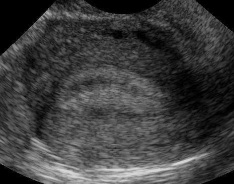
11 Normal ovary Changes during the menstrual cycle Post-menstruation Proliferative phase 3 days before ovulation Proliferative phase 1 day before ovulation Secretory phase 6 days after ovulation

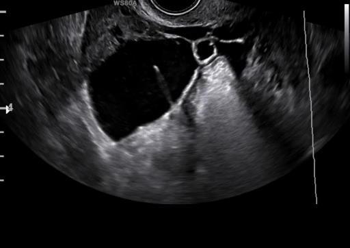
12 Some fluid in the pouch of Douglas is NORMAL before menopause


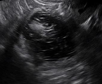
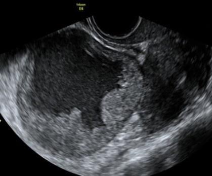
13 A corpus luteum may look different
14 Ovarian findings Normal ovary Functional cysts Benign Borderline Invasive Metastatic

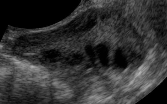
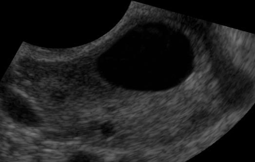
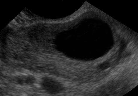
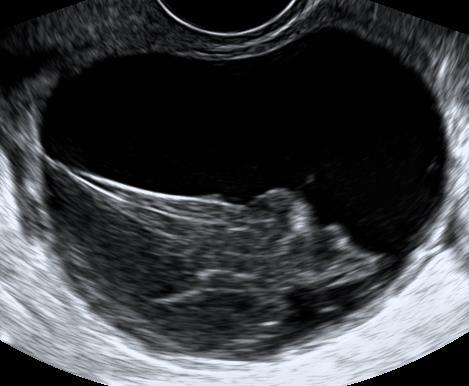
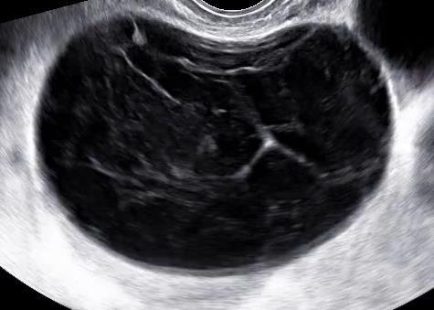
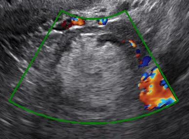
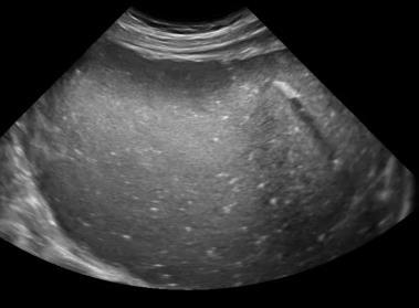
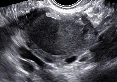
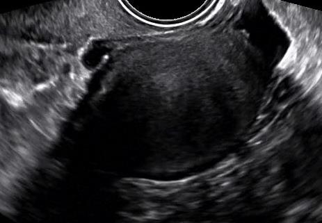
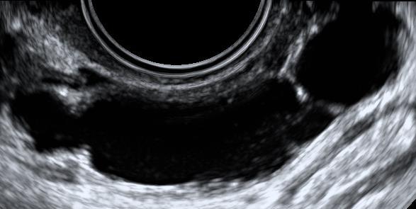
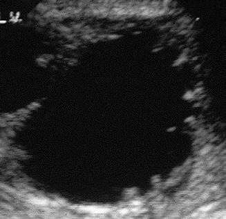
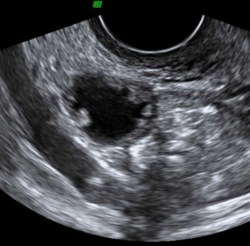
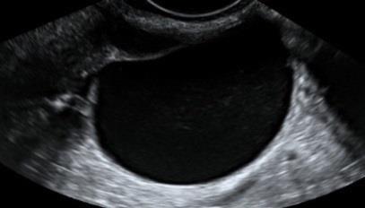
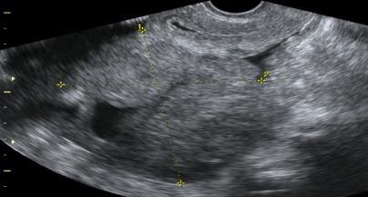
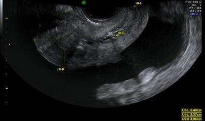
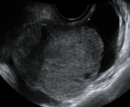
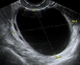
15 Functional cysts Follicular cyst / simple cyst
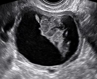
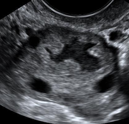
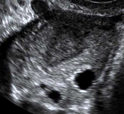
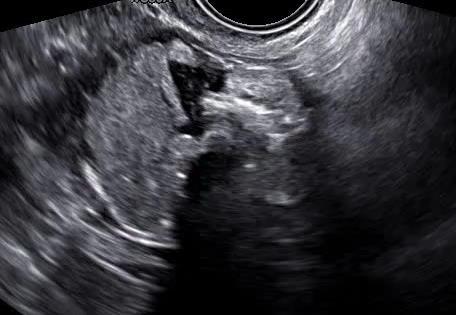
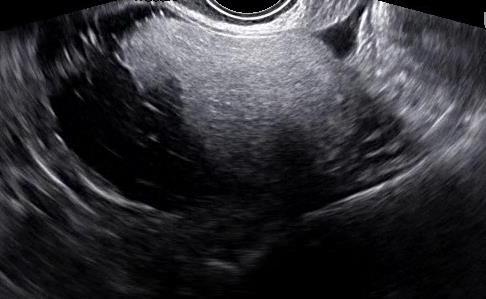
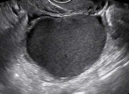
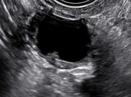
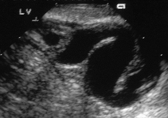
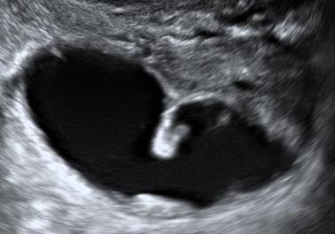
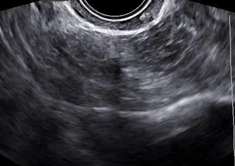
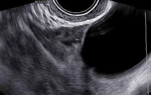
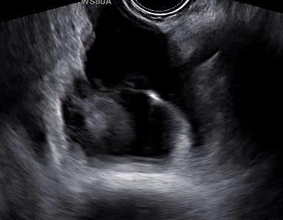
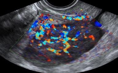
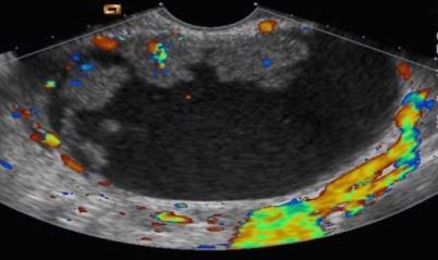
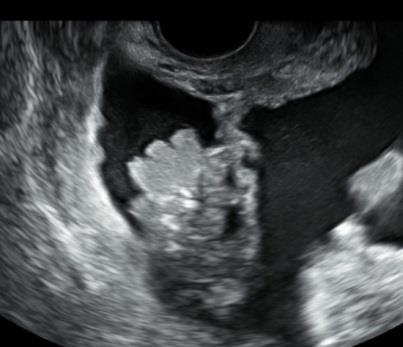
16 Functional cysts Corpus luteal cyst
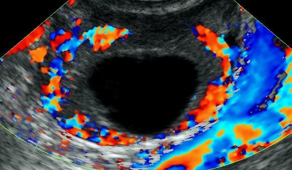
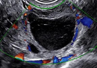
17 Functional cysts Corpus luteal cyst
18 Ovarian findings Normal ovary Functional cysts Benign Borderline Invasive Metastatic
19 Benign Common ovarian pathology Dermoid/mature teratoma Endometrioma Serous cystadenoma/cystadenofibroma Mucinous cystadenoma Fibroma

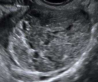
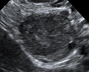
20 Benign Dermoid cyst

21 Benign Dermoid cyst
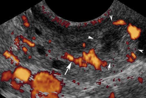
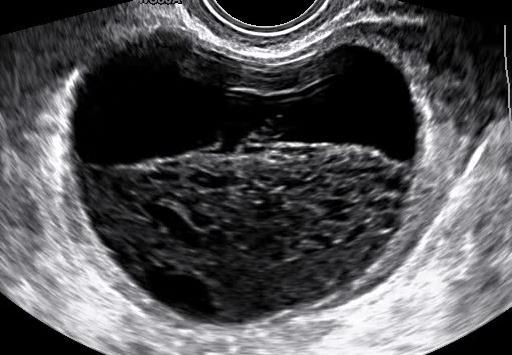
22 Benign Endometrioma

23 Benign Cystadenoma/ cystadenofibroma

24 Benign Fibroma
25 Benign Common extra-ovarian adnexal pathology Hydrosalpinx Paraovarian cysts Peritoneal inclusion cysts/ pseudocysts
26 Hydro-pyo-haemato-salpinx Sausage shape Cog wheel Beads on a string Incomplete septa Incomplete septa
27 Benign Hydrosalpinx

28 Benign Paraovarian cyst paraovarian Ovary

29 Benign Paraovarian cyst Clip paraovarian cyst TBA Ovary
30 Benign Peritoneal pseudocyst

31 Benign Peritoneal pseudocyst

32 Ovarian findings Normal ovary Functional cysts Benign Borderline Invasive Metastatic
33 Diagnostic methods to discriminate between benign and malignant adnexal pathology


34 Malignant features IOTA Simple Rules Irregular solid tumor Presence of ascites 4 papillary projections Irregular multilocular-solid tumor 100mm colour score 4 (strong blood flow) Benign features Unilocular cyst Tumor with largest solid component < 7mm Acoustic shadows Smooth multilocular tumor < 100mm colour score 1 (no blood flow)
35 Simple Rules Malignant if one or more M-features apply without presence of B-features Benign if one or more B-features apply without presence of M-features Inconclusive if no features present or if both B and M- features apply

36 Benign Tumour Borderline Tumour FIGO Stage I Ovarian cancer FIGO Stage II-IV Ovarian cancer Metastasis to the ovary
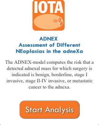
37 IOTA-ADNEX (Assessment of Different NEoplasias in the adnexa) variables Age of patient Type of centre Serum CA- 125 Six ultrasound variables


 (4) number of")

 acoustic")
38 (1) maximum diameter of lesion (mm) (2) proportion of solid tissue (3) more than 10 cyst locules (yes vs no) (4) number of papillary projections (0, 1, 2, 3, more than 3) (5) acoustic shadows (yes vs no) (6) ascites (yes vs no)


39 IOTA-ADNEX (Assessment of Different NEoplasias in the adnexa) app
40 Which patients should I refer for specialist opinion? Those in whom you are uncertain about the diagnosis (especially if you suspect malignancy)
41 Key points When in doubt: refer for second opinion
42 Acknowledgements These slides were created by: Dirk Timmerman Tom Bourne Lil Valentin
43 ISUOG Basic Training Typical ultrasound appearances of common pathologies in the adnexa
2/24/19. Ovarian pathology: IOTA ADNEXAL MASSES. Content. IOTA terms for description of an adnexal mass. IOTA terms for description of an adnexal mass
 Content Ovarian pathology: IOTA ADNEXAL MASSES X SIMPLE COMPLEX Dr DESCRIBE WHAT YOU SEE FRANZCOG, MPH, DDU, COGU Sonologist Clinically useful Benign Malignant Communication between clinicians/research
Terminology Estimate the risk of malignancy in adnexal masses - Overview
 Understanding the IOTA (International Ovarian Tumor Analysis) terminology & Classification Using the IOTA simple rules to estimate the risk of malignancy in women with adnexal masses Elisabeth Epstein,
The Adnexal Mass. Handout NCUS 3/18/2017 Suzanne Dixon, MD
 The Adnexal Mass Handout NCUS 3/18/2017 Suzanne Dixon, MD Objectives: Pelvic mass differential Characteristics of the normal ovary Standard terminology for ovarian masses Benign vs. malignant features
Gynecologic Ultrasound. Sujata Ghate, MD Associate Professor of Radiology Duke University Medical Center
 Gynecologic Ultrasound Sujata Ghate, MD Associate Professor of Radiology Duke University Medical Center Objectives Understand work-up of endometrial abnormalities Show examples of uterine and endometrial
Diane DeFriend Derriford Hospital, Plymouth
 Diane DeFriend Derriford Hospital, Plymouth Ultrasound US remains primary imaging modality for investigation of an adnexal mass Aim to characterise Benign Malignant Indeterminate 90% adnexal masses characterised
Adnexal Masses in Menopausal Women
 Adnexal Masses in Menopausal Women Surgery or Surveillance? Disclosure Frederick R. Ueland, MD Professor and Director Division of Gynecologic Oncology University of Kentucky I have no financial disclosures
Does Ovarian Cysts affect your Fertility?
 Does Ovarian Cysts affect your Fertility? Ovarian cysts are sac like structures (filled with liquid or semi-solid) develop in the ovary. Ovarian cysts are very common, mostly painless and harmless with
Adnexal Masses in Menopausal Women Surgery or Surveillance?
 Adnexal Masses in Menopausal Women Surgery or Surveillance? FREDTALK IDEASWORTHSPREADING Disclosure I am a member of Vermillion s Speakers Bureau I am NOT a paid consultant for Vermillion Inc. nor do I
A new scoring model for characterization of adnexal masses based on two-dimensional gray-scale and colour Doppler sonographic features
 FVV in ObGyn, 2014, 6 (2): 68-74 Original paper A new scoring model for characterization of adnexal masses based on two-dimensional gray-scale and colour Doppler sonographic features Ahmed M. Abbas, Kamal
OVARIAN MASSES : MANAGEMENT CHALLENGE
 SRU 2015 OVARIAN MASSES : MANAGEMENT CHALLENGE phyllis.glanc@sunnybrook.ca Sunnybrook Health Science Centre University of Toronto, Dept Medical Imaging, Obstetrics & Gynecology Thank you on behalf of the
P. SLADKEVICIUS and L. VALENTIN ABSTRACT
 Ultrasound Obstet Gynecol 2013; 41: 318 327 Published online in Wiley Online Library (wileyonlinelibrary.com). DOI: 10.1002/uog.12289 Intra- and interobserver agreement when describing adnexal masses using
Assessment of adnexal masses. Ultrasound workup of adnexal masses. symptoms. symptoms. Age. Serum tumor markers 10/1/2018
 Assessment of adnexal masses Ultrasound workup of adnexal masses Kevin Robinson, DO Department of Radiology Michigan State University October 4, 2018 Patients symptoms Age Menstrual status Serum tumor
IN THE NAME OF GOD POV: CYSTIC OVARIAN LESION
 IN THE NAME OF GOD POV: CYSTIC OVARIAN LESION CASE 1 20 years old girl with AUB and pelvic pain from 2 weeks ago Impression :Simple unilocular 6 cm ovarian cyst Next step? Almost certainly benign so FU
Title of Guideline (must include the word Guideline not protocol, policy, procedure etc)
") Title of Guideline (must include the word Guideline not protocol, policy, procedure etc) Author: Contact Name and Job Title Directorate & Speciality Assessment, referral and initial management of ultrasound
Title of Guideline (must include the word Guideline not protocol, policy, procedure etc) Author: Contact Name and Job Title Directorate & Speciality Assessment, referral and initial management of ultrasound
Category Term Definition Comments 1 Major Categories 1a
 Working Lexicon Categories, Terms & Definitions Category Term Definition Comments 1 Major Categories 1a Physiologic Category (consistent with normal ovarian physiology) Follicle Simple 3 cm in premenopausal
ISUOG Basic Training Examining the Uterus: Myometrium
 ISUOG Basic Training Examining the Uterus: Myometrium Learning objectives At the end of the lecture you will be able to: Recognise the typical ultrasound appearances of the normal myometrium Recognise
Top Tips for Gynaecological Ultrasound. Catherine Kirkpatrick Consultant Sonographer Dublin Oct 2018
 Top Tips for Gynaecological Ultrasound Catherine Kirkpatrick Consultant Sonographer Dublin Oct 2018 We can all scan a pelvis so what can we do to improve? Uterus, endometrium and ovaries, got it covered!
Simple ultrasound-based rules for the diagnosis of ovarian cancer
 Ultrasound Obstet Gynecol 2008; 31: 681 690 Published online in Wiley InterScience (www.interscience.wiley.com). DOI: 10.1002/uog.5365 Simple ultrasound-based rules for the diagnosis of ovarian cancer
Comparison of Lerner score, Doppler ultrasound examination, and their combination for discrimination between benign and malignant adnexal masses
 Ultrasound Obstet Gynecol 2000; 15: 143 147. Comparison of Lerner score, Doppler ultrasound examination, and their combination for discrimination between benign and malignant adnexal masses L. VALENTIN
M. J. KUDLA* and J. L. ALCÁZAR ABSTRACT
 Ultrasound Obstet Gynecol 2010; 35: 602 608 Published online in Wiley InterScience (www.interscience.wiley.com). DOI: 10.1002/uog.7601 Does sphere volume affect the performance of three-dimensional power
Institute of Pathology First Faculty of Medicine Charles University. Ovary
 Ovary Barrett esophagus ph in vagina between 3.8 and 4.5 ph of stomach varies from 1-2 (hydrochloric acid) up to 4-5 BE probably results from upward migration of columnar cells from gastroesophageal junction
CA125 in the diagnosis of ovarian cancer: the art in medicine
 CA125 in the diagnosis of ovarian cancer: the art in medicine Dr Marcia Hall Consultant Medical Oncology Mount Vernon Cancer Centre Hillingdon Hospital Wexham Park Hospital Epidemiology Ovarian cancer
Clinical, ultrasound parameters and tumor marker-based mathematical models and scoring systems in pre-surgical diagnosis of adnexal tumors
 REVIEW / GYNECOLOGY Ginekologia Polska 2016, vol. 87, no. 12, 824829 Copyright 2016 Via Medica ISSN 00170011 DOI: 10.5603/GP.2016.0096 Clinical, ultrasound parameters and tumor marker-based mathematical
The International Ovarian Tumour Analysis (IOTA) criteria
 criteria") The International Ovarian Tumour Analysis (IOTA) criteria Elizabeth Bullivant Specialist Sonographer Sheffield Teaching Hospitals NHS Foundation Trust Contents Ultrasound reports. What is IOTA? What the
The International Ovarian Tumour Analysis (IOTA) criteria Elizabeth Bullivant Specialist Sonographer Sheffield Teaching Hospitals NHS Foundation Trust Contents Ultrasound reports. What is IOTA? What the
IOTA and Models for Screening for Ovarian Cancer
 IOTA and Models for Screening for Ovarian Cancer Hennie Botha MARCH 2017 T H IG PY R O C F O SP EA KE R Silent Killer to Whispering Disease Listening to your body.. new, persistent, and increases in severity
Gynaecological cancers. Mr Vivek Nama MD MRCOG Consultant Gynaecological Oncologist
 Gynaecological cancers Mr Vivek Nama MD MRCOG Consultant Gynaecological Oncologist Gynaecological cancers Why do we need 2 week wait? Early/timely diagnosis of cancer Possibly less invasive treatment and
Title of Guideline (must include the word Guideline (not protocol, policy, procedure etc)
") Title of Guideline (must include the word Guideline (not protocol, policy, procedure etc) Author: Contact Name and Job Title Directorate & Speciality Assessment, referral and initial management of ultrasound
Title of Guideline (must include the word Guideline (not protocol, policy, procedure etc) Author: Contact Name and Job Title Directorate & Speciality Assessment, referral and initial management of ultrasound
CME ARTICLE. A Practical Approach to the Ultrasound Characterization of Adnexal Masses. Douglas L. Brown, MD
 CORE CURRICULUM IN SONOGRAPHY CME ARTICLE A Practical Approach to the Ultrasound Characterization of Adnexal Masses Douglas L. Brown, MD Abstract: Because pelvic ultrasound is commonly used to evaluate
The natural history of adnexal cysts incidentally detected at. transvaginal ultrasound examination in postmenopausal women
 Ultrasound Obstet Gynecol 2002; 20: 174 180 The natural history of adnexal cysts incidentally detected at Blackwell Science, Ltd transvaginal ultrasound examination in postmenopausal women L. VALENTIN*
REVIEW ARTICLE ABSTRACT
 10.5005/jp-journals-10009-1273 Jesús Utrilla-Layna et al REVIEW ARTICLE Predicting Malignancy in Entirely Solid-appearing Adnexal Masses on Gray-Scale Ultrasound Based on Additional Ultrasound Findings,
Ultrasound characteristics of different types of adnexal malignancies.
 Ultrasound characteristics of different types of adnexal malignancies. Valentin, Lil; Ameye, Lieveke; Testa, Antonia; Lécuru, Fabrice; Bernard, Jean-Pierre; Paladini, Dario; Van Huffel, Sabine; Timmerman,
Risk of Malignancy Index in the Preoperative Evaluation of Patients with Adnexal Masses among Women of Perimenopausal and Postmenopausal Age Group
 IOSR Journal of Dental and Medical Sciences (IOSR-JDMS) e-issn: 2279-0853, p-issn: 2279-0861.Volume 17, Issue 9 Ver. 8 (September. 2018), PP 20-25 www.iosrjournals.org Risk of Malignancy Index in the Preoperative
External validation of IOTA simple descriptors and simple rules for classifying adnexal masses
 Ultrasound Obstet Gynecol 2016; 48: 397 402 Published online in Wiley Online Library (wileyonlinelibrary.com). DOI: 10.1002/uog.15854 External validation of IOTA simple descriptors and simple rules for
Scanning the ovaries is no simple task.
 Second of four parts Skilled US imaging of the adnexae Part 2: The non-neoplastic mass From simple cysts to endometriomas, nonneoplastic ovarian masses can be identified through ultrasonographic observation
The key contribution of MRI in adnexal mass evaluation is in: 1. Identifying benign features. 2. Identifying malignant features.
 19 th Annual Women s Imaging Conference University of Toronto - 2016 Disclosures : None phyllis.glanc@sunnybrook.ca Sunnybrook Health Science Centre University of Toronto, Dept Medical Imaging, Obstetrics
Diagnostics guidance Published: 15 November 2017 nice.org.uk/guidance/dg31
 Tests in secondary care to identify people at high risk of ovarian cancer Diagnostics guidance Published: 15 November 2017 nice.org.uk/guidance/dg31 NICE 2017. All rights reserved. Subject to Notice of
CAN TV U/S REDUCE THE
 CAN TV U/S REDUCE THE NEED FOR SURGERY IN GYNECOLOGY? Steven R. Goldstein, M.D. Professor of Obstetrics & Gynecology New York University it School of fmedicine i Director of Gynecologic Ultrasound Co-Director
Ovarian pathology: A practical approach to imaging diagnosis and management
 Ovarian pathology: A practical approach to imaging diagnosis and management Award: Certificate of Merit Poster No.: C-1865 Congress: ECR 2015 Type: Educational Exhibit Authors: A. Castan, A. Mir Torres,
Characterizing Adnexal Masses: Pearls and Pitfalls 20 th Annual Summer Practicum SCBT-MR Jackson Hole August 11, 2010
 Characterizing Adnexal Masses: Pearls and Pitfalls 20 th Annual Summer Practicum SCBT-MR Jackson Hole August 11, 2010 Evan S. Siegelman MD University of Pennsylvania Medical Center Adnexal Masses: Pearls
MR Imaging of the Adnexal Masses: A Review
 Page54 Review of Literature NJR 2011;1(1):54 60; Available online at www.nranepal.org MR Imaging of the Adnexal Masses: A Review I Ahmad 1, S Kirmani 1, M Rashid 2, K Ahmad 3 1 Department of Radiodiagnosis,
Evaluation of the Adnexal Mass Non Neoplastic
 cta Radiológica Portuguesa, Vol.XXIII, nº 90, pág. 49-57, br.-jun., 2011 Evaluation of the dnexal Mass Non Neoplastic Jade Wong Professor of Radiology University of Maryland School of Medicine Visiting
DSJUOG ABSTRACT INTRODUCTION. Materials AND METHODS /jp-journals
 Leire Juez et al Original research 10.5005/jp-journals-10009-1398 Ultrasound Features for Determining the Risk of Malignancy in Unilocular-Solid Adnexal Masses in Premenopausal Women without Ascites and/or
Diagnosis of adnexal malignancies by using color Doppler energy imaging as a secondary test in persistent masses
 Ultrasound Obstet Gynecol 9;:277 2 Diagnosis of adnexal malignancies by using color Doppler energy imaging as a secondary test in persistent masses S. Guerriero, S. Ajossa, A. Risalvato, M. P. Lai, V.
Frequency and type of adnexal lesions in autopsy material from postmenopausal women: ultrasound study with histological correlation
 Ultrasound Obstet Gynecol 2003; 22: 284 289 Published online in Wiley InterScience (www.interscience.wiley.com). DOI: 10.1002/uog.212 Frequency and type of adnexal lesions in autopsy material from postmenopausal
3 cell types in the normal ovary
 Ovarian tumors 3 cell types in the normal ovary Surface (coelomic epithelium) the origin of the great majority of ovarian tumors (neoplasms) 90% of malignant ovarian tumors Totipotent germ cells Sex cord-stromal
Ovarian Lesion Benign vs Malignant?
 Ovarian Lesion Benign vs Malignant? Michele Keenan 1,2 Bernice Dunne 2 Mary Moran 1 Therese Herlihy 1 1. Radiography and Diagnostic Imaging, School of Medicine, University College Dublin, Ireland 2. Midland
Unexpected Gynecologic Findings at Laparotomy. Susan A. Davidson, MD University of Colorado, Denver School of Medicine
 Unexpected Gynecologic Findings at Laparotomy Susan A. Davidson, MD University of Colorado, Denver School of Medicine Adnexal Mass: Gyn Etiologies Uterine Leiomyomas Pregnancy Malignancy Tubal Pregnancy
Approach to imaging of the ovaries
 First encounter Approach to imaging of the ovaries Mariam Moshiri MD Associate professor Body Imaging Most common first encounter is via ultrasound Many of clinicians order US imaging for various female
بسم هللا الرحمن الرحيم. Prof soha Talaat
 بسم هللا الرحمن الرحيم Ovarian tumors The leading indication for gynecologic surgery. Preoperative characterization of complex solid and cystic adnexal masses is crucial for informing patients about possible
Prospective evaluation of three different models for the pre-operative diagnosis of ovarian cancer
 British Journal of Obstetrics and Gynaecology November 2000, Vol107, pp. 1347-1353 Prospective evaluation of three different models for the pre-operative diagnosis of ovarian cancer *N. Aslam Research
Performance of the IOTA ADNEX model in preoperative discrimination of adnexal masses in a gynecological oncology center
 Ultrasound Obstet Gynecol 2017; 49: 778 783 Published online 12 April 2017 in Wiley Online Library (wileyonlinelibrary.com). DOI: 10.1002/uog.15963 Performance of the IOTA ADNEX model in preoperative discrimination
Ubol Saeng-Anan, Tawiwan Pantasri, Vithida Neeyalavira, Theera Tongsong*
 DOI:http://dx.doi.org/10.7314/APJCP.2013.14.9.5409 RESEARCH ARTICLE Sonographic Pattern Recognition of Endometriomas Mimicking Ovarian Cancer Ubol Saeng-Anan, Tawiwan Pantasri, Vithida Neeyalavira, Theera
Complete Summary GUIDELINE TITLE. Gynaecological ultrasound examination. BIBLIOGRAPHIC SOURCE(S)
") Complete Summary GUIDELINE TITLE Gynaecological ultrasound examination. BIBLIOGRAPHIC SOURCE(S) Finnish Medical Society Duodecim. Gynaecological ultrasound examination. In: EBM Guidelines. Evidence-Based
Complete Summary GUIDELINE TITLE Gynaecological ultrasound examination. BIBLIOGRAPHIC SOURCE(S) Finnish Medical Society Duodecim. Gynaecological ultrasound examination. In: EBM Guidelines. Evidence-Based
Pregnancy With Huge Ovarian Cyst
 BMH Med. J. 2018;5(3):74-78 Case Report Pregnancy With Huge Ovarian Cyst Suja Ann Ranji, Usha Payyodi, Ani Praveen, Rajesh MC, Jini Chandran Baby Memorial Hospital, Kozhikode 673004 Address for Correspondence:
Disclosures. Adnexal Masses. Learning Objectives. 1. Introduction. Scanning for adnexal pathologies
 Disclosures Adnexal Masses Ilan E. Timor-Tritsch Ana Montreagudo We have no relevant financial relationships Ilan E Timor-Tritsch MD Ana Monteagudo MD Learning Objectives After completing this presentation,
A Practical Approach to Adnexal Masses
 A Practical Approach to Adnexal Masses Darcy J. Wolfman, MD Section Chief of Genitourinary Imaging American Institute for Radiologic Pathology Clinical Associate Johns Hopkins Community Radiology Division
Diagnostic accuracy of transvaginal ultrasound examination for assigning a specific diagnosis to adnexal masses
 Ultrasound Obstet Gynecol 2009; 34: 462 470 Published online 17 August 2009 in Wiley InterScience (www.interscience.wiley.com). DOI: 10.1002/uog.6444 Diagnostic accuracy of transvaginal ultrasound examination
A study of benign adnexal masses
 International Journal of Reproduction, Contraception, Obstetrics and Gynecology Manivasakan J et al. Int J Reprod Contracept Obstet Gynecol. 2012 Dec;1(1):12-16 www.ijrcog.org pissn 2320-1770 eissn 2320-1789
1. During the follicular phase of the ovarian cycle, the hypothalamus releases GnRH.
 1. During the follicular phase of the ovarian cycle, the hypothalamus releases GnRH. 2. This causes the anterior pituitary to secrete small quantities of FSH and LH. 3. At this time, the follicles in the
Pathology of the female genital tract
 Pathology of the female genital tract Common illnesses of the female genital tract Before menarche Developmental anomalies Tumors (ovarial teratoma) Amenorrhea Fertile years PCOS, ovarian cysts Endometriosis
RESEARCH. Simple ultrasound rules to distinguish between benign and malignant adnexal masses before surgery: prospective validation by IOTA group
 Simple ultrasound rules to distinguish between benign and malignant adnexal masses before surgery: prospective validation by IOTA group Dirk Timmerman, professor in obstetrics and gynaecology, 1 Lieveke
Blood Flow in Functional Cysts and Benign Ovarian Neoplasms in Premenopausal Women
 Blood Flow in Functional Cysts and Benign Ovarian Neoplasms in Premenopausal Women Juan Luis Alcazar, MD, Tania Errasti, MD, Matias Jurado, MD To assess the value of transvaginal calor Doppler sonography
The diagnosis of endometriomas using colour Doppler energy imaging
 Human Reproduction vol.13 no.6 pp.1691 1695, 1998 The diagnosis of endometriomas using colour Doppler energy imaging Stefano Guerriero, Silvia Ajossa, Valerio Mais, Andrea Risalvato, Maria Paola Lai and
Accuracy of ultrasound subjective pattern recognition for the diagnosis of borderline ovarian tumors
 Ultrasound Obstet Gynecol 2007; 29: 489 495 Published online in Wiley InterScience (www.interscience.wiley.com). DOI: 10.1002/uog.4002 Accuracy of ultrasound subjective pattern recognition for the diagnosis
Valentin, Lil; Ameye, L; Jurkovic, D; Metzger, U; Lécuru, F; Van Huffel, S; Timmerman, D
 Which extrauterine pelvic masses are difficult to correctly classify as benign or malignant on the basis of ultrasound findings and is there a way of making a correct diagnosis? Valentin, Lil; Ameye, L;
The role of ultrasound in detecting early ovarian carcinoma:the National Ovarian Cancer Early Detection Program
 The role of ultrasound in detecting early ovarian carcinoma:the National Ovarian Cancer Early Detection Program D.A. Fishman 1,2,3 L. Cohen 1,3, K. Bozorgi 1,2,3 R.Tamura 1,3 and J.R. Lurain 1,3 Ovarian
Ultrasound Evaluation of Adnexal Pathologies Jagruti Kalola 1*, Hiral Hapani 2, Anjana Trivedi 3, Jay Thakkar 4
 Scholars Journal of Applied Medical Sciences (SJAMS) Sch. J. App. Med. Sci., 2014; 2(6G):3324-3330 Scholars Academic and Scientific Publisher (An International Publisher for Academic and Scientific Resources)
ORIGINAL CONTRIBUTIONS
 477 Indian Journal of Medical Sciences (INCORPORATING THE MEDICAL BULLETIN) VOLUME 62 DECEMBER 2008 NUMBER 12 ORIGINAL CONTRIBUTIONS OVARIAN CRESCENT SIGN AND SONOMORPHOLOGICAL INDICES IN PREOPERATIVE
In The Name of GOD. Abnormal Uterine Bleeding and Gynecologic mass. Dr Shahla Danaii
 In The Name of GOD Abnormal Uterine Bleeding and Gynecologic mass Dr Shahla Danaii Causes of abnormal genital tract bleeding Genital tract disorders Pregnancy complications Trauma Drugs Systemic disease
A CLINICO -PATHOLOGICAL REVIEW OF BENIGN CYSTIC TERATOMA OF THE OVARY
 A CLINICO -PATHOLOGICAL REVIEW OF BENIGN CYSTIC TERATOMA OF THE OVARY H.C. ONG W.F. CHAN SYNOPSIS Benign cystic teratoma the ovary has a varied incidence, varying from 30 to 50 per cent all benign ovarian
Histopathological analysis of neoplastic and non neoplastic lesions of ovary: A study of one hundred cases
 Orginal Article Histopathological analysis of neoplastic and non neoplastic lesions of ovary: A study of one hundred cases 2 G Prathima, Srikanth Shastry 2 Consultant Pathologist, Image Diagnostics, Kadapa,
cysts is possible if imaging findings are correlated with appropriate clinical findings [1]. The
![cysts is possible if imaging findings are correlated with appropriate clinical findings [1]. The](/thumbs/73/68677649.jpg "cysts is possible if imaging findings are correlated with appropriate clinical findings [1]. The") Pictorial Essay Imaging of Peritoneal Inclusion Cysts Kiran. Jain1 lthough fairly common, peritoneal inclusion cysts are less well-recognized entities on imaging of the female pelvis. Peritoneal inclusion
Pictorial Essay Imaging of Peritoneal Inclusion Cysts Kiran. Jain1 lthough fairly common, peritoneal inclusion cysts are less well-recognized entities on imaging of the female pelvis. Peritoneal inclusion
Practical guidance for applying the ADNEX model from the IOTA group to discriminate between different subtypes of adnexal tumors
 Facts Views Vis Obgyn, 2015, 7 (1): 32-41 Review Practical guidance for applying the ADNEX model from the IOTA group to discriminate between different subtypes of adnexal tumors B. Van Calster 1,*, K.
The Role of Imaging for Gynecologic Emergencies
 Objectives The Role of Imaging for Gynecologic Emergencies M. Jonathon Solnik, MD, FACOG FACS Associate Professor of Obstetrics & Gynaecology Head of Gynaecology & Minimally Invasive Surgery University
Serous borderline tumor icd 10
 Note. May be used as an additional code to identify functional activity associated with a carcinoid tumor. Free, official information about 2012 (and also 2013-2015) ICD -9-CM diagnosis code 220, including
Imaging evaluation of ovarian masses.
 Imaging evaluation of ovarian masses. Poster No.: C-0988 Congress: ECR 2012 Type: Educational Exhibit Authors: M. Forment Navarro, C. La Parra Casado, A. Vera, C. Martínez 1 2 2 2 2 2 1 Rubio, M. Mazón
Evaluation of the Ovarian Crescent Sign in the Preoperative Determination of the Nature of Adnexal Masses
 OVARIAN THE IRAQI POSTGRADUATE CRESCENT SIGN MEDICAL IN ADNEXAL JOURNAL MASSES Evaluation of the Ovarian Crescent Sign in the Preoperative Determination of the Nature of Adnexal Masses Neda Salih Amen*,
Original Research Article
 Original Research Article Assessment of Adnexal Masses among Indian Women using Ultrasound: A Prospective Study Vishalkumar H Bhardava 1, Mayur V. Khandedia 2, Kalpesh K Patel 3 1 Assistant Professor,
L/O/G/O. Ovarian Tumor. Xiaoyu Niu Obstetrics and Gynecology Department Sichuan University West China Second Hospital
 L/O/G/O Ovarian Tumor Xiaoyu Niu Obstetrics and Gynecology Department Sichuan University West China Second Hospital Essentials classification of ovarian tumor clinical manifestation of ovarian tumor metastatic
Likelihood Ratio of Sonographic Findings in Discriminating Hydrosalpinx from Other Adnexal Masses
 Sonography of Adnexal Masses Women s Imaging Original Research WOMEN S IMAGING Maitray D. Patel 1 Debra L. Acord 1,2 Scott W. Young 1 Patel MD, Acord DL, Young SW Keywords: adnexal masses, hydrosalpinx,
Adolescent Gynecology: Evaluation and Management of Adnexal Mass, PCOS, and Endometriosis. Shanna M. Combs, MD
 Adolescent Gynecology: Evaluation and Management of Adnexal Mass, PCOS, and Endometriosis Shanna M. Combs, MD Adolescent Visit and Exam Adolescent Reproductive Health Visit Initial visit should take place
Reliability of preoperative evaluation of postmenopausal ovarian tumors
 Niemi et al. Journal of Ovarian Research (2017) 10:15 DOI 10.1186/s13048-017-0309-4 RESEARCH Reliability of preoperative evaluation of postmenopausal ovarian tumors Open Access Riikka Johanna Niemi 1*,
PhD Summary. J. Kaijser 1,2 Promotor:T. Bourne 1,2,3 Co-promotors: B. Van Calster 1, D. Timmerman 1,2. Abstract
 Facts Views Vis Obgyn, 2015, 7 (1): 42-59 PhD Summary Towards an evidence-based approach for diagnosis and management of adnexal masses: findings of the International Ovarian Tumour Analysis (IOTA) studies
Ovarian Masses: role of MRI in the differential diagnosis. A systematic approach.
 Ovarian Masses: role of MRI in the differential diagnosis. A systematic approach. Poster No.: C-0597 Congress: ECR 2017 Type: Educational Exhibit Authors: I. Mussetto, F. Rosa, J. Matos, G. Ficarra, D.
Ultrasound assessment in adnexal masses: an update
 For reprint orders, please contact reprints@expert-reviews.com Ultrasound assessment in adnexal masses: an update Expert Rev. Obstet. Gynecol. 7(5), 441 449 (2012) Juan Luis Alcázar* 1, María Aubá 1, Álvaro
General history. Basic Data : Age :62y/o Date of admitted: Married status : Married
 General history Basic Data : Age :62y/o Date of admitted:940510 Married status : Married General history Chief Complain : bilateral ovarian cyst incidentally being found out during pap smear. Present Illness
CASE STUDY. Presented by: Jessica Pizzo. CFCC Sonography student Class of 2018
 CASE STUDY Presented by: Jessica Pizzo CFCC Sonography student Class of 2018 Case Presentation April 4, 2017 56 yr old woman presented to ED with lower abdominal pain & swelling, along with constipation.
Diagnostic accuracy of ultrasonography with color doppler imaging techniques in adnexal masses and correlation with histopathological analysis
 Original Article Diagnostic accuracy of ultrasonography with color doppler imaging techniques in adnexal masses and correlation with histopathological analysis Neha Gupta 1*, Poonam Gupta 2, Omvati Gupta
The characterization of common ovarian cysts in premenopausal women
 Ultrasound Obstet Gynecol 2001; 17: 140 144 The characterization of common ovarian cysts in Original Blackwell Paper Science, Ltd premenopausal women K. JERMY, C. LUISE and T. BOURNE Gynaecological Ultrasound
Ovarian masses in children and adolescents in China: analysis of 203 cases
 Liu et al. Journal of Ovarian Research 2013, 6:47 RESEARCH Open Access Ovarian masses in children and adolescents in China: analysis of 203 cases Hongqian Liu 1,2,3,4, Xiangao Wang 5, Donghao Lu 1,6, Zhihong
Adnexal Diseases. Andrea Rockall and Rosemarie Forstner. 8.1 Introduction. 8.2 Imaging Modalities to Assess an Adnexal Mass. 8.2.
 Adnexal Diseases Andrea Rockall and Rosemarie Forstner 8 Learning Objectives To know the most common benign ovarian lesions To be aware of the indications for MRI of adnexal masses To know the sequences
Case 1307 Mesothelial cysts
 Case 1307 Mesothelial cysts Vinhais S, Monteiro M, Cunha TM INSTITUTO PORTUGUÊS DE ONCOLOGIA de Francisco Gentil de LISBOA Section: Gastro-Intestinal Imaging Published: 2001, Nov. 23 Patient: 44 year(s),
Sonographic Diagnosis of Ovarian Masses
 Donald School Journal of Ultrasound in Obstetrics Kohkichi Hata and Gynecology, April-June 2007;1(2):20-29 Sonographic Diagnosis of Ovarian Masses Kohkichi Hata Department of Nursing, Kagawa Prefectural
Adnexal Masses and Problem Solving Pelvic MRI
 28th Congress of the Hungarian Society of Radiologists RCR Session Budapest June 2016 Adnexal Masses and Problem Solving Pelvic MRI DrSarah Swift St James s University Hospital Leeds, UK Objectives Characterisation
Triage of Ovarian Masses. Andreas Obermair Brisbane
 Triage of Ovarian Masses Andreas Obermair Brisbane Why Triage? In ovarian cancer, best outcomes for patients can be achieved when patients are treated in tertiary centres by a multidisciplinary team led
H&E, IHC anti- Cytokeratin
 Cat No: OVC2281 - Ovary cancer tissue array Lot# Cores Size Cut Format QA/QC OVC228101 228 1.1mm 4um 12X19 H&E, IHC anti- Cytokeratin Recommended applications: For Research use only. RNA or protein ovary
Peritoneal Pseudocysts - Aetiology, Imaging Appearances and Natural History
 Peritoneal Pseudocysts - Aetiology, Imaging Appearances and Natural History Poster No.: C-0663 Congress: ECR 2012 Type: Educational Exhibit Authors: N. Bharwani, M. Crofton; London/UK Keywords: Cysts,
Icd 10 ovarian stroma
 Icd 10 ovarian stroma Struma ovarii; Micrograph of a struma ovarii. Characteristic thyroid follicles are seen on the right, and ovarian stroma on the left. H&E stain. Classification and. Free, official