Diane DeFriend Derriford Hospital, Plymouth
|
|
|
- Rosaline Fleming
- 5 years ago
- Views:
Transcription


1 Diane DeFriend Derriford Hospital, Plymouth


2 Ultrasound US remains primary imaging modality for investigation of an adnexal mass Aim to characterise Benign Malignant Indeterminate 90% adnexal masses characterised by ultrasound alone

3 Adnexal Masses- Why do we need to characterise Benign Conservative Management Discharge / Follow up imaging Surgery Minimally invasive Fertility sparing Malignant Staging laparotomy / Cytoreductive surgery Specialist gynaecological oncology surgeon Cancer Centre


4 AIMS RECOGNISE FEATURES MALIGNANCY Avoid need for repeat surgery RECOGNISE COMMON BENIGN MASSES Most adnexal masses benign May avoid unnecessary surgery INDETERMINATE MASSES Role of follow up / alternative imaging Different approaches to characterising adnexal masses Experienced examiner Subjective pattern recognition approach superior

5 Benign Ovarian Lesions BIG FIVE

6


7 Corpus Luteum Crenellated cyst Low level echoes Circumferential flow Secretory endometrium

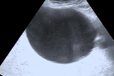
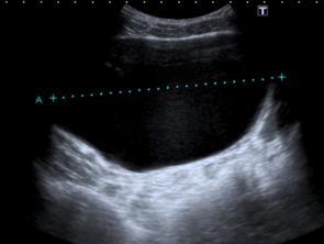
8 Benign Adnexal Masses Simple Cysts Virtually exclude malignancy Large cysts - ensure not missing small nodules serous cystadenoma occ present as simple cyst esp larger cysts / older women Most resolve 1-2 months - follow up pre-menopausal > 5cm Post-menopausal > 3cm
9


10 Haemorrhagic Cysts Varied appearance change blood products over time Typical appearances reticular pattern (fibrin strands)-strong predictor Clot may simulate solid nodule concave border absence flow Atypical premenopausal Should resolve 1-2 cycles Follow -up
11


12 Endometrioma Characteristic Well defined cyst Uni / multilocular Homogenous low level echoes - ground glass appearance Hyperechoic wall foci Atypical 15% mural irregularity Usually avascular clot Rarely flow endometrial tissue Diffuse echoes Dermoid Cyst Mucinous ovarian tumour Further imaging Characteristic - haemorrhage


13 MR T1 high signal Endometrioma FS more conspicuous excludes dermoid T2 low signal (SHADING) complete loss signal dependent layering
14

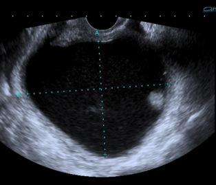
15 Polycystic Ovary 2003 Consensus report defined PCOS 2 of 3 criteria Oligo and/or anovulation Hyperandrogenism Polycystic ovary US Polycystic Ovary 12+ follicles 2-9mm and/or Ovarian volume >10cc Follicle distribution/stromal echogenicity not included? 25 follicles - Dewailly et al follicles, yrs - Lujan et al 2013

16
 Variable appearance")


 ill defined shadowing")
17 Germ Cell Tumours Cystic/Solid Mature Teratoma (Dermoid Cyst ) Variable appearance internal composition Often specific features Dermoid plug Tip of iceberg sign Rokitansky Nodule echogenic nodule / dense well defined shadowing Dermoid mesh Floating Balls Fat-Fluid level Echogenic mass (sebaceous material / hair ) ill defined shadowing obscures posterior border


18 Mature Teratoma ( Dermoid Cyst) Dermoid Mesh Multiple echogenic lines/dots Hair fibres Floating Balls Uncommon / Pathognomonic Sebaceous material around focus debris/hair Fat fluid Level non specific Often at least 2 characteristic features detected Further imaging Atypical - characteristic for fat Visualise other ovary
19


20 T2 T1 T1 FAT SAT

21

22 BIG FIVE Benign Ovarian Lesions Corpus Luteal Cysts Functional Cysts Haemorrhagic Cysts Endometriomas PCOS Dermoids


23 Benign Extra- Ovarian Lesions Hydrosalpinx Para-ovarian Cysts Peritoneal Inclusion cysts

24 Paraovarian cyst Broad ligament Paramesonephric, mesothelial, mesonephric remnants Usually simple


25 Hydrosalpinx Tubular Incomplete septations Indentations opposite sides of wall -waist sign Mural nodules cogwheel beads on a string Echoes - pyosalpinx


26 Peritoneal Inclusion Cyst Functioning ovary Adhesions surgery, endometriosis, PID Cystic mass + septa Shape conforms to adjacent pelvic structures NB normal ovary within/ periphery mass


27 Pseudocyst / Peritoneal Inclusion Cyst


28 Pitfalls


29 Pitfalls


30 Bladder - TVS


31


32
33 Classification of Ovarian Neoplasms EPITHELIAL TUMOURS 65-70% Serous Cystadenoma/Carcinoma Mucinous Cystadenoma/Carcinoma Endometrioid Clear Cell Brenner Tumour GERM CELL TUMOURS 15-20% SEX CORD STROMAL TUMOURS 5-10% Mature Teratoma (Dermoid) / Immature Teratoma Dysgerminoma Granulosa Cell Sertoli Leydig Fibroma Thecoma Metastases 5-10 %

34
35 Classification of Ovarian Neoplasms EPITHELIAL TUMOURS 65-70% Serous Cystadenoma / Carcinoma Mucinous Cystadenoma / Carcinoma Endometrioid Clear Cell Brenner Tumour 85% malignant ovarian neoplasm Serous / Mucinous Benign Peak years Malignant - Peak 6-7 th decade Cystic and Solid - varying amounts solid tissue +/-Differentiation

 few/small papillary nodules 25% malignant")
 Serous")
36 Epithelial Tumours - Cystic/Solid - Serous Predominantly cystic Largely echo-free fluid Unilocular/multilocular Papillary projections single best predictor of epithelial neoplasm 60% benign (cystadenoma) few/small papillary nodules 25% malignant (cystadenocarcinoma) Malignant more projections / solid tissue 15% borderline (resemble benign or malignant) Serous cystadenocarcinoma


37 Epithelial Tumours - Cystic/Solid - Mucinous Multilocular Cystadenoma Echogenic fluid mucin Benign thin septa Malignant thick enhancing septa / solid elements Mucinous Cystadenocarcinoma 20-25% ovarian tumours 80% benign 10% borderline 10% malignant


38 Serous Bilaterality Ca2 + common psammomatous Peritoneal carcinomatosis Mucinous Rarely bilateral Ca2+ rare linear Pseudomyoma peritonei


39 Epithelial - Cystic / Solid Endometrioid 10-15% all ovarian cancers Almost always malignant 15-30% assoc endometrial Ca / hyperplasia Clear Cell 5% all ovarian cancer Malignant Assoc with endometriosis Most common neoplasm assoc with endometriosis

40 Pitfalls


41


42


43


44


45 Decidualised Endometriotic Cyst Hypertrophy stromal cells endometrium during pregnancy Decidual change ectopic endometrium Vascularised rounded papillary projections in cyst with low level echoes Major contributory factor to incorrect diagnosis in pregnant women Resoving at follow up - surgically proven
46 Classification of Ovarian Neoplasms EPITHELIAL TUMOURS 65-70% Serous Cystadenoma/Carcinoma Mucinous Cystadenoma/Carcinoma Endometrioid Clear Cell Brenner Tumour GERM CELL TUMOURS 15-20% SEX CORD STROMAL TUMOURS 5-10% Mature Teratoma (Dermoid) / Immature Teratoma Dysgerminoma Granulosa Cell Sertoli Leydig Fibroma Thecoma Metastases 5-10 %


47 Solid Masses - Regular Regular solid masses often benign Pedunculated fibroid Typically solid Picket fence shadowing Ovarian fibroma Solid mass Marked acoustic shadowing (18-52%)
 Differential Pedunculated fibroids Brenner Tumour Brenner Tumour Rarely")
48 Solid Masses Regular Fibroma SEX CORD STROMAL TUMOURS 5-10% Granulosa Cell Sertoli Leydig Fibroma Thecoma Almost always benign Middle age women Heterogenous/homogen ous solid mass Marked acoustic shadowing (18-52%) Differential Pedunculated fibroids Brenner Tumour Brenner Tumour Rarely malignant


49 Solid Masses Regular Fibroma Further imaging Large fibroids / shadowing may limit TVS Atypical Characteristic for fibrous tissue Visualise ovaries / relationship to uterus


50 Solid Masses Regular MEIGS Syndrome Ascites Pleural Effusion Benign ovarian tumour - Fibroma


51 Solid masses - Malignant Irregular solid mass suggests malignancy Epithelial neoplasms cystic/solid rarely completely solid Solid/nearly solid malignancies Metastases Rare Tumours Sex Cord Stromal Tumours Malignant teratoma Dysgerminoma Bilateral solid masses raise concern for metastases further imaging? primary Krukenberg - gastric


52 Sex Cord Stromal - Granulosa Cell Tumours SEX CORD STROMAL TUMOURS 5-10% Granulosa Cell Sertoli Leydig Fibroma Thecoma Hormone secreting - oestrogen Rare 3% ovarian malignancies Peri /postmenopusal Unilateral - 95% US appearances non specific Cystic/solid - Solid 1/3 endometrial hyperplasia Up to 25% endometrial Ca


53 Sex Cord Stromal - Fibrothecoma SEX CORD STROMAL TUMOURS 5-10% Granulosa Cell Sertoli Leydig Fibroma Thecoma


54 Solid masses Malignant GERM CELL TUMOURS 15-20% Mature Teratoma (Dermoid) / Immature Teratoma Dysgerminoma Dysgerminoma 17 yr old Immature Teratoma 27 yr old Dysgerminoma Most common < 30 years Usually solid Immature Teratoma Most common first two decades Predominantly solid, cystic/solid Raised AFP/Beta-hCG/LDH in some malignant germ cell tumours


55 Features of Malignancy Solid Component within cystic mass Irregular solid mass / wall Thick Septations Presence of Flow Borderline Ascites Peritoneal nodules Metastases /Nodes


56 Solid Component Most important predictor of malignancy Malignant Several nodules solid tissue Echogenicity similar to cyst wall Often convex / irregular Flow Benign Solitary / small papillary nodule serous cystadenoma Hyperechoic with shadowing dermoid Clot concave border / avascular


57 Septations Septa in cystic mass suggest neoplasm Increased risk malignancy > 2-3mm flow Fibrin strands Thin weak reflectors Numerous Do not traverse entire cyst Adjacent cysts may mimic


58 Doppler Colour doppler qualitative Flow in solid component / septum Power doppler increased sensitivity / similar specificity Spectral doppler Confirm presence flow Malignant tumours Low resistance flow lower PI & RI s, higher velocity RI < 0.4; PI <1.0 prev cited as cut-off for malignancy Overlap with benign little role characterisation


59 Ascites Indirect indicator of malignancy Small amount fluid in Pouch of Douglas normal in premenopausal women >15mm AP diameter Pre-existing illness eg cirrhosis Echogenic fluid - ruptured cyst/torsion Meig s Syndrome ascites + pleural effusion + fibroma


60 Indeterminate Masses Ultrasound Typical benign lesions Sensitive malignancy Many benign lesions demonstrate features malignancy Solid areas / septations / flow Tubo-ovarian abscess Cystadenofibroma Borderline tumours
61 Risk of Malignancy Index U Score M score Feature RMI 1 Score Ultrasound features: Multilocular cyst Solid areas Bilateral lesions Ascites Intra-abdominal metastases Pre-menopausal 1 Post menopausal 3 CA125 0=none 1=one abnormality 3=2 or more abnormalities U/ml RMI score = ultrasound x menopausal x CA125 level in U/ml Sensitivity 85% Specificity 97%
62 Triage using RMI Risk RMI Women (%) Risk of cancer (%) Low <25 40 <3 Moderate High > High RMI score > referral to specialist centre (SIGN Guidelines 2012) Intermediate RMI score MRI often considered appropriate RMI heavily influenced by CA 125 CA 125 may be elevated by benign disease esp pre-menopausal RMI recommended by RCOG and systematic review 2009 Geomini P et al The Accuracy of Risk Scores in Predicting Ovarian Malignancy: A Systematic Review Obstetrics & Gynecology; Volume 113 : 2, (Part 1), February 2009, pp
63 Simple Rules Developed as part of the IOTA (International Ovarian Tumour Analysis) study Prospective, multicentre, European study patients with adnexal masses, 21 centres, 9 countries Simple rules based on clearly defined US features BENIGN - B RULES Unilocular cyst Presence solid components where largest solid component < 7mm Presence acoustic shadowing Smooth multilocular tumour with largest diameter <100mm No blood flow MALIGNANT - M RULES Irregular solid tumour Ascites At least four papillary structures Irregular multilocular solid tumour with largest diameter 100mm Very strong blood flow

64 Ultrasound in Obstetrics & Gynecology Volume 41, Issue 1, pages 9-20, 25 DEC 2012 DOI: /uog Slides Ligita Jokubkiene Jan 2013 UOG Journal club

65 A mass is unclassifiable if no M or B features or both
66 Simple Rules Could be applied in 77% 2 step strategy Simple Rules + subjective assessment by experienced examiner if unclassifiable ( MDT ) 90% sensitivity and 93% specificity to detect ovarian malignancy Equivalent to expert subjective assessment in all (sensitivity 90% /specificity 93%) misclassified fewer stage 1 malignancies than did RMI and measurement of CA125. UK - RCOG included simple rules in 2011 guideline for evaluating ovarian pathology in premenopausal women

67

68 Conclusion Clinical, Pre/Post Menopausal CA125 US Adnexal Mass Likely Malignant Adnexal Mass Indeterminate Adnexal mass Likely benign Staging Characterisation Follow up

69
2/24/19. Ovarian pathology: IOTA ADNEXAL MASSES. Content. IOTA terms for description of an adnexal mass. IOTA terms for description of an adnexal mass
 Content Ovarian pathology: IOTA ADNEXAL MASSES X SIMPLE COMPLEX Dr DESCRIBE WHAT YOU SEE FRANZCOG, MPH, DDU, COGU Sonologist Clinically useful Benign Malignant Communication between clinicians/research
The Adnexal Mass. Handout NCUS 3/18/2017 Suzanne Dixon, MD
 The Adnexal Mass Handout NCUS 3/18/2017 Suzanne Dixon, MD Objectives: Pelvic mass differential Characteristics of the normal ovary Standard terminology for ovarian masses Benign vs. malignant features
ISUOG Basic Training Typical Ultrasound Appearances of Common Pathologies in the Adnexae
 ISUOG Basic Training Typical Ultrasound Appearances of Common Pathologies in the Adnexae Learning objectives At the end of the lecture series you will be able to: Compare the differences between typical
Category Term Definition Comments 1 Major Categories 1a
 Working Lexicon Categories, Terms & Definitions Category Term Definition Comments 1 Major Categories 1a Physiologic Category (consistent with normal ovarian physiology) Follicle Simple 3 cm in premenopausal
A Practical Approach to Adnexal Masses
 A Practical Approach to Adnexal Masses Darcy J. Wolfman, MD Section Chief of Genitourinary Imaging American Institute for Radiologic Pathology Clinical Associate Johns Hopkins Community Radiology Division
Assessment of adnexal masses. Ultrasound workup of adnexal masses. symptoms. symptoms. Age. Serum tumor markers 10/1/2018
 Assessment of adnexal masses Ultrasound workup of adnexal masses Kevin Robinson, DO Department of Radiology Michigan State University October 4, 2018 Patients symptoms Age Menstrual status Serum tumor
Ovarian pathology: A practical approach to imaging diagnosis and management
 Ovarian pathology: A practical approach to imaging diagnosis and management Award: Certificate of Merit Poster No.: C-1865 Congress: ECR 2015 Type: Educational Exhibit Authors: A. Castan, A. Mir Torres,
CME ARTICLE. A Practical Approach to the Ultrasound Characterization of Adnexal Masses. Douglas L. Brown, MD
 CORE CURRICULUM IN SONOGRAPHY CME ARTICLE A Practical Approach to the Ultrasound Characterization of Adnexal Masses Douglas L. Brown, MD Abstract: Because pelvic ultrasound is commonly used to evaluate
IN THE NAME OF GOD POV: CYSTIC OVARIAN LESION
 IN THE NAME OF GOD POV: CYSTIC OVARIAN LESION CASE 1 20 years old girl with AUB and pelvic pain from 2 weeks ago Impression :Simple unilocular 6 cm ovarian cyst Next step? Almost certainly benign so FU
Pathology of Ovarian Tumours. Dr. Jyothi Ranganathan MD ( Path) AFMC Pune PDCC (Cytopathology) PGI Chandigarh
 AFMC Pune PDCC (Cytopathology) PGI Chandigarh") Pathology of Ovarian Tumours Dr. Jyothi Ranganathan MD ( Path) AFMC Pune PDCC (Cytopathology) PGI Chandigarh Outline Incidence Risk factors Classification Pathology of tumours Tumour markers Prevention
Pathology of Ovarian Tumours Dr. Jyothi Ranganathan MD ( Path) AFMC Pune PDCC (Cytopathology) PGI Chandigarh Outline Incidence Risk factors Classification Pathology of tumours Tumour markers Prevention
Terminology Estimate the risk of malignancy in adnexal masses - Overview
 Understanding the IOTA (International Ovarian Tumor Analysis) terminology & Classification Using the IOTA simple rules to estimate the risk of malignancy in women with adnexal masses Elisabeth Epstein,
Gynecologic Ultrasound. Sujata Ghate, MD Associate Professor of Radiology Duke University Medical Center
 Gynecologic Ultrasound Sujata Ghate, MD Associate Professor of Radiology Duke University Medical Center Objectives Understand work-up of endometrial abnormalities Show examples of uterine and endometrial
Top Tips for Gynaecological Ultrasound. Catherine Kirkpatrick Consultant Sonographer Dublin Oct 2018
 Top Tips for Gynaecological Ultrasound Catherine Kirkpatrick Consultant Sonographer Dublin Oct 2018 We can all scan a pelvis so what can we do to improve? Uterus, endometrium and ovaries, got it covered!
Adnexal Masses and Problem Solving Pelvic MRI
 28th Congress of the Hungarian Society of Radiologists RCR Session Budapest June 2016 Adnexal Masses and Problem Solving Pelvic MRI DrSarah Swift St James s University Hospital Leeds, UK Objectives Characterisation
بسم هللا الرحمن الرحيم. Prof soha Talaat
 بسم هللا الرحمن الرحيم Ovarian tumors The leading indication for gynecologic surgery. Preoperative characterization of complex solid and cystic adnexal masses is crucial for informing patients about possible
3 cell types in the normal ovary
 Ovarian tumors 3 cell types in the normal ovary Surface (coelomic epithelium) the origin of the great majority of ovarian tumors 90% of malignant ovarian tumors Totipotent germ cells Sex cord-stromal cells
Value of MRI in Characterizing Adnexal Masses
 The Journal of Obstetrics and Gynecology of India (July August 2015) 65(4):259 266 DOI 10.1007/s13224-015-0730-9 PHOTO ESSAY Value of MRI in Characterizing Adnexal Masses Alpana Karnik 1 Raina Anil Tembey
Pathology of the female genital tract
 Pathology of the female genital tract Common illnesses of the female genital tract Before menarche Developmental anomalies Tumors (ovarial teratoma) Amenorrhea Fertile years PCOS, ovarian cysts Endometriosis
OVARIES. MLS Basic histological diagnosis MLS HIST 422 Semester 8- batch 7 L13 Dr: Ali Eltayb.
 OVARIES MLS Basic histological diagnosis MLS HIST 422 Semester 8- batch 7 L13 Dr: Ali Eltayb. OBJECTIVES Recognize different disease of ovaries Classify ovarian cyst Describe the pathogenesis, morphology
Institute of Pathology First Faculty of Medicine Charles University. Ovary
 Ovary Barrett esophagus ph in vagina between 3.8 and 4.5 ph of stomach varies from 1-2 (hydrochloric acid) up to 4-5 BE probably results from upward migration of columnar cells from gastroesophageal junction
Diagnostic accuracy of ultrasonography with color doppler imaging techniques in adnexal masses and correlation with histopathological analysis
 Original Article Diagnostic accuracy of ultrasonography with color doppler imaging techniques in adnexal masses and correlation with histopathological analysis Neha Gupta 1*, Poonam Gupta 2, Omvati Gupta
Characterizing Adnexal Masses: Pearls and Pitfalls 20 th Annual Summer Practicum SCBT-MR Jackson Hole August 11, 2010
 Characterizing Adnexal Masses: Pearls and Pitfalls 20 th Annual Summer Practicum SCBT-MR Jackson Hole August 11, 2010 Evan S. Siegelman MD University of Pennsylvania Medical Center Adnexal Masses: Pearls
3 cell types in the normal ovary
 Ovarian tumors 3 cell types in the normal ovary Surface (coelomic epithelium) the origin of the great majority of ovarian tumors (neoplasms) 90% of malignant ovarian tumors Totipotent germ cells Sex cord-stromal
Approach to imaging of the ovaries
 First encounter Approach to imaging of the ovaries Mariam Moshiri MD Associate professor Body Imaging Most common first encounter is via ultrasound Many of clinicians order US imaging for various female
MR Imaging of the Adnexal Masses: A Review
 Page54 Review of Literature NJR 2011;1(1):54 60; Available online at www.nranepal.org MR Imaging of the Adnexal Masses: A Review I Ahmad 1, S Kirmani 1, M Rashid 2, K Ahmad 3 1 Department of Radiodiagnosis,
Evaluation of the Adnexal Mass Non Neoplastic
 cta Radiológica Portuguesa, Vol.XXIII, nº 90, pág. 49-57, br.-jun., 2011 Evaluation of the dnexal Mass Non Neoplastic Jade Wong Professor of Radiology University of Maryland School of Medicine Visiting
Gynaecological cancers. Mr Vivek Nama MD MRCOG Consultant Gynaecological Oncologist
 Gynaecological cancers Mr Vivek Nama MD MRCOG Consultant Gynaecological Oncologist Gynaecological cancers Why do we need 2 week wait? Early/timely diagnosis of cancer Possibly less invasive treatment and
The key contribution of MRI in adnexal mass evaluation is in: 1. Identifying benign features. 2. Identifying malignant features.
 19 th Annual Women s Imaging Conference University of Toronto - 2016 Disclosures : None phyllis.glanc@sunnybrook.ca Sunnybrook Health Science Centre University of Toronto, Dept Medical Imaging, Obstetrics
CAN TV U/S REDUCE THE
 CAN TV U/S REDUCE THE NEED FOR SURGERY IN GYNECOLOGY? Steven R. Goldstein, M.D. Professor of Obstetrics & Gynecology New York University it School of fmedicine i Director of Gynecologic Ultrasound Co-Director
Ovarian Lesion Benign vs Malignant?
 Ovarian Lesion Benign vs Malignant? Michele Keenan 1,2 Bernice Dunne 2 Mary Moran 1 Therese Herlihy 1 1. Radiography and Diagnostic Imaging, School of Medicine, University College Dublin, Ireland 2. Midland
L/O/G/O. Ovarian Tumor. Xiaoyu Niu Obstetrics and Gynecology Department Sichuan University West China Second Hospital
 L/O/G/O Ovarian Tumor Xiaoyu Niu Obstetrics and Gynecology Department Sichuan University West China Second Hospital Essentials classification of ovarian tumor clinical manifestation of ovarian tumor metastatic
Adnexal Masses in Menopausal Women Surgery or Surveillance?
 Adnexal Masses in Menopausal Women Surgery or Surveillance? FREDTALK IDEASWORTHSPREADING Disclosure I am a member of Vermillion s Speakers Bureau I am NOT a paid consultant for Vermillion Inc. nor do I
Case 1. Gynaecology Case Presentation. Objectives. Disclosures 22/10/ year old female Clinical history: Assess right ovarian cyst
 Gynaecology Case Presentation Organ Imaging 2016 University of Toronto Sarah Johnson 39 year old female Clinical history: Assess right ovarian cyst Clinically diagnosed endometriosis Started fertility
The Role of Imaging for Gynecologic Emergencies
 Objectives The Role of Imaging for Gynecologic Emergencies M. Jonathon Solnik, MD, FACOG FACS Associate Professor of Obstetrics & Gynaecology Head of Gynaecology & Minimally Invasive Surgery University
Complete Summary GUIDELINE TITLE. Gynaecological ultrasound examination. BIBLIOGRAPHIC SOURCE(S)
") Complete Summary GUIDELINE TITLE Gynaecological ultrasound examination. BIBLIOGRAPHIC SOURCE(S) Finnish Medical Society Duodecim. Gynaecological ultrasound examination. In: EBM Guidelines. Evidence-Based
Complete Summary GUIDELINE TITLE Gynaecological ultrasound examination. BIBLIOGRAPHIC SOURCE(S) Finnish Medical Society Duodecim. Gynaecological ultrasound examination. In: EBM Guidelines. Evidence-Based
Ubol Saeng-Anan, Tawiwan Pantasri, Vithida Neeyalavira, Theera Tongsong*
 DOI:http://dx.doi.org/10.7314/APJCP.2013.14.9.5409 RESEARCH ARTICLE Sonographic Pattern Recognition of Endometriomas Mimicking Ovarian Cancer Ubol Saeng-Anan, Tawiwan Pantasri, Vithida Neeyalavira, Theera
Adnexal Masses in Menopausal Women
 Adnexal Masses in Menopausal Women Surgery or Surveillance? Disclosure Frederick R. Ueland, MD Professor and Director Division of Gynecologic Oncology University of Kentucky I have no financial disclosures
Ultrasound Evaluation of Adnexal Pathologies Jagruti Kalola 1*, Hiral Hapani 2, Anjana Trivedi 3, Jay Thakkar 4
 Scholars Journal of Applied Medical Sciences (SJAMS) Sch. J. App. Med. Sci., 2014; 2(6G):3324-3330 Scholars Academic and Scientific Publisher (An International Publisher for Academic and Scientific Resources)
OVARIAN MASSES : MANAGEMENT CHALLENGE
 SRU 2015 OVARIAN MASSES : MANAGEMENT CHALLENGE phyllis.glanc@sunnybrook.ca Sunnybrook Health Science Centre University of Toronto, Dept Medical Imaging, Obstetrics & Gynecology Thank you on behalf of the
Risk of Malignancy Index in the Preoperative Evaluation of Patients with Adnexal Masses among Women of Perimenopausal and Postmenopausal Age Group
 IOSR Journal of Dental and Medical Sciences (IOSR-JDMS) e-issn: 2279-0853, p-issn: 2279-0861.Volume 17, Issue 9 Ver. 8 (September. 2018), PP 20-25 www.iosrjournals.org Risk of Malignancy Index in the Preoperative
Title of Guideline (must include the word Guideline not protocol, policy, procedure etc)
") Title of Guideline (must include the word Guideline not protocol, policy, procedure etc) Author: Contact Name and Job Title Directorate & Speciality Assessment, referral and initial management of ultrasound
Title of Guideline (must include the word Guideline not protocol, policy, procedure etc) Author: Contact Name and Job Title Directorate & Speciality Assessment, referral and initial management of ultrasound
Unexpected Gynecologic Findings at Laparotomy. Susan A. Davidson, MD University of Colorado, Denver School of Medicine
 Unexpected Gynecologic Findings at Laparotomy Susan A. Davidson, MD University of Colorado, Denver School of Medicine Adnexal Mass: Gyn Etiologies Uterine Leiomyomas Pregnancy Malignancy Tubal Pregnancy
Mousa. Najat kayed &Renad Al-Awamleh. Nizar Alkhlaifat
 6 Mousa Najat kayed &Renad Al-Awamleh Nizar Alkhlaifat P a g e 1 This sheet written based on record 13 on website Cover slide( 95-117 ) No need to go back to slide FALLOPIAN TUBE PATHOLOGY In general fallopian
Imaging evaluation of ovarian masses.
 Imaging evaluation of ovarian masses. Poster No.: C-0988 Congress: ECR 2012 Type: Educational Exhibit Authors: M. Forment Navarro, C. La Parra Casado, A. Vera, C. Martínez 1 2 2 2 2 2 1 Rubio, M. Mazón
Triage of Ovarian Masses. Andreas Obermair Brisbane
 Triage of Ovarian Masses Andreas Obermair Brisbane Why Triage? In ovarian cancer, best outcomes for patients can be achieved when patients are treated in tertiary centres by a multidisciplinary team led
Ovarian Masses: role of MRI in the differential diagnosis. A systematic approach.
 Ovarian Masses: role of MRI in the differential diagnosis. A systematic approach. Poster No.: C-0597 Congress: ECR 2017 Type: Educational Exhibit Authors: I. Mussetto, F. Rosa, J. Matos, G. Ficarra, D.
A new scoring model for characterization of adnexal masses based on two-dimensional gray-scale and colour Doppler sonographic features
 FVV in ObGyn, 2014, 6 (2): 68-74 Original paper A new scoring model for characterization of adnexal masses based on two-dimensional gray-scale and colour Doppler sonographic features Ahmed M. Abbas, Kamal
Title of Guideline (must include the word Guideline (not protocol, policy, procedure etc)
") Title of Guideline (must include the word Guideline (not protocol, policy, procedure etc) Author: Contact Name and Job Title Directorate & Speciality Assessment, referral and initial management of ultrasound
Title of Guideline (must include the word Guideline (not protocol, policy, procedure etc) Author: Contact Name and Job Title Directorate & Speciality Assessment, referral and initial management of ultrasound
Does Ovarian Cysts affect your Fertility?
 Does Ovarian Cysts affect your Fertility? Ovarian cysts are sac like structures (filled with liquid or semi-solid) develop in the ovary. Ovarian cysts are very common, mostly painless and harmless with
Case Fibrothecoma of the ovary
 Case 10646 Fibrothecoma of the ovary Elisa Melo Abreu, Teresa Margarida Cunha Section: Genital (Female) Imaging Published: 2015, Jan. 2 Patient: 70 year(s), female Authors' Institution Department of Radiology,
Diagnosis of adnexal malignancies by using color Doppler energy imaging as a secondary test in persistent masses
 Ultrasound Obstet Gynecol 9;:277 2 Diagnosis of adnexal malignancies by using color Doppler energy imaging as a secondary test in persistent masses S. Guerriero, S. Ajossa, A. Risalvato, M. P. Lai, V.
ACUTE PELVIC PAIN 강릉아산병원영상의학과 이은혜
 ACUTE PELVIC PAIN 강릉아산병원영상의학과 이은혜 Gynecologic PID Ruptured ovarian cyst Adnexal torsion Acute pelvic pain Pregnancy-related Ectopic pregnancy Placental abruption Nongynecologic Acute appendicitis Diverticulitis
 INTRAUTERINE DEVICE = IUD INTRAUTERINE DEVICE = IUD CONGENITAL DISORDERS Pyometra = pyometrea is a uterine infection, it is accumulation of purulent material in the uterine cavity. Ultrasound is usually
IOTA and Models for Screening for Ovarian Cancer
 IOTA and Models for Screening for Ovarian Cancer Hennie Botha MARCH 2017 T H IG PY R O C F O SP EA KE R Silent Killer to Whispering Disease Listening to your body.. new, persistent, and increases in severity
JMSCR Vol 05 Issue 06 Page June 2017
 www.jmscr.igmpublication.org Impact Factor 5.84 Index Copernicus Value: 83.27 ISSN (e)-2347-176x ISSN (p) 2455-0450 DOI: https://dx.doi.org/10.18535/jmscr/v5i6.29 MRI in Clinically Suspected Uterine and
Adnexal Diseases. Andrea Rockall and Rosemarie Forstner. 8.1 Introduction. 8.2 Imaging Modalities to Assess an Adnexal Mass. 8.2.
 Adnexal Diseases Andrea Rockall and Rosemarie Forstner 8 Learning Objectives To know the most common benign ovarian lesions To be aware of the indications for MRI of adnexal masses To know the sequences
CA125 in the diagnosis of ovarian cancer: the art in medicine
 CA125 in the diagnosis of ovarian cancer: the art in medicine Dr Marcia Hall Consultant Medical Oncology Mount Vernon Cancer Centre Hillingdon Hospital Wexham Park Hospital Epidemiology Ovarian cancer
Categorization of Ovarian Dermoids Depending Upon Their Sonographic Appearances
 IOSR Journal of Dental and Medical Sciences (IOSR-JDMS) e-issn: 2279-0853, p-issn: 2279-0861.Volume 14, Issue 12 Ver. IV (Dec. 2015), PP 34-42 www.iosrjournals.org Categorization of Ovarian Dermoids Depending
Springer Healthcare. Understanding and Diagnosing Ovarian Cancer. Concise Reference: Krishnansu S Tewari, Bradley J Monk
 Concise Reference: Understanding and Diagnosing Ovarian Cancer Krishnansu S Tewari, Bradley J Monk Extracted from: The 21 st Century Handbook of Clinical Ovarian Cancer Published by Springer Healthcare
Advances in transvaginal ultrasound scanning and their clinical application
 Advances in transvaginal ultrasound scanning and their clinical application Bill Smith, Head of Ultrasound CDS Clinical Diagnostic Services, London, UK Introduction Transvaginal ultrasound scanning (TVS)
Female Genital Tract Lab. Dr. Nisreen Abu Shahin Assistant Professor of Pathology University of Jordan
 Female Genital Tract Lab Dr. Nisreen Abu Shahin Assistant Professor of Pathology University of Jordan Ovarian Pathology A 20-year-old female presented with vague left pelvic pain. Pelvic exam revealed
The many faces of Endometriosis
 The many faces of Endometriosis Beryl Benacerraf M.D Harvard Medical School What is Endometriosis? Endometriosis is defined as the presence of normal endometrial tissue occurring outside of the endometrial
The diagnosis of endometriomas using colour Doppler energy imaging
 Human Reproduction vol.13 no.6 pp.1691 1695, 1998 The diagnosis of endometriomas using colour Doppler energy imaging Stefano Guerriero, Silvia Ajossa, Valerio Mais, Andrea Risalvato, Maria Paola Lai and
7 Mousa. Obada Zalat. Mohammad Badi
 7 Mousa Obada Zalat Mohammad Badi Tumors of the ovaries Last lecture we talked about surface epithelial tumors of the ovaries (the most common type). But there are many other types of tumors of germ cell
Ovarian fibromas/thecomas are uncommon
 Sonography of Ovarian s/thecomas Patricia A. Athey, MD, Robert S. Malone, MD The sonographic findings in 14 patients with ovarian fibromas/thecomas are described. A broad spectrum of sonographic features
Icd 10 ovarian stroma
 Icd 10 ovarian stroma Struma ovarii; Micrograph of a struma ovarii. Characteristic thyroid follicles are seen on the right, and ovarian stroma on the left. H&E stain. Classification and. Free, official
A study of benign adnexal masses
 International Journal of Reproduction, Contraception, Obstetrics and Gynecology Manivasakan J et al. Int J Reprod Contracept Obstet Gynecol. 2012 Dec;1(1):12-16 www.ijrcog.org pissn 2320-1770 eissn 2320-1789
JMSCR Vol 3 Issue 9 Page September 2015
 www.jmscr.igmpublication.org Impact Factor 3.79 ISSN (e)-2347-176x DOI: http://dx.doi.org/10.18535/jmscr/v3i9.09 A Study of Histopathological Pattern of Ovarian Neoplasms and their Age Distribution in
A Tale of Two Ovaries: Cross-Sectional Imaging Spectrum of Ovarian Emergencies
 A Tale of Two Ovaries: Cross-Sectional Imaging Spectrum of Ovarian Emergencies Presenting author A J Baxi Co-authors A M Nagar, MBBS D Rajderkar, MD V Ojili, MD Contact:ojili@uthscsa.edu Disclaimer: We
Histopathological analysis of neoplastic and non neoplastic lesions of ovary: A study of one hundred cases
 Orginal Article Histopathological analysis of neoplastic and non neoplastic lesions of ovary: A study of one hundred cases 2 G Prathima, Srikanth Shastry 2 Consultant Pathologist, Image Diagnostics, Kadapa,
CELL AND TISSUE INJURY COURSE-II PATHOLOGY LABORATORY. PATHOLOGY of MASS LESIONS and TISSUE DEFECTS -MACROSCOPY Assoc. Professor Rengin Ahıskalı
 CELL AND TISSUE INJURY COURSE-II PATHOLOGY LABORATORY PATHOLOGY of MASS LESIONS and TISSUE DEFECTS -MACROSCOPY Assoc. Professor Rengin Ahıskalı M1 - RENAL TUBERCULOSIS cavitary areas caseous necrosis fibrous
See the latest estimates for new cases of ovarian cancer and deaths in the US and what research is currently being done.
 About Ovarian Cancer Overview and Types If you have been diagnosed with ovarian cancer or are worried about it, you likely have a lot of questions. Learning some basics is a good place to start. What Is
Gynaecological Malignancies
 Gynaecological Malignancies Dr Rodney Itaki Lecturer Anatomical Pathology Discipline University of Papua New Guinea Division of Pathology School of Medicine & Health Sciences Overview Genital tract tumors
Female Reproduc.ve System. Kris.ne Kra7s, M.D.
 Female Reproduc.ve System Kris.ne Kra7s, M.D. Female Reproduc.ve System Outline Cervix Uterus Ovaries Breast Cervical Carcinoma Once the most common cancer in women now not even in top 10. Decrease due
Scanning the ovaries is no simple task.
 Second of four parts Skilled US imaging of the adnexae Part 2: The non-neoplastic mass From simple cysts to endometriomas, nonneoplastic ovarian masses can be identified through ultrasonographic observation
cysts is possible if imaging findings are correlated with appropriate clinical findings [1]. The
![cysts is possible if imaging findings are correlated with appropriate clinical findings [1]. The](/thumbs/73/68677649.jpg "cysts is possible if imaging findings are correlated with appropriate clinical findings [1]. The") Pictorial Essay Imaging of Peritoneal Inclusion Cysts Kiran. Jain1 lthough fairly common, peritoneal inclusion cysts are less well-recognized entities on imaging of the female pelvis. Peritoneal inclusion
Pictorial Essay Imaging of Peritoneal Inclusion Cysts Kiran. Jain1 lthough fairly common, peritoneal inclusion cysts are less well-recognized entities on imaging of the female pelvis. Peritoneal inclusion
Human epididymal protein 4 The role of HE4 in the management of patients presenting with pelvic mass Publication abstracts
 Human epididymal protein 4 The role of HE4 in the management of patients presenting with pelvic mass Publication abstracts Ovarian cancer is diagnosed annually in more than 200,000 women worldwide, with
ORIGINAL RESEARCH ARTICLE
 Journal of Chitwan Medical College 2016; 6(15): 16-20 Available online at: www.jcmc.cmc.edu.np ISSN 2091-2889 (Online) ISSN 2091-2412 (Print) JOURNAL OF CHITWAN MEDICAL COLLEGE JCMC ESTD 2010 ORIGINAL
Researcher 2018;10(8)
") Value of Magnetic Resonance Imaging (MRI) and Diffusion Weighted (DWI) MR in Diagnosis of Ovarian Lesions Enas ahmed a, Ikram Hamed b, Noha Abdel Shafy b, Ahmed Abdel Fattah a,* a Faculty of Medicine,
Value of Magnetic Resonance Imaging (MRI) and Diffusion Weighted (DWI) MR in Diagnosis of Ovarian Lesions Enas ahmed a, Ikram Hamed b, Noha Abdel Shafy b, Ahmed Abdel Fattah a,* a Faculty of Medicine,
Pregnancy With Huge Ovarian Cyst
 BMH Med. J. 2018;5(3):74-78 Case Report Pregnancy With Huge Ovarian Cyst Suja Ann Ranji, Usha Payyodi, Ani Praveen, Rajesh MC, Jini Chandran Baby Memorial Hospital, Kozhikode 673004 Address for Correspondence:
Clinicopathological and Histological Features of Ovarian Tumour- A Study
 IOSR Journal of Dental and Medical Sciences (IOSR-JDMS) e-issn: 2279-0853, p-issn: 2279-0861.Volume 16, Issue 9 Ver. IX (September. 2017), PP 56-60 www.iosrjournals.org Clinicopathological and Histological
Female Reproduc.ve System. Kris.ne Kra7s, M.D.
 Female Reproduc.ve System Kris.ne Kra7s, M.D. Female Reproduc.ve System Outline Cervix Uterus Ovaries Breast Female Reproduc.ve System Outline Cervix Cervical carcinoma Cervical Carcinoma Once the most
Mature Cystic Teratomas and the most common complications
 Mature Cystic Teratomas and the most common complications Poster No.: C-2230 Congress: ECR 2015 Type: Authors: Keywords: DOI: Educational Exhibit S. C. S. Silva 1, D. N. Silva 1, D. Garrido 1, I. C. S.
Likelihood Ratio of Sonographic Findings in Discriminating Hydrosalpinx from Other Adnexal Masses
 Sonography of Adnexal Masses Women s Imaging Original Research WOMEN S IMAGING Maitray D. Patel 1 Debra L. Acord 1,2 Scott W. Young 1 Patel MD, Acord DL, Young SW Keywords: adnexal masses, hydrosalpinx,
A NEW QUANTITATIVE METHOD TO EVALUATE ADNEXAL TUMORS
 ORIGINAL ARTICLE A NEW QUANTITATIVE METHOD TO EVALUATE ADNEXAL TUMORS Chung-Yuan Lee, Ching-Cheng Tseng, Chen-Bin Wang, Yu-Hsiang Lin, Chun-Hung Chen, Ting-Hung Wun, Ying-Lun Sun, Chih-Jen Tseng* Department
Prospective evaluation of three different models for the pre-operative diagnosis of ovarian cancer
 British Journal of Obstetrics and Gynaecology November 2000, Vol107, pp. 1347-1353 Prospective evaluation of three different models for the pre-operative diagnosis of ovarian cancer *N. Aslam Research
In The Name of GOD. Abnormal Uterine Bleeding and Gynecologic mass. Dr Shahla Danaii
 In The Name of GOD Abnormal Uterine Bleeding and Gynecologic mass Dr Shahla Danaii Causes of abnormal genital tract bleeding Genital tract disorders Pregnancy complications Trauma Drugs Systemic disease
Ovaries: In Sickness and Health. Mr N Pisal Consultant Gynaecologist The Portland Hospital
 Ovaries: In Sickness and Health Mr N Pisal Consultant Gynaecologist The Portland Hospital Topics for discussion How to assess ovarian function? AMH PCOS Ovarian pain Ovarian cysts Ovarian screening Menopause
Key imaging features of acute gynaecological emergencies.
 Key imaging features of acute gynaecological emergencies. Poster No.: C-2200 Congress: ECR 2014 Type: Educational Exhibit Authors: Ó. Roche, N. Bharwani, A. G. Rockall; London/UK Keywords: Acute, Complications,
A Rare Presentation of Endometriosis with Recurrent Massive Hemorrhagic Ascites which Can Mislead
 Case Report INTERNATIONAL JOURNAL OF WOMEN'S HEALTH AND REPRODUCTION SCIENCES http://www.ijwhr.net A Rare Presentation of Endometriosis with Recurrent Massive Hemorrhagic Ascites which Can Mislead Article
ORIGINAL ARTICLE CA-125 AS A SURROGATE MARKER IN A CLINICAL AND HISTOPATHOLOGICAL STUDY OF PELVIC MASS AT A TERTIARY CARE HOSPITAL
 CA-125 AS A SURROGATE MARKER IN A CLINICAL AND HISTOPATHOLOGICAL STUDY OF PELVIC MASS AT A TERTIARY CARE HOSPITAL Madhuri Kulkarni 1, Ambarish Bhandiwad 2, Sunila R 3, Sumangala 4. 1. Professor, Department
Malignant Transformation of Endometriosis: Magnetic Resonance Imaging Aspects
 Malignant Transformation of Endometriosis: Magnetic Resonance Imaging Aspects Poster No.: C-0084 Congress: ECR 2014 Type: Scientific Exhibit Authors: E. A. Yukhno, I. Trofimenko, G. Trufanov; St. Petersburg/RU
Malignant Transformation of Endometriosis: Magnetic Resonance Imaging Aspects
 Malignant Transformation of Endometriosis: Magnetic Resonance Imaging Aspects Poster No.: C-0084 Congress: ECR 2014 Type: Scientific Exhibit Authors: E. A. Yukhno, I. Trofimenko, G. Trufanov; St. Petersburg/RU
Endometrioma With Calcification Simulating a Dermoid on Sonography
 Case Report Endometrioma With Calcification Simulating a Dermoid on Sonography Kiran A. Jain, MD Several investigators have explored the sonographic diagnostic criteria of endometriomas. Endometriomas
CASE STUDY. Presented by: Jessica Pizzo. CFCC Sonography student Class of 2018
 CASE STUDY Presented by: Jessica Pizzo CFCC Sonography student Class of 2018 Case Presentation April 4, 2017 56 yr old woman presented to ED with lower abdominal pain & swelling, along with constipation.
Pelvic Pain: Overlooked
 EDUCATION EXHIBIT 3 Pelvic Pain: Overlooked and Underdiagnosed Gynecologic Conditions 1 CME FEATURE See accompanying test at http:// www.rsna.org /education /rg_cme.html LEARNING OBJECTIVES FOR TEST 1
2/9/2015. Bartholin Cyst. Vulva: Squamous epithelium skin. Vagina: Squamous epithelium mucosa. Cervix: Ectocervix: squamous Endocervix: glandular
 Vulva: Squamous epithelium skin Bartholin Cyst Vagina: Squamous epithelium mucosa Cervix: Ectocervix: squamous Endocervix: glandular Slide courtesy of Dr. Lodge Rigal Slide courtesy of Dr. Lodge Rigal
H&E, IHC anti- Cytokeratin
 Cat No: OVC2281 - Ovary cancer tissue array Lot# Cores Size Cut Format QA/QC OVC228101 228 1.1mm 4um 12X19 H&E, IHC anti- Cytokeratin Recommended applications: For Research use only. RNA or protein ovary
Adolescent Gynecology: Evaluation and Management of Adnexal Mass, PCOS, and Endometriosis. Shanna M. Combs, MD
 Adolescent Gynecology: Evaluation and Management of Adnexal Mass, PCOS, and Endometriosis Shanna M. Combs, MD Adolescent Visit and Exam Adolescent Reproductive Health Visit Initial visit should take place