Mako Partial Knee Patellofemoral
|
|
|
- Scot Osborne
- 5 years ago
- Views:
Transcription


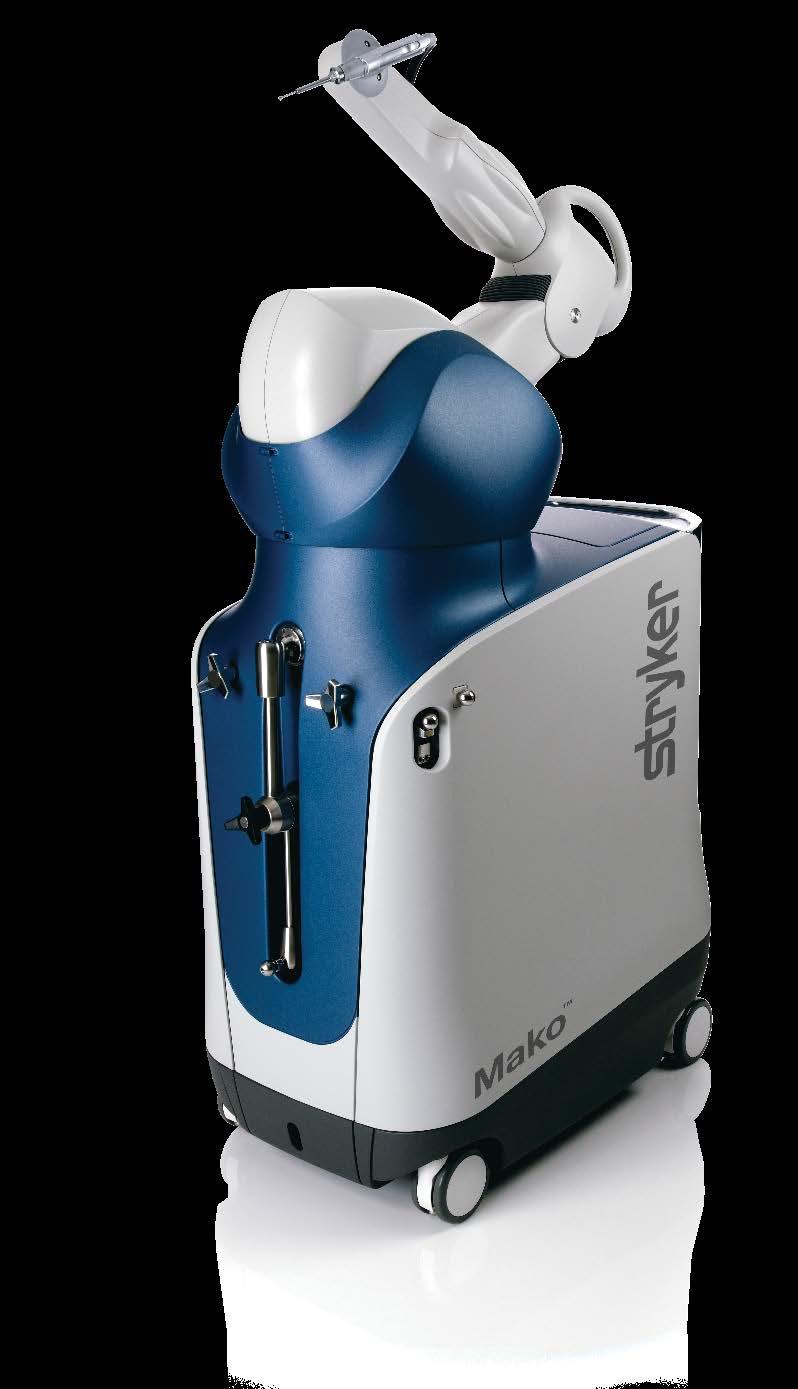


1 Mako Partial Knee Patellofemoral Mako Robotic-Arm Assisted Surgery Surgical reference guide
2 Table of contents Implant compatibility Pre-operative implant planning... 4 Intra-operative planning Planning technique tips Initial exposure and array placement Bone registration and gap balancing Fine-tune implant positioning and bone resection. 12 Trialing, soft tissue balancing and implant insertion This publication sets forth detailed recommended procedures for using Stryker s devices and instruments. It offers guidance that you should heed, but as with any such technical guide, each surgeon must consider the particular needs of each patient and make appropriate adjustments when and as required. the information provided in this document is not to be used as the surgical technique when completing a Mako Partial Knee procedure. Please refer to the Mako PKA Surgical Guide (PN ) for detailed intended use, contraindications, and other essential product information. 2

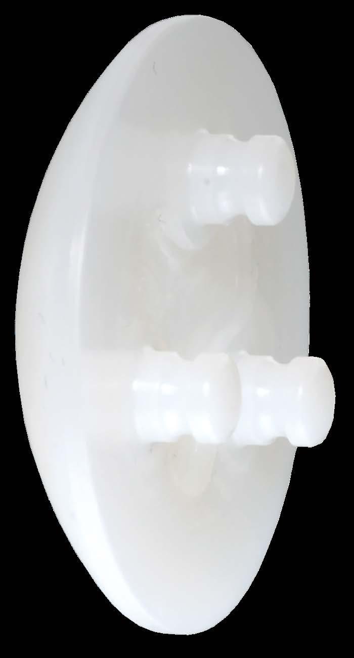
3 Implant compatibility Medial Patellofemoral Lateral Medial bicompartmental Mako MCK Implant System Options Sizes Part number Femoral implant Left medial/right lateral X Right medial/left lateral X Tibial baseplate Left medial/right lateral X Right medial/left lateral X Poly insert 8,9,10,12mm X-X X3 insert 8,9,10,11,12mm X-X Patella implant 3mm increments 26-41mm X Patellofemoral implant Left X Right X 3
 component that does")
4 Pre-operative implant planning The goals of pre-operative implant planning are: 1. Size: Determine appropriate implant size 2. Fit: Define appropriate implant position and orientation Implant size In the quadrant of the computer screen showing the transverse view, select the largest patellofemoral (PF) component that does not overhang the bone in the medial and lateral direction. It should be no more than 2mm inset on either side (figure 1). Although the positioning limits allow for internal rotation, the preferred setting is 0 internal, even for a prominent lateral trochlear ridge. In these conditions, the lateral edge of the component will be recessed into the bone (not proud of bone), which is acceptable (figure 2). Use of 3 pre-set views of the patellofemoral component in the software application is recommended. Alternatively, the user may elect to manually scroll through the appropriate slices of the bone model to verify proper size and fit of the PF implant. Appendix 1 shows the anatomical planes in the 3 pre-set PF views. Lateralizing the trochlear groove improves patella tracking. Externally rotating the patellofemoral component will lateralize the trochlear groove, but it will also decrease lateral patella jump height (decrease lateral patella constraint), and therefore is not recommended. Internally rotating the patellofemoral component will increase lateral patella jump height (increase lateral patella constraint), but it will also undesirably medialize the trochlear groove. Any internal rotation of the component should be accompanied by lateral translation to offset the medialization due to internal rotation. Incorrect selection of the medial and lateral epicondyle CT landmarks will impact the internal/ external rotation values, it is important to confirm these are correct. Use of the PF Primary pre-set view can be very useful because the transverse plane of this view goes through the widest section of the component. Figure 1 Figure 2 4
5 Implant fit overview Scroll through the transverse slices of the bone model to ensure proper implant fit. Then, verify that in the sagittal quadrant the trochlear groove of the implant is 1-2mm proud (approximate thickness of healthy cartilage) of Whiteside s line, and that the distal tongue of the component does not interfere with the ACL. The distal tip of the component should be anterior to Blumensaat s line (figure 3). In the sagittal plane, the implant is 3mm thick superiorly and 4mm thick distally. Figure 3 This can be accomplished using the PF Verify 2 pre-set view in which the sagittal plane sections the implant through the deepest point of the trochlear groove. Implant fit: Overview An implant with proper fit meets the following requirements: 1. The superior-lateral edge of the anterior flange is split midway by bone, and the superiormedial edge of the anterior flange is at least contacting bone. 2. The component trochlear groove is 1-2mm proud of the bone trochlear groove. 3. The distal tongue is centered on the intercondylar notch. Figure 4 Implant fit: Details 1. In the sagittal view, adjust the component in the anterior/posterior dimension until the lateral edge of the anterior flange is split midway by bone (figure 4). In the PF Primary pre-set view the sagittal plane crosses the implant through the superior-lateral edge. Similarly, in the PF Verify 1 pre-set view the sagittal plane cuts the implant through the superior-medial edge. Use of PF Primary and PF Verify 1 pre-set views to position the superior edges of the implant may expedite the process. 5 5

6 Implant fit overview 2. To fit the PF component 1-2mm proud of the trochlear groove bone, place the crosshair at the superior edge of the component to set the rotation anchor, manually scroll medially to the deepest point of the trochlear groove, and adjust flexion/extension until the implant is in the desired position. The preferred rotation value is 0-5 flexion (figure 5). Figure 5 For this purpose, the sagittal quadrant of the PF Verify 2 pre-set view can be useful. The sagittal plane of this pre-set view goes through the trochlear groove of the component. 3. In the coronal view, position the component so that it is centered on the notch, and its pink transition zones are 2mm proud of bone (approximate thickness of healthy cartilage). Adjust medial/lateral and varus/valgus as necessary (figure 6). Placing the component in valgus will undesirably medialize the trochlear groove. Any valgus rotation should be accompanied by lateral translation to offset the medialization due to valgus rotation. If the component cannot be translated any more laterally, the preferred setting is 0-2 varus. Figure 6 In both the PF Primary and PF Verify 1 pre-set views, the coronal plane cuts the implant at the start of the distal tongue. Either of the two pre-set views can be used in this step. 6

7 Intra-operative planning Confirmation of overall plan Scroll through the slices of the bone model in all planes to verify proper implant fit, and that virtual implant position is in accordance to the recommended positioning limits. The recommended positioning limit ranges are as follows: 4 internal to 0 external rotation; implant proud 1-2mm of bone at the trochlear groove (the preferred rotation is 0 external) 3 varus to 2 valgus; implant distal tongue center on the intercondylar notch (the preferred rotation is 0-2 varus) 5 flexion to 5 extension; from superior to inferior the bone line splits the implant (the preferred rotation is 0-5 flexion) Intra-operative implant planning The goals of intra-operative implant planning are: 1. Map cartilage surfaces 2. Fine-tune the implant position and orientation for proper implant proudness, and smooth transition from the component to the mapped cartilage surfaces Cartilage mapping 1. After bone registration is completed, use the green probe to collect points on the superior edges of the virtual component (one medial and one lateral). 2. Collect a minimum of five cartilage points along the deepest points of the trochlear groove (Whiteside s line). 3. Collect three cartilage points on each of the medial and lateral distal PF transition zones (Figure 7). Collect points on the superior edges. Collect a minimum of five cartilage points. Collect three cartilage points on each lateral and medial PF transition zones. Patellofemoral (trochlear) intra-op cartilage mapping 1. Map 2 points at the medial and lateral superior edges 2. Map 5 cartilage points along the deepest point of the trochlear groove (most anterior to most distal) 3. Map 3 cartilage points along each of the medial and lateral transition zones Figure 7 7

 show the position of the mapped cartilage surface in proximity to the virtual implant.")
8 Fine-tune implant position The cartilage surfaces mapped in the preceding section are now used to fine-tune the virtual implant position to ensure smooth transition from the implant to the cartilage. The areas of interest are: 1. Medial and lateral superior edges of the component 2. Trochlear groove 3. Medial and lateral distal transition zones from the component to the femoral condyles Below are specific instructions on how to finetune and establish the final implant position. 1. On the computer screen, the yellow surfaces (or lines) show the position of the mapped cartilage surface in proximity to the virtual implant. In the sagittal quadrant, translate the component in the anterior/posterior direction to ensure that the superior-lateral edge of the implant is placed midway through the mapped surface (figure 8). Verify that the superiormedial edge of the implant is contacting the medial mapped surface. Figure 8 2. In the sagittal quadrant, the deepest point of the trochlear groove of the implant should rest on the cartilage mapped in the trochlear groove. To fine-tune, place the crosshair at the superior-lateral flange tip of the implant to set the rotation anchor, manually scroll to the deepest point of the trochlear groove, and adjust flexion/extension as required (figure 9a). Use of the PF Primary and PF Verify 1 pre-set views can save time in fine-tuning the position of the lateral and medial superior implant tips, respectively. 8 Figure 9a
. Figure 9b 3.")


9 In the same quadrant, verify that there is no interference with the ACL. The PF component should lie anteriorly to Blumensaat s line (Figure 9b). Figure 9b 3. Distally, in the sagittal and coronal quadrants, the virtual implant should be flush (or slightly recessed to the cartilage). The mapped cartilage is represented as yellow lines. Adjust superior/ inferior and varus/valgus of the implant to create a smooth transition from the distallateral edge of the component to the lateral cartilage of the femoral condyles. The preferred varus/valgus setting is 0-2 varus (figures 10, 11a and 11b). By selecting the PF Verify 2 pre-set view, the bone model is dissected in the sagittal plane right at the trochlear groove. Use of the PF Primary and PF Verify 1 pre-set views can be useful in fine-tuning the position of the component for smooth medial and lateral transition zones, respectively. PF Primary is useful for lateral and PF Verify 1 for the medial transition zones. Figure 10 Figure 11a Figure 11b 9
10 Implant planning tips Do not compromise fit on the lateral transition zone in favor of the medial transition zone. Since the patella tracks primarily laterally, the lateral transition zone is the most important, and the medial transition zone is secondary. Removing the pink transition zones from the visual display helps avoid confusion with the yellow lines representing the mapped cartilage surfaces. Planning tips A. Occasionally it is difficult for the implant to perfectly match the anatomy. Below is the order of importance for the implant to match the mapped cartilage areas: B. Patellofemoral joint overstuffing is one of the most common sources of anterior knee pain post-operatively. Proper procedure planning and careful execution allows for precise placement of the PF component. Anatomic resurfacing of the patella is also needed to ensure good kinematics. Exact reconstruction of the patella thickness and shape is as important as proper PF component placement. If the PF component must seat proud on the native trochlear groove, the thickness of the resurfaced patella may need to be adjusted (reduced) to prevent overstuffing. A smooth transition, particularly to the lateral femoral condyle, is very important for good kinematics and to help prevent clunking. 1. Trochlear groove and lateral femoral condyle transition zone 2. Medial femoral condyle transition zone 3. Superior (proximal) edges of component Matching the trochlear groove may help prevent overstuffing of the joint and allow for an anatomical reconstruction. A smooth transition, particularly to the lateral femoral condyle, is very important for good kinematics and to help prevent clunking. Contact between the patella and the superior section of the component occurs during full extension when there is little tension in the patellar tendon. Therefore, matching the superior areas is less important. 10

.")
11 Appendix - Pre-set PF views PF Primary (figure A) This pre-set view is the most useful because it is used to set size, as well as 5 out of the 6 degrees of freedom. Transverse plane: Sections the PF component through its widest medial/lateral extent. This is useful for setting size, medial/lateral position, and internal/external rotation. Coronal plane: Sections the PF component through the distal tongue (patella transition zone). This is useful for setting superior/inferior position and varus/valgus rotation. Sagittal plane: Sections the PF component through the most superior-lateral flange tip. This is useful for setting anterior/posterior position. Figure A 11

. This is useful for verifying superior/ inferior position and varus/valgus rotation.")
12 PF Verify 1 (figure B) This pre-set view is identical to PF Primary except that the sagittal plane sections through the superior-medial flange tip. It is used to verify that the superior-medial flange is contacting bone and that the patella transition is still good even if adjustments are made. Transverse plane: Sections the PF component through its widest medial/lateral extent. This is useful for verifying size, and medial/lateral position, and internal/external rotation. Coronal plane: Sections the PF component through the distal tongue (patella transition zone). This is useful for verifying superior/ inferior position and varus/valgus rotation. Sagittal plane: Sections the PF component through the most superior-medial flange tip. This is useful for verifying anterior/posterior position. This pre-set view is identical to PF Primary except that the sagittal plane sections through the superior-medial flange tip. Figure B 12


13 PF Verify 2 (figure C) This pre-set view is important for setting the depth of the PF trochlear groove to the bone trochlear groove, and to verify that the patella transition is still good even if adjustments were made. Transverse plane: Sections the PF component just inferior to the widest medial/lateral extent. This is useful for verifying patella transition. Coronal plane: Sections the PF component just anterior to the distal tongue (patella transition zone). This is useful for verifying patella transition. Sagittal plane: Sections the PF component through the deepest point of the trochlear groove. This is useful for setting flexion/ extension rotation. This pre-set view is important for setting the depth of the PF trochlear groove to the bone trochlear groove. Figure C 13
14 Notes 14
15 Notes 15
16 325 Corporate Drive Mahwah, NJ t: A surgeon must always rely on his or her own professional clinical judgment when deciding whether to use a particular product when treating a particular patient. Stryker does not dispense medical advice and recommends that surgeons be trained in the use of any particular product before using it in surgery. The information presented is intended to demonstrate the breadth of Stryker s product offerings. A surgeon must always refer to the package insert, product label and/or instructions for use before using any of Stryker s products. The products depicted are CE marked according to the Medical Device Directive 93/42/EEC. Products may not be available in all markets because product availability is subject to the regulatory and/or medical practices in individual markets. Please contact your sales representative if you have questions about the availability of Stryker s products in your area. Stryker Corporation or its divisions or other corporate affiliated entities own, use or have applied for the following trademarks or service marks: Mako, Stryker. All other trademarks are trademarks of their respective owners or holders. MAKPKA-PG-4 Copyright 2016 Stryker.
Mako Partial Knee Medial bicompartmental
 Mako Partial Knee Medial bicompartmental Surgical reference guide Mako Robotic-Arm Assisted Surgery Table of contents Implant compatibility.... 3 Pre-operative planning.... 4 Intra-operative planning....
B i co m pa r t m e n ta l Knee Ar t h r o p l a s t y. Surgical Technique Overview
 B i co m pa r t m e n ta l Knee Ar t h r o p l a s t y Surgical Technique Overview Femoral array Medial parapatellar incision Tibial array 1. Perform a medial incision and parapatellar arthrotomy to expose
Single Axis Revision Knee System
 Orthopaedics Scorpio TS Single Axis Revision Knee System Scorpio TS Trial Cutting Guide Surgical Protocol Orthopaedics Scorpio TS Single Axis Revision Knee System Scorpio TS Trial Cutting Guide Surgical
P a rt i a l Knee Ar t h r o p l a s t y (PKA) Surgical Technique Overview
 Surgical Technique Overview") P a rt i a l Knee Ar t h r o p l a s t y (PKA) Surgical Technique Overview Femoral array Medial parapatellar incision Tibial array 1. Perform a medial incision and parapatellar arthrotomy to expose the
P a rt i a l Knee Ar t h r o p l a s t y (PKA) Surgical Technique Overview Femoral array Medial parapatellar incision Tibial array 1. Perform a medial incision and parapatellar arthrotomy to expose the
TRUMATCH PERSONALIZED SOLUTIONS with the SIGMA High Performance Instruments
 TRUMATCH PERSONALIZED SOLUTIONS with the SIGMA High Performance Instruments Resection Guide System SURGICAL TECHNIQUE RESECTION GUIDE SURGICAL TECHNIQUE The following steps are an addendum to the SIGMA
AVS ARIA TM Product Overview
 AVS ARIA TM Product Overview Table of Contents Indication 3 Adaptive Sizing 4-5 Wedge Nose Design 6 Graft Volumes 7 Efficient Fixation 8 Lordotic Descriptions 9 Anatomical Descriptions 10 Visualization
Mako Total Hip Direct anterior approach
 Mako Total Hip Direct anterior approach Mako Robotic-Arm Assisted Surgery Table of contents Implant compatibility...................... 4... 6 Acetabular shell planning... 6 Femoral stem planning... 8
OrthoMap Express Knee Product Guide. OrthoMap Express Knee Navigation Software 2.0
 OrthoMap Express Knee Product Guide OrthoMap Express Knee Navigation Software 2.0 Product Guide 1 Introduction Introduction The Stryker OrthoMap Express Knee Navigation System is providing surgeons with
VersiTomic Flexible Reaming System
 VersiTomic Flexible Reaming System Joint Preservation: VersiTomic Flexible Reaming System Is Designed for... Versatile tunnel preparation with current ACL techniques. Better anatomic placement. Bending
Patient-specific bicompart mental knee resurfacing system
 iduog2 Patient-specific bicompart mental knee resurfacing system Superior implant fit and performance require a patient-specific approach. The ConforMIS Partial Knee Resurfacing Systems use proprietary
ANATOMIC. Navigated Surgical Technique 4 in 1 TO.G.GB.016/1.0
 ANATOMIC Navigated Surgical Technique 4 in 1 TO.G.GB.016/1.0 SCREEN LAYOUT Take screenshot Surgical step Dynamic navigation zone Information area and buttons 2 SCREEN LAYOUT Indicates action when yellow
10/31/18. How Do I Get Out of this Jam? David Halsey, MD. Intra-operative problem solving. Femoral side
 How Do I Get Out of this Jam? David Halsey, MD Intra-operative problem solving My systematic approach to patellar tracking problems during a primary total knee arthroplasty: Pre-op plan Femur alignment
Flexibility In Action. ACL Instrumentation
 Flexibility In Action ACL Instrumentation ACL Tunnel-Preparation Instrumentation Set Reproducible graft placement with stable fixation. Stable ACL Tunnel-Preparation The Stryker Universal ACL Instrumentation
RESECTION GUIDE SYSTEM. TRUMATCH Personalized Solutions Surgical Technique with ATTUNE Knee INTUITION Instruments
 RESECTION GUIDE SYSTEM TRUMATCH Personalized Solutions Surgical Technique with ATTUNE Knee INTUITION Instruments RESECTION GUIDE SURGICAL TECHNIQUE The following steps are an addendum to the ATTUNE Knee
TKA Gap Planning. Supporting healthcare professionals
 TKA Gap Planning The NAVIO TKA Gap Planning stage helps you adjust the plan based on gap information between the femur and tibia implants. Supporting healthcare professionals Interactive Views Four interactive
Revolution. Unicompartmental Knee System
 Revolution Unicompartmental Knee System While Total Knee Arthroplasty (TKA) is one of the most predictable procedures in orthopedic surgery, many patients undergoing TKA are in fact excellent candidates
ACL Reconstruction Cross-Pin Technique
 ACL Reconstruction Cross-Pin Technique Surgical Technique Lonnie E. Paulos, MD Salt Lake City, Utah 325 Corporate Drive Mahwah, NJ 07430 t: 201 831 5000 www.stryker.com A surgeon should always rely on
Surgical Technique. VISIONAIRE FastPak Instruments for the LEGION Total Knee System
 Surgical Technique VISIONAIRE FastPak Instruments for the LEGION Total Knee System VISIONAIRE FastPak for LEGION Instrument Technique* Nota Bene The technique description herein is made available to the
TRUMATCH PERSONALIZED SOLUTIONS with the SIGMA High Performance Instruments
 TRUMATCH PERSONALIZED SOLUTIONS with the SIGMA High Performance Instruments Pin Guide System SURGICAL TECHNIQUE PIN GUIDE SURGICAL TECHNIQUE The following steps are an addendum to the SIGMA High Performance
PAL Pelvic Alignment Level
 PAL Pelvic Alignment Level Surgical Protocol For consistency during surgery Pelvic Alignment Level (PAL) Features Pelvic Alignment Level Surgical Protocol To Table To Floor 1. Patient Positioning & Preparation
Enhanced Microfracture
 Enhanced Microfracture Introduction Enhanced Microfracture The Stryker MicroFX OCD system enhances the way microfracture holes are created. Using an efficient fluted drill, the MicroFX system is designed
Masterclass. Tips and tricks for a successful outcome. E. Verhaven, M. Thaeter. September 15th, 2012, Brussels
 Masterclass Tips and tricks for a successful outcome September 15th, 2012, Brussels E. Verhaven, M. Thaeter Belgium St. Nikolaus-Hospital Orthopaedics & Traumatology Ultimate Goal of TKR Normal alignment
TrueSight Personalized Planning & Targeting System. Operative technique
 TrueSight Personalized Planning & Targeting System Operative technique TrueSight Personalized Planning & Targeting System Contents Introduction.... 2 Indications and contraindications... 3 Design rationale...
Constrained Posterior Stabilized (CPS) Surgical Technique
 Surgical Technique") Constrained Posterior Stabilized (CPS) Surgical Technique Constrained Posterior Stabilized (CPS) Surgical Technique INTRO Introduction The Constrained Posterior Stabilized (CPS) articular surfaces can
Constrained Posterior Stabilized (CPS) Surgical Technique Constrained Posterior Stabilized (CPS) Surgical Technique INTRO Introduction The Constrained Posterior Stabilized (CPS) articular surfaces can
Constrained Posterior Stabilized (CPS)
") Constrained Posterior Stabilized (CPS) Persona The Personalized Knee Surgical Technique Table of Contents Introduction... 2 Constraint Options Initial Knee Assessment... 3 Femoral Box Cut CPS Tibial Bearing
Constrained Posterior Stabilized (CPS) Persona The Personalized Knee Surgical Technique Table of Contents Introduction... 2 Constraint Options Initial Knee Assessment... 3 Femoral Box Cut CPS Tibial Bearing
Surgical Technique. VISIONAIRE Disposable Instruments for the LEGION Total Knee System
 Surgical Technique VISIONAIRE Disposable Instruments for the LEGION Total Knee System VISIONAIRE and LEGION Disposable instrument technique* Note: All disposable instruments are interchangeable with the
Zimmer FuZion Instruments. Surgical Technique (Beta Version)
") Zimmer FuZion Surgical Technique (Beta Version) INTRO Surgical Technique Introduction Surgical goals during total knee arthroplasty (TKA) include establishment of normal leg alignment, secure implant fixation,
Zimmer FuZion Surgical Technique (Beta Version) INTRO Surgical Technique Introduction Surgical goals during total knee arthroplasty (TKA) include establishment of normal leg alignment, secure implant fixation,
Pin Guide System. Surgical Technique
 Pin Guide System Surgical Technique Pin Guide Surgical Technique The following steps are an addendum to the SIGMA High Performance (HP) Instruments, Fixed Reference Surgical Technique (Cat. No. 0612-87-510
Asnis. Micro Cannulated screw system. Xpress operative technique
 Asnis Micro Cannulated screw system Xpress operative technique Asnis Micro Cannulated screw system Table of contents Indications, precautions & contraindications 3 Operative technique 4 This publication
Persona. The Personalized Knee. Trabecular Metal Tibia. Surgical Technique
 Persona The Personalized Knee Trabecular Metal Tibia Surgical Technique Table of Contents Resect the Tibia... 4 Size and Finish the Tibia... 4 Trial Fit... 6 Component Implantation... 7 Inserter/Implant
Technique Guide. VersiTomic. ReelX STT Double-Row Achilles G-Lok. J. Martin Leland III, M.D. J. Martin Leland III, M.D. Proximal Biceps Tenodesis
 Technique Guide VersiTomic ReelX STT Double-Row Achilles G-Lok Tendon Sub-Pectoral Repair Proximal Biceps Tenodesis J. Martin Leland III, M.D. J. Martin Leland III, M.D. The opinions expressed are those
PPS P I N P O S I T I O N I N G S Y S T E M GMK EFFICIENCY VERSION. Hip Knee Spine Navigation
 PPS P I N P O S I T I O N I N G S Y S T E M D ESIGNED FOR YOU BY YOU GMK EFFICIENCY VERSION Surgical Surgical Technique Hip Knee Spine Navigation MyKnee Surgical Technique Hip Knee Spine Navigation I N
Zimmer NexGen MIS Tibial Component. Cemented Surgical Technique IMAGE TO COME
 Zimmer NexGen MIS Tibial Component Cemented Surgical Technique IMAGE TO COME Zimmer NexGen MIS Tibial Component Cemented Surgical Technique 1 Zimmer NexGen MIS Tibial Component Cemented Surgical Technique
TOTAL KNEE ARTHROPLASTY SYSTEM
 SURGICAL TECHNIQUE TOTAL KNEE ARTHROPLASTY SYSTEM 90-SRK-700000 B.0 0 Contents 1. Implant Sizing 2. Surgical Technique a. Incision and Exposure b. Distal Femoral Resection c. Tibial Resection d. Femoral
CONTRIBUTING SURGEON. Barry Waldman, MD Director, Center for Joint Preservation and Replacement Sinai Hospital of Baltimore Baltimore, MD
 CONTRIBUTING SURGEON Barry Waldman, MD Director, Center for Joint Preservation and Replacement Sinai Hospital of Baltimore Baltimore, MD System Overview The EPIK Uni is designed to ease the use of the
PIN GUIDE SYSTEM SURGICAL TECHNIQUE. with the SIGMA High Performance Instruments System. This publication is not intended for distribution in the USA.
 PIN GUIDE SYSTEM with the SIGMA High Performance Instruments System This publication is not intended for distribution in the USA. SURGICAL TECHNIQUE Pin Guide Surgical Technique The following steps are
Craniomaxillofacial. Customized Mandible Reconstruction Plate. Patient Specific Design Powered by BluePrint Technology
 Craniomaxillofacial Customized Mandible Reconstruction Plate Patient Specific Design Powered by BluePrint Technology We re putting the control in your hands Customized Mandible gives you the flexibility
CableFIX Xpress Carpometacarpal Fixation System. Operative technique
 CableFIX Xpress Carpometacarpal Fixation System Operative technique CableFIX Xpress Carpometacarpal Fixation System CableFIX Xpress Carpometacarpal Fixation System Contents 1. Indications and contraindications...
Foot & Ankle. Smart Toe II. Intramedullary Implant. Operative Technique. Foot & Ankle
 Foot & Ankle Smart Toe II Intramedullary Implant Operative Technique Foot & Ankle Smart Toe This publication sets forth detailed recommended procedures for using Stryker Osteosynthesis devices and instruments.
Customized Mandible Reconstruction Plate
 Craniomaxillofacial Customized Mandible Reconstruction Plate Patient Specific Design Powered by BluePrint Technology We re putting the control in your hands Customized Mandible gives you the flexibility
Figure 3 Figure 4 Figure 5
 Figure 1 Figure 2 Begin the operation with examination under anesthesia to confirm whether there are any ligamentous instabilities in addition to the posterior cruciate ligament insufficiency. In particular
Aero -C. StraightForward insertion Remarkable compression 1. Featuring Aerofoil Compression Technology
 Aero -C StraightForward insertion Remarkable compression 1 Featuring Aerofoil Compression Technology StraightForward insertion Introducing StraightForward instruments Make insertion StraightForward with
EasyStep. Operative technique
 Operative technique EasyStep - Step staple This publication sets forth detailed recommended procedures for using Stryker Osteosynthesis devices and instruments. It offers guidance that you should heed,
Contact your Zimmer representative or visit us at
 Please refer to the package inserts for complete product information, including contraindications, warnings, precautions, and adverse effects. Contact your Zimmer representative or visit us at www.zimmer.com
Unicondylar Surgical Technique
 Unicondylar Surgical Technique Contents Indications, Contra-indications and X-ray Templating 2 Approach and Exposure 3 Proximal Tibial Resection 4 Tibial Jig Alignment 6 Tibial Sizing 9 Balancing 10 Distal
Mako. Total Knee with Triathlon. Surgical protocol
 Mako Total Knee with Triathlon Surgical protocol Mako Total Knee with Triathlon Surgical protocol Contents Introduction.... 4 Femoral.... 9 Femoral trial assessment... 14 Final tibial and trialing...
Zimmer Patient Specific Instruments. Surgical Technique for Gender Solutions Natural-Knee Flex System
 Zimmer Patient Specific Instruments Surgical Technique for Gender Solutions Natural-Knee Flex System TOC Zimmer Patient Specific Instruments Surgical Technique for Gender Solutions Natural-Knee Flex Table
Different types of Patellar-Femoral prosthesis
 7th Advanced Course on Knee Surgery - 2018: Different types of Patellar-Femoral prosthesis Presenter: Anders Troelsen, MD, ph.d., dr.med., Professor Indications/Contraindications for PF UKA Bone-on-bone
ANATOMIC SURGICAL TECHNIQUE. 5 in 1. Conventional instrumentation 07/11/2013
 ANATOMIC SURGICAL TECHNIQUE 5 in 1 Conventional instrumentation PRO.GB.933/1.0 Octobre 2013 2 Tibial step 3 Intramedullary technique - Based on the preoperative plan, drill the medullary canal with the
LAMINA SPREADER SURGICAL TECHNIQUE
 LAMINA SPREADER SURGICAL TECHNIQUE Balanced and appropriate external rotation of the femoral component is important for tibio-femoral stability in flexion and patello-femoral tracking/function. Depending
Triathlon Knee System. Universal Baseplate Surgical Protocol
 Triathlon Knee System Universal Baseplate Surgical Protocol Table of Contents Acknowledgments..........................................................2 Introduction...............................................................2
EXACTECH KNEE. Operative Technique ADDENDUM. Metaphyseal Cones. Surgeon focused. Patient driven. TM
 EXACTECH KNEE Operative Technique ADDENDUM Metaphyseal Cones Surgeon focused. Patient driven. TM TABLE OF CONTENTS INTRODUCTION...1 DESCRIPTION...1 DETAILED OPERATIVE TECHNIQUE Initial Preparation and
EGR Endoscopic Gastrocnemius Recession. Operative technique
 Endoscopic Gastrocnemius Recession Operative technique Endoscopic Gastrocnemius Recession Table of contents Introduction 3 Operative technique 4 This publication sets forth detailed recommended procedures
Triathlon Knee System
 Triathlon Knee System Express Instruments Surgical Protocol Posterior Stabilized & Cruciate Retaining TriathlonKneeSystem Express Instruments Surgical Protocol Acknowledgments..........................................................2
ConforMIS, Inc. 28 Crosby Drive Bedford, MA Phone: Fax:
 ConforMIS, Inc. 28 Crosby Drive Bedford, MA 01730 Phone: 781.345.9001 Fax: 781.345.0147 www.conformis.com 0086 Authorized Representative: Medical Device Safety Service, GMBH Schiffgraben 41, 30175 Hannover,
MIS Cemented Tibial Component
 MIS Cemented Tibial Component NexGen Complete Knee Solution Surgical Technique Table of Contents Surgical Exposure... 2 Finish the Tibia... 2 Position Based on Anatomic Landmarks... 3 Lateral Posterior
MEDPOR. Oral maxillofacial surgery
 MEDPOR Oral maxillofacial surgery MEDPOR biomaterial MEDPOR has been a trusted name in the industry since 1985, with hundreds of thousands of procedures performed, and hundreds of published clinical reports
4Fusion. Shape Memory Implant. Operative Technique
 4Fusion Shape Memory Implant Operative Technique 4Fusion This publication sets forth detailed recommended procedures for using Stryker devices and instruments. It offers guidance that you should heed,
SIGMA High Performance Partial Knee. Unicondylar. Surgical Technique
 SIGMA High Performance Partial Knee Unicondylar Surgical Technique Table of Contents Surgical Technique X-ray Templating 3 Approach and Exposure 4 Proximal Tibial Resection 5 Tibial Jig Alignment 7 Tibial
Biomechanical Effects of Femoral Component Axial Rotation in Total Knee Arthroplasty (TKA)
") Biomechanical Effects of Femoral Component Axial Rotation in Total Knee Arthroplasty (TKA) Mohammad Kia, PhD, Timothy Wright, PhD, Michael Cross, MD, David Mayman, MD, Andrew Pearle, MD, Peter Sculco,
Biomechanical Effects of Femoral Component Axial Rotation in Total Knee Arthroplasty (TKA) Mohammad Kia, PhD, Timothy Wright, PhD, Michael Cross, MD, David Mayman, MD, Andrew Pearle, MD, Peter Sculco,
TOTAL KNEE ARTHROPLASTY (TKA)
") TOTAL KNEE ARTHROPLASTY (TKA) 1 Anatomy, Biomechanics, and Design 2 Femur Medial and lateral condyles Convex, asymmetric Medial larger than lateral 3 Tibia Tibial plateau Medial tibial condyle: concave
TOTAL KNEE ARTHROPLASTY (TKA) 1 Anatomy, Biomechanics, and Design 2 Femur Medial and lateral condyles Convex, asymmetric Medial larger than lateral 3 Tibia Tibial plateau Medial tibial condyle: concave
The information contained in this document is intended for healthcare professionals only.
 The information contained in this document is intended for healthcare professionals only. Orthopaedics GMRS Distal Femoral Surgical Protocol Global Modular Replacement System Acknowledgements Stryker would
Silicone PIP, MCP & MCP-X (PreFlex)
") Silicone PIP, MCP & MCP-X (PreFlex) Finger Joint Arthroplasty Operative Technique Silicone PIP Silicone MCP Silicone PreFlex (MCP-X) Stryker Disclaimer This publication sets forth detailed recommended
Silicone PIP, MCP & MCP-X (PreFlex) Finger Joint Arthroplasty Operative Technique Silicone PIP Silicone MCP Silicone PreFlex (MCP-X) Stryker Disclaimer This publication sets forth detailed recommended
Scorpio TS Single Axis Revision Knee System. Scorpio Total Stabilizer Revision Knee System Surgical Protocol
 Scorpio TS Single Axis Revision Knee System Scorpio Total Stabilizer Revision Knee System Surgical Protocol Table of Contents Scorpio Total Stabilizer Revision Knee System Surgical Protocol Component
STIFFNESS AFTER TKA PRE, PER AND POST OPERATIVE CAUSING FACTORS
 STIFFNESS AFTER TKA PRE, PER AND POST OPERATIVE CAUSING FACTORS Patrick DJIAN INTRODUCTION Stiffness is one of the most common complications following TKR, causing frustration to both the surgeon and the
ClassiQ. Scorpio. Anterior Referencing Surgical Protocol. Anterior Referencing. For use with Scorpio ClassiQ Instrument System
 Scorpio ClassiQ Anterior Referencing Surgical Protocol For use with Scorpio ClassiQ Instrument System For use with Scorpio ClassiQ Single Radius Total Knee System AR Anterior Referencing This document
Zimmer Patient Specific Instruments. Surgical Techniques for NexGen Complete Knee Solution
 Zimmer Patient Specific Instruments Surgical Techniques for NexGen Complete Knee Solution INTRO Zimmer Patient Specific Instruments Surgical Techniques for NexGen Complete Knee Solution Table of Contents
TRK REVISION KNEE Surgical Technique
 1 TRK REVISION KNEE Surgical Technique 1. 2. 3. 4. 5. 6. 7. 8. 9. 10. INTERCONDYLAR RESECTION...... page FEMORAL STEM...... page NON CEMENTED FEMORAL STEM...... page TRIAL FEMORAL COMPONENTS...... page
The AperFix II System
 The AperFix II System A Complete Anatomic Solution Transtibial Surgical Technique 2 AperFix II System Transtibial Surgical Technique Figure 1 A Complete Anatomic Solution The Cayenne Medical AperFix and
Medial Patellofemoral Ligament (MPFL) Surgical Technique
 Surgical Technique") Medial Patellofemoral Ligament (MPFL) Surgical Technique Medial Patellofemoral Ligament The medial patellofemoral complex, consisting of the medial patellofemoral ligament (MPFL) and the medial patellotibial
Medial Patellofemoral Ligament (MPFL) Surgical Technique Medial Patellofemoral Ligament The medial patellofemoral complex, consisting of the medial patellofemoral ligament (MPFL) and the medial patellotibial
Knee Joint Anatomy 101
 Knee Joint Anatomy 101 Bone Basics There are three bones at the knee joint femur, tibia and patella commonly referred to as the thighbone, shinbone and kneecap. The fibula is not typically associated with
Surgical Technique. Poly UHMWPE. Bio Hyaluronic Acid. Advancing Materials. Advancing Outcomes.
 Surgical Technique Bio Hyaluronic Acid Poly UHMWPE Advancing Materials. Advancing Outcomes. Surgical 2 Assessment 1. Proper implant sizing is determined by placing the appropriate sized trial over the
KneeAlign System Surgical Technique Guide
 KneeAlign System Surgical Technique Guide Table of Contents Step 1 System Assembly... 1 Step 2 System Assembly... 2 Step 3 System Assembly... 2 Step 4 System Assembly... 2 Step 5 Sensor Pairing... 2 Step
P. F. C. Sigma RP Knee System surgical technique
 P. F. C. Sigma RP Knee System surgical technique I N T E L L I G E N T O R T H O P A E D I C S Contents Ci System Connections 2 Ci System Set-up 4 Start-up Procedure 5 Ci Menu Buttons 6 Ci Procedure Set-up
NATURAL MOTION TECHNOLOGY SURGICAL TECHNIQUE. EMPOWR 3D Knee. EMPOWR PS Knee
 NATURAL MOTION TECHNOLOGY EMPOWR 3D Knee EMPOWR PS Knee SURGICAL TECHNIQUE Contents System Features.... 3 Indications and Contraindications.... 4 Surgical Snap Shot.... Preoperative Planning.... Surgical
Luminus Flex Total Knee System (Fixed Type) The Innovative Technology for Knee Replacement
 The Innovative Technology for Knee Replacement") Luminus Flex Total Knee System (Fixed Type) The Innovative Technology for Knee Replacement Intro The Luminus-Flex Total Knee system takes its name from the English word "Luminous" An adjective meaning
Luminus Flex Total Knee System (Fixed Type) The Innovative Technology for Knee Replacement Intro The Luminus-Flex Total Knee system takes its name from the English word "Luminous" An adjective meaning
AFX. Femoral Implant. System. The AperFix. AM Portal Surgical Technique Guide. with the. The AperFix System with the AFX Femoral Implant
 The AperFix System AFX with the Femoral Implant AM Portal Surgical Technique Guide The Cayenne Medical AperFix system with the AFX Femoral Implant is the only anatomic system for soft tissue ACL reconstruction
MRH Knee System Modular Peg Baseplate Surgical Protocol
 MRH Knee System Modular Peg Baseplate Surgical Protocol Using Monogram IM Revision Instruments 4 N 4 N Modular Rotating Hinge Knee System Using Monogram IM Revision Instruments Mr C R Howie FRCS Consultant
Multi-Guide II Mandibular Distractor
 Multi-Guide II Mandibular Distractor Ordering Information Basic Set Cat.No. Quantity Description Sterilization, Organization, Storage 29-10270 1 Storage Container Instrumentation 01-01305 1 Pin Driver
Rotating Platform. stabilityinmotion
 Rotating Platform stabilityinmotion BRINGING PATENTED TECHNOLOGIES TO A SEAMLESS SYSTEM, FROM PRIMARY THROUGH REVISION The ATTUNE Revision Rotating Platform Knee System is a comprehensive system that is
JOINT RULER. Surgical Technique For Knee Joint JRReplacement
 JR JOINT RULER Surgical Technique For Knee Joint JRReplacement INTRODUCTION The Joint Ruler * is designed to help reduce the incidence of flexion, extension, and patellofemoral joint problems by allowing
Distal Cut First Femoral Preparation
 Surgical Technique Distal Cut First Femoral Preparation Primary Total Knee Arthroplasty LEGION Total Knee System Femoral preparation Contents Introduction...3 DCF femoral highlights...4 Preoperative planning...6
iunig2 SURGICAL TECHNIQUE GUIDE Patient-specific UNICOMPARTMENTALknee resurfacing system
 iunig2 SURGICAL TECHNIQUE GUIDE Patient-specific UNICOMPARTMENTALknee resurfacing system iunig2 UNICOMPARTMENTAL Table of Contents Introduction...page 3 Pre-Operative Image Review...page 6 Step 1: Site
PATIENT MATCHED TECHNOLOGY IN KNEE REPLACEMENT
 PATIENT MATCHED TECHNOLOGY IN KNEE REPLACEMENT DESIGNED FOR YOU BY YOU Surgical Technique Hip Knee Spine Navigation MyKnee Surgical Technique Hip Knee Spine Navigation INTRODUCTION This brochure describes
Correction System. Surgical Technique
 Re+Line Bunion Correction System Surgical Technique Bunion Correction System Easy insertion and medial placement accuracy using Landmark Guide technology 1 mm compression slot and fixed tines to encourage
A novel cementless option. Zimmer NexGen Trabecular Metal Primary Patella Surgical Technique
 A novel cementless option Zimmer NexGen Trabecular Metal Primary Patella Surgical Technique Zimmer Trabecular Metal Primary Patella 1 Zimmer NexGen Trabecular Metal Primary Patella Surgical Technique
Uniglide. Unicompartmental Knee Replacement Mk III surgical technique
 Uniglide Unicompartmental Knee Replacement Mk III surgical technique Uniglide Contents Operative summary 4 Pre-operative assessment 6 Preparation 7 Incision 7 Approach 7 Medial procedure 8 Tibial preparation
Technique Guide. VersiTomic G-Lok. J. Martin Leland III, M.D. Sub-Pectoral Proximal Biceps Tenodesis
 Technique Guide VersiTomic G-Lok Sub-Pectoral Proximal Biceps Tenodesis J. Martin Leland III, M.D. The opinions expressed are those of Dr. Leland and are not necessarily those of Stryker. Sub-Pectoral
ACL reconstruction with the ACUFEX Director Drill Guide and. ENDOBUTTON CL Fixation System. *smith&nephew. Knee Series Technique Guide ENDOBUTTON CL
 Knee Series Technique Guide *smith&nephew ENDOBUTTON CL Fixation System ACL reconstruction with the ACUFEX Director Drill Guide and ENDOBUTTON CL Fixation System Thomas D. Rosenberg, MD ACL Reconstruction
rhead System Extended stems Operative technique
 rhead System Extended stems rhead System rhead System Extended stems Contents 1. Indications and contraindications... 3 2..... 4 The initial incision...4 Capsular exposure...5 Using the radial head resection
KneeTec PFJ. Surgical Technique
 KneeTec PFJ KneeTec PFJ Surgical Technique CONTENTS INDICATIONS p.3 CONTRAINDICATIONS p.4 INSTRUCTION FOR USE p.5 Anterior Resection p.6 Sizing and Positioning p.11 Femoral Preparation p.14 Distal and
Aesculap Orthopaedics Columbus MIOS
 Aesculap Orthopaedics Columbus MIOS Minimally Invasive Orthopaedic Solutions Manual TKA Surgical Technique MIOS 4-in-1 Cutting Block MIOS Distal Femoral Cutting Block MIOS Tibial Left and Right Cutting
Patient Specific Instruments. Surgical Technique
 Patient Specific Instruments Surgical Technique Patient Specific Instruments Surgical Technique INTRO Introduction General Considerations Zimmer Patient Specific Instruments (PSI) are not designed for
Surgical Technique Final Trial Reduction and Component Implantation of
 Surgical Technique Final Trial Reduction and Component Implantation of TC *smith&nephew TC-PLUS PRIMARY Mobile Bearing TC-PLUS PRIMARY Mobile Bearing Final Trial Reduction and Component Implantation of
POSTERIOR REFERENCE NEXGEN COMPLETE KNEE SOLUTION. Multi-Reference 4-in-1 Femoral Instrumentation Posterior Reference Surgical Technique
 POSTERIOR REFERENCE NEXGEN COMPLETE KNEE SOLUTION Multi-Reference 4-in-1 Femoral Instrumentation Posterior Reference Surgical Technique For NexGen Cruciate Retaining & Legacy Posterior Stabilized Knees
SURGICAL TECHNIQUE VISUALIZE FEMORAL FIXATION 360 GRAFT TO BONE CONTACT INCREASED PULL-OUT STRENGTH
 SURGICAL TECHNIQUE VISUALIZE FEMORAL FIXATION 360 GRAFT TO BONE CONTACT INCREASED PULL-OUT STRENGTH PINN-ACL CROSSPIN SYSTEM SURGICAL TECHNIQUE INTRODUCTION The ConMed Linvatec Pinn-ACL CrossPin System
Exeter. Technical Guide
 Exeter Technical Guide Exeter Hip System Anatomic Restoration The Exeter hip system allows interoperative changes in offset, leg length and version, all independent of one another. This flexibility facilitates
ANTERIOR REFERENCE NEXGEN COMPLETE KNEE SOLUTION. Multi-Reference 4-in-1 Femoral Instrumentation Anterior Reference Surgical Technique
 ANTERIOR REFERENCE NEXGEN COMPLETE KNEE SOLUTION Multi-Reference 4-in-1 Femoral Instrumentation Anterior Reference Surgical Technique For NexGen Cruciate Retaining & Legacy Posterior Stabilized Knees INTRODUCTION
Fixed Bearing. stabilityinmotion
 Fixed Bearing stabilityinmotion BRINGING PATENTED TECHNOLOGIES TO A SEAMLESS SYSTEM, FROM PRIMARY THROUGH TO REVISION The ATTUNE Revision Fixed Bearing Knee System is a comprehensive system that is designed