PPS P I N P O S I T I O N I N G S Y S T E M GMK EFFICIENCY VERSION. Hip Knee Spine Navigation
|
|
|
- Jodie Nichols
- 5 years ago
- Views:
Transcription


1 PPS P I N P O S I T I O N I N G S Y S T E M D ESIGNED FOR YOU BY YOU GMK EFFICIENCY VERSION Surgical Surgical Technique Hip Knee Spine Navigation


2 MyKnee Surgical Technique Hip Knee Spine Navigation I N T R O D U C T I O N This document describes the Surgical Technique to implant the GMK Total Knee System using the MyKnee PPS blocks. MyKnee PPS blocks are patient-specific pin positioners. They are created utilizing MRI images of the patient s knee and pre-operative 3D planning. Resections are performed through the standard cutting blocks which are positioned using the holes of the MyKnee PPS blocks, according to the surgeon s pre-operative planning. This technique applies only to surgical procedures that are to be carried out with GMK Efficiency Single Use Instruments. GMK Efficiency can be used in association with all MyKnee PPS tibial blocks, but a special version of the MyKnee PPS femoral blocks must be used. The compatibility with GMK Efficiency is indicated on the label of these blocks.. 2

3 C A U T I O N Use GMK Efficiency EXCLUSIVELY in association with the dedicated MyKnee PPS femoral blocks. The compatibility with GMK Efficiency is indicated on the label of these blocks. Always check that the description on the label states Femur Pin Positioner Efficiency prior to use. GMK For the complete list of MyKnee PPS femoral blocks that are compatible with Efficiency, please refer to the 'MyKnee PPS femoral blocks Efficiency' section ( 15). MyKnee PPS femoral blocks that are not listed are NOT COMPATIBLE with GMK Efficiency Single Use Instruments. S Y M B O L S Throughout the surgical technique you will find the following symbol: The MSS box descriptions refer to instruments that have been specifically designed for Muscle Sparing approaches. SS 3
4 MyKnee Surgical Technique Hip Knee Spine Navigation I N D E X 1 INDICATIONS CONTRAINDICATIONS PRE-OPERATIVE PL ANNING SURGICAL APPROACH DISTAL FEMORAL RESECTION MyKnee Femoral PPS Block Positioning Preparing the Distal Cutting Block Fixation Holes Preparing the 4in1 Cutting Block Fixation Holes Performing the Distal Resection TIBIAL RESECTION MyKnee Tibial PPS Block Positioning Preparing the Tibial Cutting Block Fixation Holes Performing the Tibial Resection EXTENSION GAP CONTROL ANTERIOR CUT, POSTERIOR CUT AND CHAMFERS Anterior Reference Femoral Peg Reference FEMORAL FINISHING
5 10 TIBIAL FINISHING PATELLA TRIALS SELECTION OF THE PROSTHETIC COMPONENTS SIZE MATCHING FINAL IMPLANTS MYKNEE PPS FEMORAL BLOCKS EFFICIENCY MYKNEE PPS TIBIAL BLOCKS
6 MyKnee Surgical Technique Hip Knee Spine Navigation 1 INDICATIONS MyKnee Pin Positioners are intended to be used as anatomical pin positioners specifically designed for a single patient to assist in the positioning of total knee replacement components intra-operatively and in guiding the marking of bone before cutting. MyKnee Pin Positioners are intended for use with GMK Total Knee System when the clinical evaluation complies with its cleared indications for use. 2 CONTRAINDICATIONS Contraindications for using MyKnee instrumentation are the same as the situations when a total knee replacement is contraindicated. It is the surgeon s responsibility to verify that the patient is not allergic to the material of which the MyKnee PPS blocks are made (Polyamide PA12). 3 PREOPERATIVE PL ANNING The pre-operative planning is managed through the website in cooperation between the surgeon and Medacta International. Please contact Medacta International to gain access to the website. The goal of the pre-operative planning is to assess the surgical parameters regarding femoral and tibial implantation in order to manufacture dedicated patient specific single use pin positioners. Parameters are planned by the surgeon and include: Femoral implant size Tibial implant size Femoral resections - Posterior cut height, on both condyles (medial and lateral) - Distal cut height, on both condyles (medial and lateral) Femoral angles - Varus/valgus - Flexion/extension Femoral rotation - Internal/external rotation vs posterior condylar axis or vs epicondylar axis Tibial resection - Proximal cut height related to both plateaus (medial and lateral) Tibial angles - Varus/valgus - Posterior slope. MRI imaging is used to create 3D bone models which replicate the patient s natural anatomy. This bone model is used to create the anatomic PPS blocks, which are specific to the patient s knee morphology, without using any alignment jigs for positioning. NOTICE Please refer to the official MRI protocol available on the website myknee.medacta.com. Scans taken with different protocols may lead to unusable images. Before using MyKnee s procedures, every radiology center must be registered. Please contact Medacta International to register your Radiological Center. 6


7 The surgeon will receive a MyKnee Surgical Planning Report (ref.no. M 08.59). This report indicates the surgical parameters, according to their default profile, previously set on the MyKnee website. An example of this report is located on the following page. It is the surgeon s responsibility to validate the preliminary planning or set different parameters according to their own assessment. Both validation and changes to the planning must be communicated via the MyKnee website (see picture on the next page). After the planning has been validated by the surgeon, MyKnee PPS blocks are manufactured and delivered to the associated representative. MyKnee PPS blocks can be supplied sterile or non-sterile (see pictures below). Where supplied non-sterile, it is the healthcare institution s responsibility to clean and sterilize them before use. Please read the Note for sterilization included at the end of this surgical technique. The surgical technique requires the use of a few reusable metal instruments. Please refer to the dedicated GMK Efficiency surgical technique for the list of reusable metal instruments required to complete the surgical procedure. It is recommended that a full conventional metallic instrument set be available and ready for the operation as a back up to a MyKnee PPS GMK Efficiency case. The compatibility between GMK Efficiency and metal instruments is described in the dedicated GMK Efficiency surgical technique. In the surgical technique described hereafter, the resections are performed in the following order: Distal femoral resection Tibial resection A/P femoral resections and chamfers* NOTICE * The surgeon can change the resection order according to his/her preferences**. ** Distal femoral resection must be performed before the A/P Femoral resections and chamfers. Federal law (USA) restricts this device to sale by or on the order of physician. Some specific instruments are fixed to the bone by means of dedicated pins. Before using the pins, ensure that they are intact and fully functional. BENT OR DEFECTIVE PINS CANNOT BE USED AND MUST BE REPLACED BY NEW ONES. When extracting pins it is important to avoid any bending as this results in axial alignment between the pin and the dedicated extractor. It is strongly recommended not to impact or hammer on any instruments unless otherwise specified in the surgical technique. For detailed instructions contact your local Medacta sales representative. 7

8 8 MyKnee Surgical Technique Hip Knee Spine Navigation

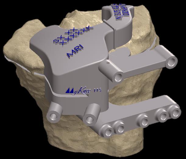
9 4 SURGICAL APPROACH The most commonly used surgical approach is the medial parapatellar approach. Other approaches may be used depending on the surgeon s practice. S Do not remove any osteophytes from the tibia or femur. This will ensure the boney references, utilized by the MyKnee PPS blocks, are not altered. 5 DISTAL FEMORAL RESECTION 5.1 MyKnee Femoral PPS Block Positioning Each MyKnee femoral PPS block shows the following information: 1- patient ID 2- MyKnee femoral PPS block reference and lot number. 1 2 A lateral parapatellar approach may be indicated for some patients. Different MyKnee tibial PPS blocks are available depending whether a medial or lateral surgical approach is used. The femoral block will suit both medial and lateral approaches. The surgeon must indicate in the surgery planning which kind of surgical approach will be used. Before starting the surgery, please check the accuracy of the data that is specific to the patient. An example of the patient ID is shown below: N_SUR_XTK_SN_DDMMYYYY N= first letter of patient s given name SUR = first three letters of patient s family name XTK = side operated, left (LTK) or right (RTK) SN= first letters of the surgeon s given and family name DDMMYYYY= patient s birth date (DD=day, MM=month, YYYY=year). DO NOT use the PPS block if it does not clearly indicate the patient identification string. Should this occur, please contact Medacta staff immediately. DO NOT use MyKnee PPS blocks on a patient for whom the pre-operative planning has not been carried out. A MyKnee PPS block used on a different patient will lead to unpredictable total knee replacement outcomes. DO NOT remove any osteophytes from or around the trochlear groove before positioning the femoral PPS block on the bone. 9


10 MyKnee Surgical Technique Hip Knee Spine Navigation Before use, visibly inspect the MyKnee PPS block to ensure it is intact and in good working order. A 3D bone model of the patient s femoral bone will be supplied with the MyKnee femoral PPS block. Matching the PPS block with the 3D bone model allows for an additional check of the PPS block s integrity before use. The 3D bone model can be supplied sterile or non-sterile. Where supplied nonsterile, it must be sterilized by the healthcare institution. Please read the Note for sterilisation included at the end of this surgical technique. The 3D femoral bone model allows for an accurate simulation of the position of the MyKnee femoral PPS block. A secondary verification can be completed by using the angel wing to verify the resection level and compare this resection to the planned femoral resection level marked on the bone model. The block must be in contact with the trochlea, the anterior part of the condyles and the distal part of the condyles. To ensure maximum stability, verify that contact areas between the MyKnee femoral PPS block and the femur are flush. If bone models are available ensure that contact areas between the MyKnee PPS block and the bone are in the same position as the areas marked on the bone model. The block is positioned manually on the distal femur. Due to the anatomical shape of the block, only one orientation is possible. Correct placement corresponds to maximum stability of the block. MRI-based MyKnee femoral PPS block contact area Before positioning the MyKnee femoral PPS block, remove the soft tissues from the femur without damaging the osteophytes. Inaccurate positioning may lead to cut parameters that do not match the planning. Once the MyKnee femoral PPS block has been properly positioned on the femur, the cut parameters are automatically set for the knee that is undergoing surgery according to the pre- operative planning (see par.3). 10


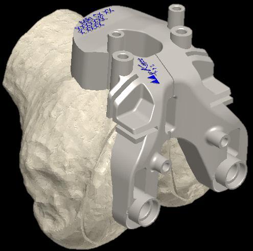
11 TIP The telescopic alignment rod can be used to check the proper positioning of the MyKnee femoral PPS block on the bone. Secure the block in position, and connect the rod to the guide using the green holes in the figure below. A visual check on the distal and anterior cut levels can be carried out using the angel wing as per the yellow slots in the figure below. If bone models are available, verify the cut height with the planned resection level that is marked on the bone model. To avoid malalignment, do not alter the MyKnee femoral PPS block position whilst drilling the pin holes. Pin the MyKnee femoral PPS guide by inserting two 3.2 mm diameter pins into the drilled holes. 5.3 Preparing the 4in1 Cutting Block Fixation Holes Before removing the MyKnee femoral PPS block, use the dedicated drills to prepare the holes for the 4in1 cutting block fixation. Two alternative options are available: Anterior reference parallel pins Femoral pegs Alignment rod holes Angel wing slots Check the varus/valgus alignment with the telescopic alignment rod only after final positioning of the MyKnee femoral PPS block. Do not use the telescopic rod to position the MyKnee PPS block. 5.2 Preparing the Distal Cutting Block Fixation Holes Once the position of the femoral PPS block is deemed satisfactory, prepare the holes for GMK Efficiency distal cutting block by drilling the dedicated parallel pin holes as shown in the picture below. To avoid malalignment, do not alter the MyKnee femoral PPS block position whilst drilling the pin or peg holes. Distal cutting block pin holes 11
12 MyKnee Surgical Technique Hip Knee Spine Navigation 5.4 Performing the Distal Resection Remove the MyKnee femoral PPS block by sliding it up and off the two pins previously placed. If the block is difficult to remove, remove one or both pins, slide the block off and reinsert the pin(s). Slide the GMK Efficiency distal cutting block onto the pins through the holes marked with a line. The picture on the right shows the correspondence between the MyKnee femoral PPS block row of pins and the distal cutting block pin holes. Before cutting, visually double check the cut height using the standard angel wing through the sawblade slot and the varus/valgus alignment with the telescopic alignment rod (as per the green holes in the figure on the right). Fix the distal cutting block and perform the distal resection following the procedure described in the dedicated GMK Efficiency surgical technique. After the resection, thoroughly rinse the joint before positioning both the trial and final implants. If a distal recut is necessary, follow the same procedure described in the dedicated GMK Efficiency surgical technique. Otherwise, remove the guide and the pins. Standard slotted distal cutting block (Ref ) Parallel positioning holes Alignment rod holes/ sawblade guide holes 12 1

13 6 TIBIAL RESECTION 6.1 MyKnee Tibial PPS Block Positioning Each MyKnee tibial PPS block shows the following information: 1-patient ID 2-MyKnee tibial PPS block reference and lot number. 2 Before starting the surgery, please check the accuracy of the data that is specific to the patient. An example of the patient ID is shown below: N_SUR_XTK_SN_DDMMYYYY 1 DO NOT remove any osteophytes from the tibial bone. Before use, visibly ensure that the MyKnee tibial PPS block is intact and in good working order. A 3D bone model of the patient s tibia will be supplied with the MyKnee tibial PPS block. Matching the PPS block with the 3D bone model allows for an additional check of the PPS block integrity before use. The 3D bone model can be supplied sterile or non-sterile. Where supplied non-sterile, it must be sterilized by the healthcare institution. Please read the Note for sterilization included at the end of this surgical technique. The 3D tibial bone model allows for an accurate simulation of the correct position of the MyKnee tibial PPS block. A secondary verification can be accomplished using the angel wing to verify the resection level, as the planned tibial resection level is marked on the bone model. The block is positioned manually on tibial plateaus. N= first letter of patient s given name SUR = first three letters of patient s family name XTK = side operated, left (LTK) or right (RTK) SN= first letters of surgeon s given and family name DDMMYYYY= patient s birth date (DD=day, MM=month, YYYY=year). DO NOT use the PPS block if it does not clearly indicate the patient identification string. Should this occur, please contact Medacta immediately. DO NOT use the MyKnee tibial PPS block on a patient for whom the pre-operative planning has not been carried out. A MyKnee tibial PPS block used on a different patient will lead to unpredictable total knee replacement outcomes. Due to the anatomical shape of the block, only one orientation is possible. Correct placement corresponds to maximum stability of the block. To ensure maximum stability, verify that the points of contact between the MyKnee tibial PPS block and the tibial bone are flush. Use the bone models to ensure that the contact points between the MyKnee tibial PPS block and bone are in the same position as the areas marked on the bone model. 13


14 MyKnee Surgical Technique Hip Knee Spine Navigation Medial approach MRI-based MyKnee tibial PPS blocks contact area Lateral approach MRI-based MyKnee tibial PPS blocks contact area Before positioning the MyKnee tibial PPS block remove the soft tissues from the tibia without damaging the osteophytes. Inaccurate positioning may lead to cut parameters that do not match the planning. Once the MyKnee tibial PPS guide has been properly positioned on the tibia, the cut parameters are automatically set for the knee that is undergoing surgery according to the pre- operative planning (see par.3). Alignment rod holes Angel wing slots Check the tibial varus/valgus with the telescopic alignment rod only after proper positioning the MyKnee tibial PPS block. Do not use the telescopic rod to position the MyKnee block. TIP The telescopic alignment rod can be used to check the proper positioning of the MyKnee tibial PPS block on the bone. Secure the block in position, and connect the rod to the guide using the green holes in the figure to the right. A visual check on the proximal tibial cut level can be carried out using the angel wing as per the yellow slots in the figure below. If bone model is available, verify the cut height with the planned resection level that is marked on the bone model. 14 1

. Position the corresponding GMK Efficiency tibia cutting block on the pins in the holes marked with a line.")
15 6.2 Preparing the Tibial Cutting Block Fixation Holes Once the position of the MyKnee tibial PPS guide is deemed satisfactory, prepare the holes for the GMK Efficiency tibial cutting block by drilling the dedicated pin holes as shown in the picture below. 6.3 Performing the Tibial Resection Pin the MyKnee tibial PPS guide by inserting two 3.2 mm diameter pins into the drilled holes. To remove the MyKnee tibia PPS block, slide the block over the two pins previously placed. If it is difficult to remove the MyKnee tibia PPS block, remove one or both pins. Then slide the guide up and off and reinsert the pin(s). Position the corresponding GMK Efficiency tibia cutting block on the pins in the holes marked with a line. The picture below shows the correspondence between the MyKnee tibial PPS block pin holes and the tibial cutting block pin holes. Before cutting, visually double check the cut height and the posterior slope using the standard angel wing through the dedicated slot on the guide. Also check the varus/valgus alignment with the telescopic alignment rod (as per the green holes in the picture below). Fix the tibia cutting block and perform the tibial resection following the procedure described in the dedicated GMK Efficiency surgical technique. After the resection, thoroughly rinse the joint before positioning both the trial and final implants. Standard tibial cutting guide pin holes MIS tibial cutting guide pin holes Tibial slope pin hole TIP The slope hole direction corresponds to the sagittal plane. Marking the position of this hole on the bone can be useful to detect the center of the tibial plateau. Should a tibial recut be necessary, follow the procedure described in the dedicated GMK Efficiency surgical technique. Otherwise remove the guide and the pins. Whilst drilling the pin holes, do not alter the position of the MyKnee tibial PPS block as this could cause the guide to misalign. 15

 MIS tibial cutting block left side (Ref.")
 Parallel positioning holes MIS guide")

16 MyKnee Surgical Technique Hip Knee Spine Navigation Standard tibial cutting block (Ref ) MIS tibial cutting block left side (Ref ) MIS tibial cutting block left side (Ref ) Parallel positioning holes MIS guide Alignment rod holes Parallel positioning holes Standard guide Parallel positioning hole MIS guide Alignment rod holes 16 1
17 7 EXTENSION GAP CONTROL Follow the procedure described in the dedicated GMK Efficiency surgical technique. 8 ANTERIOR CUT, POSTERIOR CUT AND CHAMFERS To perform the anterior, posterior and chamfer resections the GMK Efficiency 4in1 cutting block of the planned femoral size is required. The 4in1 cutting block can be fixed to the femur by the following methods: Further stabilization can be obtained using oblique fixation holes or cancellous bone screw holes, as indicated in the figure below. Anterior reference Femoral peg reference 8.1 Anterior Reference After the MyKnee femoral PPS block has been removed from the femur and the distal resection has been performed (see 5.4), position the anterior referenced parallel pins into the corresponding holes using the dedicated pin impactor. Slide the predetermined 4in1 cutting block onto the femur along the corresponding zero reference line indicated on the 4in1 cutting block. 4in1 cutting block holes Parallel positioning (Anterior Referencing) Oblique fixation holes Cancellous bone screw holes Once the 4in1 cutting block has been properly fixed to the femur, visually double check the cut height using the standard angel wing before cutting. Check the correct femoral external rotation of the 4in1 cutting blocks using the rotation guide (horseshoe). To perform the cuts, follow the procedure described in the dedicated GMK Efficiency surgical technique. TIP Parallel positioning holes (Anterior Referencing) The anterior reference method allows for correction of the 4in1 cutting block position. To correct the position move the block onto a different row of parallel pin holes as indicated in the picture below. 17


18 MyKnee Surgical Technique Hip Knee Spine Navigation 8.2 Femoral Peg Reference After the femoral PPS block has been removed from the femur and the distal resection has been performed (see 5.4), assemble the pegs to the 4in1 cutting block of the correct size and position the guide flush to the distal resection respecting the corresponding pre-drilled holes. 4in1 cutting block Parallel +2/-2mm repositioning holes (Anterior Referencing) Femoral peg holes The posterior reference method DOES NOT allow the 4in1 cutting block position to be corrected. Once the 4in1 cutting block has been properly fixed to the femur, visually double check the cut height using the standard angel wing before cutting. Check the correct femoral external rotation of the 4in1 cutting blocks, using the rotation guide (horseshoe). To perform the cuts, follow the procedure described in the dedicated GMK Efficiency surgical technique. 18 1
19 9 FEMORAL FINISHING Follow the procedure described in the dedicated GMK Efficiency surgical technique. 10 TIBIAL FINISHING Follow the procedure described in the dedicated GMK Efficiency surgical technique. 11 PATELLA Follow the procedure described in the dedicated GMK Efficiency surgical technique. 12 TRIALS Follow the procedure described in the dedicated GMK Efficiency surgical technique. 13 SELECTION OF THE PROSTHETIC COMPONENTS - SIZE MATCHING Follow the procedure described in the dedicated surgical technique of the Medacta knee implant. 14 FINAL IMPLANTS Follow the procedure described in the dedicated surgical technique of the Medacta knee implant. 19
20 MyKnee Surgical Technique Hip Knee Spine Navigation 15 MYKNEE PPS FEMORAL BLOCKS EFFICIENCY The following table summarises the MyKnee Efficiency PPS block reference codes, depending on the knee undergoing surgery (Right or Left). The references are divided into non-sterile and sterile versions. Description NON-STERILE Reference N. STERILE Reference N. MyKnee Efficiency STD Femur PPS - MRI - Left - Size S MyKnee Efficiency STD Femur PPS - MRI - Left - Size S MyKnee Efficiency STD Femur PPS - MRI - Left - Size S MyKnee Efficiency STD Femur PPS - MRI - Left - Size S MyKnee Efficiency STD Femur PPS - MRI - Left - Size S MyKnee Efficiency STD Femur PPS - MRI - Left - Size S MyKnee Efficiency STD Femur PPS - MRI - Left - Size S MyKnee Efficiency STD Femur PPS - MRI - Left - Size M SM MyKnee Efficiency STD Femur PPS - MRI - Left - Size M SM MyKnee Efficiency STD Femur PPS - MRI - Left - Size M SM MyKnee Efficiency STD Femur PPS - MRI - Left - Size M SM FEMUR MRI MEDIAL/LATERAL APPROACH MyKnee Efficiency STD Femur PPS - MRI - Left - Size M SM MyKnee Efficiency STD Femur PPS - MRI - Left - Size M SM MyKnee Efficiency STD Femur PPS - MRI - Right - Size S MyKnee Efficiency STD Femur PPS - MRI - Right - Size S MyKnee Efficiency STD Femur PPS - MRI - Right - Size S MyKnee Efficiency STD Femur PPS - MRI - Right - Size S MyKnee Efficiency STD Femur PPS - MRI - Right - Size S MyKnee Efficiency STD Femur PPS - MRI - Right - Size S MyKnee Efficiency STD Femur PPS - MRI - Right - Size S MyKnee Efficiency STD Femur PPS - MRI - Right - Size M SM MyKnee Efficiency STD Femur PPS - MRI - Right - Size M SM MyKnee Efficiency STD Femur PPS - MRI - Right - Size M SM MyKnee Efficiency STD Femur PPS - MRI - Right - Size M SM MyKnee Efficiency STD Femur PPS - MRI - Right - Size M SM MyKnee Efficiency STD Femur PPS - MRI - Right - Size M SM 20 2
21 16 MYKNEE PPS TIBIAL BLOCKS The following table summarises the MyKnee PPS tibial block reference codes, depending on the surgical approach (medial or lateral) and the knee undergoing surgery (Right or Left). The references are divided into non-sterile and sterile versions. Description NON-STERILE Reference N. STERILE Reference N. TIBIA MRI MEDIAL APPROACH TIBIA MRI LATERAL APPROACH MyKnee Tibial Pin Positioner Block - MRI Left Medial - Size S MyKnee Tibial Pin Positioner Block - MRI Left Medial - Size S MyKnee Tibial Pin Positioner Block - MRI Left Medial - Size S MyKnee Tibial Pin Positioner Block - MRI Left Medial - Size S MyKnee Tibial Pin Positioner Block - MRI Left Medial - Size S MyKnee Tibial Pin Positioner Block - MRI Left Medial - Size S MyKnee Tibial Pin Positioner Block - MRI Right Medial - Size S MyKnee Tibial Pin Positioner Block - MRI Right Medial - Size S MyKnee Tibial Pin Positioner Block - MRI Right Medial - Size S MyKnee Tibial Pin Positioner Block - MRI Right Medial - Size S MyKnee Tibial Pin Positioner Block - MRI Right Medial - Size S MyKnee Tibial Pin Positioner Block - MRI Right Medial - Size S MyKnee Tibial Pin Positioner Block - MRI Left Lateral - Size S MyKnee Tibial Pin Positioner Block - MRI Left Lateral - Size S MyKnee Tibial Pin Positioner Block - MRI Left Lateral - Size S MyKnee Tibial Pin Positioner Block - MRI Left Lateral - Size S MyKnee Tibial Pin Positioner Block - MRI Left Lateral - Size S MyKnee Tibial Pin Positioner Block - MRI Left Lateral - Size S MyKnee Tibial Pin Positioner Block - MRI Right Lateral - Size S MyKnee Tibial Pin Positioner Block - MRI Right Lateral - Size S MyKnee Tibial Pin Positioner Block - MRI Right Lateral - Size S MyKnee Tibial Pin Positioner Block - MRI Right Lateral - Size S MyKnee Tibial Pin Positioner Block - MRI Right Lateral - Size S MyKnee Tibial Pin Positioner Block - MRI Right Lateral - Size S 21
22 MyKnee Surgical Technique Hip Knee Spine Navigation Description NON-STERILE Reference N. STERILE Reference N. MyKnee MIS Tibial Pin Positioner Block - MRI Left Medial - Size S MyKnee MIS Tibial Pin Positioner Block - MRI Left Medial - Size S MyKnee MIS Tibial Pin Positioner Block - MRI Left Medial - Size S MyKnee MIS Tibial Pin Positioner Block - MRI Left Medial - Size S MyKnee MIS Tibial Pin Positioner Block - MRI Left Medial - Size S TIBIA MRI MEDIAL APPROACH TIBIA MRI LATERAL APPROACH MyKnee MIS Tibial Pin Positioner Block - MRI Left Medial - Size S MyKnee MIS Tibial Pin Positioner Block - MRI Right Medial - Size S MyKnee MIS Tibial Pin Positioner Block - MRI Right Medial - Size S MyKnee MIS Tibial Pin Positioner Block - MRI Right Medial - Size S MyKnee MIS Tibial Pin Positioner Block - MRI Right Medial - Size S MyKnee MIS Tibial Pin Positioner Block - MRI Right Medial - Size S MyKnee MIS Tibial Pin Positioner Block - MRI Right Medial - Size S MyKnee MIS Tibial Pin Positioner Block - MRI Left Lateral - Size S MyKnee MIS Tibial Pin Positioner Block - MRI Left Lateral - Size S MyKnee MIS Tibial Pin Positioner Block - MRI Left Lateral - Size S MyKnee MIS Tibial Pin Positioner Block - MRI Left Lateral - Size S MyKnee MIS Tibial Pin Positioner Block - MRI Left Lateral - Size S MyKnee MIS Tibial Pin Positioner Block - MRI Left Lateral - Size S MyKnee MIS Tibial Pin Positioner Block - MRI Right Lateral - Size S MyKnee MIS Tibial Pin Positioner Block - MRI Right Lateral - Size S MyKnee MIS Tibial Pin Positioner Block - MRI Right Lateral - Size S MyKnee MIS Tibial Pin Positioner Block - MRI Right Lateral - Size S MyKnee MIS Tibial Pin Positioner Block - MRI Right Lateral - Size S MyKnee MIS Tibial Pin Positioner Block - MRI Right Lateral - Size S 22 2
23 Part numbers subject to change. N O T E F O R S T E R I L I S A T I O N Note for sterilization: in case the instrumentation is not sterile upon delivery, it must be cleaned before use and sterilized in an autoclave respecting the US regulations, directives where applicable and following the instructions for use of the autoclave manufacturer. For detailed instructions please refer to the document Recommendations for cleaning decontamination and sterilization of Medacta International reusable orthopedic devices available at Medacta, GMK and MyKnee are registered trademarks of Medacta International SA, Castel San Pietro, Switzerland. 23

24 MyKnee Surgical Technique Hip Knee Spine Navigation Medacta International SA Strada Regina Castel San Pietro Switzerland Phone Fax info@medacta.ch D I S T R I B U T E D Medacta USA, Inc West Carroll Avenue - Chicago Illinois Phone Fax info@medacta.us.com BY MyKnee PPS Efficiency Surgical Technique ref: 99.MY26E.12PPSUS rev. 00 Last update: November
PATIENT MATCHED TECHNOLOGY IN KNEE REPLACEMENT
 PATIENT MATCHED TECHNOLOGY IN KNEE REPLACEMENT DESIGNED FOR YOU BY YOU Surgical Technique Hip Knee Spine Navigation MyKnee Surgical Technique Hip Knee Spine Navigation INTRODUCTION This brochure describes
DESIGNED FOR YOU BY YOU. Surgical Technique
 DESIGNED FOR YOU BY YOU Surgical Technique Joint Spine Sports Med MyKnee MIS Surgical Technique 2 INDEX 1. INTRODUCTION 4 1.1 Knee minimally invasive surgery: benefits 4 1.2 Mini-subvastus: the muscle
Zimmer Patient Specific Instruments. Surgical Technique for Gender Solutions Natural-Knee Flex System
 Zimmer Patient Specific Instruments Surgical Technique for Gender Solutions Natural-Knee Flex System TOC Zimmer Patient Specific Instruments Surgical Technique for Gender Solutions Natural-Knee Flex Table
Zimmer Patient Specific Instruments. Surgical Techniques for NexGen Complete Knee Solution
 Zimmer Patient Specific Instruments Surgical Techniques for NexGen Complete Knee Solution INTRO Zimmer Patient Specific Instruments Surgical Techniques for NexGen Complete Knee Solution Table of Contents
Patient Specific Instruments. Surgical Technique
 Patient Specific Instruments Surgical Technique Patient Specific Instruments Surgical Technique INTRO Introduction General Considerations Zimmer Patient Specific Instruments (PSI) are not designed for
TRUMATCH PERSONALIZED SOLUTIONS with the SIGMA High Performance Instruments
 TRUMATCH PERSONALIZED SOLUTIONS with the SIGMA High Performance Instruments Resection Guide System SURGICAL TECHNIQUE RESECTION GUIDE SURGICAL TECHNIQUE The following steps are an addendum to the SIGMA
G K SPHERE. Surgical Technique STABILITY FOR LIFE. Hip Knee Spine Navigation
 G K SPHERE M E D I A L L Y S T A B I L I Z E D K N E E STABILITY FOR LIFE Surgical Technique Hip Knee Spine Navigation GMK SPHERE Surgical Technique Hip Knee Spine Navigation INTRODUCTION This brochure
Surgical Technique. VISIONAIRE FastPak Instruments for the LEGION Total Knee System
 Surgical Technique VISIONAIRE FastPak Instruments for the LEGION Total Knee System VISIONAIRE FastPak for LEGION Instrument Technique* Nota Bene The technique description herein is made available to the
CC TRIO VERSAFITCUP. Surgical Technique. each to their own. Hip Knee Spine Navigation
 VERSAFITCUP CC TRIO each to their own Surgical Technique Hip Knee Spine Navigation Versafitcup CC TRIO Surgical Technique Hip Knee Spine Navigation EACH TO THEIR OWN The Versafitcup CC Trio is a range
THE NATURAL FIT. Surgical Technique. Hip Knee Spine Navigation
 THE NATURAL FIT Surgical Technique Hip Knee Spine Navigation MiniMAX Surgical Technique Hip Knee Spine Navigation INTRODUCTION The MiniMAX TM is a cementless anatomic stem available in 9 right sizes and
RESECTION GUIDE SYSTEM. TRUMATCH Personalized Solutions Surgical Technique with ATTUNE Knee INTUITION Instruments
 RESECTION GUIDE SYSTEM TRUMATCH Personalized Solutions Surgical Technique with ATTUNE Knee INTUITION Instruments RESECTION GUIDE SURGICAL TECHNIQUE The following steps are an addendum to the ATTUNE Knee
Surgical Technique. VISIONAIRE Disposable Instruments for the LEGION Total Knee System
 Surgical Technique VISIONAIRE Disposable Instruments for the LEGION Total Knee System VISIONAIRE and LEGION Disposable instrument technique* Note: All disposable instruments are interchangeable with the
STABILITY FOR LIFE. Surgical Technique
 STABILITY FOR LIFE Surgical Technique Joint Spine Sports Med GMK Sphere Surgical Technique 2 INDEX 1. INTRODUCTION 4 1.1 Indications 4 1.2 Contraindications 4 1.3 Preoperative planning 5 1.4 Surgical approach
Distal Cut First Femoral Preparation
 Surgical Technique Distal Cut First Femoral Preparation Primary Total Knee Arthroplasty LEGION Total Knee System Femoral preparation Contents Introduction...3 DCF femoral highlights...4 Preoperative planning...6
M.I.S. MAKE IT SMART IN ONE SYSTEM. Surgical Technique. Hip Knee Spine Navigation
 M.I.S. MAKE IT SMART IN ONE SYSTEM Surgical Technique Hip Knee Spine Navigation M.U.S.T. Mini Open Surgical Technique Hip Knee Spine Navigation 2 C O N T E N T S 1 INTRODUCTION 4 2 SURGICAL TECHNIQUE 5
TRIAL COMPONENTS SURGICAL TECHNIQUE REAMING THE TIBIAL AND FEMORAL INTRAMEDULLARY CANAL
 Ref. no. 99.27.2TRIALS TRIAL COMPONENTS SURGICAL TECHNIQUE CLINICAL INDICATION This technique is indicated in the case of revisions with significant bone loss when minimal or any resection of bone is required.
Persona. The Personalized Knee. Trabecular Metal Tibia. Surgical Technique
 Persona The Personalized Knee Trabecular Metal Tibia Surgical Technique Table of Contents Resect the Tibia... 4 Size and Finish the Tibia... 4 Trial Fit... 6 Component Implantation... 7 Inserter/Implant
ANATOMIC. Navigated Surgical Technique 4 in 1 TO.G.GB.016/1.0
 ANATOMIC Navigated Surgical Technique 4 in 1 TO.G.GB.016/1.0 SCREEN LAYOUT Take screenshot Surgical step Dynamic navigation zone Information area and buttons 2 SCREEN LAYOUT Indicates action when yellow
PIN GUIDE SYSTEM SURGICAL TECHNIQUE. with the SIGMA High Performance Instruments System. This publication is not intended for distribution in the USA.
 PIN GUIDE SYSTEM with the SIGMA High Performance Instruments System This publication is not intended for distribution in the USA. SURGICAL TECHNIQUE Pin Guide Surgical Technique The following steps are
Pin Guide System. Surgical Technique
 Pin Guide System Surgical Technique Pin Guide Surgical Technique The following steps are an addendum to the SIGMA High Performance (HP) Instruments, Fixed Reference Surgical Technique (Cat. No. 0612-87-510
OrthoMap Express Knee Product Guide. OrthoMap Express Knee Navigation Software 2.0
 OrthoMap Express Knee Product Guide OrthoMap Express Knee Navigation Software 2.0 Product Guide 1 Introduction Introduction The Stryker OrthoMap Express Knee Navigation System is providing surgeons with
TRUMATCH PERSONALIZED SOLUTIONS with the SIGMA High Performance Instruments
 TRUMATCH PERSONALIZED SOLUTIONS with the SIGMA High Performance Instruments Pin Guide System SURGICAL TECHNIQUE PIN GUIDE SURGICAL TECHNIQUE The following steps are an addendum to the SIGMA High Performance
Signature Personalized Patient Care*
 Signature Personalized Patient Care* Surgical Technique Addendum Vanguard Complete Knee System One Surgeon. One Patient. Over 1 million times per year, Biomet helps one surgeon provide personalized care
UNIQUE ANATOMIES PATIENT-MATCHED SOLUTIONS. Surgical Technique
 UNIQUE ANATOMIES PATIENT-MATCHED SOLUTIONS Surgical Technique Joint Spine Sports Med MySpine Surgical Technique Joint Spine Sports Med 2 INTRODUCTION MySpine is a patient matched, pedicle targeted technology
OPERATIVE TECHNIQUE SKS Total Knee Replacement
 OPERATIVE TECHNIQUE SKS Total Knee Replacement Femoral preparation A40577 1 2 5 1: Alignment rod A30049 + A30124 2: Centromedullary rod A40224 4: T-handle A40232 6: Femoral measuring device A40411 + A40414
pact SYSTEM Surgical Technique HEMISPHERICAL CEMENTLESS CUP SYSTEM MULTI-HOLE & RIM-HOLE Hip Knee Spine Navigation
 pact SYSTEM HEMISPHERICAL CEMENTLESS CUP SYSTEM MULTI-HOLE & RIM-HOLE Surgical Technique Hip Knee Spine Navigation Mpact Surgical Technique Hip Knee Spine Navigation PREFACE The Mpact Multi-hole and the
Revolution. Unicompartmental Knee System
 Revolution Unicompartmental Knee System While Total Knee Arthroplasty (TKA) is one of the most predictable procedures in orthopedic surgery, many patients undergoing TKA are in fact excellent candidates
LAMINA SPREADER SURGICAL TECHNIQUE
 LAMINA SPREADER SURGICAL TECHNIQUE Balanced and appropriate external rotation of the femoral component is important for tibio-femoral stability in flexion and patello-femoral tracking/function. Depending
for Standard version (STD) for Postero-stabilized version (PS) for Ultra-Congruent version (UC)
 for Postero-stabilized version (PS) for Ultra-Congruent version (UC)") A L L I N O N E s u r g i c a l t e c h n i q u e I N T R O D u c t i O N This document describes the Surgical Technique for all the three version of Evolis Total Knee System (Standard, Postero-stabilized
A L L I N O N E s u r g i c a l t e c h n i q u e I N T R O D u c t i O N This document describes the Surgical Technique for all the three version of Evolis Total Knee System (Standard, Postero-stabilized
TOTAL KNEE ARTHROPLASTY SYSTEM
 SURGICAL TECHNIQUE TOTAL KNEE ARTHROPLASTY SYSTEM 90-SRK-700000 B.0 0 Contents 1. Implant Sizing 2. Surgical Technique a. Incision and Exposure b. Distal Femoral Resection c. Tibial Resection d. Femoral
TRK REVISION KNEE Surgical Technique
 1 TRK REVISION KNEE Surgical Technique 1. 2. 3. 4. 5. 6. 7. 8. 9. 10. INTERCONDYLAR RESECTION...... page FEMORAL STEM...... page NON CEMENTED FEMORAL STEM...... page TRIAL FEMORAL COMPONENTS...... page
asterloc Surgical Technique HIP SYSTEM UNDERSTANDING TRADITION, MASTERING INNOVATION Hip Knee Spine Navigation
 asterloc HIP SYSTEM UNDERSTANDING TRADITION, MASTERING INNOVATION Surgical Technique Hip Knee Spine Navigation Masterloc Surgical Technique Hip Knee Spine Navigation INTRODUCTION This document describes
LUMBAR POSTERIOR MINIMALLY INVASIVE SYSTEM. Surgical Technique
 LUMBAR POSTERIOR MINIMALLY INVASIVE SYSTEM Surgical Technique Joint Spine Sports Med M.U.S.T. Mini Open Surgical Technique Joint Spine Sports Med CAUTION Federal law (USA) restricts this device to sale
KneeAlign System Surgical Technique Guide
 KneeAlign System Surgical Technique Guide Table of Contents Step 1 System Assembly... 1 Step 2 System Assembly... 2 Step 3 System Assembly... 2 Step 4 System Assembly... 2 Step 5 Sensor Pairing... 2 Step
Triathlon Knee System
 Triathlon Knee System Express Instruments Surgical Protocol Posterior Stabilized & Cruciate Retaining TriathlonKneeSystem Express Instruments Surgical Protocol Acknowledgments..........................................................2
KneeAlign 2 System. Surgical Technique Manual Tibia and Distal Femur Navigation. About OrthAlign, Inc.
 About OrthAlign, Inc. KneeAlign 2 System Surgical Technique Manual and Distal Femur Navigation OrthAlign is committed to providing surgeons with user-friendly, cost-effective, surgical navigation products
CONTRIBUTING SURGEON. Barry Waldman, MD Director, Center for Joint Preservation and Replacement Sinai Hospital of Baltimore Baltimore, MD
 CONTRIBUTING SURGEON Barry Waldman, MD Director, Center for Joint Preservation and Replacement Sinai Hospital of Baltimore Baltimore, MD System Overview The EPIK Uni is designed to ease the use of the
For Distal Femur Fractures. 95º Condylar Plate. Quick Reference Chart
 For Distal Femur Fractures 95º Condylar Plate Quick Reference Chart 95 Condylar Plate. Quick reference chart for distal femur fractures. Insert guide wires Fix condylar fragments with 6.5 mm cancellous
U2 PSA. Revision Knee. Surgical Protocol
 U2 PSA TM Revision Knee Surgical Protocol Table of Contents 1 Component Removal... 1 2 Tibial Preparation... 1 2.1 Tibial Canal Preparation... 1 2.2 Proximal Tibial Resection... 2 2.3 Non Offset Tibial
ANATOMIC SURGICAL TECHNIQUE. 5 in 1. Conventional instrumentation 07/11/2013
 ANATOMIC SURGICAL TECHNIQUE 5 in 1 Conventional instrumentation PRO.GB.933/1.0 Octobre 2013 2 Tibial step 3 Intramedullary technique - Based on the preoperative plan, drill the medullary canal with the
Intramedullary Tibial Preparation
 Surgical Technique Intramedullary Tibial Preparation Primary Total Knee Arthroplasty LEGION Total Knee System Intramedullary tibial preparation Contents Introduction...2 IM tibial highlights...3 Preoperative
Extramedullary Tibial Preparation
 Surgical Technique Extramedullary Tibial Preparation Primary Total Knee Arthroplasty LEGION Total Knee System Extramedullary tibial preparation Contents Introduction...2 EM tibial highlights...3 Preoperative
Trabecular Metal Femoral Component. Surgical Technique
 Trabecular Metal Femoral Component Surgical Technique INTRO Trabecular Metal Femoral Component Surgical Technique Introduction The Persona Trabecular Metal Femoral Component is designed to unite stable
Zimmer PSI Knee System For Use with Persona The Personalized Knee System Surgical Technique
 Zimmer PSI Knee System For Use with Persona The Personalized Knee System Surgical Technique TOC Zimmer PSI Knee Surgical Technique Zimmer PSI Knee Surgical Technique Table of Contents Intra-Operative Guide
Zimmer FuZion Instruments. Surgical Technique (Beta Version)
") Zimmer FuZion Surgical Technique (Beta Version) INTRO Surgical Technique Introduction Surgical goals during total knee arthroplasty (TKA) include establishment of normal leg alignment, secure implant fixation,
Zimmer FuZion Surgical Technique (Beta Version) INTRO Surgical Technique Introduction Surgical goals during total knee arthroplasty (TKA) include establishment of normal leg alignment, secure implant fixation,
Zimmer Trabecular Metal Ankle Interpositional Spacer and Trabecular Metal Ankle Fusion Spacer
 Zimmer Trabecular Metal Ankle Interpositional Spacer and Trabecular Metal Ankle Fusion Spacer Surgical Technique 2 Zimmer Trabecular Metal Ankle Interpositional Spacer and Trabecular Metal Ankle Fusion
Zimmer NexGen Trabecular Metal Tibial Tray
 Zimmer NexGen Trabecular Metal Tibial Tray Surgical Technique Zimmer NexGen Trabecular Metal Tibial Tray Surgical Technique Give Bone A Solid Hold Zimmer NexGen Trabecular Metal Tibial Tray Surgical Technique
POSTERIOR REFERENCE NEXGEN COMPLETE KNEE SOLUTION. Multi-Reference 4-in-1 Femoral Instrumentation Posterior Reference Surgical Technique
 POSTERIOR REFERENCE NEXGEN COMPLETE KNEE SOLUTION Multi-Reference 4-in-1 Femoral Instrumentation Posterior Reference Surgical Technique For NexGen Cruciate Retaining & Legacy Posterior Stabilized Knees
1. Pre-Operative Planning Skin Incision and Arthrotomy
 Stage of Operation 1. Pre-Operative Planning --------------------------1 2. Skin Incision and Arthrotomy ------------------1 3. Femoral valgus angle confirmation ------------2 4. Distal Femur Cutting -----------------------------3
KneeAlign 2 System. Surgical Technique Manual Tibia and Distal Femur Navigation
 KneeAlign 2 System Surgical Technique Manual Tibia and Distal Femur Navigation KneeAlign 2 System precise alignment simplified This technique guide describes the proper use of the KneeAlign 2 System. This
EXACTECH KNEE. Operative Technique ADDENDUM. Metaphyseal Cones. Surgeon focused. Patient driven. TM
 EXACTECH KNEE Operative Technique ADDENDUM Metaphyseal Cones Surgeon focused. Patient driven. TM TABLE OF CONTENTS INTRODUCTION...1 DESCRIPTION...1 DETAILED OPERATIVE TECHNIQUE Initial Preparation and
EVOLVING OUR HERITAGE, MEETING YOUR NEEDS. Surgical Technique
 EVOLVING OUR HERITAGE, MEETING YOUR NEEDS Surgical Technique Joint Spine Sports Med Mpact DM Surgical Technique Joint Spine Sports Med INTRODUCTION The Mpact DM is part of the Mpact Acetabular System and
UNDERSTANDING TRADITION, MASTERING INNOVATION. Surgical Technique
 UNDERSTANDING TRADITION, MASTERING INNOVATION Surgical Technique Joint Spine Sports Med MasterLoc Surgical Technique Joint Spine Sports Med INTRODUCTION This document describes the Surgical Technique for
ANTERIOR REFERENCE NEXGEN COMPLETE KNEE SOLUTION. Multi-Reference 4-in-1 Femoral Instrumentation Anterior Reference Surgical Technique
 ANTERIOR REFERENCE NEXGEN COMPLETE KNEE SOLUTION Multi-Reference 4-in-1 Femoral Instrumentation Anterior Reference Surgical Technique For NexGen Cruciate Retaining & Legacy Posterior Stabilized Knees INTRODUCTION
Zimmer NexGen MIS Tibial Component. Cemented Surgical Technique IMAGE TO COME
 Zimmer NexGen MIS Tibial Component Cemented Surgical Technique IMAGE TO COME Zimmer NexGen MIS Tibial Component Cemented Surgical Technique 1 Zimmer NexGen MIS Tibial Component Cemented Surgical Technique
Mako Partial Knee Medial bicompartmental
 Mako Partial Knee Medial bicompartmental Surgical reference guide Mako Robotic-Arm Assisted Surgery Table of contents Implant compatibility.... 3 Pre-operative planning.... 4 Intra-operative planning....
Contact your Zimmer representative or visit us at
 Please refer to the package inserts for complete product information, including contraindications, warnings, precautions, and adverse effects. Contact your Zimmer representative or visit us at www.zimmer.com
.U.S.T. MINI OPEN. Surgical Technique LUMBAR POSTERIOR MINIMALLY INVASIVE SYSTEM. Hip Knee Spine Navigation
 .U.S.T. MINI OPEN LUMBAR POSTERIOR MINIMALLY INVASIVE SYSTEM Surgical Technique Hip Knee Spine Navigation M.U.S.T. Mini Open Surgical Technique Hip Knee Spine Navigation CAUTION Federal law (USA) restricts
MIS Cemented Tibial Component
 MIS Cemented Tibial Component NexGen Complete Knee Solution Surgical Technique Table of Contents Surgical Exposure... 2 Finish the Tibia... 2 Position Based on Anatomic Landmarks... 3 Lateral Posterior
Single Axis Revision Knee System
 Orthopaedics Scorpio TS Single Axis Revision Knee System Scorpio TS Trial Cutting Guide Surgical Protocol Orthopaedics Scorpio TS Single Axis Revision Knee System Scorpio TS Trial Cutting Guide Surgical
TKA Gap Planning. Supporting healthcare professionals
 TKA Gap Planning The NAVIO TKA Gap Planning stage helps you adjust the plan based on gap information between the femur and tibia implants. Supporting healthcare professionals Interactive Views Four interactive
14 mm x +30 mm Stem Extension
 14 mm x +30 mm Stem Extension Persona The Personalized Knee Surgical Technique Table of Contents Introduction...2 Preoperative Planning Surgical Approach Patient Preparation Magnet Usage Symbols Screw/Pin
Surgical Technique Final Trial Reduction and Component Implantation of
 Surgical Technique Final Trial Reduction and Component Implantation of TC *smith&nephew TC-PLUS PRIMARY Mobile Bearing TC-PLUS PRIMARY Mobile Bearing Final Trial Reduction and Component Implantation of
ClassiQ. Scorpio. Anterior Referencing Surgical Protocol. Anterior Referencing. For use with Scorpio ClassiQ Instrument System
 Scorpio ClassiQ Anterior Referencing Surgical Protocol For use with Scorpio ClassiQ Instrument System For use with Scorpio ClassiQ Single Radius Total Knee System AR Anterior Referencing This document
BIOTECH FUTURE KNEE Minimal Invasive (and classic) Surgical Technique
 Surgical Technique") BIOTECH FUTURE KNEE Minimal Invasive (and classic) Surgical Technique Page No. Product description 2. Pre-operational planning 3. 1st step Femoral resection 4. P/S Femoral component 7. 2nd step Proximal
BIOTECH FUTURE KNEE Minimal Invasive (and classic) Surgical Technique Page No. Product description 2. Pre-operational planning 3. 1st step Femoral resection 4. P/S Femoral component 7. 2nd step Proximal
MRH Knee System Modular Peg Baseplate Surgical Protocol
 MRH Knee System Modular Peg Baseplate Surgical Protocol Using Monogram IM Revision Instruments 4 N 4 N Modular Rotating Hinge Knee System Using Monogram IM Revision Instruments Mr C R Howie FRCS Consultant
Case Report. Antegrade Femur Lengthening with the PRECICE Limb Lengthening Technology
 Case Report Antegrade Femur Lengthening with the PRECICE Limb Lengthening Technology S. Robert Rozbruch, MD Hospital for Special Surgery New York, NY, USA ABSTRACT This is a case illustrating a 4.5 cm
NATURAL MOTION TECHNOLOGY SURGICAL TECHNIQUE. EMPOWR 3D Knee. EMPOWR PS Knee
 NATURAL MOTION TECHNOLOGY EMPOWR 3D Knee EMPOWR PS Knee SURGICAL TECHNIQUE Contents System Features.... 3 Indications and Contraindications.... 4 Surgical Snap Shot.... Preoperative Planning.... Surgical
Total Knee Original System Primary Surgical Technique
 Surgical Procedure Total Knee Original System Primary Surgical Technique Where as a total hip replacement is primarily a bony operation, a total knee replacement is primarily a soft tissue operation. Excellent
Medial Rotation Knee. Forever Active. Landmark Operative Technique Supplement: No.1 All-Polyethylene Tibia (Cemented)
") Medial Rotation Knee Landmark Operative Technique Supplement: No.1 All-Polyethylene Tibia (Cemented) Physiological Stability and Mobility for the Active Knee Without Compromise Forever Active Contents
Medial Rotation Knee Landmark Operative Technique Supplement: No.1 All-Polyethylene Tibia (Cemented) Physiological Stability and Mobility for the Active Knee Without Compromise Forever Active Contents
SCORE Revision. Revision Total Knee Arthroplasty Cemented. Surgical Technique With Conventional Instrumentation Primary cases
 SCORE Revision Revision Total Knee Arthroplasty Cemented Surgical Technique With Conventional Instrumentation Primary cases SCORE REVISION FOR PRIMARY CASES The SCORE Revision is a revision total knee
Aesculap Orthopaedics Columbus MIOS
 Aesculap Orthopaedics Columbus MIOS Minimally Invasive Orthopaedic Solutions Manual TKA Surgical Technique MIOS 4-in-1 Cutting Block MIOS Distal Femoral Cutting Block MIOS Tibial Left and Right Cutting
FIRST STEM SPECIFICALLY DESIGNED FOR AMIS. Surgical Technique
 FIRST STEM SPECIFICALLY DESIGNED FOR AMIS Surgical Technique Joint Spine Sports Med AMIStem Surgical Technique Joint Spine Sports Med INTRODUCTION This document describes the Surgical Technique for the
Arthrex Open Wedge Osteotomy Technique Designed in conjunction with:
 Arthrex Open Wedge Osteotomy Technique Designed in conjunction with: Dr. Giancarlo Puddu, M.D. Dr. Peter Fowler, M.D. Dr. Ned Amendola, M.D. To treat pain and instability associated with lower extremity
FLK167 02/08. Biomet UK Ltd Waterton Industrial Estate Bridgend, South Wales CF31 3XA, United Kingdom. Tel Fax:
 FLK167 02/08 Biomet UK Ltd Waterton Industrial Estate Bridgend, South Wales CF31 3XA, United Kingdom Tel. 01656 655221 Fax: 01656 645454 Premier Instrumentation CR or PS Surgical Technique Vanguard Premier
NexGen Cruciate Retaining (CR) and Revision Instrumentation. Surgical Technique
 and Revision Instrumentation. Surgical Technique") NexGen Cruciate Retaining (CR) and Revision Instrumentation Surgical Technique Table of Contents Introduction... 4 Revision Arthroplasty... 4 Multi-Reference 4-in-1 Instrumentation System MICRO-MILL Instrumentation
NexGen Cruciate Retaining (CR) and Revision Instrumentation Surgical Technique Table of Contents Introduction... 4 Revision Arthroplasty... 4 Multi-Reference 4-in-1 Instrumentation System MICRO-MILL Instrumentation
P. F. C. Sigma RP Knee System surgical technique
 P. F. C. Sigma RP Knee System surgical technique I N T E L L I G E N T O R T H O P A E D I C S Contents Ci System Connections 2 Ci System Set-up 4 Start-up Procedure 5 Ci Menu Buttons 6 Ci Procedure Set-up
This surgical technique describes how to perform an anatomic total shoulder arthroplasty implanting a short stem.
 INTRODUCTION This surgical technique describes how to perform an anatomic total shoulder arthroplasty implanting a short stem. CAUTION Federal law (USA) restricts this device to sale distribution and use
Persona The Personalized Knee. Surgical Technique
 Persona The Personalized Knee Surgical Technique Table of Contents Introduction... 2 Constraint Options Preoperative Planning Surgical Approach Patient Preparation Magnet Usage Symbols Screw/Pin Information
Surgical Technique i.m.a.g.e. Patient Specific Instrumentation 5-in-1
 Surgical Technique i.m.a.g.e. Patient Specific Instrumentation -in- SCORE Primary Total Knee System Mobile bearing Cemented or cementless Overview of SCORE TKS Overview of SCORE TKS The SCORE TKS is a
Surgical Technique Joint Spine Sports Med
 Surgical Technique Joint Spine Sports Med MOTO Medial Surgical Technique INTRODUCTION This brochure describes the surgical technique of the MOTO TM Partial Knee System for a Medial Unicompartmental Replacement.
Signature Personalized Patient Care
 Surgical Technique Acetabular Guide System Contents One Surgeon. One Patient. Over 1 million times per year, Biomet helps one surgeon provide personalized care to one patient. The science and art of medical
Patella Planing System
 REFERENCE GUIDE AND SURGICAL TECHNIQUE Patella Planing System SPECIALIST INSTRUMENTS AN ONLAY PATELLA PREPARATION SYSTEM TECHNIQUE FOR PRIMARY PATELLAR RESURFACING The Specialist 2 Patellar Planer System
Surgical Technique. CONQUEST FN Femoral Neck Fracture System
 Surgical Technique CONQUEST FN Femoral Neck Fracture System Table of Contents Introduction... 3 Indications... 3 Product Overview... 4 Surgical Technique... 5 Patient Positioning... 5 Reduce the Fracture...
Mako Partial Knee Patellofemoral
 Mako Partial Knee Patellofemoral Mako Robotic-Arm Assisted Surgery Surgical reference guide Table of contents Implant compatibility.... 3 Pre-operative implant planning... 4 Intra-operative planning....
NexGen Trabecular Metal Tibial Tray. Surgical Technique
 NexGen Trabecular Metal Tibial Tray Surgical Technique Table of Contents Surgical Technique...2 Resect the Tibia...2 Finish the Tibia...2 Position Based Anatomic Landmarks...3 Position Based on Trial
FOREWORD PRESERVATION UNICOMPARTMENTAL KNEE SYSTEM
 Surgical Technique FOREWORD PRESERVATION UNICOMPARTMENTAL KNEE SYSTEM Our surgeon design team first implanted the Preservation Unicompartmental Knee System in 2001. The system was developed with over
Zimmer NexGen Tibial Stem Extension & Augmentation. Surgical Technique IMAGE TO COME. Stem Extensions and Augments
 Zimmer NexGen Tibial Stem Extension & Augmentation Surgical Technique IMAGE TO COME Stem Extensions and Augments Zimmer NexGen Tibial Stem Extension & Augmentation Surgical Technique 1 Zimmer NexGen Tibial
Orthopedic Bone Nail System - Distal Femoral Nail Surgical Technique Manual
 Orthopedic Bone Nail System - Distal Femoral Nail Surgical Technique Manual Note: The surgical procedures should be performed under the guidance of qualified skilled orthopedic surgeons, and this surgical
Resurfacing Distal Femur. Orthopaedic Salvage System
 Resurfacing Distal Femur Orthopaedic Salvage System Primary Arthroplasty OSS 3cm Resurfacing Distal Femur Distal Femoral Resection Drill and ream the distal femur in the following sequence: (Figure 1)
OPTIONAL/ADDITIONAL INSTRUMENTS
 20 Elevated 6320-36-22 20 Elevated Rim Liner, 36mm OD x 22mm ID 6320-80-22 20 Elevated Rim Liner, 80mm OD x 22mm ID 6320-42-26 20 Elevated Rim Liner, 42mm OD x 26mm ID 6320-80-26 20 Elevated Rim Liner,
Surgical Technique Final Trial Reduction and Component Implantation of
 Surgical Technique Final Trial Reduction and Component Implantation of TC *smith&nephew TC-PLUS PRIMARY Fixed Bearing TC-PLUS PRIMARY Fixed Bearing Final Trial Reduction and Component Implantation of
Improve your quality of life after medially stabilized knee arthroplasty
 G K SPHERE MEDIALLY STABILIZED KNEE Improve your quality of life after medially stabilized knee arthroplasty IMPROVE YOUR QUALI 2 All trademarks and registered trademarks are the property of their respective
Integra. DigiFuse Cannulated Intramedullary Fusion System SURGICAL TECHNIQUE
 Integra DigiFuse Cannulated Intramedullary Fusion System SURGICAL TECHNIQUE Table of Contents Design Rationale... 2 System Features... 2 Indications... 2 Contraindications... 2 Surgical Technique...3 Step
Zimmer Unicompartmental High Flex Knee. Intramedullary, Spacer Block Option and Extramedullary Minimally Invasive Surgical Techniques
 Zimmer Unicompartmental High Flex Knee Intramedullary, Spacer Block Option and Extramedullary Minimally Invasive Surgical Techniques ZIMMER UNICOMPARTMENTAL HIGH FLEX KNEE INTRAMEDULLARY, SPACER BLOCK
NexGen Complete Knee Solution. Cruciate Retaining (CR) and Revision Instrumentation Surgical Technique for Cruciate Retaining Augmentable (CRA) Knees
 and Revision Instrumentation Surgical Technique for Cruciate Retaining Augmentable (CRA) Knees") NexGen Complete Knee Solution Cruciate Retaining (CR) and Revision Instrumentation Surgical Technique for Cruciate Retaining Augmentable (CRA) Knees INTRODUCTION This surgical technique document combines
NexGen Complete Knee Solution Cruciate Retaining (CR) and Revision Instrumentation Surgical Technique for Cruciate Retaining Augmentable (CRA) Knees INTRODUCTION This surgical technique document combines
Unicondylar Surgical Technique
 Unicondylar Surgical Technique Contents Indications, Contra-indications and X-ray Templating 2 Approach and Exposure 3 Proximal Tibial Resection 4 Tibial Jig Alignment 6 Tibial Sizing 9 Balancing 10 Distal
Masterclass. Tips and tricks for a successful outcome. E. Verhaven, M. Thaeter. September 15th, 2012, Brussels
 Masterclass Tips and tricks for a successful outcome September 15th, 2012, Brussels E. Verhaven, M. Thaeter Belgium St. Nikolaus-Hospital Orthopaedics & Traumatology Ultimate Goal of TKR Normal alignment
MEDACTA UNCONSTRAINED SCREW TECHNOLOGY - REDUCTION SCREWS. Surgical Technique. Hip Knee Spine Navigation
 .U.S.T. MEDACTA UNCONSTRAINED SCREW TECHNOLOGY - REDUCTION SCREWS Hip Knee Spine Navigation M.U.S.T. Hip Knee Spine Navigation INTRODUCTION The Medacta Unconstrained Screw Technology [M.U.S.T.] Pedicle