GRAVITY Plantar Plate Repair System SURGIC AL TECHNIQUE
|
|
|
- Diana Shavonne Harrington
- 5 years ago
- Views:
Transcription


1 GRAVITY Plantar Plate Repair System SURGIC AL TECHNIQUE
2 Contents PREFACE Preface Chapter Appendix 14 Introduction Positioning Indications Contraindications Surgical Technique Step 1: Incise, Dissect and Ligament Release Step 2: Distal Metatarsal Osteotomy Step 3: Distraction Step 4: Suturing Step 5: Phalanx Drilling and Suture Passing Step 6: Metatarsal Head Fixation, Tensioning and Knot Tying Explant Information Post-Operative Care Ordering Information Proper surgical procedures and techniques are the responsibility of the medical professional. The following guidelines are furnished for information purposes only. Each surgeon must evaluate the appropriateness of the procedures based on his or her personal medical training and experience. Prior to use of the system, the surgeon should refer to the product package insert for complete warnings, precautions, indications, contraindications and adverse effects. Package inserts are also available by contacting the manufacturer. Contact information can be found on the back of this surgical technique and the package insert is available on the website listed. Caution: Federal law restricts this device to sale by or on the order of a physician. Please contact your local Wright representative for product availability.
3
4 Introduction chapter 1 For DPMS, DOs and MDs, the GRAVITY Plantar Plate Repair System addresses a common difficulty encountered by foot and ankle surgeons: an attenuated or torn plantar plate. This system surgical technique eliminates two typical surgical steps - suture reloading and sequential suture retrieval through the phalanx - which can potentially reduce OR time. This technique further uses a novel GRAVITY DOUBLE BARREL Drill Guide* that enables reproducibility and can enable greater visualization while addressing the phalanx. The system incorporates two low profile GRAVITY Needle Drivers* with a tapered tip corkscrew needle that are pre-loaded with loops of #0 Force Fiber * ultra-high molecular weight polyethylene (UHMWPE) suture, as well as two CHARLOTTE Snap-Off Screws for permanent osteotomy fixation. 4 Chapter 1 Introduction
5 Indications The GRAVITY Soft Tissue Repair System is indicated for the following: GRAVITY Plantar Plate Repair System:»» Distal metatarsal osteotomy (screw)»» Plantar plate repair (suture) GRAVITY Needle Driver*:»» Approximation of soft tissues, including the use of allograft tissue for orthopaedic surgeries Contraindications There are no product-specific contraindications. Prior to use of the system, the surgeon should refer to the product package insert for complete warnings, precautions, indications, contraindications and adverse effects. Package inserts are also available by contacting the manufacturer. Contact information can be found on the back of this surgical technique and the package insert is available on the website listed. Preoperative Planning Preoperative planning is based upon surgeon discretion Chapter 1 Description of Chapter 5
 joint.")

6 Surgical Technique chapter 2 Step 1: Incise, Dissect and Ligament Release The patient is placed in a supine position on the operating room table. A dorsal longitudinal incision is centered over the metatarsophalangeal (MTP) joint. The extensor digitorum longus and brevis are lengthened or split and a self-retaining rectractor is placed deep of the tendons to allow for visualization of the dorsal capsule of the MTP joint. After entering the MTP joint, the dorsal capsule and collateral ligaments are released from the proximal phalanx. A McGlamry Elevator (86PP0001) is used plantarly to release the plantar plate from the distal metatarsal. This step will aid in the necessary exposure and advancement of the plantar plate. Figure 1 Figure 1 NOTE Care is taken when releasing the plantar plate tissue from the distal metatarsal so as not to advance the McGlamry Elevator excessively proximally, medially or laterally in order to spare sufficient ligament to bone connection. 6 Chapter 2 Surgical Technique


7 Step 2: Distal Metatarsal Osteotomy A distal metatarsal osteotomy is performed to increase the working area and to shorten the metatarsal. It is recommended not to remove the metatarsal dorsal lip until after the permanent osteotomy fixation in a later step. Once the osteotomy is complete, the Metatarsal Head Pusher (86PP0002) is used to translate the capital fragment far enough proximally in order to provide a large working area in the joint space. Figure 2 The capital fragment is temporarily fixed with a.062in / 1.6mm dual-trocar K-Wire. The K-Wire should be sufficiently proximal on the dorsal fragment in order to provide ample room for permanent fixation. Do not penetrate the plantar surface of the capital fragment. Figure 2 NOTE To reduce waste and cost, all K-Wires in the system are sharp on both ends so that they can be cut in half with one being used for the metatarsal head and one used for the proximal phalanx. Chapter 2 Surgical Technique 7


8 Step 3: Distraction The GRAVITY Plantar Plate Repair System Distractor (86PP0003) is utilized to gain optimal visualization of the plantar plate. First, remove the selfretaining retractor (if used). Second, pass one of the proximal distractor holes over the.062 in / 1.6mm K-Wire previously used to temporarily secure the osteotomy. Next, extend the Distractor until the distal holes are sufficiently distal to the articular surface of the proximal phalanx. This ensures optimal room for drilling bone tunnels. Fourth, place a second.062 in / 1.6mm K-Wire through one of the two distal Distractor holes in order to fixate the proximal phalanx. Fifth, distract in order to gain dorsal access to the plantar capsular tissue of the MTP joint. Finally, replace the retractor over the arms of the Distractor (if desired). If a partial tear of the distal plantar plate is observed, completion of the tear with a scalpel as distally as possible is recommended before proceeding. This step may allow for additional distraction. NOTE Care is taken not to transect the flexor digitorum longus tendon which runs immediately plantar to the plantar plate ligament. Figure 3 NOTE Ensure the Distractor is completely seated against the bone before distracting in order to avoid bending the K-Wires during distraction. Optional Secondary Fixation Cut the two.062 in / 1.6mm K-Wires in half. Do not yet discard the cut halves. To fully lock the phalanx in place and to prevent medial/lateral laxity, the saved K-Wire halves may be used. First, rotate the phalanx to the desired orientation, then drive one or both of the saved halves through the remaining Distractor holes. If this optional secondary fixation step is not necessary, the cut K-Wire halves may be discarded. 8 Chapter 2 Surgical Technique


9 Step 4: Suturing The two included GRAVITY Needle Drivers* are pre-loaded with a #0 Force Fiber ultra-high molecular weight polyethylene (UHMWPE) suture loop and a corkscrew needle. They are designed to make a single pass through tissue where workspace is limited and to create a luggage tag knot. First, stabilize the plantar plate tissue with a forceps or similar device. Second, hold the Needle Driver* like a pencil and use a twisting or rotating motion to insert the corkscrew needle into the medial or lateral half of the tissue. A surgeon can pass the needle from dorsal to plantar or plantar to dorsal based upon anatomy and surgeon preference. Figure 4 It is recommended to not approach the joint space strictly dorsally with the Needle Driver*, approach from the medial or lateral side as much as possible to ensure the corkscrew needle twists back into view where it can be grabbed in the next step. Figure 4 NOTE Use a twisting or rotating motion to pass the needle through the tissue. Avoid pulling on the Needle Driver *. Third, grasp the needle with a forceps or other similar device. Fourth, press the release button on the Needle Driver* then gently pull the Needle Driver* away from the joint space. Now, the needle is free of the Needle Driver* housing and the suture contained within the Needle Driver* can be seen. Next, pass the needle through the open loop of suture and Chapter 2 Surgical Technique 9
.")

10 create a luggage tag knot around the tissue. Finally, repeat these suturing steps using the second Needle Driver* for either the medial or lateral half of the plantar plate (whichever was not done in the previous steps). Figure 5 Figure 5 10 Chapter 2 Surgical Technique NOTE The needles may be left attached to the suture to aid in later passing through the Suture Retriever* snares. Additionally, one Needle Driver* will contain a blue Force Fiber suture loop and the other will contain a white Force Fiber suture loop. This color differentiation aids in distinguishing the medial suture from the lateral suture. Optional Needle Driver* Reloading (if necessary) In situations where the user unintentionally pushes the release button early and the needle prematurely unseats, it is useful to have the ability to reseat the needle by retightening the preloaded suture within the Needle Driver*. First, turn the cap counterclockwise one quarter turn and fully remove the Driver. Now, the preloaded suture is exposed from beneath the cap. Next, gently pull on this exposed suture to return the needle to its housing. It may be necessary to rotate the needle to ensure that it fully seats back into the Needle Driver*. Finally, with the needle properly seated, maintain tension on the suture and press the release button back to its original locked position. If the preloaded suture and needle are deployed too far to be visible when the cap is removed, or if the needle and suture are fully deployed and completely separated from the Needle Driver*, there is a sterile, individually packed Needle Driver* (86PP00N0) available. The Needle Driver* should only be used to pass the preloaded needle and suture one time. After a successful pass and deployment of the needle and suture from the Needle Driver*, the driver housing should be discarded.
 is placed around the base of the proximal phalanx and clamped tightly.")


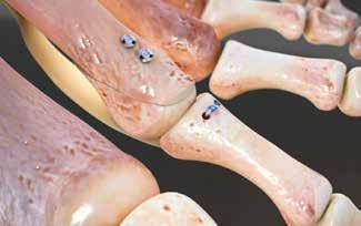
11 Figure 6 Figure 7 Step 5: Phalanx Drilling and Suture Passing Once suturing of the Plantar Plate is complete, the Distractor and the phalangeal K-Wires can be removed in preparation for drilling the bone tunnels. A GRAVITY DOUBLE BARREL Drill Guide* (86PP0004) is placed around the base of the proximal phalanx and clamped tightly. The Drill Trajectory Guide (86PP0005) is used in conjunction with the GRAVITY DOUBLE BARREL Drill Guide*. When viewed sagitally, the guide post indicates the path of the drill holes. Use the Drill Trajectory Guide to ensure the drill holes will not violate the articular surface of the phalanx. The guide post may be placed medially or laterally to assess positioning of the Drill Guide*. Adjust Drill Guide* positioning as necessary. Utilize the leverage gained with the GRAVITY DOUBLE BARREL Drill Guide* to plantarflex the phalanx 90 degrees. Using a rongeur or curette, remove residual soft tissue from the plantar surface of the phalanx. While maintaining plantarflexion, utilize the 3rd.062in / 1.6mm K-Wire to drill two holes in the proximal phalanx through the holes in the GRAVITY DOUBLE BARREL Drill Guide*. Ensure the K-Wire completely exits the plantar surface of the proximal phalanx. Depicted in Figure 6 is the 2nd proximal phalanx plantarflexed 90 degrees. Figure 6 NOTE Remove any residual plantar soft tissue obstructing the bone tunnel exits to allow full passing of both Suture Retriever* diamondshaped snares in the next step. While maintaining 90 degree phalanx plantarflexion, insert the Suture Retriever* with its diamond snares retracted into the GRAVITY DOUBLE BARREL Drill Guide* until fully seated. Next, slide the button toward the phalanx. As the button is slid forward, the two diamond-shaped snares will advance through the bone tunnels and then exit the bone tunnels. Figure 7 Now, feed the lateral needle of the Force Fiber suture through the lateral diamond-shaped snare, and the medial needle through the medial diamond-shaped snare. If the needles have already been removed, utilize the suture color for medial/lateral distinction. Next, pull adequate suture through the diamond-shaped snares. Finally, cut the needles off the sutures (if not previously done) and gently pull the Suture Retriever* out of the GRAVITY DOUBLE BARREL Drill Guide* until the suture ends are completely through the snares. Figure 8 Unclamp and remove the GRAVITY DOUBLE BARREL Drill Guide* and dispose of the Suture Retriever*. Figure 8 Chapter 2 Surgical Technique 11
 is used to maintain compression and prevent rotation between the fragments until fixation is achieved.")
.")
 before tensioning the suture and tying knots.")

12 Step 6: Metatarsal Head Fixation, Tensioning and Knot Tying Remove the K-Wires from the Metatarsal. Translate the capital fragment of the metatarsal to the desired final position. The Osteotomy Clamp (86PP0006) is used to maintain compression and prevent rotation between the fragments until fixation is achieved. For reference, a groove is located at 3mm on the plantar tip of the clamp over which the dorsal metatarsal lip will protrude. Fixate the osteotomy with a 2.0x12mm and/or 2.7x13mm CHARLOTTE Snap-Off Screw across the fragments using a CHARLOTTE Snap-Off Screwdriver ( ). Figure 9 Using a rongeur, remove the protruding lip of the distal metatarsal fragment until flush with the capital fragment. NOTE If using the hole left by the.062in /.062mm K-Wire for permanent fixation, use of the larger 2.7x13mm screw is recommended for improved purchase. Figure 9 Figure 10 NOTE It is recommended to perform any adjunct procedure (e.g. PIP fusion with PHALINX Hammertoe Fixation) before tensioning the suture and tying knots. This will ensure proper plantar plate approximation is not altered after knots have been tied. Please reference the PHALINX surgical technique for PIP fusion. After completing any adjunct procedures, reduce the MTP joint to the desired position. Pull the sutures tight to ensure any slack is removed and to advance the plantar plate onto the proximal phalanx. The sutures are tied over the cortical bridge between the two drill holes. A double throw, followed by five single throws (surgeon s knot followed by two square knots) is recommended, but specific knot tying technique is at surgeon discretion. Proceed with capsular repair, extensor tendon repair (if applicable) and wound closure per surgeon preference. Figure Chapter 2 Surgical Technique
13 Explant Information To remove the CHARLOTTE Snap-Off screws, engage the screws with the CHARLOTTE Snap-Off Screwdriver ( ) and rotate counterclockwise. Explantation of the Force Fiber suture requires no product specific instrumentation. Cut the suture if necessary and remove. If removal of the implant is required due to revision or failure of the device, the surgeon should contact the manufacturer using the contact information located on the back cover of this surgical technique to receive instructions for returning the explanted device to the manufacturer for investigation. Post-Operative Care Post-operative care is based upon surgeon discretion. Chapter 2 Surgical Technique 13

14 Ordering Information APPENDIX Instruments P/N 86PPKIT1 86PP PP PP PP PP PP0006 Description GRAVITY Plantar Plate Instrument Set McGlamry Elevator Met Head Pusher Distractor GRAVITY DOUBLE BARREL Drill Guide* Drill Trajectory Guide Osteotomy Clamp CHARLOTTE Snap-Off Screwdriver Implants P/N 86PPS1N0 86PP00N0 Description GRAVITY Plantar Plate Implant Set GRAVITY Needle Driver* Osteotomy Clamp 86PP0006 GRAVITY DOUBLE BARREL Drill Guide* 86PP0004 Distractor 86PP0003 Drill Trajectory Guide 86PP0005 McGlamry Elevator Size 11 86PP0001 CHARLOTTE Snap-Off Screwdriver Met Head Pusher 86PP GRAVITY Plantar Plate Repair System Ordering Information


15 Packaging Guide APPENDIX STERILE CONSUMABLES Implant Set* 86PPS1N0 One 2.0 X 12 mm and one 2.7 X 13mm CHARLOTTE Snap-Off Screw Bundle of three dual-trocar K-wires with tip protectors Two GRAVITY Needle Drivers*, one with blue & one with white Force Fiber ** suture Suture Retriever* with twin nitinol diamond snares GRAVITY Needle Driver* 86PP00N0 GRAVITY Needle Driver* with blue Force Fiber suture *The Implant Set contains all consumables needed to complete the GRAVITY Plantar Plate Repair System surgical technique. **Force Fiber is a Registered Trademark of Teleflex, Inc. GRAVITY Plantar Plate Repair System Ordering Information 15

845 833 4435 161 Rue")
4 76 61 35 00 and denote Trademarks and Registered")

16 1023 Cherry Road Memphis, TN Kingston Road Staines-upon-Thames Surrey TW18 4NL United Kingdom +44 (0) Rue Lavoisier Montbonnot Saint Martin France +33 (0) and denote Trademarks and Registered Trademarks of Wright Medical Group N.V. or its affiliates Wright Medical Group N.V. or its affiliates. All Rights Reserved A_22-Jan-2018
DARCO. Bow 2 Plate SURGIC AL TECHNIQUE
 DARCO Bow 2 Plate SURGIC AL TECHNIQUE Contents 2 Preface 3 Chapter 1 4 Chapter 2 5 6 7 8 9 Appendix 10 10 11 Intended Use Indications/Contraindications Design Rationale Preoperative Planning Surgical Technique
CANNULINK. Intraossous Fixation System SURGICAL TECHNIQUE
 CANNULINK Intraossous Fixation System SURGICAL TECHNIQUE Contents Chapter 1 4 Introduction The CANNULINK Advantage Indications for Use Preoperative Planning Chapter 2 5 Surgical Technique CANNULINK Standard
7% - 15% Single Tunnel Repair of Plantar Plate. Savings of. are common with EcoSMART *Patent Pending
 * Provide Value Sterile kit packaging provides high quality, surgical grade instruments and implants making your O.R. more efficient. No hassles or expense from cleaning, storing, sterilizing, and maintaining
MICA. Minimally Invasive Foot Surgery CHEVRON OSTEOTOMY SURGIC AL TECHNIQUE
 MICA Minimally Invasive Foot Surgery CHEVRON OSTEOTOMY SURGIC AL TECHNIQUE Contents Chapter 1 4 Introduction Chapter 2 5 Indications and Warnings Chapter 3 6 Patient Positioning and Set Up Chapter 4 7
ARTHROTUNNELER TUNNELPRO SYSTEM
 TORNIER ARTHROTUNNELER TUNNELPRO SYSTEM Transosseous Rotator Cuff Repair SURGICAL TECHNIQUE ARTHROTUNNELER TUNNELPRO SYSTEM ARTHROTUNNELER Surgical Technique Step 1. Drill medial tunnel(s) to a positive
Contents. Chapter 1 4 Chapter Chapter Chapter Chapter 5 15
 Contents Chapter 1 4 Chapter 2 5 5 Chapter 3 6 6 7 Chapter 4 8 8 8 8 9 10 10 11 12 13 14 14 Chapter 5 15 Introduction Intended Use Indications Device Description Implant Options and Sizing Instrumentation
PHALINX. Hammertoe Fixation SURGICAL TECHNIQUE
 PHALINX Hammertoe Fixation SURGICAL TECHNIQUE Contents Chapter 1 4 Product Information 4 Device Description Chapter 2 5 Intended Use 5 Indications 5 Contraindications Chapter 3 6 Surgical Technique 6
CHARLOTTE Lisfranc. Recontruction System SURGICAL TECHNIQUE
 CHARLOTTE Lisfranc Recontruction System SURGICAL TECHNIQUE Contents Preface 3 Chapter 1 4 Chapter 2 4 4 4 4 4 Chapter 3 5 5 5 6 6 6 7 7 8 Chapter 4 8 8 9 10 Appendix 10 Introduction Indications and Contraindications
Correction System. Surgical Technique
 Nextra Hammertoe Correction System Surgical Technique Maximized Bone Purchase* Stable and Secure Phalanx Optimized Screw Design Adjustable Bone-to-Bone Apposition Progressive Ratchet Tightening Mechanism
Foot and Ankle Technique Guide Metatarsophalangeal (MTP) Bilateral Joint Repair
 Bilateral Joint Repair") Surgical Technique Foot and Ankle Technique Guide Metatarsophalangeal (MTP) Bilateral Joint Repair Prepared in consultation with: Phinit Phisitkul, MD Department of Orthopedics and Rehabilitation University
Surgical Technique Foot and Ankle Technique Guide Metatarsophalangeal (MTP) Bilateral Joint Repair Prepared in consultation with: Phinit Phisitkul, MD Department of Orthopedics and Rehabilitation University
ORTHOLOC 3Di. Foot Reconstruction System SURGIC AL TECHNIQUE
 ORTHOLOC 3Di Foot Reconstruction System S C R E W TA R G E T I N G G U I D E SURGIC AL TECHNIQUE SURGEON DESIGN TEAM The ORTHOLOC 3Di Foot Reconstruction System was developed in conjuction with: ORTHOLOC
LOWER EXTREMITIES SINGLE-USE INSTRUMENTATION SURGICAL IMPLANTATION TECHNIQUE. First Metatarsal Phalangeal Joint FOOT THE DIFFERENCE IS MOVING.
 LOWER EXTREMITIES SINGLE-USE INSTRUMENTATION SURGICAL IMPLANTATION TECHNIQUE First Metatarsal Phalangeal Joint FOOT THE DIFFERENCE IS MOVING. CARTIVA SYNTHETIC CARTILAGE IMPLANT SURGICAL IMPLANTATION TECHNIQUE
Foot and Ankle Technique Guide Metatarsophalangeal (MTP) Unilateral Joint Repair
 Unilateral Joint Repair") Surgical Technique Foot and Ankle Technique Guide Metatarsophalangeal (MTP) Unilateral Joint Repair Prepared in consultation with: Phinit Phisitkul, MD Department of Orthopedics and Rehabilitation University
Surgical Technique Foot and Ankle Technique Guide Metatarsophalangeal (MTP) Unilateral Joint Repair Prepared in consultation with: Phinit Phisitkul, MD Department of Orthopedics and Rehabilitation University
CHARLOTTE. Snap-Off Screws SURGICAL TECHNIQUE
 CHARLOTTE Snap-Off Screws SURGICAL TECHNIQUE CHARLOTTE snap-off screws surgical technique SURGICAL ADVISORS ROBERT ANDERSON, MD BRUCE COHEN, MD W. HODGES DAVIS, MD Proper surgical procedures and techniques
ORTHOLOC 3Di. Foot Reconstruction System Midfoot Fusion Plate SURGICAL TECHNIQUE
 ORTHOLOC 3Di Foot Reconstruction System Midfoot Fusion Plate SURGICAL TECHNIQUE ORTHOLOC Foot Reconstruction System Midfoot Fusion Plate SURGICAL TECHNIQUE Surgeon Design Team The ORTHOLOC 3Di Foot Reconstruction
ORTHOLOC. 3Di. Ankle Fusion Plating System SYSTEM OVERVIEW
 ORTHOLOC with 3Di Technology Ankle Fusion Plating System SYSTEM OVERVIEW ORTHOLOC with 3Di Technology Ankle Fusion Plating System The ORTHOLOC Ankle Fusion Plating System with 3Di Technology contains plates
Arthroscopic Shoulder Repair Using the Smith & Nephew KINSA Suture Anchor
 Shoulder Series Technique Guide *smith&nephew KINSA Suture Anchor Arthroscopic Shoulder Repair Using the Smith & Nephew KINSA Suture Anchor Reviewed by: Larry D. Field, MD Mississippi Sports Medicine and
Correction System. Surgical Technique
 Nextra Hammertoe Correction System Surgical Technique Maximized Bone Purchase* Stable and Secure Phalanx Optimized Screw Design Adjustable Bone-to-Bone Apposition Progressive Ratchet Tightening Mechanism
Foot and Ankle Technique Guide Metatarsal Shortening Osteotomy
 Surgical Technique Foot and Ankle Technique Guide Metatarsal Shortening Osteotomy Prepared in consultation with: Phinit Phisitkul, MD Department of Orthopedics and Rehabilitation University of Iowa Iowa
Merete PlantarMAX Lapidus Plate Surgical Technique. Description of Plate
 Merete PlantarMAX Lapidus Plate Surgical Technique Description of Plate Merete Medical has designed the PlantarMax; a special Plantar/Medial Locking Lapidus plate which places the plate in the most biomechanically
ORTHOFLEX. Silicone Hammertoe Implant SURGICAL TECHNIQUE
 ORTHOFLEX Silicone Hammertoe Implant SURGICAL TECHNIQUE Contents Chapter 1 4 Product Information 4 Device Description Chapter 2 4 Intended Use 5 Indications 5 Contraindications Chapter 3 6 Surgical Technique
DYNAMIC, TRANSVERSE COMPRESSION. Low Profile, Anatomic Design, Type II Anodized. Mechanical Compression Designed to Stimulate the Fusion Process
 CoLink_XP_ST_080218.pdf 1 8/2/18 8:36 AM SURGICAL TECHNIQUE CoLink XP Plates DYNAMIC, TRANSVERSE COMPRESSION C M Y CM MY CY CMY K MTP Std. Plate MTP Revision Plate Lapidus Standard +1mm and +2mm Y Plate
CrossTIE. PIP Arthrodesis Implant. surgical technique ordering information PEEK IMPLANTS PEEK IMPLANTS ALLOGRAFT IMPLANTS CROSSTIE INSERTER
 ordering information x x 0o 1443-3301 1444-3300 x x 10o 1443-3311 1444-3310 2.5mm x 2.5mm x 0o 1443-2501 1444-2500 2.5mm x 2.5mm x 10o 1443-2511 1444-2510 ALLOGRAFT IMPLANTS * Sterile Packaged in Saline,
Implantable K-wire SURGIC A L T ECHNIQUE
 Implantable K-wire SURGIC A L T ECHNIQUE Contents Chapter 1 4 Product Information 4 Device Description 4 Indications 4 Contraindications Chapter 2 5 Surgical Technique 5 Hammertoe Correction 6 MTP Joint
Integra. Silicone PIP Implant SURGICAL TECHNIQUE
 Integra Silicone PIP Implant SURGICAL TECHNIQUE Table of Contents Indications For Use...2 Contraindications...2 Warnings and Precautions...2 Surgical Technique Preoperative Assessment... 3 Step 1: Initial
CSS. Cannulated Screw System SURGICAL TECHNIQUE
 CSS Cannulated Screw System SURGICAL TECHNIQUE Contents Chapter 1 4 Product Information 4 Device Description Chapter 2 5 Intended Use 5 Indications 5 Contraindications Chapter 3 6 Surgical Technique Appendix
MULTIFIX S Knotless Implants
 Technique Guide MULTIFIX S Knotless Implants The MULTIFIX S system offers 5.5mm and 6.5mm knotless, all-peek, poundin implants. The system provides multiple fixation options, a streamlined technique, and
AFX. Femoral Implant. System. The AperFix. AM Portal Surgical Technique Guide. with the. The AperFix System with the AFX Femoral Implant
 The AperFix System AFX with the Femoral Implant AM Portal Surgical Technique Guide The Cayenne Medical AperFix system with the AFX Femoral Implant is the only anatomic system for soft tissue ACL reconstruction
Twin Tail TightRope System
 Open Stabilization of Acute Acromioclavicular Joint Dislocation using the Twin Tail TightRope System Surgical Technique Twin Tail TightRope System Open Stabilization of Acute Acromioclavicular Joint Dislocation
Hemi Phalangeal Implant SURGICAL TECHNIQUE
 SURGICAL TECHNIQUE SIZING AND ORDERING HEAD WIDTH A HEAD HEIGHT B HPI-0001 1 16.9 12 HPI-0002 2 19.5 13.8 HPI-0003 3 22.4 15.9 PART NO. A B SIZE DIMENSIONS ARE IN MILLIMETERS. The HPI stemmed prosthesis,
JuggerLoc Bone-to-Bone System for Ankle Syndesmosis Fixation. Surgical Technique
 JuggerLoc Bone-to-Bone System for Ankle Syndesmosis Fixation Surgical Technique Table of Contents Position and Preparation... 2 Incision... 2 Fracture Reduction... 2 Drill Fibula and Tibia... 3 JuggerLoc
SureLock All-Suture Anchor System
 SureLock All-Suture Anchor System Guide for Bankart Repair Surgical Technique 2 SureLock All-Suture System Bankart Repair Surgical Technique Figure 1 *Curved and straight drill guides are available for
PROstep Minimally Invasive Surgery HALLUX VALGUS CORRECTION USING PROSTEP MICA MINIMALLY INVASIVE FOOT SURGERY: TWO CASE STUDIES
 PROstep Minimally Invasive Surgery HALLUX VALGUS CORRECTION USING PROSTEP MICA MINIMALLY INVASIVE FOOT SURGERY: TWO CASE STUDIES AS PRESENTED BY: JOEL VERNOIS M.D. 016798A Case Study 1 PROstep Minimally
Technique Guide. *smith&nephew N8TIVE ACL Anatomic ACL Reconstruction System
 Technique Guide *smith&nephew N8TIVE ACL Anatomic ACL Reconstruction System N8TIVE ACL System The N8TIVE ACL Anatomic Reconstruction System provides a novel and simple approach to ACL repair. The N8TIVE
GENERAL TECHNIQUE GUIDE
 GENERAL TECHNIQUE GUIDE PRODUCT OVERVIEW The Eclipse Soft Tissue Anchor employs MedShape s shape memory PEEK Altera technology for soft tissue fixation procedures in the shoulder, elbow, knee, hand & wrist,
DuaFit. Proximal Interphal angeal Impl ant
 DuaFit Proximal Interphal angeal Impl ant DUAFIT - TABLE OF CONTENTS PRODUCT DESCRIPTION 3 INDICATIONS 6 SURGICAL TECHNIQUE #1 7 Without guide wire (DuaFit 0 10 17 ) SURGICAL TECHNIQUE #2 14 With guide
Surgical Technique. Customer Service:
 Patent and Patent Pending CAUTION: Federal Law (USA) restricts this device to sale by or on the order of a physician. INDICATIONS FOR USE The Axis Charcot Fixation System in diameters of 5.5, 6.5 and 7.5mm
Lesser MPJ Hemi Implant
 Lesser MPJ Hemi Implant Surgical Technique Contents Product The BioPro Lesser MPJ Hemi Implant is a simple, durable, metallic hemiarthroplasty resurfacing prosthesis for the treatment of arthritis, Freiberg
Technique Guide Hammertoe Correction System
 Technique Guide Hammertoe Correction System TM The ToeMATE Hammertoe Correction System is an easy to implant bone screw system intended for the correction of hammertoe deformity. It is provided in a complete
Locking Ankle Plating System. Surgical Technique
 Locking Ankle Plating System Surgical Technique Acumed is a global leader of innovative orthopaedic and medical solutions. We are dedicated to developing products, service methods, and approaches that
MSP TM Metatarsal Shortening System. Surgical Technique
 MSP TM Metatarsal Shortening System Surgical Technique Metaphyseal/Diaphyseal osteotomy does not result in plantar displacement of metatarsal head Osteotomy guide integrated into implant allows for controlled
CHARLOTTE. Compression Staple SURGIC A L T ECHNIQUE
 CHARLOTTE Compression Staple SURGIC A L T ECHNIQUE CHARLOTTE Compression Staple SURGICAL TECHNIQUE Surgical Technique as described by: Robert Anderson, MD Bruce Cohen, MD W. Hodges Davis, MD Contents Chapter
Foot Reconstruction System SURGICAL TECHNIQUE
 ORTHOLOC 3Di Foot Reconstruction System SURGICAL TECHNIQUE SURGEON DESIGN TEAM The ORTHOLOC 3Di Foot Reconstruction System was developed in conjuction with: Robert B. Anderson, MD OrthoCarolina Charlotte,
Technique Guide KISSloc Suture System
 Technique Guide KISSloc Suture System The KISSloc Suture System consists of a strong self-cinching suture assembly, a unique load-dispersing Arrow Plate and procedure specific instrumentation. Simple,
Foot and Ankle Technique Guide Proximal Inter-Phalangeal (PIP) Fusion
 Fusion") Surgical Technique Foot and Ankle Technique Guide Proximal Inter-Phalangeal (PIP) Fusion Prepared in consultation with: Phinit Phisitkul, MD Department of Orthopedics and Rehabilitation University of Iowa
Surgical Technique Foot and Ankle Technique Guide Proximal Inter-Phalangeal (PIP) Fusion Prepared in consultation with: Phinit Phisitkul, MD Department of Orthopedics and Rehabilitation University of Iowa
Femoral Fixation for ACL Reconstruction. Surgical Protocol by Mark Gittins, D.O.
 Femoral Fixation for ACL Reconstruction Surgical Protocol by Mark Gittins, D.O. Features A unique weave in which a single strand of braided polyethylene is woven through itself twice in opposite directions.
CARTIVA. Synthetic Cartilage Implant SURGICAL IMPLANTATION TECHNIQUE. First Metatarsal Phalangeal Joint THE DIFFERENCE IS MOVING.
 CARTIVA Synthetic Cartilage Implant SURGICAL IMPLANTATION TECHNIQUE First Metatarsal Phalangeal Joint THE DIFFERENCE IS MOVING. CARTIVA SYNTHETIC CARTILAGE IMPLANT TABLE OF CONTENTS Introduction... 3 Cartiva
MICRONAIL. Intramedullary Distal Radius System SURGICAL TECHNIQUE
 MICRONAIL II Intramedullary Distal Radius System SURGICAL TECHNIQUE Contents Introduction 3 4 Chapter 1 5 Chapter 2 6 Appendix A 18 Appendix B 19 Surgeon Design Team Introduction Product Information Surgical
Distal Radius Plate Instrument and Implant Set. Discontinued December 2017 DSUS/TRM/0916/1063(1)
") Distal Radius Plate Instrument and Implant Set Surgical Technique Discontinued December 2017 DSUS/TRM/0916/1063(1) The Distal Radius Plates Indications For fixation of fractures and osteotomies, including
Distal Radius Plate Instrument and Implant Set Surgical Technique Discontinued December 2017 DSUS/TRM/0916/1063(1) The Distal Radius Plates Indications For fixation of fractures and osteotomies, including
The Ceterix NovoStitch Disposable Suture Passer. Do Not Reuse
 The Ceterix NovoStitch Disposable Suture Passer Ceterix Orthopaedics 959 Hamilton Avenue Menlo Park, CA 94025 Customer Service: (650) 241-1748 IK Sterile D Do Not Reuse i See Instructions for Use THE CETERIX
BTB ACL Reconstruction with the ToggleLoc Fixation Device with ZipLoop Technology. Surgical Technique by James R. Andrews, M.D.
 BTB ACL Reconstruction with the ToggleLoc Fixation Device with ZipLoop Technology Surgical Technique by James R. Andrews, M.D. Table of Contents Femoral Tunnel Preparation... 4 Prepare ToggleLoc Device...
Surgical Technique 4.5/8.5MM BEAMING SYSTEM. Customer Service:
 Patent and Patent Pending CAUTION: Federal Law (USA) restricts this device to sale by or on the order of a physician. INDICATIONS FOR USE The 4.5/8.5 screw system is intended for fixation arthrodesis of
ALLOPURE. Bicortical Allograft Wedges Specific for Evans and Cotton Osteotomies SURGICAL TECHNIQUE
 ALLOPURE Bicortical Allograft Wedges Specific for Evans and Cotton Osteotomies SURGICAL TECHNIQUE Contents Chapter 1 4 Chapter 2 5 5 5 Chapter 3 6 6 7 8 8 Appendix A 9 Introduction Intended Use Indications
Surgical Technique Guide
 Guide CAUTION: Federal Law (USA) restricts this device to sale by or on the order of a physician. INDICATIONS FOR USE The Align Anterior Ankle Fusion Plate is intended to facilitate arthrodesis of the
TABLE OF CONTENTS. 2 (8144 Rev 2)
") 1 (8144 Rev 2) TABLE OF CONTENTS Introduction Conventus CAGE TM - Proximal Humerus...3 Indications and Contraindications...4 Surgical Summary...5 Patient Positioning & Approach...6 Surgical Technique Plate
1 (8144 Rev 2) TABLE OF CONTENTS Introduction Conventus CAGE TM - Proximal Humerus...3 Indications and Contraindications...4 Surgical Summary...5 Patient Positioning & Approach...6 Surgical Technique Plate
Surgical Technique. Forearm Fracture Solutions
 Surgical Technique Forearm Fracture Solutions Acumed is a global leader of innovative orthopaedic and medical solutions. We are dedicated to developing products, service methods, and approaches that improve
BIOKNOTLESSRC ROTATOR CUFF REPAIR SUTURE ANCHOR SURGICAL TECHNIQUE. Surgical Technique for Arthroscopic Rotator Cuff Repair. Raymond Thal, M.D.
 SURGICAL TECHNIQUE ROTATOR CUFF REPAIR BIOKNOTLESSRC SUTURE ANCHOR Surgical Technique for Arthroscopic Rotator Cuff Repair Raymond Thal, M.D. Town Center Orthopaedic Associates Reston, Virginia Surgical
JuggerKnot Soft Anchor 1.0 mm Mini
 JuggerKnot Soft Anchor 1.0 mm Mini Ulnar Collateral Ligament (UCL) Repair of the Thumb Surgical Technique Surgical Protocols by Mark Rekant, M.D. A. Lee Osterman, M.D. One Surgeon. One Patient. Over 1
Surgical Technique. Foot and Ankle Technique Guide Ankle Syndesmosis Repair, Operative Technique
 Surgical Technique Foot and Ankle Technique Guide Ankle Syndesmosis Repair, Operative Technique INVISIKNOT Ankle Syndesmosis Repair Surgical Technique The following technique guide was prepared under close
CHARLOTTE. 3.0 and 4.3 Multi-Use Compression Screws SURGIC A L T ECHNIQUE
 CHARLOTTE 3.0 and 4.3 Multi-Use Compression Screws SURGIC A L T ECHNIQUE CHARLOTTE 3.0 and 4.3 Multi-Use Compression Screws SURGICAL TECHNIQUE Surgical Technique as described by: Robert Anderson, MD Bruce
CHARLOTTE. Compression Staple
 CHARLOTTE Compression Staple CHARLOTTE compression staple surgical technique SURGICAL ADVISORS ROBERT ANDERSON, MD BRUCE COHEN, MD W. HODGES DAVIS, MD Proper surgical procedures and techniques are the
JuggerKnot Soft Anchor 1.0 mm Mini. Scapholunate Ligament Repair/Reconstruction. Brochure and Surgical Technique
 JuggerKnot Soft Anchor 1.0 mm Mini Scapholunate Ligament Repair/Reconstruction Brochure and Surgical Technique One Surgeon. One Patient. Over 1 million times per year, Biomet helps one surgeon provide
HammerFUZE HAMMERTOE COMPRESSION SYSTEM SURGICAL TECHNIQUE
 HammerFUZE HAMMERTOE COMPRESSION SYSTEM SURGICAL TECHNIQUE HammerFUZE HAMMERTOE COMPRESSION SYSTEM SURGICAL TECHNIQUE Design Rationale The HammerFUZE Hammertoe Compression System was designed to simplify
Biomet Large Cannulated Screw System
 Biomet Large Cannulated Screw System s u r g i c a l t e c h n i q u e A Complete System for Simplified Fracture Fixation 6.5mm & 7.3mm The Titanium, Self-drilling, Self-tapping Large Cannulated Screw
Technique Guide. 6.5 mm Midfoot Fusion Bolt. For intramedullary fixation of the medial column of the foot.
 Technique Guide 6.5 mm Midfoot Fusion Bolt. For intramedullary fixation of the medial column of the foot. Table of Contents Introduction 6.5 mm Midfoot Fusion Bolt 2 AO Principles 4 Indications 5 Surgical
The AperFix II System
 The AperFix II System A Complete Anatomic Solution Transtibial Surgical Technique 2 AperFix II System Transtibial Surgical Technique Figure 1 A Complete Anatomic Solution The Cayenne Medical AperFix and
CHARLOTTE. 7.0 Multi-Use Compression Screw System SURGICAL TECHNIQUE
 CHARLOTTE 7.0 Multi-Use Compression Screw System SURGICAL TECHNIQUE Contents Chapter 1 1 Chapter 2 2 2 3 4 Appendix A 5 6 Appendix B 7 8 Preoperative Planning Surgical Technique Subtalar Arthrodesis Guide
SALVATION 3Di. Plating System SURGICAL TECHNIQUE
 SALVATION 3Di Plating System SURGICAL TECHNIQUE Contents Chapter 1 4 Introduction Chapter 2 5 Intended use 5 Indications 5 Contraindications Chapter 3 6 Device Description Chapter 4 7 Preoperative Planning
Technique Guide. 3.5 mm LCP Low Bend Medial Distal Tibia Plate Aiming Instruments. Part of the 3.5 mm LCP Percutaneous Instrument System.
 Technique Guide 3.5 mm LCP Low Bend Medial Distal Tibia Plate Aiming Instruments. Part of the 3.5 mm LCP Percutaneous Instrument System. Table of Contents Introduction 3.5 mm LCP Low Bend Medial Distal
DEVELOPED BY MEDSHAPE, INC. IN CONJUNCTION WITH PATRICK ST. PIERRE, M.D. BICEPS TENODESIS ARTHROSCOPIC AND SUBPECTORAL SURGICAL TECHNIQUE
 ! SURGICAL TECHNIQUE! DEVELOPED BY MEDSHAPE, INC. IN CONJUNCTION WITH PATRICK ST. PIERRE, M.D. ARTHROSCOPIC AND SUBPECTORAL BICEPS TENODESIS SURGICAL TECHNIQUE BICEPS TENODESIS Indications Tenodesis of
Tibial Fixation. with TunneLoc Device. Surgical Technique by Mark J. Albritton, M.D. and Sherwin Ho, M.D.
 Tibial Fixation with TunneLoc Device Surgical Technique by Mark J. Albritton, M.D. and Sherwin Ho, M.D. Table of Contents Surgical Technique... 4 Ordering Information... 11 Indications For Use... 12 Contraindications...
JuggerKnot Soft Anchor 1.0 mm Mini
 JuggerKnot Soft Anchor 1.0 mm Mini Ulnar Collateral Ligament (UCL) Repair of the Thumb Surgical Technique Surgical Protocols by Mark Rekant, M.D. A. Lee Osterman, M.D. One Surgeon. One Patient. Over 1
RFS. Resorbable Fixation System SURGICAL TECHNIQUE
 RFS Resorbable Fixation System SURGICAL TECHNIQUE Product Information RFS Resorbable Material - PLGA - What is it? The RFS Pins and Solid/Cannulated Screws are made of PLGA, a bioabsorbable poly lactic/glycolic
DARCO. LPS Plate SURGICAL TECHNIQUE
 DARCO LPS Plate SURGICAL TECHNIQUE Contents Preface 3 Chapter 1 4 Chapter 2 5 5 6 7 9 Appendix 10 10 11 Design Rationale Preoperative Planning Surgical Technique Surgical Approach Joint Preparation Surgical
Technique Guide. *smith&nephew MAGNUM 2 Knotless Implant
 Technique Guide *smith&nephew MAGNUM 2 Knotless Implant MAGNUM 2 knotless implant The MAGNUM 2 implant increases surgeon control by combining independent cortical bone lock, suture lock, and incremental
ORTHOLOC Calcaneal Fracture System SURGIC AL TECHNIQUE
 ORTHOLOC Calcaneal Fracture System SURGIC AL TECHNIQUE Contents Chapter 1 4 Chapter 2 5 Chapter 3 6 7 8 9 Appendix 1 10-12 ORTHOLOC Calcaneal Fracture System - Introduction - ORTHOLOC Polyaxial Locking
SWANSON. Flexible Hinge Toe Implant SURGIC AL TECHNIQUE
 SWANSON Flexible Hinge Toe Implant SURGIC AL TECHNIQUE SWANSON flexible HINGE TOE IMPLANT with SWANSON Flexible Hinge Grommet surgical technique presented by ALFRED B. SWANSON, MD, FACS, GRAND RAPIDS,
Conventus CAGE PH Surgical Techniques
 Conventus CAGE PH Surgical Techniques Conventus Orthopaedics The Conventus CAGE PH (PH Cage) is a permanent implant comprised of an expandable scaffold, made from nitinol and titanium, which is deployed
Subpectoral Biceps Tenodesis using Cortical Buttons Surgical Technique
 Subpectoral Biceps Tenodesis using Cortical Buttons Surgical Technique Subpectoral Biceps Tenodesis Subpectoral Biceps Tenodesis using Cortical Buttons Introduction Subpectoral biceps tenodesis using cortical
SURGICAL TECHNIQUE GUIDE: JONES FRACTURE USING THE PRECISION JONES FRACTURE SCREW SYSTEM
 PRODUCT DESCRIPTION The PRECISION Jones Fracture Screw System offers extensive options of Type II Anodized Titanium screws. System-specific instrumentation is designed to address procedural challenges
Acu-Loc Wrist Spanning Plate System. Surgical Technique
 Acu-Loc Wrist Spanning Plate System Surgical Technique Acumed is a global leader of innovative orthopaedic and medical solutions. We are dedicated to developing products, service methods, and approaches
CHARLOTTE. 7.0 Multi-Use Compression Screw System SURGICAL TECHNIQUE
 CHARLOTTE 7.0 Multi-Use Compression Screw System SURGICAL TECHNIQUE Contents Chapter 1 1 Chapter 2 2 Chapter 3 3-9 Chapter 4 10-12 Appendix A 13-14 Design Rationale Screw Behavior Surgical Technique Procedure
Correction System. Surgical Technique
 Re+Line Bunion Correction System Surgical Technique Bunion Correction System Easy insertion and medial placement accuracy using Landmark Guide technology 1 mm compression slot and fixed tines to encourage
Clover Staple. Surgical Technique
 Clover Staple Surgical Technique Contents Product The BioPro Clover Staple is a permanent implant that removes minimal cross-sectional fusion area, allows easy radiographical monitoring and may permit
JuggerKnot Soft Anchor 1.4/1.5 mm with Percutaneous Instrumentation for Low Profile/Trans-Cuff SLAP Repair
 JuggerKnot Soft Anchor 1.4/1.5 mm with Percutaneous Instrumentation for Low Profile/Trans-Cuff SLAP Repair Surgical Technique by Shabi Kahn, MD One Surgeon. One Patient. Over 1 million times per year,
Passer. Sterile. D i. Do Not PASSER DESCRIPTION. The Ceterix. Page 1
 The Ceterix NovoStitch Disposable Suture Passer Ceterix Orthopaedic cs 959 Hamilton Avenue Menlo Park, CA 94025 Customer Service: (650) 241 1748 IK D i Sterile Do Not Reusee See Instructions for Use THE
STS. Subtalar Spacer System SURGICAL TECHNIQUE
 STS Subtalar Spacer System SURGICAL TECHNIQUE Contents Chapter 1 4 Introduction 4 Device Description Chapter 2 5 Intended Use 5 Indications Chapter 3 6 Surgical Technique 9 Postoperative Protocol 9 Explant
Technique Guide Small Bone Fusion System
 Technique Guide Small Bone Fusion System The Pinit Plate Small Bone Fusion System is a super low profile, modular bone plate and screw system designed to stabilize a bunionectomy with a medial to lateral
ToggleLoc. Fixation Device. Surgical Technique. Femoral Fixation for ACL Reconstruction SPORTS MEDICINE. Surgical Protocol by Mark Gittins, D.O.
 ToggleLoc Fixation Device Femoral Fixation for ACL Reconstruction Surgical Technique Surgical Protocol by Mark Gittins, D.O. SPORTS MEDICINE One Surgeon. One Patient. Over 1 million times per year, Biomet
Integra. DigiFuse Cannulated Intramedullary Fusion System SURGICAL TECHNIQUE
 Integra DigiFuse Cannulated Intramedullary Fusion System SURGICAL TECHNIQUE Table of Contents Design Rationale... 2 System Features... 2 Indications... 2 Contraindications... 2 Surgical Technique...3 Step
SURGICAL TECHNIQUE THE TENDON ANCHOR SYSTEM
 SURGICAL TECHNIQUE THE TENDON ANCHOR SYSTEM TABLE OF CONTENTS Introduction PAGE 1 Features and Benefits PAGE 1-2 Applications PAGE 3 Ordering Information PAGE 4 Indications & Contra-indications PAGE 5
Technique Guide. *smith&nephew SPEEDSCREW Fully Threaded Knotless Implant
 Technique Guide *smith&nephew SPEEDSCREW Fully Threaded Knotless Implant SPEEDSCREW system Fully threaded knotless implant system The SPEEDSCREW system is specifically designed for rotator cuff repair
BLUEPRINT. 3D Planning + PSI SURGIC AL TECHNIQUE V 2.1 T I TA N I U M
 TO R N I E R BLUEPRINT 3D Planning + PSI SURGIC AL TECHNIQUE V 2.1 T I TA N I U M Contents 4 5 6 7 9 Patient-Specific Instrumentation Overview 3D Planning PSI Guide Creation and Order Use of PSI Guide
AcUMEDr. FoREARM ROD SYSTEM
 AcUMEDr FoREARM ROD SYSTEM FoREARM ROD SYSTEM Since 1988 Acumed has been designing solutions to the demanding situations facing orthopedic surgeons, hospitals and their patients. Our strategy has been
AcUMEDr. Olecranon Threaded Compression Rod
 AcUMEDr Olecranon Threaded Compression Rod Olecranon Threaded Compression Rod Since 1988, Acumed has been designing solutions to the demanding situations facing orthopaedic surgeons, hospitals and their
HemiEDGE. Patent No. 8,845,750. Surgical Technique
 HemiEDGE Patent No. 8,845,750 Surgical Technique Contents Product Based on the clinical success of our First MPJ Hemi Implant, the HemiEDGE, incorporates an overlapping edge extending around the medial,
Anchorage. Foot & Ankle. Plating System. Operative Technique
 Foot & Ankle Anchorage Plating System Operative Technique Foot & Ankle Anchorage Plate System MTP Plates Lapidus Plates Lisfranc Plates Basal Osteotomy Plates Utility Plates Anchorage Cross Plate System
PEDUS-L. Locking Plantar Lapidus Plate
 PEDUS-L Locking Plantar Lapidus Plate Page 1 PEDUS-L - Locking Plantar Lapidus Plate Table of Contents Implants 3 System 4 Operation manual 5 Approach 5 Identification of the TMT 1 joint with a cannula
Foot & Ankle. EasyClip. Osteosynthesis Compression Staples. Foot & Ankle
 Foot & Ankle EasyClip Osteosynthesis Compression Staples Foot & Ankle Operative Technique EasyClip Osteosynthesis Compression Staples 2 This publication sets forth detailed recommended procedures for using
Technique Guide. VersiTomic. ReelX STT Double-Row Achilles G-Lok. J. Martin Leland III, M.D. J. Martin Leland III, M.D. Proximal Biceps Tenodesis
 Technique Guide VersiTomic ReelX STT Double-Row Achilles G-Lok Tendon Sub-Pectoral Repair Proximal Biceps Tenodesis J. Martin Leland III, M.D. J. Martin Leland III, M.D. The opinions expressed are those