1/19/2018. Winter injuries to the shoulder and elbow. Highgate Private Hospital (Whittington Health NHS Trust)
|
|
|
- Corey Watts
- 5 years ago
- Views:
Transcription
 Highgate Private Hospital (Whittington Health NHS Trust) E: ShoulderUnit@denovomedic.co.")

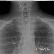
1 Winter injuries to the shoulder and elbow Omar Haddo Consultant Orthopaedic Surgeon, Shoulder, Elbow, Hand & Wrist Specialist MBBS, BmedSci, FRCS(Orth) Highgate Private Hospital (Whittington Health NHS Trust) E: Objectives Introduction Clavicle fracture/dislocation Humerus Head fracture/dislocation Shaft fracture Suprachondylar fracture Elbow Anterior/Posterio dislocation Olecrenon fracture Radial head fracture Chorocoid fracture General Comments Zone of Injury 1

2 General Comments In the field ABCs Airway Breathing Circulation Always assess neurovascular status (CMS = circulation, motor and sensory) Control any bleeding Do not move victim until stabilized General Comments If possible, always ask the patient where it hurts the most. Remove jewellery, etc before splinting Patient will self-splint the upper extremity (internal rotation, elbow flexed and adducted to body) Self-splinting 2


3 ARMS Appearance and alignment Pulse Motor function and mechanism of injury Sensation Prevention Upper extremity injuries Snowboarding 3


4 Upper extremity injury Skiing Clavicle injuries 4
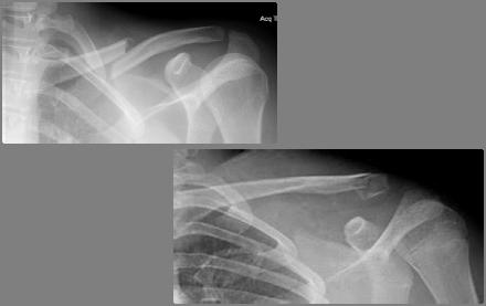

5 5


6 Humerus Injuries 6

 Supraconylar")


7 Humeral Fractures MOI Head - Direct trauma to the humerus from collision with an object or fall directly onto the bone Shaft bent forces like breaking a stick (shear or torsion) Supraconylar upper transmission of force on outstretched hand Humeral Head fracture Diagnosis Upper humeral fractures usually involve the surgical neck of the bone extracapsular low incidence of avascular necrosis (AVN) Anatomical Neck intracapsular higher incidence of AVN Humeral Head Fractures NEER Classification 7


 Rule out")
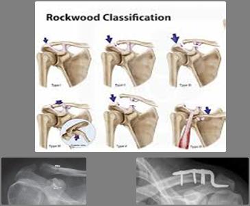
8 Humeral Head Fractures Treatment One part fractures (no fracture fragments displaced < 1cm or 45 deg) Non-operative immobilization in sling1-2 weeks Early motion started immediately 75% good to excellent results; 10% poor Any other fracture Closed reduction with percutaneous pinning ORIF 2-6 weeks to allow pain free movement Humeral Fractures Complications Avascular Necrosis of Humeral Head Especially at risk with 4 part fractures Non-union 3-6 mos after injury Shoulder stiffness with prolonged immobilization Shoulder Dislocation Most commonly anterior Examine for associated injuries (vascular or neurological) Rule out associated fracture Reduce asap 8


9 Shoulder Dislocation Younger patient Bankart lesion Older patient RC tear Shoulder Dislocation Younger patient: 1st time Reduce Mobilise after 2-3 weeks Reassess at 6 weeks if still apprehensive then consider surgery Recurrent Reduce Surgery Shoulder Dislocation Older patient: Reduce Reassess after 6 weeks Repair of RC if unable to elevate arm 9


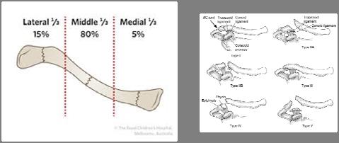
10 Humeral Shaft Fracture Diagnosis Fractures of the shaft of the humerus 1 3% of all fractures Up to 20% have radial n palsy Humeral Shaft Fracture Humeral Fractures Treatment Non-operative Acceptable alignment AP anglulation 20 deg Varus 30 deg <30mm shortening 70 80% with % union rates Time consuming and requires cooperative patient Collar and cuff; coaptation splint; hanging cast; functional bracing Weight of forearm provides traction 10

 Commonly")
11 Humeral Fractures Treatment Operative Absolute Indications Failure of closed treatment Associated articular involvement Vascular injuries Ipsilateral forearm fractures Pathological fractures Open fractures Polytrauma Relative Indications Short oblique or transverse fracture in an active individual Body habitus Patient compliance Staff considerations Radial nerve palsy Most at risk distal 1/3 fractures Occurs up to 20% of fractures 90% neurapraxias and heal in 3 4 mos Exploration indicated No recovery in 3 4 mos (clinical or EMG) Loss of function with closed reduction Open fractures Holstein Lewis distal 1/3 spiral fractures Supracondylar fracture Diagnosis Supracondylar fractures Most common pediatric elbow fracture (65% of fractures and dislocations of the elbow) Commonly associated with neurovascular injury 11


12 Supracondylar fractures Diagnosis Classification Type I non displaced Type II angulated but not translated in the sagittal plane with hinging of the posterior cortex of the humerus Type III posteriorly displaced with IIIA being posteromedial and type IIIB being posterolateral Supracondylar Fractures Diagnosis Radiology AP view Baumann s angle Medial epichondylar epiphyseal angle (MEE) Lateral view Humerotrochlear angle Oblique Supracondylar Fracture Treatment Non-displaced fxs cast immobilization Displaced fxs close reduction with percutaneous pinning 12


13 Suprachondylar fracture Complications Vascular injury brachial aa Neurologic deficits median nerve; possible radial nerve Volkmann s contracture Cubitus varus Humerus Injuries Emergency Care Sling Ladder splint Elbow Injuries 13

14 Radial Anatomy Radial head articulates with capitellum Radial neck tapers to radial tuberosity which is insertion for biceps brachii tendon Ulnar Anatomy Sigmoid/semilunar/ trochlear notch Anteriorly composed of coronoid process Posteriorly composed of olecranon process Articulates with trochlea of humerus Elbow Joint Articulation Elbow consists of articulations: Ulnohumeral (elbow flexion/extension) Radiohumeral (forearm pronation/supination) Radioulnar (forearm pronation/supination) 14


15 Elbow Injuries MOI Fall onto outstretched hand with elbow extended or direct trauma Elbow dislocation Diagnosis Second to shoulder dislocations Posterior dislocation account for 80-90% Most occur without fracture Elbow dislocation Treatment Immediate reduction vs splint and refer Children should be splinted; increase incidence of fractures Need for radiographs After relocation Assess neurovascular status Assess joint stability Rehab early 15


16 Elbow fracture Radial head 30% Olecrenon 20% Coronoid fractures 10 to 15% of elbow dislocations Elbow fat pads Elbow Fat Pads 16
 Longer immobilization (1 2 weeks) removal of")

 ORIF Comminuted (Type III) ORIF Elbow Fractures")

17 Elbow Fractures Treatment Radial Head Non displaced (type I) sling and or splint until no pain Displaced (type II) Longer immobilization (1 2 weeks) removal of bone fragments if necessary Comminuted (Type III) Surgery to remove bone fragments Repair ligament damage Elbow Fractures Treatment Olecrenon Fracture Non displaced (type I) Sling, splint and or cast for 3 4 weeks Follow by x ray for dislocation of fracture Displaced (type II) ORIF Comminuted (Type III) ORIF Elbow Fractures Treatment Coronoid Fracture Type 1 Immobilization for 2 weeks Type 2 Immobilization for 2 weeks Displaced or humeroulnar joint instability may consider ORIF Type 3 ORIF 17


18 Elbow dislocation or fracture Emergency Care Immobilize Sling Posterior elbow splint or brace ice Conclusion Accurate assessment and rapid transport critical (60 rule) Immobilize in the position found Sling is good immobilizer for upper extremity injuries Every patient should be advised to seek the care of a physician regardless of injury, especially if symptoms persist > 24 hrs. Thank -You 18
Fractures of the shoulder girdle, elbow and fractures of the humerus. H. Sithebe 2012
 Fractures of the shoulder girdle, elbow and fractures of the humerus H. Sithebe 2012 Fractures of the Clavicle (mid-shaft). Fractures of the clavicle Fractures of the clavicle Treatment- conservative.
Fractures and dislocations around elbow in adult
 Lec: 3 Fractures and dislocations around elbow in adult These include fractures of distal humerus, fracture of the capitulum, fracture of the radial head, fracture of the olecranon & dislocation of the
Pediatric Fractures. Objectives. Epiphyseal Complex. Anatomy and Physiology. Ligaments. Bony matrix
 1 Pediatric Fractures Nicholas White, MD Assistant Professor of Pediatrics Eastern Virginia Medical School Attending, Pediatric Emergency Department Children s Hospital of The King s Daughters Objectives
Upper Extremity Fractures
 Upper Extremity Fractures Ranie Whatley, RN,FNP-C David W. Gray, MD Skeletal Trauma 10 to 15 % of all Childhood Injuries Physeal (Growth Plate) Injuries are ~ 15% of all Skeletal Injuries Orthopaedic Assessment
Upper Extremity Injury Management. Jonathan Pirie MD, Med, FRCPC, FAAP
 Upper Extremity Injury Management Jonathan Pirie MD, Med, FRCPC, FAAP Learning Objectives At the end of this session, you will be able to manage common fractures of the: 1. Humerus 2. Elbow 3. Forearm
Upper Extremity Trauma.
 Upper Extremity Trauma www.fisiokinesiterapia.biz Topics Clavicle Shoulder Dislocation Humerus Elbow Forearm Distal Radius Clavicle Fractures Clavicle Fractures Mechanism Fall onto shoulder (87%) Direct
The Elbow and Radioulnar Joints Kinesiology. Dr Cüneyt Mirzanli Istanbul Gelisim University
 The Elbow and Radioulnar Joints Kinesiology Dr Cüneyt Mirzanli Istanbul Gelisim University 1 The Elbow & Radioulnar Joints Most upper extremity movements involve the elbow & radioulnar joints. Usually
Rehabilitation after Total Elbow Arthroplasty
 Rehabilitation after Total Elbow Arthroplasty Total Elbow Atrthroplasty Total elbow arthroplasty (TEA) Replacement of the ulnohumeral articulation with a prosthetic device. Goal of TEA is to provide pain
MANAGEMENT OF INTRAARTICULAR FRACTURES OF ELBOW JOINT. By Dr B. Anudeep M. S. orthopaedics Final yr pg
 MANAGEMENT OF INTRAARTICULAR FRACTURES OF ELBOW JOINT By Dr B. Anudeep M. S. orthopaedics Final yr pg INTRAARTICULAR FRACTURES Intercondyar fracture Elbow dislocation Capitellum # Trochlea # Radial head
Ligaments of Elbow hinge: sagittal plane so need lateral and medial ligaments
 Ligaments of Elbow hinge: sagittal plane so need lateral and medial ligaments Ulnar Collateral ligament on medial side; arising from medial epicondyle and stops excess valgus movement (lateral movement)
Other Upper Extremity Trauma. Inje University Sanggye Paik Hospital Yong-Woon Shin
 Other Upper Extremity Trauma Inje University Sanggye Paik Hospital Yong-Woon Shin Forearm Fractures Forearm fractures - the most common orthopaedic injuries in children - 30-50% of all pediatric fractures
Functional Anatomy of the Elbow
 Functional Anatomy of the Elbow Orthopedic Institute Daryl C. Osbahr, M.D. Chief of Sports Medicine, Orlando Health Chief Medical Officer, Orlando City Soccer Club Orthopedic Consultant, Washington Nationals
Goals. Initial management skeletal trauma. Physical Exam ABC OF PRIMARY CARE MEDICINE FRACTURE MANAGEMENT 12/4/2010
 ABC OF PRIMARY CARE MEDICINE FRACTURE MANAGEMENT Brian Feeley, MD UCSF Sports Medicine and Shoulder Surgery Goals Discuss common fractures and initial management, treatment guidelines Let your patients
The Elbow and the cubital fossa. Prof Oluwadiya Kehinde
 The Elbow and the cubital fossa Prof Oluwadiya Kehinde www.oluwadiya.com Elbow and Forearm Anatomy The elbow joint is formed by the humerus, radius, and the ulna Bony anatomy of the elbow Distal Humerus
Proximal Humerus Fractures
 Proximal Humerus Fractures Trafford General Hospital, June 2010 Nehmat Singh, Jawad Sultan Anatomy of the Proximal Humerus Consists of four parts: humeral head, surgical neck and greater and lesser tubercles
Recurrent subluxation or dislocation after surgical
 )263( COPYRIGHT 2017 BY THE ARCHIVES OF BONE AND JOINT SURGERY CASE REPORT Persistent Medial Subluxation of the Ulna with Radiotrochlear Articulation Amir R. Kachooei, MD; David Ring, MD, PhD Research
Surgical Care at the District Hospital. EMERGENCY & ESSENTIAL SURGICAL CARE
 Surgical Care at the District Hospital 1 18 Orthopedic Trauma Key Points 2 18.1 Upper Extremity Injuries Clavicle Fractures Diagnose fractures from the history and by physical examination Treat with a
The Elbow Scanning Protocol
 The Elbow Scanning Protocol Diagnostic Imaging of the Elbow: Introduction The elbow maybe considered as consisting of four quadrants, anterior, medial, lateral and posterior. Ultrasound would normally
Sports Medicine Unit 16 Elbow
 Sports Medicine Unit 16 Elbow I. Bones a. b. c. II. What movements does the elbow perform? a. Flexion b. c. Pronation d. III. Muscles in motion a. FLEXION (supinated) i Brachialis (pronated) ii (neutral)
RADIOGRAPHY OF THE ELBOW & HUMERUS
 RADIOGRAPHY OF THE ELBOW & HUMERUS Patient Position: ELBOW AP Projection in same plane Part Position: Hand in ; patient Centered to Humeral epicondyles Central Ray: Structures Shown: AP Elbow Criteria
Orthopedics in Motion Tristan Hartzell, MD January 27, 2016
 Orthopedics in Motion 2016 Tristan Hartzell, MD January 27, 2016 Humerus fractures Proximal Shaft Distal Objectives 1) Understand the anatomy 2) Epidemiology and mechanisms of injury 3) Types of fractures
Elbow Elbow Anatomy. Flexion extension. Pronation Supination. Anatomy. Anatomy. Romina Astifidis, MS., PT., CHT
 Elbow Elbow Anatomy Romina Astifidis, MS., PT., CHT Curtis National Hand Center Baltimore, MD October 6-8, 2017 Link between the arm and forearm to position the hand in space Not just a hinge Elbow = 70%
PEDIATRIC ELBOW FRACTURES.
 PEDIATRIC ELBOW FRACTURES www.fisiokinesiterapia.biz INCIDENCE SECOND MOST COMMON PEDIATRIC INJURY OSSIFICATION 1. CAPITELLUM (6 mo. - 2 yrs.) 2. MED. EPICONDYLE (5-9 yrs.) 3. TROCHLEA (7-13 yrs.) 4. LAT.
St Mary Orthopaedic Conference. Steven A. Caruso, MD Trenton Orthopaedic Group Trauma and Complex Fracture Surgeon October 25, 2014
 St Mary Orthopaedic Conference Steven A. Caruso, MD Trenton Orthopaedic Group Trauma and Complex Fracture Surgeon October 25, 2014 Nothing to disclose Goals To discuss common orthopaedic pathologies and
Elbow Joint Anatomy ELBOW ANATOMY, BIOMECHANICS. Bone Anatomy. Bone Anatomy. Property of VOMPTI, LLC
 ELBOW ANATOMY, BIOMECHANICS AND PATHOLOGY Kristin Kelley, DPT, OCS, FAAOMPT Elbow Joint Anatomy Joint articulations Humeroulnar Radiohumeral Radioulnar (proximal and distal) Orthopaedic Manual Physical
Upper limb injuries in children. Key points, # & dislocations 7/23/2009 (MIMIC)
") Upper limb injuries in children (MIMIC) Key points, # & dislocations Before the age of 16 around 50% of boys & 25% of girls will sustain a # Dislocations are very uncommon Children s bones are less brittle
Upper limb injuries in children (MIMIC) Key points, # & dislocations Before the age of 16 around 50% of boys & 25% of girls will sustain a # Dislocations are very uncommon Children s bones are less brittle
Surgical Complications
 Surgical Complications Complications are common even with ideal management. Recently, Bashyal performed a retrospective review of 622 patients treated for supracondylar fractures and evaluated the complications
Traumatic injuries of the paediatric elbow: A pictorial review
 Traumatic injuries of the paediatric elbow: A pictorial review Poster No.: C-750 Congress: ECR 2009 Type: Educational Exhibit Topic: Pediatric Authors: A. M. Veitch, J. Harington, K. Franklin ; Plymouth/UK,
Osteology of the Elbow and Forearm Complex. The ability to perform many activities of daily living (ADL) depends upon the elbow.
 depends upon the elbow.") Osteology of the Elbow and Forearm Complex The ability to perform many activities of daily living (ADL) depends upon the elbow. Activities of Daily Living (ADL) Can you think of anything that you do to
Osteology of the Elbow and Forearm Complex The ability to perform many activities of daily living (ADL) depends upon the elbow. Activities of Daily Living (ADL) Can you think of anything that you do to
1 Humeral fractures 1.13 l Distal humeral fractures Treatment with a splint
 1 Executive Editor: Chris Colton Authors: Mariusz Bonczar, Daniel Rikli, David Ring 1 Humeral fractures 1.13 l Distal humeral fractures Treatment with a splint Indication All 13-A type fractures, excluding
The Biomechanics of the Human Upper Extremity-The Elbow Joint C. Mirzanli Istanbul Gelisim University
 The Biomechanics of the Human Upper Extremity-The Elbow Joint C. Mirzanli Istanbul Gelisim University Structure of The Elbow Joint A simple hinge joint, actually categorized as a trochoginglymus joint
HUMERAL SHAFT FRACTURES. Fractures of the shaft of the humerus are common, especially in the elderly.
 HUMERAL SHAFT FRACTURES Introduction Fractures of the shaft of the humerus are common, especially in the elderly. The majority can be treated conservatively but patient coping issues may be significant.
Elbow Problems.
 Elbow Problems www.fisiokinesiterapia.biz Anatomy Hinged joint formed by humerus and ulna produces flexion and extension Rotation producing pronation and supination from radial head and humerus Assessment
OCCUPATIONAL INJURIES OF THE ELBOW
 PLEASE STAND BY WEBINAR WILL BEGIN AT 12:00 PM PST FOR AUDIO: CALL 866-740-1260 / ACCESS CODE: 764-4915# JAMES VAN DEN BOGAERDE, MD OCCUPATIONAL INJURIES OF THE ELBOW Conflict of Interest Disclosure I,
7/23/2018 DESCRIBING THE FRACTURE. Pattern Open vs closed Location BASIC PRINCIPLES OF FRACTURE MANAGEMENT. Anjan R. Shah MD July 21, 2018.
 BASIC PRINCIPLES OF FRACTURE MANAGEMENT Anjan R. Shah MD July 21, 2018 DESCRIBING THE FRACTURE Pattern Open vs closed Location POLL OPEN HOW WOULD YOU DESCRIBE THIS FRACTURE PATTERN? 1 Spiral 2 Transverse
Upper limb fractures. Mithun Nambiar Orthopaedic Resident Royal Melbourne Hospital
 Upper limb fractures Mithun Nambiar Orthopaedic Resident Royal Melbourne Hospital http://janeaustensworld.files.wordpress.com/2010/10/17_skeleton.jpg Principles of fracture management Restoration of anatomy
Trauma Films for Upper Body. LCDR. Naruebade Rungrattanawilai RTN M.D., LL.B. FRCOST, DMOC
 Trauma Films for Upper Body LCDR. Naruebade Rungrattanawilai RTN M.D., LL.B. FRCOST, DMOC Objective A 42 year-old housekeeper with history of motorcycle accident. There was no external wound but she have
Basic Care of Common Fractures Utku Kandemir, MD
 Basic Care of Common Fractures Utku Kandemir, MD Assistant Clinical Professor Trauma & Sports Medicine Dept. of Orthopaedic Surgery UCSF / SFGH History Physical Exam Radiology Treatment History Acute trauma
Paediatric fractures in the Emergency Department. October 2012
 Paediatric fractures in the Emergency Department October 2012 Victorian Paediatric Orthopaedic Network What this presentation covers Paediatric bone anatomy Buckle injury of distal radius Supracondylar
PEM GUIDE CHILDHOOD FRACTURES
 PEM GUIDE CHILDHOOD FRACTURES INTRODUCTION Skeletal injuries account for 10-15% of all injuries in children; 20% of those are fractures, 3 out of 4 fractures affect the physis or growth plate. Always consider
Diaphyseal Humerus Fractures. OTA Course Dallas, TX 1/20/17 Ellen Fitzpatrick MD
 Diaphyseal Humerus Fractures OTA Course Dallas, TX 1/20/17 Ellen Fitzpatrick MD OBJECTIVES TREATMENT OPTIONS SURGICAL INDICATIONS CONTROVERSIES IN MANAGEMENT Humerus Fractures Treatment Goals: Functional
REHABILITATION FOR SHOULDER FRACTURES & SURGERIES. Clavicle fractures Proximal head of humerus fractures
 REHABILITATION FOR SHOULDER FRACTURES & SURGERIES Clavicle fractures Proximal head of humerus fractures By Dr. Mohamed Behiry Lecturer Department of physical therapy for Orthopaedic and its surgery. Delta
J of Evolution of Med and Dent Sci/ eissn , pissn / Vol. 4/ Issue 50/ June 22, 2015 Page 8632
 MANAGEMENT OF DISPLACED SUPRACONDYLAR FRACTURES OF THE HUMERUS IN CHILDREN BY CLOSED REDUCTION AND PERCUTANEOUS PINNING P. L. Srinivas 1, K. Jagadish 2, B. Mahesh 3 HOW TO CITE THIS ARTICLE: P. L. Srinivas,
11/5/14. I will try to make this painless. Great, a Fracture, Now What? Objectives. Basics for Fracture Workup. Basics for Fracture Workup
 Great, a Fracture, Now What? I will try to make this painless Mary Greve MS, PA-C Department of Orthopedic Surgery Trauma Team University of Iowa Hospitals and Clinics Mary-Greve@uiowa.edu Pager 2121 Objectives
Proximal radioulnar translocation associated with elbow dislocation and radial neck fracture in child: a case report and review of literature
 DOI 10.1007/s00402-013-1820-8 TRAUMA SURGERY Proximal radioulnar translocation associated with elbow dislocation and radial neck fracture in child: a case report and review of literature Hong Kee Yoon
Main Menu. Elbow and Radioulnar Joints click here. The Power is in Your Hands
 1 The Elbow and Radioulnar Joints click here Main Menu K.4 http://www.handsonlineeducation.com/classes//k4entry.htm[3/23/18, 1:29:53 PM] Bones Ulna is much larger proximally than radius Radius is much
THE ELBOW. The elbow is a commonly injured joint in both children and adults.
 ABC of Emergency Radiology FIG i-lateral radiograph of elbow and line THE ELBOW D A Nicholson, P A Driscoll The elbow is a commonly injured joint in both children and adults. Interpretation of elbow radiographs
PEDIATRIC UPPER EXTREMITY FRACTURE MANAGEMENT JULIA RAWLINGS, MD SPORTS MEDICINE SYMPOSIUM: THE PEDIATRIC ATHLETE 2 MARCH 2018
 PEDIATRIC UPPER EXTREMITY FRACTURE MANAGEMENT JULIA RAWLINGS, MD SPORTS MEDICINE SYMPOSIUM: THE PEDIATRIC ATHLETE 2 MARCH 2018 DISCLOSURE I have nothing to disclose. 2 OBJECTIVES Discuss the diagnosis,
MEDIAL EPICONDYLE FRACTURES
 MEDIAL EPICONDYLE FRACTURES Demographic 20% of elbow fractures 60% of which are associated with elbow dislocation. 75% in boys between 6-12 years 20% of elbow dislocation with ME fracture, the ME is incarcerated
HUMERAL SHAFT FRACTURES: ORIF, IMN, NONOP What to do?
 HUMERAL SHAFT FRACTURES: ORIF, IMN, NONOP What to do? TRAUMA 101 2018 FRACTURE CARE FOR THE COMMUNITY ORTHOPEDIST William W. Cross III, MD Assistant Professor Division of Orthopaedic Trauma Chair, Division
Elbow injuries.
 Elbow injuries www.fisiokinesiterapia.biz Objectives Revise a wee bit anatomy Learn elbow movements Know common injuries Know management of those injuries Anatomy Examination Inspection Palpation Movements
Chapter 6 The Elbow and Radioulnar Joints
 The Elbow & Radioulnar Chapter 6 The Elbow and Radioulnar Manual of Structural Kinesiology R.T. Floyd, EdD, ATC, CSCS Most upper extremity movements involve the elbow & radioulnar joints Usually grouped
Złamania trzonu kości ramiennej kiedy nie operować?
 Złamania trzonu kości ramiennej kiedy nie operować? Jakub Kamiński, Damian Kusz. Humeral shaft fractures when not to operate? Jakub Kamiński, Damian Kusz. Humeral shaft fx general considerations Incidence
Pediatric Elbow Radiology. Seema Awatramani, MD Friday, April 5, 2018 ACOEP Spring Seminar
 Pediatric Elbow Radiology Seema Awatramani, MD Friday, April 5, 2018 ACOEP Spring Seminar Disclosure I have no relevant financial relationships with the manufacturer(s) of any commercial product(s) and/or
Title Protocol for the Management of Shoulder Injuries in MIUs and WICs
 Document Control Title in MIUs and WICs Author Author s job title Professional Lead, Minor Injuries Unit Directorate, Logistics and Resilience Department Emergency Department Version Date Issued Status
Index. B Backslap technique depth assessment, 82, 83 diaphysis distal trocar, 82 83
 Index A Acromial impingement, 75, 76 Aequalis intramedullary locking avascular necrosis, 95 central humeral head, 78, 80 clinical and functional outcomes, 95, 96 design, 77, 79 perioperative complications,
4/28/2010. Fractures. Normal Bone and Normal Ossification Bone Terms. Epiphysis Epiphyseal Plate (physis) Metaphysis
 Metaphysis") Fractures Normal Bone and Normal Ossification Bone Terms Epiphysis Epiphyseal Plate (physis) Metaphysis Diaphysis 1 Fracture Classifications A. Longitudinal B. Transverse C. Oblique D. Spiral E. Incomplete
Fractures Normal Bone and Normal Ossification Bone Terms Epiphysis Epiphyseal Plate (physis) Metaphysis Diaphysis 1 Fracture Classifications A. Longitudinal B. Transverse C. Oblique D. Spiral E. Incomplete
Elbow & Forearm H O W V I T A L I S T H E E L B O W T O O U R D A I L Y L I V E S?
 Elbow & Forearm H O W V I T A L I S T H E E L B O W T O O U R D A I L Y L I V E S? Clarification of Terms The elbow includes: 3 bones (humerus, radius, and ulna) 2 joints (humeroulnar and humeroradial)
Connects arm to thorax 3 joints. Glenohumeral joint Acromioclavicular joint Sternoclavicular joint
 Connects arm to thorax 3 joints Glenohumeral joint Acromioclavicular joint Sternoclavicular joint Scapula Elevation Depression Protraction (abduction) Retraction (adduction) Downward Rotation Upward Rotation
Shoulder and Elbow ORTHOPAEDIC SYPMPOSIUM APRIL 8, 2017 DANIEL DOTY MD
 Shoulder and Elbow ORTHOPAEDIC SYPMPOSIUM APRIL 8, 2017 DANIEL DOTY MD Shoulder Articulations Glenohumeral Joint 2/3 total arc of motion Shallow Ball and Socket Joint Allows for excellent ROM Requires
A Patient s Guide to Adult Radial Head (Elbow) Fractures
 Fractures") A Patient s Guide to Adult Radial Head (Elbow) Fractures 2321 Coronado Idaho Falls, ID 83404 Phone: 208-227-1100 jpond@summitortho.net 1 DISCLAIMER: The information in this booklet is compiled from a variety
A Patient s Guide to Adult Radial Head (Elbow) Fractures 2321 Coronado Idaho Falls, ID 83404 Phone: 208-227-1100 jpond@summitortho.net 1 DISCLAIMER: The information in this booklet is compiled from a variety
Proximal Humerus Fractures: contemporary perspectives
 Proximal Humerus Fractures: contemporary perspectives Diego L Fernandez M.D Professor of Orthopaedic Surgery Department of Orthopaedic Surgery Lindenhof Hospital, Berne, Switzerland www.diegofernandez.ch
RADIAL HEAD FRACTURES. It is far more common in adults than in children, (who more commonly fracture their neck of radius).
.") RADIAL HEAD FRACTURES Introduction Fractures of the head of the radius are relatively common. The injury can be subtle unless specifically looked for. It is far more common in adults than in children,
RADIAL HEAD FRACTURES Introduction Fractures of the head of the radius are relatively common. The injury can be subtle unless specifically looked for. It is far more common in adults than in children,
Common Orthopaedic Injuries in Children
 Common Orthopaedic Injuries in Children Rakesh P. Mashru, M.D. Division of Orthopaedic Trauma Cooper University Hospital Cooper Medical School of Rowan University December 1, 2017 1 Learning Objectives
The Shoulder Complex. Anatomy. Articulations 12/11/2017. Oak Ridge High School Conroe, Texas. Clavicle Collar Bone Scapula Shoulder Blade Humerus
 The Shoulder Complex Oak Ridge High School Conroe, Texas Anatomy Clavicle Collar Bone Scapula Shoulder Blade Humerus Articulations Sternoclavicular SC joint. Sternum and Clavicle. Acromioclavicular AC
FOOSH It sounded like a fun thing at the time!
 FOOSH It sounded like a fun thing at the time! Evaluating acute hand and wrist injuries Larry Collins, MPAS, PA-C, ATC, DFAAPA Assistant Professor, Physician Assistant Program Assistant Professor, Department
Elbow. Chapter 2 LISTEN. Mechanism of Injury (If Applicable) Pain
 Pain") Chapter 2 Elbow LISTEN Mechanism of Injury (If Applicable) Patient usually remembers their position at the time of injury Certain mechanisms of injury result in characteristic patterns Fall on outstretched
Chapter 2 Elbow LISTEN Mechanism of Injury (If Applicable) Patient usually remembers their position at the time of injury Certain mechanisms of injury result in characteristic patterns Fall on outstretched
CHAPTER 6: THE UPPER EXTREMITY: THE ELBOW, FOREARM, WRIST, AND HAND
 CHAPTER 6: THE UPPER EXTREMITY: THE ELBOW, FOREARM, WRIST, AND HAND KINESIOLOGY Scientific Basis of Human Motion, 12 th edition Hamilton, Weimar & Luttgens Presentation Created by TK Koesterer, Ph.D.,
Injuries of the upper extremity
 Injuries of the upper extremity Dep. of Traumatology M.Szebeny Egon Schiele Sternoclavicular dislocation Direction: towards the outside or inside (what is behind the dislocation!) Reduction? Retention?
Hand and wrist emergencies
 Chapter1 Hand and wrist emergencies Carl A. Germann Distal radius and ulnar injuries PEARL: Fractures of the distal radius and ulna are the most common type of fractures in patients younger than 75 years.
Shoulder Trauma (Fractures and Dislocations)
") Shoulder Trauma (Fractures and Dislocations) Trauma to the shoulder is common. Injuries range from a separated shoulder resulting from a fall onto the shoulder to a high-speed car accident that fractures
Shoulder Trauma (Fractures and Dislocations) Trauma to the shoulder is common. Injuries range from a separated shoulder resulting from a fall onto the shoulder to a high-speed car accident that fractures
Osteology of the Elbow and Forearm Complex
 Osteology of the Elbow and Forearm Complex The ability to perform m any activities of daily living (ADL) d epends upon the elbow. Activities of Daily Living (ADL) Can you think of anything that you do
NE Nebraska Trauma Conference Tristan Hartzell, MD November 8, 2017
 NE Nebraska Trauma Conference 2017 Tristan Hartzell, MD November 8, 2017 Traumatic arm injuries in the elderly Fractures Hand Wrist Elbow Shoulder Soft tissue injuries Definitions Elderly? old or aging
FOOSH It sounded like a fun thing at the time!
 FOOSH It sounded like a fun thing at the time! Evaluating acute hand and wrist injuries Larry Collins, MPAS, PA-C, ATC, DFAAPA Assistant Professor, Physician Assistant Program Assistant Professor, Department
Is Closed Manipulative Reduction and Percutaneous Kirschner Wiring of Supracondylar Humeral Fracture in Children as Day-Care Surgery a Safe Procedure?
 Doi:http://dx.doi.org/10.5704/MOJ.1307.006 Is Closed Manipulative Reduction and Percutaneous Kirschner Wiring of Supracondylar Humeral Fracture in Children as Day-Care Surgery a Safe Procedure? Ashok R
Elbow, forearm injuries. K. Fekete
 Elbow, forearm injuries K. Fekete 1. Outline: Fractures of the elbow Dislocation of the elbow Fractures of the forearm Special injuries 2. ANATOMY 3. Lennard Funk Anatomical reminder Three joints: Humero-ulnar
Adam J. Seidl, MD Assistant Professor University of Colorado School of Medicine Shoulder & Elbow Surgery Division of Sports Medicine and Shoulder
 Adam J. Seidl, MD Assistant Professor University of Colorado School of Medicine Shoulder & Elbow Surgery Division of Sports Medicine and Shoulder Surgery Division of Hand, Wrist, and Elbow Surgery Anatomy
Chapter 8. The Pectoral Girdle & Upper Limb
 Chapter 8 The Pectoral Girdle & Upper Limb Pectoral Girdle pectoral girdle (shoulder girdle) supports the arm consists of two on each side of the body // clavicle (collarbone) and scapula (shoulder blade)
11/15/2017. Biceps Lesions. Highgate Private Hospital (Whittington Health NHS Trust) E: LHB Anatomy.
 E: LHB Anatomy.") Biceps Lesions Mr Omar Haddo (Consultant Orthopaedic Surgeon MBBS, BmedSci, FRCS(Orth) ) Highgate Private Hospital (Whittington Health NHS Trust) E: admin@denovomedic.co.uk LHB Anatomy Arise from superior
Biceps Lesions Mr Omar Haddo (Consultant Orthopaedic Surgeon MBBS, BmedSci, FRCS(Orth) ) Highgate Private Hospital (Whittington Health NHS Trust) E: admin@denovomedic.co.uk LHB Anatomy Arise from superior
Chapter XIX.1. Fractures May 2002
 Case Based Pediatrics For Medical Students and Residents Department of Pediatrics, University of Hawaii John A. Burns School of Medicine Chapter XIX.1. Fractures May 2002 Annemarie Uliasz The skeletal
Nearly all of these fractures are displaced, given the paucity of soft tissue attachments.
 CAPITELLAR FRACTURE Vasu Pai Nearly all of these fractures are displaced, given the paucity of soft tissue attachments. Nonsurgical management is fraught with complications including chronic pain, mechanical
A clinical study of closed reduction and percutaneous Kirschner wire fixation of displaced supracondylar fractures of humerus in children
 International Journal of Research in Orthopaedics Asimuddin M et al. Int J Res Orthop. 2017 Mar;3(2):282-286 http://www.ijoro.org Original Research Article DOI: http://dx.doi.org/10.18203/issn.2455-4510.intjresorthop20170787
Chapter 30 - Musculoskeletal_Trauma
 Introduction to Emergency Medical Care 1 OBJECTIVES 30.1 Define key terms introduced in this chapter. Slides 11 12, 19 20, 22 23, 37 30.2 Describe the anatomy of elements of the musculoskeletal system.
#12. Joint نبيل خوري
 #12 30 Anatomy Joint هيام الر جال 9/10/2015 نبيل خوري Salam Awn Some notes before starting : ** Not all slides are included, so I recommend having a look at the slides beside this sheet ** If you find
David G. Simpson, Ph.D.
 David G. Simpson, Ph.D. ARM & CUBITAL FOSSA Revised 7/08 Text References Moores 3 rd ed., p402 408, 436 439, 439 443, 478, 481 LEARNING OBJECTIVES: 1. Describe the humerus, indicating the sites of muscle
Sick Call Screener Course
 Sick Call Screener Course Musculoskeletal System Upper Extremities (2.7) 2.7-2-1 Enabling Objectives 1.46 Utilize the knowledge of musculoskeletal system anatomy while assessing a patient with a musculoskeletal
BCCH Emergency Department UPPER LIMB INJURIES Resource pack Developed by: RENA HEATHCOTE RN
 - 1 - BCCH Emergency Department UPPER LIMB INJURIES Resource pack Developed by: RENA HEATHCOTE RN - 2 - FRACTURES The shoulder Dislocation +/_ fracture of humeral head A dislocated shoulder generally follows
Chapter 53 - Shoulder Episode Overview
 Chapter 53 - Shoulder Episode Overview 1. List 3 views of shoulder 2. Describe the sensory and motor components of the brachial plexus 3. List 6 indications for orthopedic consultation for clavicle fractures
Results of lateral pin fixation for the displaced supracondylar fracture of humerus in children
 Journal of College of Medical Sciences-Nepal, 2012, Vol-8, No-1,13-17 Original Article Results of lateral pin fixation for the displaced supracondylar fracture of humerus in children H.K. Gupta 1, K.D.
Radial Head Fractures Save or Replace?
 Radial Head Fractures Save or Replace? Current Solutions in Orthopedic Trauma Sepember 19, 2015 Jorge L. Orbay MD. Disclosure Skeletal Dynamics Elbow Joint Ulno-Humeral and Radio-Capitellar The key joint
Radial head fractures; ORIF radial head; radial head arthroplasty; coronoid process fracture; ligament repair Elbow Anatomy Spectrum of injuries
 Radial head fractures; ORIF radial head; radial head arthroplasty; coronoid process fracture; ligament repair This information aims to help you understand your condition and gain maximum benefit from your
Cross pinning versus lateral pinning in type III supracondylar fracture: a retrospective analysis
 International Journal of Research in Orthopaedics http://www.ijoro.org Research Article DOI: http://dx.doi.org/10.18203/issn.2455-4510.intjresorthop20162619 Cross pinning versus lateral pinning in type
A Patient s Guide to Adult Humerus Shaft Fractures
 A Patient s Guide to Adult Humerus Shaft Fractures Glendale Adventist Medical Center 1509 Wilson Terrace Glendale, CA 91206 Phone: (818) 409-8000 1 DISCLAIMER: The information in this booklet is compiled
A Patient s Guide to Adult Olecranon (Elbow) Fractures
 Fractures") A Patient s Guide to Adult Olecranon (Elbow) Fractures 2350 Royal Boulevard Suite 200 Elgin, IL 60123 Phone: 847.931.5300 Fax: 847.931.9072 1 DISCLAIMER: The information in this booklet is compiled from
A Patient s Guide to Adult Olecranon (Elbow) Fractures 2350 Royal Boulevard Suite 200 Elgin, IL 60123 Phone: 847.931.5300 Fax: 847.931.9072 1 DISCLAIMER: The information in this booklet is compiled from
Episode 121 Elbow Injuries Pitfalls in Diagnosis and Management
 Radial head fracture mechanism of injury Episode 121 Elbow Injuries Pitfalls in Diagnosis and Management With Arun Sayal & Dale Dantzer Prepared by Lorraine Lau & Shaun Mehta, February 2019 Key concepts
A Patient s Guide to Elbow Dislocation
 A Patient s Guide to Elbow Dislocation 2 Introduction When the joint surfaces of an elbow are forced apart, the elbow is dislocated. The elbow is the second most commonly dislocated joint in adults (after
Practical 2 Worksheet
 Practical 2 Worksheet Upper Extremity BONES 1. Which end of the clavicle is on the lateral side (acromial or sternal)? 2. Describe the difference in the appearance of the acromial and sternal ends of the