RADIOGRAPHY OF THE ELBOW & HUMERUS
|
|
|
- Samantha Maxwell
- 5 years ago
- Views:
Transcription
1 RADIOGRAPHY OF THE ELBOW & HUMERUS


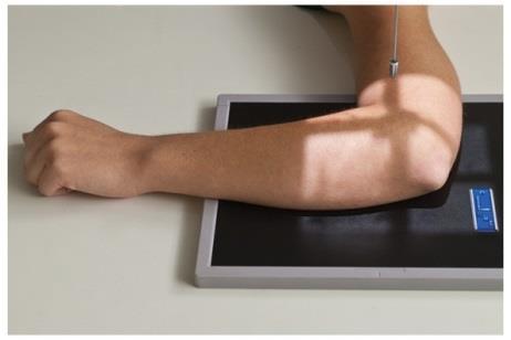
2 Patient Position: ELBOW AP Projection in same plane Part Position: Hand in ; patient Centered to Humeral epicondyles

3 Central Ray: Structures Shown:

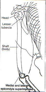
4 AP Elbow Criteria for Evaluation The humeral epicondyles are seen. The is superimposed over the lateral aspect of the The coronoid process is
5 AP Elbow Criteria for Evaluation The capitulum-radius joint should. The radial tuberosity should be seen. The humerus and forearm should other.
6 AP elbow Error; rotation of elbow. of head of radius is superimposed on and the humerus and forearm are. To correct: Rotate the elbow until the humeral epicondyles are to the IR. Align Good image

7 AP elbow Error: rotation of elbow. Correction: overlap of head of radius on ulna. To correct: Rotate the elbow until the humeral epicondyles are with the IR Good image

8 AP elbow Error: Elbow not, and humerus & forearm. To correct: elbow if patient is able. If patient is unable of the AP. Humerus and forearm should. Good image
 NOTE: Used when")

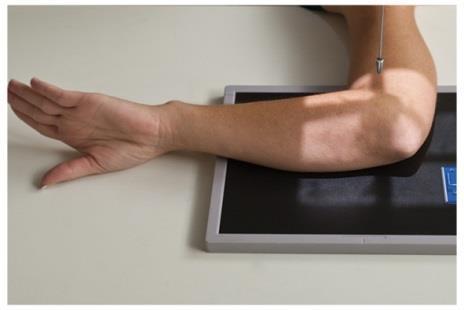
9 ELBOW Partial Flexion Views AP Projections (2) NOTE: Used when the patient is Distal Humerus: Entire humerus to IR Forearm is

10 Central Ray: Structures shown


11 Proximal Forearm: Entire to IR Hand is Central Ray Structures Shown

 to place elbow at º to IR Center")
12 ELBOW AP Oblique Projection Patient Position: Lateral Rotation Arm for AP projection Part Position: Rotate hand ( ) to place elbow at º to IR Center to

13 Central Ray: Structures Shown:

14 ELBOW Method Demonstrates the radial,, & (same as in projection)

")
15 ELBOW AP Oblique Projection Patient Position: Medial Rotation Arm projection Part Position: hand Rotate elbow ( ) - º Center to

16 Central Ray: Structures Shown:

17 ELBOW Lateral (lateromedial) Projection Patient in same plane Part Position: Elbow º; Hand Center to Humeral epicondyles

18 Central Ray: Structures Shown:

19 Elbow only flexed º if soft tissue structures are in question

20 Lateral Elbow Criteria for Evaluation The epicondyles should. Arm should be The elbow joint should be and the radial head should. fat pads seen.

21 Lateral elbow Error: Elbow. To correct degrees. Good image

22 Lateral elbow Error: humerus. Hand. To correct: degrees. Good image


23 Error: Humerus is Correct image Trochlea Capitulum


24 ELBOW Acute Flexion Method NOTE: Used when that radiographs are made with Distal Humerus: projection CR to humerus Shows


25 Proximal Forearm: Projection CR Shows

26 METHOD Trauma Axial Lateral NOTE: Used when patient cannot projections Patient Position:


27 Part Position and CR for radial head : Elbow flexed ; hand CR angled shoulder


28 Part Position and CR for coronoid process: Elbow flexed ; hand CR angled from shoulder

29 ELBOW Radial Head Views (4) Patient Position: in same plane Part Position: Elbow
30 Make separate exposures with the following changes in hand positions:

31 Central Ray: Structures Shown: Eversion Lateral Pronation Inversion
 Top of IR Humeral epicondyles to")
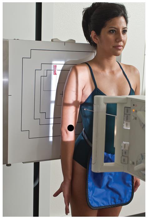
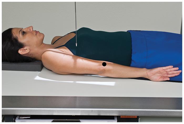
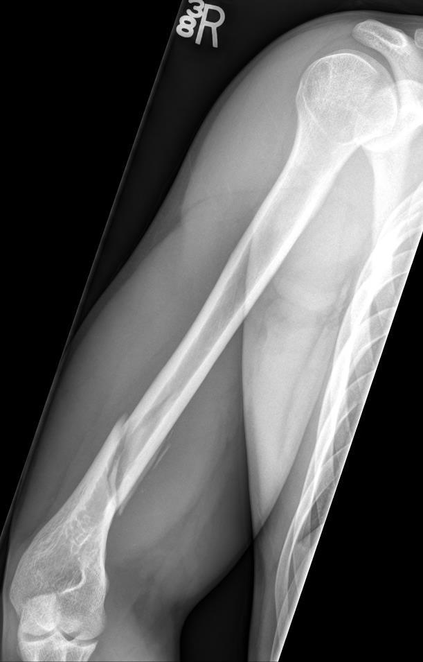
32 HUMERUS AP Projection Patient Position: Part Position: Arm ; position Hand in ( rotation) Top of IR Humeral epicondyles to IR

33 Central Ray: Structures Shown:


34 Patient Position: HUMERUS Lateral Projection Part Position: Arm rotated to position hand in rotation Rotate patient - affected side Humeral epicondyles Elbow if possible
35


36

37 Central Ray: Structures Shown:


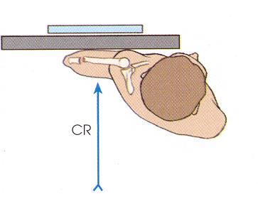
38 TRAUMA VIEWS of HUMERUS AP Proximal humerus: AP in


39 PA distal humerus - Patient is, but arm is. CR is, so that it is to the plane.


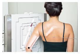
40 TRAUMA VIEWS of HUMERUS Lateral Proximal humerus: lateral (erect in rotation)


41 TRAUMA VIEWS of the HUMERUS Lateral Proximal humerus: Transthoracic lateral (in rotation; with beam)


42 TRAUMA VIEWS of HUMERUS Lateral Distal humerus projection with beam
43 Situation: An AP radiograph of the elbow demonstrates the radius directly superimposed over the ulna and the coronoid process in profile. Solution: This indicates that the projection of the elbow has been performed.
44 Situation: A radiograph of an AP oblique elbow with medial rotation reveals that theradial head is superimposed over part of the coronoid process. Solution: This indicates that the elbow is has excessive rotation.
45 Situation: A radiograph of an AP projection of the elbow reveals that there is complete separation of the proximal radius and ulna. What positioning error has been committed? Solution: The elbow is in excessive rotation (humeral epicondyles not parallel to IR).
46 Situation: A patient enters the ER in severe pain with a possible dislocation of the elbow. The patient has the elbow flexed more than 90. Solution: The projections to be performed to confirm the diagnosis are and limited.
47 Situation: A patient enters the ER with an elbow injury. The partially flexed AP and lateral positions reveal a possible fracture of the coronoid process. The patient s elbow is partially flexed and he refuses to extend it further. Solution: The method should be performed to confirm the coronoid process fracture. The elbow is flexed degrees and the CR is angled 45 the shoulder.
48 Situation: A young child comes to radiology with an elbow injury. The basic elbowprojections demonstrate a possible nondisplaced fracture of the radial head. The patient s elbow is partially flexed and he is unable to extend it. Solution: The method should be performed to confirm the radial head fracture. The elbow is flexed degrees and the CR is angled 45 the shoulder.
49 Situation: A radiograph of a transthoracic lateral projection reveals that it is difficult to visualize the proximal humerus due to the ribs and lung markings. The exposure was made on suspended respiration. Solution: Using a technique would improve the quality of the image.
50 Situation: A radiograph for an AP projection with external rotation of the proximal humerus reveals that the greater tubercle is profiled laterally. Is a repeat exposure necessary? Solution:
51 Situation: A patient enters the ER with a midshaft humeral fracture. The AP projection taken on the stretcher demonstrates another fracture near the surgical neck of the humerus. The patient is unable to stand or rotate the humerus due to the extent of the trauma. What other projection should be taken for this patient? Solution: A lateral projection of the humerus with a horizontal beam should be taken on this patient.
RADIOGRAPHY OF THE WRIST
 RADIOGRAPHY OF THE WRIST Patient Position: WRIST PA Projection, elbow in same plane Part Position: Hand ; fingers centered to IR Central Ray: Structures Shown: NOTE: Optional AP projection best demonstrates
RADIOGRAPHY OF THE HAND, FINGERS & THUMB
 RADIOGRAPHY OF THE HAND, FINGERS & THUMB FINGERS (2nd 5th) - PA Projection Patient Position: Seated; hand ; elbow on IR table top Part Position: Fingers centered to IR unless protocol is Central Ray: Perpendicular
Hands PA; Obl. Lat.; Norgaard s Thumb AP; Lat. PA. PA; Lat.: Obls.; Elongated PA with ulnar deviation
 Projections Region Basic projections Additional / Modified projections Upper Limbs Hands PA; Obl. Lat.; Norgaard s Thumb ; Lat. PA Fingers PA; Lat. Wrist PA; Lat. Obls. Scaphoid Lunate Trapezium Triquetral
RADIOGRAPHY OF THE ANKLE and LOWER LEG
 RADIOGRAPHY OF THE ANKLE and LOWER LEG Patient Position: ANKLE AP Projection Part Position: True Slight to place foot s long axis Center to Central Ray: to IR Midway Note: Ankle joint is to tips of malleoli
Radiographic Positioning Summary (Basic Projections RAD 222)
") Lower Extremity Radiographic Positioning Summary (Basic Projections RAD 222) AP Pelvis AP Hip (Unilateral) (L or R) AP Femur Mid and distal AP Knee Lateral Knee Pt lies supine on table Align MSP to Center
Lower Extremity Radiographic Positioning Summary (Basic Projections RAD 222) AP Pelvis AP Hip (Unilateral) (L or R) AP Femur Mid and distal AP Knee Lateral Knee Pt lies supine on table Align MSP to Center
Upper Limb Imaging Requirements
 Imaging Requirements Upper Limb Imaging Requirements Instructions for Measurement Radiography and CT Scans Please read before commencing radiography Stanmore Implants 210 Centennial Avenue Centennial Park
Osteology of the Elbow and Forearm Complex. The ability to perform many activities of daily living (ADL) depends upon the elbow.
 depends upon the elbow.") Osteology of the Elbow and Forearm Complex The ability to perform many activities of daily living (ADL) depends upon the elbow. Activities of Daily Living (ADL) Can you think of anything that you do to
Osteology of the Elbow and Forearm Complex The ability to perform many activities of daily living (ADL) depends upon the elbow. Activities of Daily Living (ADL) Can you think of anything that you do to
The Elbow Scanning Protocol
 The Elbow Scanning Protocol Diagnostic Imaging of the Elbow: Introduction The elbow maybe considered as consisting of four quadrants, anterior, medial, lateral and posterior. Ultrasound would normally
LESSON ASSIGNMENT. Positioning for Exams of the Upper Extremities. After completing this lesson, you should be able to:
 LESSON ASSIGNMENT LESSON 5 Positioning for Exams of the Upper Extremities. LESSON ASSIGNMENT Paragraphs 5-1 through 5-25. LESSON OBJECTIVES After completing this lesson, you should be able to: 5-1. Identify
Pediatric Elbow Radiology. Seema Awatramani, MD Friday, April 5, 2018 ACOEP Spring Seminar
 Pediatric Elbow Radiology Seema Awatramani, MD Friday, April 5, 2018 ACOEP Spring Seminar Disclosure I have no relevant financial relationships with the manufacturer(s) of any commercial product(s) and/or
The Biomechanics of the Human Upper Extremity-The Elbow Joint C. Mirzanli Istanbul Gelisim University
 The Biomechanics of the Human Upper Extremity-The Elbow Joint C. Mirzanli Istanbul Gelisim University Structure of The Elbow Joint A simple hinge joint, actually categorized as a trochoginglymus joint
Radiographic Positioning for Dogs
 Radiographic Positioning for Dogs Elbow Radiographs: Lateral View A routine elbow exam consists of a lateral, flexed lateral and craniocaudal view. When performing elbow radiographs, a quality control
P V S MEMORIAL HOSPITAL LTD.
 SHOULDER XRAYS Instability Series o True AP (Grashey s) o Axillary o Stryker Notch view o True AP in Internal rotation o Scapular Y view o West Point view for Bony Bankart ( looks like modif axillary view)
Radiology Positioning Practical Test #2 Table (By Jung Park):
:") Radiology Positioning Practical Test #2 Table (By Jung Park): (Lower Extremity): patient is fully gowned / no artifacts / properly shielded (exposure for femur and below : hold still, don t move ) (exposure
Radiology Positioning Practical Test #2 Table (By Jung Park): (Lower Extremity): patient is fully gowned / no artifacts / properly shielded (exposure for femur and below : hold still, don t move ) (exposure
ORTHOSCAN MOBILE DI POSITIONING GUIDE
 ORTHOSCAN MOBILE DI POSITIONING GUIDE Table of Contents SHOULDER A/P of Shoulder... 4 Tangential (Y-View) of Shoulder... 5 Lateral of Proximal Humerus... 6 ELBOW A/P of Elbow... 7 Extended Elbow... 8 Lateral
The Elbow and the cubital fossa. Prof Oluwadiya Kehinde
 The Elbow and the cubital fossa Prof Oluwadiya Kehinde www.oluwadiya.com Elbow and Forearm Anatomy The elbow joint is formed by the humerus, radius, and the ulna Bony anatomy of the elbow Distal Humerus
RADIOGRAPHY OF THE KNEE, PATELLA, and FEMUR
 RADIOGRAPHY OF THE KNEE, PATELLA, and FEMUR KNEE AP Projection Patient Position: Part Position: Leg in Center Femoral condyles Central Ray: - Asthenic patient - if ASIS to tabletop is < 19 cm Sthenic patient
Multiple Choice Identify the letter of the choice that best completes the statement or answers the question.
 RA202 positioning class three- EXM Multiple Choice Identify the letter of the choice that best completes the statement or answers the question. 1. Which of the following hand projections would be used
Osteology of the Elbow and Forearm Complex
 Osteology of the Elbow and Forearm Complex The ability to perform m any activities of daily living (ADL) d epends upon the elbow. Activities of Daily Living (ADL) Can you think of anything that you do
Chapter 8. The Pectoral Girdle & Upper Limb
 Chapter 8 The Pectoral Girdle & Upper Limb Pectoral Girdle pectoral girdle (shoulder girdle) supports the arm consists of two on each side of the body // clavicle (collarbone) and scapula (shoulder blade)
The Elbow 3/5/2015. The Elbow Scanning Sequence. * Anterior Joint (The anterior Pyramid ) * Lateral Epicondyle * Medial Epicondyle * Posterior Joint
 * Lateral Epicondyle * Medial Epicondyle * Posterior Joint") Scanning Sequence * Anterior Joint (The anterior Pyramid ) * Lateral Epicondyle * Medial Epicondyle * Posterior Joint Anterior Elbow Pyramid Courtesy of Jay Smith, MD. Vice chair PMR Mayo Clinic Rochester,
Scanning Sequence * Anterior Joint (The anterior Pyramid ) * Lateral Epicondyle * Medial Epicondyle * Posterior Joint Anterior Elbow Pyramid Courtesy of Jay Smith, MD. Vice chair PMR Mayo Clinic Rochester,
Proteus XR/f Patient positioning guide
 Proteus XR/f Patient positioning guide PROTEUS XR/F Now a single digital x-ray room accommodates nearly all your radiographic studies. With extended tube coverage and wireless detectors, Proteus XR/f gives
THE ELBOW. The elbow is a commonly injured joint in both children and adults.
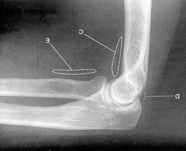
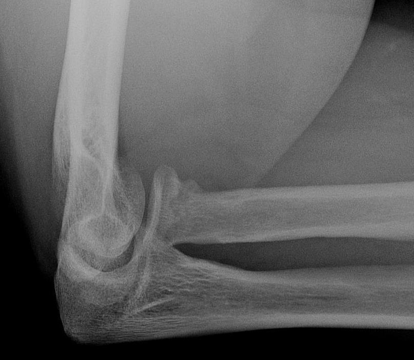
 ABC of Emergency Radiology FIG i-lateral radiograph of elbow and line THE ELBOW D A Nicholson, P A Driscoll The elbow is a commonly injured joint in both children and adults. Interpretation of elbow radiographs
Practical 2 Worksheet
 Practical 2 Worksheet Upper Extremity BONES 1. Which end of the clavicle is on the lateral side (acromial or sternal)? 2. Describe the difference in the appearance of the acromial and sternal ends of the
The Elbow and Radioulnar Joints Kinesiology. Dr Cüneyt Mirzanli Istanbul Gelisim University
 The Elbow and Radioulnar Joints Kinesiology Dr Cüneyt Mirzanli Istanbul Gelisim University 1 The Elbow & Radioulnar Joints Most upper extremity movements involve the elbow & radioulnar joints. Usually
Main Menu. Elbow and Radioulnar Joints click here. The Power is in Your Hands
 1 The Elbow and Radioulnar Joints click here Main Menu K.4 http://www.handsonlineeducation.com/classes//k4entry.htm[3/23/18, 1:29:53 PM] Bones Ulna is much larger proximally than radius Radius is much
Upper limb injuries in children. Key points, # & dislocations 7/23/2009 (MIMIC)
") Upper limb injuries in children (MIMIC) Key points, # & dislocations Before the age of 16 around 50% of boys & 25% of girls will sustain a # Dislocations are very uncommon Children s bones are less brittle
Upper limb injuries in children (MIMIC) Key points, # & dislocations Before the age of 16 around 50% of boys & 25% of girls will sustain a # Dislocations are very uncommon Children s bones are less brittle
Fractures and dislocations around elbow in adult
 Lec: 3 Fractures and dislocations around elbow in adult These include fractures of distal humerus, fracture of the capitulum, fracture of the radial head, fracture of the olecranon & dislocation of the
Figure 1: Bones of the upper limb
 BONES OF THE APPENDICULAR SKELETON The appendicular skeleton is composed of the 126 bones of the appendages and the pectoral and pelvic girdles, which attach the limbs to the axial skeleton. Although the
1/19/2018. Winter injuries to the shoulder and elbow. Highgate Private Hospital (Whittington Health NHS Trust)
") Winter injuries to the shoulder and elbow Omar Haddo Consultant Orthopaedic Surgeon, Shoulder, Elbow, Hand & Wrist Specialist MBBS, BmedSci, FRCS(Orth) Highgate Private Hospital (Whittington Health NHS
Winter injuries to the shoulder and elbow Omar Haddo Consultant Orthopaedic Surgeon, Shoulder, Elbow, Hand & Wrist Specialist MBBS, BmedSci, FRCS(Orth) Highgate Private Hospital (Whittington Health NHS
THE SKELETAL SYSTEM. Focus on the Pectoral Girdle
 THE SKELETAL SYSTEM Focus on the Pectoral Girdle Appendicular Skeleton 126 bones Includes bones of the limbs (arms and legs) Pectoral girdle (shoulder) Pelvic girdle (hip) Pectoral Girdle (the shoulder)
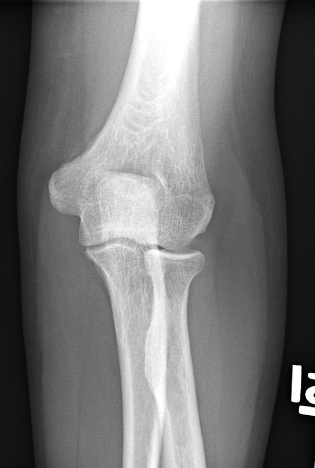
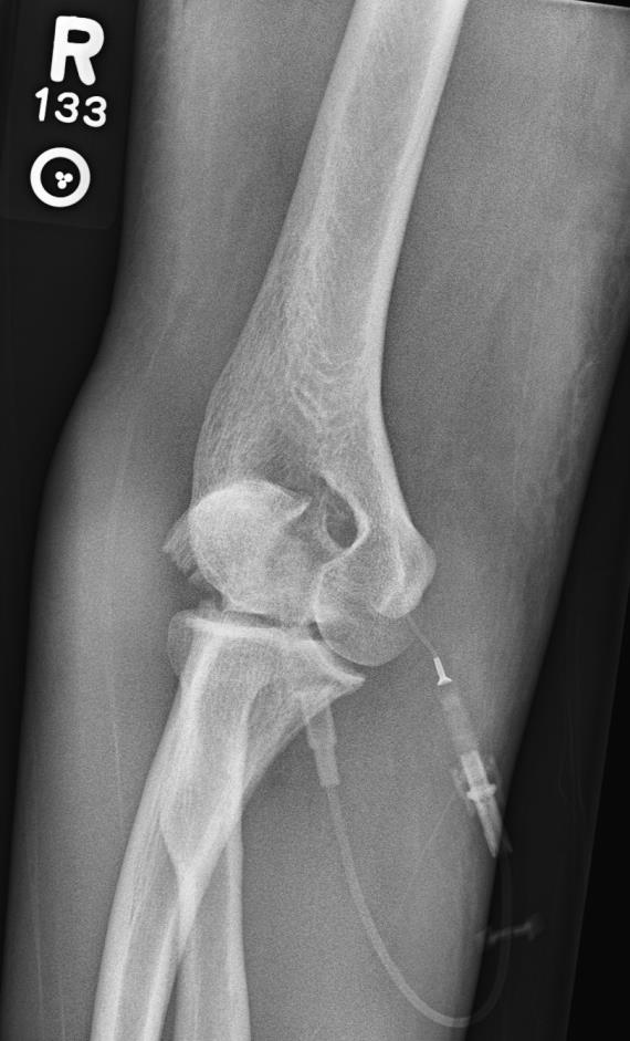
RADIAL HEAD FRACTURES. It is far more common in adults than in children, (who more commonly fracture their neck of radius).
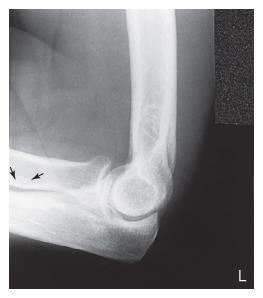
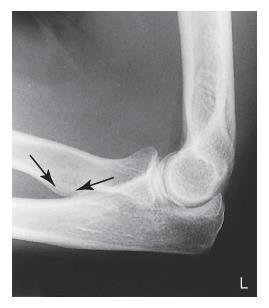
.") RADIAL HEAD FRACTURES Introduction Fractures of the head of the radius are relatively common. The injury can be subtle unless specifically looked for. It is far more common in adults than in children,
RADIAL HEAD FRACTURES Introduction Fractures of the head of the radius are relatively common. The injury can be subtle unless specifically looked for. It is far more common in adults than in children,
Elbow Anatomy, Growth and Physical Exam. Donna M. Pacicca, MD Section of Sports Medicine Division of Orthopaedic Surgery Children s Mercy Hospital
 Elbow Anatomy, Growth and Physical Exam Donna M. Pacicca, MD Section of Sports Medicine Division of Orthopaedic Surgery Children s Mercy Hospital Contributing Factors to Elbow Injury The elbow is affected
Country Health SA Medical Imaging
 Country Health SA Medical Imaging REMOTE OPERATORS POSITIONING GUIDE Contents Image Evaluation Page 4 Positioning Guides Section 1 - THORAX 1.1 Chest Page 5 1.2 Bedside Chest Page 7 1.3 Ribs Page 8 Section
WEEKEND 2 Elbow. Elbow Range of Motion Assessment
 Virginia Orthopedic Manual Physical Therapy Institute - 2016 Technique Manual WEEKEND 2 Elbow Elbow Range of Motion Assessment - Patient Positioning: Sitting or supine towards the edge of the bed - Indications:
Fractures of the shoulder girdle, elbow and fractures of the humerus. H. Sithebe 2012
 Fractures of the shoulder girdle, elbow and fractures of the humerus H. Sithebe 2012 Fractures of the Clavicle (mid-shaft). Fractures of the clavicle Fractures of the clavicle Treatment- conservative.
Connects arm to thorax 3 joints. Glenohumeral joint Acromioclavicular joint Sternoclavicular joint
 Connects arm to thorax 3 joints Glenohumeral joint Acromioclavicular joint Sternoclavicular joint Scapula Elevation Depression Protraction (abduction) Retraction (adduction) Downward Rotation Upward Rotation
region of the upper limb between the shoulder and the elbow Superiorly communicates with the axilla.
 1 region of the upper limb between the shoulder and the elbow Superiorly communicates with the axilla. Inferiorly, a number of important structures pass between arm & forearm through cubital fossa. 2 medial
Gross Anatomy Questions That Should be Answerable After October 27, 2017
 Gross Anatomy Questions That Should be Answerable After October 27, 2017 1. The inferior angle of the scapula of a woman who was recently in an automobile accident seems to protrude making a ridge beneath
4/28/2010. Fractures. Normal Bone and Normal Ossification Bone Terms. Epiphysis Epiphyseal Plate (physis) Metaphysis
 Metaphysis") Fractures Normal Bone and Normal Ossification Bone Terms Epiphysis Epiphyseal Plate (physis) Metaphysis Diaphysis 1 Fracture Classifications A. Longitudinal B. Transverse C. Oblique D. Spiral E. Incomplete
Fractures Normal Bone and Normal Ossification Bone Terms Epiphysis Epiphyseal Plate (physis) Metaphysis Diaphysis 1 Fracture Classifications A. Longitudinal B. Transverse C. Oblique D. Spiral E. Incomplete
Trauma Films for Upper Body. LCDR. Naruebade Rungrattanawilai RTN M.D., LL.B. FRCOST, DMOC
 Trauma Films for Upper Body LCDR. Naruebade Rungrattanawilai RTN M.D., LL.B. FRCOST, DMOC Objective A 42 year-old housekeeper with history of motorcycle accident. There was no external wound but she have
Elbow & Forearm H O W V I T A L I S T H E E L B O W T O O U R D A I L Y L I V E S?
 Elbow & Forearm H O W V I T A L I S T H E E L B O W T O O U R D A I L Y L I V E S? Clarification of Terms The elbow includes: 3 bones (humerus, radius, and ulna) 2 joints (humeroulnar and humeroradial)
Elbow Elbow Anatomy. Flexion extension. Pronation Supination. Anatomy. Anatomy. Romina Astifidis, MS., PT., CHT
 Elbow Elbow Anatomy Romina Astifidis, MS., PT., CHT Curtis National Hand Center Baltimore, MD October 6-8, 2017 Link between the arm and forearm to position the hand in space Not just a hinge Elbow = 70%
Body Planes & Positions
 Learning Objectives Objective 1: Identify and utilize anatomical positions, planes, and directional terms. Demonstrate what anatomical position is and how it is used to reference the body. Distinguish
Functional Anatomy of the Elbow
 Functional Anatomy of the Elbow Orthopedic Institute Daryl C. Osbahr, M.D. Chief of Sports Medicine, Orlando Health Chief Medical Officer, Orlando City Soccer Club Orthopedic Consultant, Washington Nationals
David G. Simpson, Ph.D.
 David G. Simpson, Ph.D. ARM & CUBITAL FOSSA Revised 7/08 Text References Moores 3 rd ed., p402 408, 436 439, 439 443, 478, 481 LEARNING OBJECTIVES: 1. Describe the humerus, indicating the sites of muscle
Traumatic injuries of the paediatric elbow: A pictorial review
 Traumatic injuries of the paediatric elbow: A pictorial review Poster No.: C-750 Congress: ECR 2009 Type: Educational Exhibit Topic: Pediatric Authors: A. M. Veitch, J. Harington, K. Franklin ; Plymouth/UK,
Proximal radioulnar translocation associated with elbow dislocation and radial neck fracture in child: a case report and review of literature
 DOI 10.1007/s00402-013-1820-8 TRAUMA SURGERY Proximal radioulnar translocation associated with elbow dislocation and radial neck fracture in child: a case report and review of literature Hong Kee Yoon
Evidence- Based Examination of the Shoulder Presented by Eric Hegedus, PT, DPT, MHSC, OCS, CSCS Practice Sessions/Skill Check- offs
 Evidence- Based Examination of the Shoulder Practice Session & Skills Check- offs Evidence- Based Examination of the Shoulder Presented by Eric Hegedus, PT, DPT, MHSC, OCS, CSCS Practice Sessions/Skill
Rad Tech 4643 MRI Torso and Extremities
 Rad Tech 4643 MRI Torso and Extremities Prostate Cancer Leiomyoma Retroverted Anteverted Ovarian Cyst Gone Wrong Fibroid (Leiomyoma) IUD Ovary Hysterectomy? What are we to see when imaging a female pelvis
Upper Extremity Fractures
 Upper Extremity Fractures Ranie Whatley, RN,FNP-C David W. Gray, MD Skeletal Trauma 10 to 15 % of all Childhood Injuries Physeal (Growth Plate) Injuries are ~ 15% of all Skeletal Injuries Orthopaedic Assessment
Lab Activity 11: Group II
 Lab Activity 11: Group II Muscles Martini Chapter 11 Portland Community College BI 231 Origin and Insertion Origin: The place where the fixed end attaches to a bone, cartilage, or connective tissue. Insertion:
Pediatric Fractures. Objectives. Epiphyseal Complex. Anatomy and Physiology. Ligaments. Bony matrix
 1 Pediatric Fractures Nicholas White, MD Assistant Professor of Pediatrics Eastern Virginia Medical School Attending, Pediatric Emergency Department Children s Hospital of The King s Daughters Objectives
Anatomy and Physiology II. Review Shoulder Girdle New Material Upper Extremities - Bones
 Anatomy and Physiology II Review Shoulder Girdle New Material Upper Extremities - Bones Anatomy and Physiology II Shoulder Girdle Review Questions From Last Lecture Can you identify the following muscles?
11/5/14. I will try to make this painless. Great, a Fracture, Now What? Objectives. Basics for Fracture Workup. Basics for Fracture Workup
 Great, a Fracture, Now What? I will try to make this painless Mary Greve MS, PA-C Department of Orthopedic Surgery Trauma Team University of Iowa Hospitals and Clinics Mary-Greve@uiowa.edu Pager 2121 Objectives
Surgical Care at the District Hospital. EMERGENCY & ESSENTIAL SURGICAL CARE
 Surgical Care at the District Hospital 1 18 Orthopedic Trauma Key Points 2 18.1 Upper Extremity Injuries Clavicle Fractures Diagnose fractures from the history and by physical examination Treat with a
EMERGENCY PITFALLS IN ORTHOPAEDIC TRAUMA. Thierry E. Benaroch, MD, FRCS MCH Trauma Rounds February 9, 2009
 EMERGENCY PITFALLS IN ORTHOPAEDIC TRAUMA Thierry E. Benaroch, MD, FRCS MCH Trauma Rounds February 9, 2009 MORAL OF THE STORY Fracture distal radius and intact ulna W/O radius fracture will most likely
Medical Terminology. Anatomical Position, Directional Terms and Movements
 Medical Terminology Anatomical Position, Directional Terms and Movements What we will cover... Content Objectives Students will be able to gain a better understanding and application of medical terminology
Copyright 2003 Pearson Education, Inc. publishing as Benjamin Cummings. Dr. Nabil khouri
 Dr. Nabil khouri Appendicular Skeleton The appendicular skeleton is made up of the bones of the upper and lower limbs and their girdles Two girdles: Pectoral girdles attach the upper limbs to the body
CHAPTER 6: THE UPPER EXTREMITY: THE ELBOW, FOREARM, WRIST, AND HAND
 CHAPTER 6: THE UPPER EXTREMITY: THE ELBOW, FOREARM, WRIST, AND HAND KINESIOLOGY Scientific Basis of Human Motion, 12 th edition Hamilton, Weimar & Luttgens Presentation Created by TK Koesterer, Ph.D.,
Pectoral girdle, SUPERIEUR ARM AND HAND. Danil Hammoudi.MD
 Pectoral girdle, SUPERIEUR ARM AND HAND Danil Hammoudi.MD The pectoral girdle is the set of bones which connect the upper limb to the axial skeleton on each side. It consists of the clavicle scapula in
ARM Brachium Musculature
 ARM Brachium Musculature Coracobrachialis coracoid process of the scapula medial shaft of the humerus at about its middle 1. flexes the humerus 2. assists to adduct the humerus Blood: muscular branches
#12. Joint نبيل خوري
 #12 30 Anatomy Joint هيام الر جال 9/10/2015 نبيل خوري Salam Awn Some notes before starting : ** Not all slides are included, so I recommend having a look at the slides beside this sheet ** If you find
PEDIATRIC UPPER EXTREMITY FRACTURE MANAGEMENT JULIA RAWLINGS, MD SPORTS MEDICINE SYMPOSIUM: THE PEDIATRIC ATHLETE 2 MARCH 2018
 PEDIATRIC UPPER EXTREMITY FRACTURE MANAGEMENT JULIA RAWLINGS, MD SPORTS MEDICINE SYMPOSIUM: THE PEDIATRIC ATHLETE 2 MARCH 2018 DISCLOSURE I have nothing to disclose. 2 OBJECTIVES Discuss the diagnosis,
Elbow Joint Anatomy ELBOW ANATOMY, BIOMECHANICS. Bone Anatomy. Bone Anatomy. Property of VOMPTI, LLC
 ELBOW ANATOMY, BIOMECHANICS AND PATHOLOGY Kristin Kelley, DPT, OCS, FAAOMPT Elbow Joint Anatomy Joint articulations Humeroulnar Radiohumeral Radioulnar (proximal and distal) Orthopaedic Manual Physical
SUPERIEUR ARM AND HAND
 Pectoral girdle, SUPERIEUR ARM AND HAND Danil Hammoudi.MD The pectoral girdle is the set of bones which connect the upper limb to the axial skeleton on each side. It consists of the clavicle scapula in
The Language of Anatomy. (Anatomical Terminology)
") The Language of Anatomy (Anatomical Terminology) Terms of Position The anatomical position is a fixed position of the body (cadaver) taken as if the body is standing (erect) looking forward with the upper
The Language of Anatomy (Anatomical Terminology) Terms of Position The anatomical position is a fixed position of the body (cadaver) taken as if the body is standing (erect) looking forward with the upper
MUSCLES. Anconeus Muscle
 LAB 7 UPPER LIMBS MUSCLES Anconeus Muscle anconeus origin: distal end of dorsal surface of humerus insertion: lateral surface of ulna from distal margin of the semilunar notch to proximal end of the olecranon
An Introduction to the Appendicular Skeleton
 An Introduction to the Appendicular Skeleton The Appendicular Skeleton is composed of the 126 bones of the appendages (limbs) and the pectoral and pelvic girdles, which attach to the axial skeleton. Each
Joints. Vi Michelle Austin
 Joints Vi Michelle Austin Joints Overview A joint, otherwise known as an articulation, is a point at which points connect. They are constructed to allow movement (except for skull bones) and provide mechanical
Chapter 6 The Elbow and Radioulnar Joints
 The Elbow & Radioulnar Chapter 6 The Elbow and Radioulnar Manual of Structural Kinesiology R.T. Floyd, EdD, ATC, CSCS Most upper extremity movements involve the elbow & radioulnar joints Usually grouped
Joints Dr. Ali Ebneshahidi
 Joints Dr. Ali Ebneshahidi Function of Joints 1. Serve as functional junctions between bones. 2. Bind bones, strokes, and other related tissues together. 3. Allow bone growth to occur. 4. Permit certain
Acute Elbow Trauma in Children: Spectrum of Injury Revealed by MR Imaging Not Apparent on Radiographs
 James F. Griffith 1 Derek J. Roebuck 1,2 Jack C. Y. Cheng 3 Yu Leung Chan 1 Timothy H. Rainer 4 Bobby K. W. Ng 3 Constantine Metreweli 1 Received December 14, 1999; accepted after revision June 8, 2000.
ORIGINAL PAPER. Department of Hand Surgery, Nagoya University School of Medicine ABSTRACT
 Nagoya J. Med. Sci. 74. 167 ~ 171 2012 ORIGINAL PAPER TILT OF THE RADIUS FROM FOREARM ROTATIONAL AXIS RELIABLY PREDICTS ROTATIONAL IMPROVEMENT AFTER CORRECTIVE OSTEOTOMY FOR MALUNITED FOREARM FRACTURES
Rehabilitation after Total Elbow Arthroplasty
 Rehabilitation after Total Elbow Arthroplasty Total Elbow Atrthroplasty Total elbow arthroplasty (TEA) Replacement of the ulnohumeral articulation with a prosthetic device. Goal of TEA is to provide pain
Lecture 9: Forearm bones and muscles
 Lecture 9: Forearm bones and muscles Remember, the region between the shoulder and the elbow = brachium/arm, between elbow and wrist = antebrachium/forearm. Forearm bones : Humerus (distal ends) Radius
Human Anatomy Biology 351
 1 Human Anatomy Biology 351 Upper Limb Exam Please place your name on the back of the last page of this exam. You must answer all questions on this exam. Because statistics demonstrate that, on average,
Medical Terminology. Unit 2
 Medical Terminology Unit 2 Students will apply medical terminology. Objective 1: Identify and utilize anatomical positions, planes, and directional terms. Demonstrate what anatomical position is and how
Muscles in the Shoulder, Chest, Arm, Stomach, and Back
 Muscles in the Shoulder, Chest, Arm, Stomach, and Back Shoulder Muscles Deltoid Supraspinatus Infraspinatus Teres Major Teres Minor Subscapularis Deltoid (Delts) Function: Raises the upper arm Origin:
Bipolar Radial Head System
 Bipolar Radial Head System Katalyst Surgical Technique DESCRIPTION The Katalyst Telescoping Bipolar Radial Head implant restores the support and bearing surface of the radial head in the face of fracture,
The Arm and Cubital Fossa
 The Arm and Cubital Fossa Dr. Andrew Gallagher School of Anatomical Sciences University of the Witwatersrand Introduction The ARM (BRACHIUM) is the most proximal segment of the upper limb musculoskeletal
Anatomy and Physiology 2016
 Anatomy and Physiology 2016 O = Temporal line I = coronoid process (Mandible) A = elevates mandible (chewing) O = galea aponeurotica (layer of dense fibrous tissue which covers the upper part of the cranium)
Radiographic Procedures 1
 Western Technical College 10526149 Radiographic Procedures 1 Course Outcome Summary Course Information Textbooks Description Career Cluster Instructional Level Total Credits 5 Prepares radiography students
Evidence-Based Examination of the Elbow, Wrist, and Hand
 Evidence-Based Examination of the Elbow, Wrist, and Hand Presented by Chad Cook, PT, PhD, MBA, FAAOMPT Practice Sessions/Skill Check-offs Chapter Five: Movement Examination of the Elbow, Wrist, and Hand
Ligaments of Elbow hinge: sagittal plane so need lateral and medial ligaments
 Ligaments of Elbow hinge: sagittal plane so need lateral and medial ligaments Ulnar Collateral ligament on medial side; arising from medial epicondyle and stops excess valgus movement (lateral movement)
Chapter 8 The Skeletal System: The Appendicular Skeleton. Copyright 2009 John Wiley & Sons, Inc.
 Chapter 8 The Skeletal System: The Appendicular Skeleton Appendicular Skeleton It includes bones of the upper and lower limbs Girdles attach the limbs to the axial skeleton The pectoral girdle consists
GENERAL SCOPE AND USES OF PHYSICAL/BIOLOGICAL ANTHROPOLOGY. Paper No. & Title: B.A./B.Sc. (Honours) 2 dn semester. (Practical)
 2 dn semester. (Practical)") GENERAL SCOPE AND USES OF PHYSICAL/BIOLOGICAL ANTHROPOLOGY Course name: Physical Anthropology Paper No. & Title: B.A./B.Sc. (Honours) 2 dn semester (Practical) Topic No. & Title: 5/12 (Part-I) Drawing
GENERAL SCOPE AND USES OF PHYSICAL/BIOLOGICAL ANTHROPOLOGY Course name: Physical Anthropology Paper No. & Title: B.A./B.Sc. (Honours) 2 dn semester (Practical) Topic No. & Title: 5/12 (Part-I) Drawing
Bony Thorax. Anatomy and Procedures of the Bony Thorax Edited by M. Rhodes
 Bony Thorax Anatomy and Procedures of the Bony Thorax 10-526-191 Edited by M. Rhodes Anatomy Review Bony Thorax Formed by Sternum 12 pairs of ribs 12 thoracic vertebrae Conical in shape Narrow at top Posterior
Integra. Modular Radial Head System SURGICAL TECHNIQUE
 Integra Modular Radial Head System SURGICAL TECHNIQUE Table of Contents System Overview...2 Indications and Contraindications... 3 Modular Radial Head Implant Technique...4 Component Dimensions...8 Implant
1 Humeral fractures 1.13 l Distal humeral fractures Treatment with a splint
 1 Executive Editor: Chris Colton Authors: Mariusz Bonczar, Daniel Rikli, David Ring 1 Humeral fractures 1.13 l Distal humeral fractures Treatment with a splint Indication All 13-A type fractures, excluding
REHABILITATION FOR SHOULDER FRACTURES & SURGERIES. Clavicle fractures Proximal head of humerus fractures
 REHABILITATION FOR SHOULDER FRACTURES & SURGERIES Clavicle fractures Proximal head of humerus fractures By Dr. Mohamed Behiry Lecturer Department of physical therapy for Orthopaedic and its surgery. Delta
Radial Head Fractures Save or Replace?
 Radial Head Fractures Save or Replace? Current Solutions in Orthopedic Trauma Sepember 19, 2015 Jorge L. Orbay MD. Disclosure Skeletal Dynamics Elbow Joint Ulno-Humeral and Radio-Capitellar The key joint
Integra. Katalyst Bipolar Radial Head System SURGICAL TECHNIQUE
 Integra Katalyst Bipolar Radial Head System SURGICAL TECHNIQUE Surgical Technique As the manufacturer of this device, Integra does not practice medicine and does not recommend this or any other surgical
Goniometry. Wrist Flexion: Pt seated with forearm resting on table (use olecranon process & midline of ulna as reference for stationary arm)
") Goniometry Wrist Flexion: Pt seated with forearm resting on table (use olecranon process & midline of ulna as reference for stationary arm) Wrist Extension: Pt seated with forearm resting on table (Goniometer
Goniometry Wrist Flexion: Pt seated with forearm resting on table (use olecranon process & midline of ulna as reference for stationary arm) Wrist Extension: Pt seated with forearm resting on table (Goniometer
Anatomy of the Shoulder Girdle. Prof Oluwadiya Kehinde FMCS (Orthop)
") Anatomy of the Shoulder Girdle Prof Oluwadiya Kehinde FMCS (Orthop) www.oluwadiya.com Bony Anatomy Shoulder Complex: Sternum(manubrium) Clavicle Scapula Proximal humerus Manubrium Sterni Upper part of
Anatomy of the Shoulder Girdle Prof Oluwadiya Kehinde FMCS (Orthop) www.oluwadiya.com Bony Anatomy Shoulder Complex: Sternum(manubrium) Clavicle Scapula Proximal humerus Manubrium Sterni Upper part of
Joints of the upper limb II
 Joints of the upper limb II Prof. Abdulameer Al-Nuaimi E-mail: a.al-nuaimi@sheffield.ac.uk E. mail: abdulameerh@yahoo.com Elbow joint The elbow joint is connecting the upper arm to the forearm. It is classed
Biceps Brachii. Muscles of the Arm and Hand 4/4/2017 MR. S. KELLY
 Muscles of the Arm and Hand PSK 4U MR. S. KELLY NORTH GRENVILLE DHS Biceps Brachii Origin: scapula Insertion: radius, fascia of forearm (bicipital aponeurosis) Action: supination and elbow flexion Innervation:
Basic Radiographic Principles Part II
 Basic Radiographic Principles Part II Kristopher Avant, D.O. October 19 th, 2016 I have no disclosures relevant to the material presented in this discussion. Good Stuff!!! 1 Really? Really! Musculoskeletal
Traditional Thai Acupressure Points. The anterior aspect of the body THE ANATOMICAL ATLAS
 Traditional Thai Acupressure Points The anterior aspect of the body THE ANATOMICAL ATLAS lines of the SHOULDER BLADES AND POSTERIOR ARM Scapula Line This line runs through landmarks: 1. Above the midpoint