onotube TRIAX EXTERN AL FIXATION SYSTEM
|
|
|
- Dominick Dean
- 5 years ago
- Views:
Transcription

1 onotube EXTERN AL FIXATION SYSTEM
2 onotube
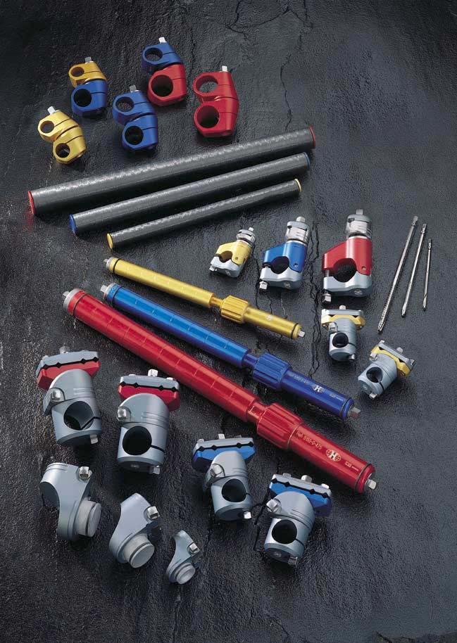
3 Overview The Monotube Triax external fixation system was developed in cooperation with Prof. J. Lazo-Zbikowski and Prof. J. Cañadell. Its design consists of a dynamic axial fixator designed to maintain rotational and angular stability while allowing the bone to share the load in the axial plane. The Monotube Triax has been designed to handle a wide variety of orthopaedic and trauma applications keeping in mind the need for controlled dynamisation. The built in dynamisation collar allows 0-1mm axial compression thus allowing the surgeon to initiate the natural callus formation that is critical to the healing process. Its absolute modularity and unrestricted reposition possibilities allow restoration of the correct anatomy in three planes. The system consists of three basic components. The dynamic tube, the pin clamp, and the single pin clamp. Speciality clamps and accessories are also available.
4 The Monotube Triax system can be dynamically loaded upon muscular contraction and weightbearing. This allows the system to be converted from a static to a dynamic mode by activating the built in dynamisation collar. This will stimulate and enhance callus formation and promote faster healing. 1 The Monotube Triax is minimally invasive using only percutaneous Apex Pins which are non conical and therefore avoid radial stresses at the bone to pin interface and reduce soft tissue disruption. The Monotube Triax simple design characteristics make its use extremely versatile and easy to apply thanks to universal instrumentation and few basic components. The Monotube Triax is simple to use, by sliding the desired clamps anywhere along the tube. Fine tuning the reduction is simple, and requires no additional components. The Monotube Triax system is available in three sizes allowing the surgeon to choose the appropriate fixator according to patient size and indication requirements. All the adjustments on the tube and clamp use 7mm square head screws in the red and blue and 5mm square head screws in the yellow and require only one instrument. The Monotube Triax is functional, allowing patients partial weightbearing from the beginning of the treatment.


5 onotube The Monotube Triax allows increased fracture visualisation through the design of the pin clamps, which permit X-ray visualisation in both antero-posterior and medio-lateral planes, unlike other competitive systems which are restricted to one visual plane. The Monotube Triax system provides the ability to reduce fractures with reduction handles. Post-operative correction and handling are simple. The Monotube Triax has a built in fracture compressiondistraction system. Monotube Triax The Monotube Triax has variable pin positioning allowing for multiple pin placement and good bone purchase. The Monotube Triax system is compatible with other Stryker Howmedica Fixators. The Monotube Triax system is colour coded for easy intra-operative management. Triax Clamps The Monotube Triax is made of lightweight anodised aluminium. Triax Single Pin Clamp Triax Carbon Monotube 3


, and")


6 onotube Controlled Dynamisation Bio-Spring adjustment screw Dynamisation collar The objective of external fixation is to bring stability to a long bone that has sustained a fracture. This process must be as physiological as possible and therefore, the fixation device used must be stable enough to prevent reduction loss and resist movement in lateral bending and torque, yet be dynamic enough to allow minute and controlled amounts of movement at the fracture site during weight bearing. Dynamisation is the term given to the transmission of forces across the fracture (without the frame causing distraction of the fragments), and with the aim of promoting healing. Healing of the bone fractures using external fixation takes place by indirect consolidation, and fractures treated by micromovement heal more quickly than those treated using a rigid external fixator (13 weeks as compared to 18). 1 The precise amount of weight bearing is dependant on the type of fracture, soft tissue damage, total weight of the patient, presence of multiple trauma, pain threshold and the patient's overall condition. Dynamisation should occur at the first sign of periosteal callus formation and the fixator should be set for 1mm of micromovement at the fracture site (see protocol on page 6). The Monotube Triax is designed to provide a high degree of stability, yet permit dynamic movement due to its built in dynamisation module. The Monotube Triax can easily be converted from static to dynamic mode by activating the dynamisation module. The biology of the fracture can be modified by adjusting the external fixator. Rigid external fixators produce primary bone healing, whereas elastic fixators produce a greater volume of periosteal callus. 2 4
7 Biocompression The Monotube Triax system is designed to permit two types of dynamisation, Biocompression and Adjustable Bio-Spring. The first is an axial telescopic function in which an outer tube slides over an inner tube, allowing a recommended 1mm micromovement at the fracture site. Biocompression allows the surgeon to dial in the amount of compression at the fracture site by simply opening the dynamisation collar from 0 1mm from the beginning. This micromotion is controlled by a specially designed needle-bearing technology that allows the tube to slide effortlessly even if not axially aligned. Other systems have a tendency to jam in this situation. This specialised technology guarantees appropriate dynamisation and therefore, callus formation leading to faster healing. Biocompression is recommended for stable fractures or in fractures where there is substantial cortical contact. Rigid Fixation Force After Callus Biocompression Force Force After Callus Gap 1mm Gap 1mm Immediate transmission of the forces through the fracture

8 Dynamic Monotube BIO-SPRING ADJUSTMENT SCREW "O"-RING DYNAMISATION COLLAR LENGTHENING SEGMENT DYNAMISATION SEGMENT "O"-RING COMPRESSION/DISTRACTION ADJUSTMENT SCREW

9 Adjustable Bio-Spring onotube The second type of dynamisation is referred to as ADJUSTABLE BIO-SPRING, and is the combination of an axial telescopic function and a compressive spring. The objective of this shock absorbing mechanism is to counteract muscle tension and prevent the collapse of the fragments in an unstable fracture or a leg lengthening procedure, yet at the same time to guarantee accurate and effective dynamisation. The Adjustable Bio-Spring allows the surgeon to dial in the amount of compression at the fracture site by simply opening the dynamisation collar from 0 1mm, and set the compressive spring to full tension, based on the fracture type and the patients weight. Adjustable Bio-Spring is recommended in unstable fractures or fractures in which there is no cortical contact and leg lengthening procedures. Adjustable Bio-spring Force applied to bone 0 Kg 15 Kg 30 Kg 75 Kg Needle Bearings Adjustable Bio-spring Gap 1mm 30 Kg Spring Tension Gap 2mm Gap 0.5mm Gap 1mm Gap 0mm 30 Kg 30 Kg 30 Kg 30Kg 45Kg Gap 0mm Dynamisation Collar 5
. Use the dynamisation wrench if necessary.")

10 onotube Dynamisation Protocol Stable Fracture patterns Biocompression is recommended for stable fracture patterns. The frame may be dynamised anytime after application. The following steps should be followed: 1. Open the dynamisation collar to 1mm (one complete revolution of the dynamisation collar equates to 1mm). Use the dynamisation wrench if necessary. Note: The one millimetre setting on the collar may or may not be visible. Unstable Fracture patterns Adjustable Bio-Spring is recommended for unstable fracture patterns and leg lengthening. The frame may be dynamised once callus is evident on the X-ray, approximately 21 to 28 days postoperatively. To dynamise the frame, the following sequence should be followed: 1. Advance Adjustable Bio-Spring tension screw clockwise until fully compressed, about 7 1 /2 full turns. The screw will recede into the tube as it compresses the spring. 2. Open the dynamisation collar to one millimetre. Use the dynamisation wrench if necessary. 3. Reduce Bio-Spring tension one full turn anti-clockwise each week. Be sure to check that biocompression collar is still set at 1mm after each spring adjustment. The force required to dynamise the frame will be progressively reduced (see technique guide). Note: X-ray visualisation is required to determine callus formation. Monotube Lengths and Pin Options Tube Colour (Dynamic) Dynamic Monotube Pin Size dia Spring Tension Yellow dia 15mm x 180mm Fully Closed dia 15mm x 250mm Fully Opened 3mm & 4mm 20kg Blue dia 20mm x 250mm Fully Closed dia 20mm x 350mm Fully Opened 4mm & 5mm 30kg Red dia 25mm x 320mm Fully Closed dia 25mm x 470mm Fully Opened 5mm & 6mm 45kg 6


 Tibia Femur Humerus Forearm Ulna Radius Colles Simple/ stable Oblique/ comminuted/ unstable")
11 Distraction/Compression onotube The Monotube Triax has a built in compression distraction mechanism, which can be used in lengthening procedures and to adjust fracture gaps. Compression and Distraction are achieved by turning the Lengthening Screw turning the 7mm squared headed screw anti-clockwise will lengthen the tube and distract the fracture. Turning clockwise will shorten the tube and compress the fracture. The end of the tube is marked with a visual reference each 1 /4 turn 4 dots 3 dots 2 dots 1 dot The Monotube Triax system is available in three sizes which are colour coded Yellow, Blue, Red. To determine the proper frame components the size of tube selected is determined by: Location / type of fracture Patient s weight Length of tube required Dynamisation requirements Selecting Proper Dynamic Monotube Size (Yellow, Blue, or Red) Tibia Femur Humerus Forearm Ulna Radius Colles Simple/ stable Oblique/ comminuted/ unstable Simple/ stable Oblique/ comminuted/ unstable Simple/ stable Oblique/ comminuted/ unstable Patient s Weight in Kg (1kg=2.2lb) Y Y Y Y Y Y Y B B Y Y Y Y Y Y Y Y Y Y Y Y Y Y Y Y Y Y Y Y Y B B B B B B Y Y B B B B B B B Y B B B R R R R R Y B B R R R R R R Y Y B B B B B B R Y B B B B R R R R Y=Yellow tube and clamps B=Blue tube and clamps R=Red tube and clamps 7


12 onotube Triax Standard Pin Clamps The Monotube Triax Pin Clamp allows independent control of the fracture reduction in three planes. Clamps may be placed anywhere along the length of the tube at virtually any angle, wherever the best bone purchase may be available. A unique compression sleeve allows for excellent mobility in rotation preventing deviation of the pin clamp while guaranteeing maximum stability. The two outer square head screws A when tightened hold the pins in the clamp. Each screw can independently lock up to two pins in place. The red and blue Triax Clamps have four pin positions, and the yellow Triax Clamp has two pin positions. The centre square head screw B allows for +/- 20 degrees of angulation in the sagittal plane. The side square head screw C allows for 360 of rotation in the coronal plane. A B A The square head screw D at the bottom of the clamp allows for translation along the length of the tube and for 360 rotation around the axis of the tube. Note: All square head screws need to be tightened to the appropriate torque to maintain adequate stability using the torque wrench (picture of use). Yellow 5Nm C Blue Red 9Nm 11Nm D 8


13 Triax T-Clamp onotube T-clamps are beneficial when treating proximal and distal fractures or fractures that span a joint. The T-clamp allows for perpendicular placement of pins. Standard Monotube Triax Clamps can be converted to T-clamps utilising the T-adapter. Be sure you select the appropriate adapter for the size of clamp to be modified. Assembly Instructions 1. Select the appropriate T-adapter. 2. Remove body screw and compression sleeve from T-adapter. 3. Remove body screw and compression sleeve from Standard Pin Clamp. Separate pin connector from tube coupling. 4. Insert T-adapter into tube coupling and replace compression sleeve. Body screws C should be introduced through the recessed portion of clamp until fully seated and finger tight. 5. Insert pin connector onto T-adapter and replace compression sleeves. Body screw C should be introduced through recessed portion of the clamp until fully seated and finger tight. A B A C C C D NOTE: Body screws and compression sleeves are exactly alike and will fit in either position. However, if body screws are not started from recessed side of the clamp, they will not fully seat. In addition, the T-adapter should always be connected to the tube coupling before the pin connector. 9


14 onotube Triax Single Pin Clamp The Monotube Triax Single Pin Clamps are designed for independent pin placement. They allow for capture and stabilisation of fracture fragments. They are also intended for use in pathologies that require non parallel pin position for additional stability of the frame. The Single Pin Clamp features a snap-fit fixation mechanism which ensures an easy and firm grip to the fixation pins. A serrated interlocking mechanism between the jaws of the Pin Clamp provides superior rotational locking. A 7mm square head screw allows easy access. Pins can be placed parallel or convergent to improve stability. The Monotube Triax Single Pin Clamps come in three sizes and accept the following Apex pins Yellow 3/4mm dia. Blue 4/5mm dia. Red 5/6mm dia. Top locking for adjustable control of the pin placement. The Single Pin Clamp allows 360 of rotation in the coronal plane. The square head screw positioned at the bottom of the pin clamp allows for translation along the length of the tube and 360 around the axis of the tube. Triax Tube-to-Tube The Monotube Triax tube-to-tube Clamps allow connection of Red, Blue, and Yellow tubes in various combinations to treat complex trauma or pelvic fractures. Monotube Triax clamps are available in the following configurations Red/Red Red/Blue Blue/Blue Blue/Yellow Yellow/Yellow 10


15 Triax Hybrid Frame onotube The Monotube Triax can be connected to the TenXor carbon ring by using a ring-to-tube clamp. Such a Triax Hybrid Frame was designed to address C type fracture of the proximal and distal tibia using K-wires. It combines the principles of the circular ring and unilateral frame fixation to better neutralise the forces acting upon the fracture. The TenXor carbon ring is available in various sizes and the ring-totube clamp is available in 2 sizes for blue and red Monotube Triax. Its main features are: Possibility of sliding along the ring in order to determine the best position according to frame configuration Integrated snap-fit mechanism for "clicking" the clamp on the ring Positioning the clamp inside or outside the ring 3 axes of rotation 7mm square head locking screws compatible with Hoffmann II and blue/red Monotube Triax wrenches Easy access of locking screws Manufactured from stainless steel and aluminium Additional information is available in the TenXor brochure and operative technique. Triax Tube-to-Tube The clamp consists of two aluminium jaws that hold the tubes and allow 360 of rotation. A single 7mm square head screw positioned at the top of the clamp enables the device to be locked. There are 100 radially serrated teeth between the jaws which allow precise positioning of the tube components, each tooth movement represents 3.6 of rotation. NOTE: This device cannot be dismantled 11
16 onotube Humeral Proximal Humerus Wrist Diaphyseal Humerus Distal Humerus Knee Joint Humeral Lengthening Radial Shaft Bridging Floating Knee Knee Arthrodesis Femur Proximal Femur Femoral Shaft Segmental Femur Femoral Lengthening 12

17 Tibial onotube Proximal Tibia* Tibial Plateau Diaphyseal Tibia* * * Segmental Tibia Hybrid Distal Tibia Ankle Tibial Bone Transport Tibia Lengthening Pelvis Proximal Tibia Ankle Fracture Pelvic Fracture See page 7 for colour classification. *May also be placed antero-medial 13


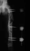
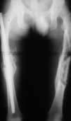

18 onotube X-rays Pre-Op Intra-Operative Post-Op Humeral Pre-Op Intra-Operative Post-Op Femur 14 Pre-Op Intra-Operative Post-Op Femur


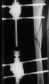
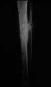
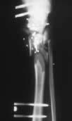
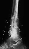
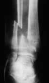
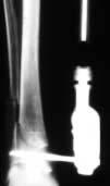

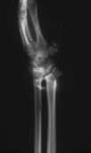
19 X-rays onotube Pre-Op Tibial Intra-Operative Post-Op Intra-Operative Tibial Post-Op Pre-Op Intra-Operative Tibial Pre-Op Wrist Intra-Operative Intra-Operative Post-Op Ankle 15


20 onotube Triax Carbon Tubes If no dynamisation is required or a radiotranslucent frame is needed, Triax Carbon Tubes can be used in place of the Dynamic Tube. Triax Carbon Tubes are available in 3 diameters; Yellow 15, Blue 20, Red 25 and in lengths from mm The use of Triax Carbon Tubes does not alter the surgical protocol, except that the colour coded end caps must be removed before placement of the Monotube Triax Clamps. Unlike the Dynamic Monotube Triax the Triax Carbon Tubes require an external compression/distraction device. To use the device, position the chariot over the back of the standard Triax pin clamp, and wrap the swivel clamp around the tube. Tighten the 7mm square head screw A, then proceed to loosen pin clamp square head screw D. Compression or distraction can now be achieved. Maximum distraction/compression is:- Yellow 20mm Blue 30mm Red 30mm 1 revolution of square head screw B provides 1mm of compression or distraction. After distraction or compression is completed, re-tighten pin clamp square head screw D. The external compression distraction device may now be moved. Chariot A Wrench D Clamp Screw B Thumbwheel 16


21 Economical & Radiolucent onotube An uncompromising alternative. When fracture dynamisation is not necessary or radiolucency is desired, the Carbon Monotube Triax offers an efficient and economic solution. Carbon Monotube Triax is available in three colour-coded diameters and a variety of lengths. The Monotube Triax Clamps can be placed anywhere along the tube to achieve the desired pin positioning while making final reduction possible after the frame is attached to the pins! If needed, Single Pin Clamps, T adapter, and tube-to-tube clamps, can be used with Triax Carbon Tubes. 17
 5150-0-470 Dynamic Tube Blue (20mm) 5150-0-475 Dynamic Tube Red (25mm) 5150-3-065 Pin Clamp Yellow (15mm) 5150-3-070 Pin Clamp Blue (20mm) 5150-3-075 Pin Clamp Red (25mm) 5150-3-165")
22 2 Products Catalogue Number Description Dynamic Tube Assembly Yellow (15mm) Dynamic Tube Assembly Blue (20mm) Dynamic Tube Assembly Red (25mm) Dynamic Tube Yellow (15mm) Dynamic Tube Blue (20mm) Dynamic Tube Red (25mm) Pin Clamp Yellow (15mm) Pin Clamp Blue (20mm) Pin Clamp Red (25mm) Single Pin Clamp Yellow (15mm) Single Pin Clamp Blue (20mm) Single Pin Clamp Red (25mm) T-Adapter Yellow (15mm) T Adapter Blue (20mm) T-Adapter Red (25mm) Tube-to-Tube Clamp Yellow (15mm) Tube-to-Tube Clamp Blue/Blue (20mm) Tube-to-Tube Clamp Red/Red (25mm) Tube-to-Tube Clamp Yellow (15mm)/Blue (20mm) Tube-to-Tube Clamp Blue (20mm)/Red (25mm) Please refer to the Tenxor brochure (Cat. Number ) for the components of the Triax Hybrid Frame 18
23 Products onotube Catalogue Number Description Carbon Tube Assembly Yellow Ø15/150mm Carbon Tube Assembly Yellow Ø15/200mm Carbon Tube Assembly Yellow Ø15/250mm Carbon Tube Assembly Yellow Ø15/300mm Carbon Tube Yellow Ø15/150mm Carbon Tube Yellow Ø15/200mm Carbon Tube Yellow Ø15/250mm Carbon Tube Yellow Ø15/300mm Carbon Tube Blue Ø20/200mm Carbon Tube Blue Ø20/250mm Carbon Tube Blue Ø20/300mm Carbon Tube Blue Ø20/350mm Carbon Tube Red Ø25/250mm Carbon Tube Red Ø25/300mm Carbon Tube Red Ø25/350mm Carbon Tube Red Ø25/400mm Instruments Long Handle Wrench Yellow Long Handle Wrench Blue Long Handle Wrench Red Reduction Handles Blue/Red Clamps Spanner Wrench Compression Distraction Device Yellow (15mm) Compression Distraction Device Blue (20mm) Compression Distraction Device Red (25mm) Dynamisation Wrench Yellow Dynamisation Wrench Blue Dynamisation Wrench Red Monotube Triax Yellow case Monotube Triax Red/Blue Case Stryker, Hoffmann, Hoffmann II, Apex, Monotube, Triax, Compact and Tenxor are trademarks of Stryker Corporation. Hoffmann II Swiss Patent Application: /94-3. Other Patents Pending. * Patents: EU 385,929; 374,093; Canada 1,193,506; U.S. 5,160,335 and 5,207,676. ** Swiss Patent Application: /94-3. Other Patents Pending. *** Patents: EU 230,856; Swiss CH 671,150; U.S. 4,978,350. Data on file at Stryker Howmedica Osteonics USA. 19
24 E X T E R N A L F I X A T I O N S Y S T E M Unilateral frame system designed to handle a wide variety of fractures and limb-lengthening applications. This simple, colour-coded system offers both dynamic and carbon tubes for individualised performance and economy. True simplicity, versatility, and economy. Modular frames which allow for true independent pin placement. Completely compatible with Original Hoffmann components, this new system improves flexibility and ease-of-use, while enhancing frame economics through minimal componentry. It s external fixation with a snap. ** Designed to complement the anatomy of the distal radius by allowing independent movement of its clamps in multiple planes. Standard unilateral or bi-lateral bridging frames for intra-articular fractures and peri-articular non-bridging frames for extraarticular fractures. Fully compatible with the Hoffmann II System, based on a spring-loaded snap-fit mechanism that improves flexibility and ease-of-use. * The DJD II is a Dynamic Elbow Joint Distractor. Fully compatible with the Hoffmann II Compact System, it is designed to treat posttraumatic elbow stiffness as well as acute elbow trauma cases. The Tenxor System is a hybrid system providing advanced technology and ease of application. Fully compatible with Hoffmann II and Monotube Triax, based on a spring-loaded, snap-fit mechanism that improves flexibility and ease of use. *** Every Fixator incorporates the high quality pin-to-bone interface provided by Apex Pins. The Apex Pin cuts more sharply with less torque, friction and heat upon insertion improving purchase while minimising the risk of pin tract problems. Available in self-drilling and blunt tip designs, only from Stryker Howmedica Osteonics! MANUFACTURER: Manufacturing location 5 chemin des Aulx CH-1228 Plan-les-Ouates Geneva, Switzerland Tel: Fax: Cat No: Stryker Corporation. All rights reserved.
TRIAX EXTERNAL FIXATION SYSTEM
 onotube onotube TRIAX E X T E R N A L F I X A T I O N S Y S T E M onotube The Monotube Triax allows increased fracture visualisation through the design of the pin clamps, which permit X-ray visualisation
Monotube Triax. External Fixation System. External Fixation. Operative Technique
 Monotube Triax External Fixation System Operative Technique External Fixation 1 Monotube Triax This publication sets forth detailed recommended procedures for using Stryker Osteosynthesis devices and instruments.
EXTERNAL FIXATION SYSTEM
 EXTERNAL FIXATION SYSTEM 2 3 1 4 5 1. DJD II Body 2. Humeral Guide 3. Pin Insertion Guides 4. Hoffmann II Compact Instruments 5. Hoffmann II Compact Components and Apex Pins 2 Overview The DJD II is a
EXTERNAL FIXATION SYSTEM
 EXTERNAL FIXATION SYSTEM The Apex Pin Fixation System is a one step procedure reducing insertion time and reducing insertion temperature. There are three types of fixation pins: Self-drilling / Self-tapping
Hoffmann II Compact External Fixation System
 Trauma Hoffmann II Compact External Fixation System Modular System for Upper Extremity Foot Introduction In 1938, Raoul Hoffmann, a surgeon from Geneva, Switzerland, designed a revolutionary External Fixation
Hoffmann II External Fixation System
 Hoffmann II External Fixation System Modular System for Long Bones Pelvis Introduction In 1938, Raoul Hoffmann, a surgeon from Geneva, Switzerland, designed a revolutionary External Fixation System. The
Hoffmann II Compact External Fixation System. Modular System for
 Hoffmann II Compact External Fixation System Modular System for Introduction In 1938, Raoul Hoffmann, a surgeon from Geneva, Switzerland, designed a revolutionary External Fixation System. The basic features
Monolateral External Fixation System for Trauma and Orthopaedics
 MEFiSTO Monolateral External Fixation System for Trauma and Orthopaedics Surgical Technique Original Instruments and Implants of the Association for the Study of Internal Fixation AO/ASIF MEFiSTO Table
External Fixator Brochure
 External Fixator Brochure Response Ortho is a global orthopaedic trauma solutions manufacturer offering premium products created under its founding principles of innovation, excellence by design and functional
Knee spanning solutions
 Knee spanning solutions System features Indications Intended to be used on adults or pediatric patients as required for fracture fixation (open or closed); post-traumatic joint contracture which has resulted
TIPMED EXTERNAL FIXATION SYSTEMS
 TIPMED EXTERNAL FIXATION SYSTEMS ANATOMICAL LOCATIONS FOR EXTERNAL FIXATION SYSTEMS Humeral Dynamic Axial Fixator Elbow Fixator Pelvic Dynamic Axial Fixator Pennig Wrist Fixator Hand Fixator Finger Fixator
Hoffmann II MRI External Fixation Systems. Hoffmann II MRI Hoffmann II Compact MRI Modular Systems for Upper Extremity Lower Extremity Pelvis
 Hoffmann II MRI External Fixation Systems Hoffmann II MRI Hoffmann II Compact MRI Modular Systems for Upper Extremity Lower Extremity Pelvis Introduction In the ever changing world of medicine, new technology
Hoffmann II Micro External Fixation System. Indications for the Hand
 Hoffmann II Micro External Fixation System Indications for the Hand Features & Benefits The instrumentation is Simple and User-Friendly, and the system is Color Coded, so there is no lost time or confusion
Hoffmann II Micro External Fixation System
 Trauma Hoffmann II Micro External Fixation System Indications for the Hand Components 9 3 4 5 1 2 7 8 6 1 Hoffmann II Micro Multi-Pin Clamp 2 Hoffmann II Micro 90 Multi-Pin Clamp 3 Hoffmann II Micro Rod
Hoffmann LRF Circular External Fixation. Operative technique
 Hoffmann LRF Circular External Fixation Operative technique Hoffmann LRF Circular External Fixation Operative technique Hoffmann LRF Circular External Fixation Contents 1. Indications and contraindications...
EXTERNAL FIXATION SYSTEM OPERATIVE TECHNIQUE
 EXTERNAL FIXATION SYSTEM OPERATIVE TECHNIQUE 2 3 1 4 5 Dynamic Joint Distractor II B. F. Morrey, M.D. 1. DJD II Body 2. Humeral Guide 3. Pin Insertion Guides 4. Hoffmann II Compact Instruments 5. Hoffmann
Surgical Technique. Targeter Systems Overview
 Surgical Technique Targeter Systems Overview PERI-LOC Locked Plating System Targeter Systems Overview Table of contents Product overview... 2 Introduction... 2 Indications... 2 Design features and benefits...
MEFiSTO. Monolateral External Fixation System for Trauma and Orthopaedics.
 MEFiSTO. Monolateral External Fixation System for Trauma and Orthopaedics. Surgical Technique This publication is not intended for distribution in the USA. Instruments and implants approved by the AO Foundation.
AxSOS Locking Plate System
 AxSOS Locking Plate System Operative Technique Small Fragment Basic Fragment 1 2 Contents Page 1. Introduction 4 2. Features & Benefits 5 4 and 5mm Compression Plates 5 Reconstruction and 1/3 Tubular Locking
QUICK REFERENCE GUIDE. The XCaliber Meta-Diaphyseal Fixator
 17 The XCaliber Meta-Diaphyseal Fixator GENERAL POINTS The XCaliber Fixator is made of radiolucent material for unobstructed X-ray visualization. The metallic bolts and the cam and bush of each ball-joint,
MEFiSTO. Monolateral External Fixation System for Trauma and Orthopaedics.
 MEFiSTO. Monolateral External Fixation System for Trauma and Orthopaedics. Surgical Technique This publication is not intended for distribution in the USA. Instruments and implants approved by the AO Foundation.
Technique Guide. The Distraction Osteogenesis Ring System. Nonarticular tibia frame.
 Technique Guide The Distraction Osteogenesis Ring System. Nonarticular tibia frame. Table of Contents Introduction The Distraction Osteogenesis Ring System 2 AO Principles 4 Indications 5 Surgical Technique
TransFx External Fixation System Large and Intermediate Surgical Technique
 TransFx External Fixation System Large and Intermediate Surgical Technique TransFx External Fixation System Large and Intermediate Surgical Technique 1 Surgical Technique For TransFx External Fixation
Small External Fixator Nonspanning Wrist Frame. For the treatment of wrist fractures.
 Small External Fixator Nonspanning Wrist Frame. For the treatment of wrist fractures. Technique Guide Part of the Small External Fixation System Small External Fixator Nonspanning Wrist Frame When to use
TransFx External Fixation System Large and Intermediate Surgical Technique
 TransFx External Fixation System Large and Intermediate Surgical Technique Choice, Simplicity, Transition TransFx External Fixation System Large and Intermediate Surgical Technique 1 Surgical Technique
Elbow Hinge Fixator. Guided Flexion/Extension for Unstable Elbow Fractures.
 Elbow Hinge Fixator. Guided Flexion/Extension for Unstable Elbow Fractures. Surgical Technique MR Safe Radiolucent Table of Contents System Description 3 Indications and Contraindications 4 Fixation Components
LCP Medial Distal Tibia Plate, without Tab. The Low Profile Anatomic Fixation System with Angular Stability and Optimal Screw Orientation.
 LCP Medial Distal Tibia Plate, without Tab. The Low Profile Anatomic Fixation System with Angular Stability and Optimal Screw Orientation. Technique Guide LCP Small Fragment System Table of Contents Introduction
QUICK REFERENCE GUIDE. The PreFix Fixator (92000 Series) ALWAYS INNOVATING
 ALWAYS INNOVATING") 21 The PreFix Fixator (92000 Series) ALWAYS INNOVATING INTRODUCTION The PreFix fixator is designed to provide temporary external fixation. This may be needed when local facilities or the condition of the
21 The PreFix Fixator (92000 Series) ALWAYS INNOVATING INTRODUCTION The PreFix fixator is designed to provide temporary external fixation. This may be needed when local facilities or the condition of the
Mini External Fixator.
 Mini External Fixator. Assembly and Surgical Technique This publication is not intended for distribution in the USA. Instruments and implants approved by the AO Foundation. Image intensifier control Warning
LCP Medial Proximal Tibial Plate 3.5. Part of the Synthes small fragment Locking Compression Plate (LCP) system.
 system.") LCP Medial Proximal Tibial Plate 3.5. Part of the Synthes small fragment Locking Compression Plate (LCP) system. Technique Guide This publication is not intended for distribution in the USA. Instruments
LCP Medial Proximal Tibial Plate 3.5. Part of the Synthes small fragment Locking Compression Plate (LCP) system. Technique Guide This publication is not intended for distribution in the USA. Instruments
DFS STANDARD FIXATOR DFS ANKLE CLAMP DFS T-CLAMP
 DFS STANDAD FIXATO DFS ANKLE CLAMP DFS T-CLAMP SUGICAL TECHNIQUE Dr. James V. Nepola Professor of Orthopaedics University of Iowa Hospitals and Clinics Iowa City, Iowa Patent No. 5,662,650 C ontents DynaFix
External Skeletal Fixation (ESF)
") External Skeletal Fixation (ESF) Technique for fracture repair in animals Introduction External Skeletal Fixation is a versatile and effective technique for fracture repair in animals, rigidly stabilizing
External Skeletal Fixation (ESF) Technique for fracture repair in animals Introduction External Skeletal Fixation is a versatile and effective technique for fracture repair in animals, rigidly stabilizing
Surgical Technique. 3.5mm and 4.5mm Lateral Proximal Tibia Locking Plates
 Surgical Technique 3.5mm and 4.5mm Lateral Proximal Tibia Locking Plates PERI-LOC Periarticular Locked Plating System 3.5mm and 4.5mm Lateral Proximal Tibia Locking Plate Surgical Technique Contents Product
Small External Fixator Wrist Spanning Frame. For the treatment of wrist fractures.
 Small External Fixator Wrist Spanning Frame. For the treatment of wrist fractures. Technique Guide Part of the Small External Fixation System Small External Fixator Wrist Spanning Frame When to use The
Hoffmann 3 Modular External Fixation. Operative technique
 Hoffmann 3 Modular External Fixation Operative technique Hoffmann 3 External Fixation Contents Indications and contraindications............... 4 Technical details & MRI Information.... 5 Components...
Medical Devices HUMERUS. WRIStX
 Medical Devices DDDAF HUMERUS WRIStX DAF HUMERUS FIXATOR It is a single plan external fixation tool and used for the treatment of the fractures of humerus, radius and ulna for adults. It is also used at
Surgical Technique. Anterolateral and Medial Distal Tibia Locking Plates
 Surgical Technique Anterolateral and Medial Distal Tibia Locking Plates PERI-LOC Periarticular Locked Plating System Anterolateral and Medial Distal Tibia Locking Plates Surgical Technique Contents Product
Surgical Technique. Distal Radius and Foot
 Surgical Technique Distal Radius and Foot JET-X BAR Unilateral Fixator Distal Radius and Foot Surgical Technique Contents Design Features...2 Distal Radius Surgical Technique Indications...10 Surgical
LCP Low Bend Medial Distal Tibia Plates 3.5 mm. Anatomic plates with low profile head for intra- and extraarticular fractures.
 LCP Low Bend Medial Distal Tibia Plates 3.5 mm. Anatomic plates with low profile head for intra- and extraarticular fractures. Surgical Technique This publication is not intended for distribution in the
External Distal Radius Fixator. Supplement to the 8 mm rod fixator system
 External Distal Radius Fixator. Supplement to the 8 mm rod fixator system Surgical technique This publication is not intended for distribution in the USA. Instruments and implants approved by the AO Foundation
Humerus Block. Discontinued December 2016 DSEM/TRM/0115/0296(1) Surgical Technique. This publication is not intended for distribution in the USA.
 Surgical Technique. This publication is not intended for distribution in the USA.") Humerus Block Surgical Technique Discontinued December 2016 DSEM/TRM/0115/0296(1) This publication is not intended for distribution in the USA. Instruments and implants approved by the AO Foundation. Contents
Humerus Block Surgical Technique Discontinued December 2016 DSEM/TRM/0115/0296(1) This publication is not intended for distribution in the USA. Instruments and implants approved by the AO Foundation. Contents
Technique Guide. 3.5 mm LCP Low Bend Medial Distal Tibia Plate Aiming Instruments. Part of the 3.5 mm LCP Percutaneous Instrument System.
 Technique Guide 3.5 mm LCP Low Bend Medial Distal Tibia Plate Aiming Instruments. Part of the 3.5 mm LCP Percutaneous Instrument System. Table of Contents Introduction 3.5 mm LCP Low Bend Medial Distal
OPERATIVE TECHNIQUE. Knee Hinge (LRS Advanced System)
") OPERTIVE TECHNIQUE Knee Hinge (LRS dvanced System) 1 1 2 4 6 INTRODUCTION INDICTIONS FETURES ND BENEFITS EQUIPMENT REQUIRED KNEE HINGE SSEMBLY 8 17 TRUM KNEE DISLOCTION Orthofix wishes to thank the following
OPERTIVE TECHNIQUE Knee Hinge (LRS dvanced System) 1 1 2 4 6 INTRODUCTION INDICTIONS FETURES ND BENEFITS EQUIPMENT REQUIRED KNEE HINGE SSEMBLY 8 17 TRUM KNEE DISLOCTION Orthofix wishes to thank the following
Choice, Simplicity, Transition. TransFx External Fixation System Small and Mini Surgical Technique
 Choice, Simplicity, Transition TransFx External Fixation System Small and Mini Surgical Technique TransFx External Fixation System Small and Mini Surgical Technique 1 Surgical Technique For TransFx External
Technique Guide. LCP Proximal Femoral Hook Plate 4.5/5.0. Part of the LCP Periarticular Plating System.
 Technique Guide LCP Proximal Femoral Hook Plate 4.5/5.0. Part of the LCP Periarticular Plating System. Table of Contents Introduction Features and Benefits 2 AO ASIF Principles 4 Indications 5 Surgical
M.I.S. MAKE IT SMART IN ONE SYSTEM. Surgical Technique. Hip Knee Spine Navigation
 M.I.S. MAKE IT SMART IN ONE SYSTEM Surgical Technique Hip Knee Spine Navigation M.U.S.T. Mini Open Surgical Technique Hip Knee Spine Navigation 2 C O N T E N T S 1 INTRODUCTION 4 2 SURGICAL TECHNIQUE 5
Biomet Carbon Rail Deformity System. Surgical Technique
 Biomet Carbon Rail Deformity System Surgical Technique Contents Introduction... Page 1 System Components... Page 2 Indications, Contraindications, and Deformity Planning... Page 4 Deformity Planning-Identifying...
pediatric orthopedic implants fixator
 pediatric orthopedic implants DDDAF fixator DAF HUMERUS FIXATOR It is a single plan external fixation tool and used for the treatment of the fractures of humerus, radius and ulna for adults. It is also
Large External Fixator Delta Frame Ankle Bridge. For staged fixation of the distal tibia.
 Large External Fixator Delta Frame Ankle Bridge. For staged fixation of the distal tibia. Technique Guide Part of the Large External Fixation System Large External Fixator Delta Frame Ankle Bridge Technique
Introduction to the Taylor Spatial Frame Hardware. Trademark of Smith & Nephew. Certain marks Reg. US Pat. & TM Off.
 Introduction to the Taylor Spatial Frame Hardware Trademark of Smith & Nephew. Certain marks Reg. US Pat. & TM Off. What is the Taylor Spatial Frame? Next generation circular fixator capable of 6 axes
OPERATING MANUAL AND TECHNIQUE GUIDE FOR TITANIUM FEMORAL AND TIBIAL NAILING SYSTEMS
 OPERATING MANUAL AND TECHNIQUE GUIDE FOR TITANIUM FEMORAL AND TIBIAL NAILING SYSTEMS ORTHO-MEDICAL GMBH TITANIUM FEMORAL NAIL OPERATIVE TECHNIQUE Introduction: Why a new type of femoral nail? The latest
OPERATIVE TECHNIQUE. Limb Reconstruction System. Part B: Correction of Deformities. By Dr. S. Nayagam
 OPERATIVE TECHNIQUE 11 Limb Reconstruction System Part B: Correction of Deformities By Dr. S. Nayagam CONTENTS LIMB RECONSTRUCTION SYSTEM Part B: Correction of Deformities QUICK REFERENCE GUIDE... Page
Large External Fixator Delta Frame Ankle Bridge. Using pin clamps with outrigger posts.
 Large External Fixator Delta Frame Ankle Bridge. Using pin clamps with outrigger posts. Technique Guide Part of the Large External Fixation System Large External Fixator Delta Frame Ankle Bridge Technique
Operative Technique. by PROF. NAYAGAM. LIMB RECONSTRUCTION SYSTEM Part B: Correction of Deformities
 Operative Technique by PROF. NAYAGAM LIMB RECONSTRUCTION SYSTEM Part B: Correction of Deformities 11 Quick Reference Guide CONTENTS LIMB RECONSTRUCTION SYSTEM Part A: General Principles Page N o I Introduction
LCP Distal Tibia Plate
 Surgical Technique LCP Locking Compression Plate Original Instruments and Implants of the Association for the Study of Internal Fixation AO/ASIF Table of contents Indications 3 Implants/Instruments 5 Surgical
Technique Guide. Adjustable Distal Radius Fixator. Part of the Synthes External Fixation Systems.
 Technique Guide Adjustable Distal Radius Fixator. Part of the Synthes External Fixation Systems. Table of Contents Introduction Adjustable Distal Radius Fixator 2 Indications 3 Surgical Technique Configure
Surgical Technique International Version
 Surgical Technique International Version PERI-LOC VLP Variable-Angle Locked Plating System Surgical Technique Table of Contents Product Overview...2 Introduction...2 Indications and Contraindications...3
OPERATIVE TECHNIQUE. The Ring Fixator System. The Sheffield Ring Fixator - Limb Reconstruction. and Complex Trauma. By Prof. M.
 OPERATIVE TECHNIQUE 12 The Ring Fixator System Part C: The Sheffield Ring Fixator - Limb Reconstruction and Complex Trauma By Prof. M. Saleh CONTENTS Page N INTRODUCTION...................................................................................................................................
GAMMA LOCKING NAIL INSTRUMENTS OPERATIVE TECHNIQUE
 GAMMA LOCKING NAIL INSTRUMENTS OPERATIVE TECHNIQUE FEATURES AND BENEFITS The One Shot Device is a new component of the Gamma Locking Nail instrumentation system determining the correct position of the
QUICK REFERENCE GUIDE. Arthrodiatasis. Articulated Joint Distraction
 4 Arthrodiatasis Articulated Joint Distraction ARTHRODIATASIS OF THE HIP To prepare the assembly, remove the female component and replace it with the ProCallus articulated body for the hip. Remove cam
EBI FIX DYNAFIX SYSTEM VISION EXTERNAL FIXATION SURGICAL TECHNIQUE. Patent 6,277,119
 EBI DYNAFIX FIX SYSTEM DYNAFIX VISION EXTERNAL FIXATION SYSTEM SURGICAL TECHNIQUE Patent 6,277,119 1 CONTENTS Basic Principles and Biomechanical Concepts...Page 2 Introduction...Page 2 Component Review...
LCP Superior Clavicle Plate. The anatomically precontoured fixation system with angular stability for clavicle shaft and lateral clavicle.
 Technique Guide LCP Superior Clavicle Plate. The anatomically precontoured fixation system with angular stability for clavicle shaft and lateral clavicle. Table of Contents Introduction LCP Superior Clavicle
Large Distractor Femur
 Fracture Reduction and Provisional Stabilization Large Distractor Femur Surgical Technique Table of Contents Introduction Standard Femoral Distraction 2 Large Distractor System 4 Surgical Technique Prepare
VLIFT System Overview. Vertebral Body Replacement System
 VLIFT System Overview Vertebral Body Replacement System VLIFT System System Description The VLIFT Vertebral Body Replacement System consists of a Distractible In Situ (DIS) implant, which enables the surgeon
S U R G I C A L T E C H N I Q U E David A. McQueen, MD Return to Menu
 S U R G I C A L T E C H N I Q U E David A. McQueen, MD TOTAL KNEE INSTRUMENTS Wichita Fusion Nail Introduction...1 Preoperative Planning...2 Surgical Technique...3-8 Wichita Fusion Nail Surgical Technique
LCP Medial Proximal Tibial Plate 4.5/5.0. Part of the Synthes LCP periarticular plating system.
 LCP Medial Proximal Tibial Plate 4.5/5.0. Part of the Synthes LCP periarticular plating system. Technique Guide This publication is not intended for distribution in the USA. Instruments and implants approved
Knee Surgical Technique
 Knee Surgical Technique COMPASS Universal Hinge by Jimmy Tucker, M.D. Orthopaedic Surgeon Director, Arkansas Sports Medicine, P.A. Little Rock, Arkansas Table of contents Design features 3 Indications
Surgical Technique. Cannulated Angled Blade Plate 3.5 and 4.5, 90
 Surgical Technique Cannulated Angled Blade Plate 3.5 and 4.5, 90 Cannulated Angled Blade Plate 3.5 and 4.5, 90 Table of contents Indications/Contraindications 2 Implants 3 Surgical technique 5 Implant
Conventus CAGE PH Surgical Techniques
 Conventus CAGE PH Surgical Techniques Conventus Orthopaedics The Conventus CAGE PH (PH Cage) is a permanent implant comprised of an expandable scaffold, made from nitinol and titanium, which is deployed
Technique Guide. 3.5 mm LCP Low Bend Medial Distal Tibia Plates. Part of the Synthes locking compression plate (LCP) system.
 system.") Technique Guide 3.5 mm LCP Low Bend Medial Distal Tibia Plates. Part of the Synthes locking compression plate (LCP) system. Table of Contents Introduction 3.5 mm LCP Low Bend Medial Distal Tibia Plates
Technique Guide 3.5 mm LCP Low Bend Medial Distal Tibia Plates. Part of the Synthes locking compression plate (LCP) system. Table of Contents Introduction 3.5 mm LCP Low Bend Medial Distal Tibia Plates
Pre-Operative Planning. Positioning of the Patient
 Surgical Technique Pre-Operative Planning Decide upon the size and angle of the barrel plate to be used from measuring the x-rays. To maximise the sliding action when using shorter lag screws, the Short
Distal Cut First Femoral Preparation
 Surgical Technique Distal Cut First Femoral Preparation Primary Total Knee Arthroplasty LEGION Total Knee System Femoral preparation Contents Introduction...3 DCF femoral highlights...4 Preoperative planning...6
Large External Fixator Modular Knee Bridge. Using multi-pin clamps.
 Large External Fixator Modular Knee Bridge. Using multi-pin clamps. Technique Guide Part of the Large External Fixation System Large External Fixator Modular Knee Bridge Technique Overview Insert Schanz
Surgical Technique. Proximal Humerus Locking Plate
 Surgical Technique Proximal Humerus Locking Plate PERI-LOC Upper Extremity Locked Plating System 3.5mm & 4.5mm Proximal Humerus Locking PlatesCatalog Infor Table of Contents Introduction.........................................................2
Types of Plates 1. New Dynamic Compression Plate: Diaphyseal fracture: Radius, Ulna, Humerus, Rarely tibia
 Types of Plates 1. New Dynamic Compression Plate: DCP Diaphyseal fracture: Radius, Ulna, Humerus, Rarely tibia 1. Undercut adjacent to the holes low contact: less stress shield 2. Undercut at the undersurface
Periarticular Aiming Arm Instruments for LCP Proximal Tibial Plate 4.5/5.0. Part of the LCP Periarticular Aiming Arm Instrument System (large).
.") Technique Guide Periarticular Aiming Arm Instruments for LCP Proximal Tibial Plate 4.5/5.0. Part of the LCP Periarticular Aiming Arm Instrument System (large). Image intensifier control Warning This description
Technique Guide Periarticular Aiming Arm Instruments for LCP Proximal Tibial Plate 4.5/5.0. Part of the LCP Periarticular Aiming Arm Instrument System (large). Image intensifier control Warning This description
Surgical Technique International Version
 Surgical Technique International Version PERI-LOC PFP 4.5mm Proximal Femur Locking Plate Surgical Technique Table of contents Product overview...2 Introduction...2 Indications...3 Case examples...4 Design
Zimmer MIS Periarticular 3.5mm Proximal Tibial Locking Plate
 Zimmer MIS Periarticular 3.5mm Proximal Tibial Locking Plate Surgical Technique The Science of the Landscape Zimmer MIS Periarticular 3.5mm Proximal Tibial Locking Plate Surgical Technique 1 Zimmer MIS
LISS DF and LISS PLT. Less Invasive Stabilization Systems for Distal Femur and Proximal Lateral Tibia.
 LISS DF and LISS PLT. Less Invasive Stabilization Systems for Distal Femur and Proximal Lateral Tibia. LISS DF and LISS PLT. Less Invasive Stabilization Systems for Distal Femur and Proximal Lateral Tibia.
FLT105 12/02.
 FLT105 12/02 www.biometmerck.co.uk Disclaimer Biomet Merck Ltd, as the manufacturer of this device, does not practice medicine and does not recommend this or any other surgical technique for use on a
Medium External Fixator Humeral Shaft Frame. Modular frame for upper extremity use.
 Medium External Fixator Humeral Shaft Frame. Modular frame for upper extremity use. Technique Guide Part of the Medium External Fixation System MRI Information Synthes Medium External Fixation devices
A locking plate system that expands a surgeon s options in trauma surgery. Zimmer NCB Plating System
 A locking plate system that expands a surgeon s options in trauma surgery Zimmer NCB Plating System The Power of Choice The power of having true intraoperative options is at your fingertips. Using standard
Instructions For Use. Surgical Technique
 Instructions For Use Surgical Technique English Instructions for use Intended use INTELLIGENT ORTHOPAEDICS instruments consist of manual surgical instruments and devices intended for use in surgical procedures.
Extron External Fixator
 Operative Technique Extron External Fixator The disposable set for a Distal Radius Fracture The EXTRON-External Fixator made by tantum provides you with a new generation of supply engineering for distal
Femur. Monoaxial Locking Plate System. Operative Technique. Distal Lateral Femur Universal Holes Targeting Instrumentation.
 Femur AxSOS 3 Titanium Monoaxial Locking Plate System Femur Fractures Operative Technique Distal Lateral Femur Universal Holes Targeting Instrumentation This publication sets forth detailed recommended
PROXIMAL TIBIAL PLATE
 SURGICAL NÁSTROJE TECHNIQUE PRO ARTROSKOPII PROXIMAL INSTRUMENTS TIBIAL FOR PLATE ARTHROSCOPY Proximal Tibial Plate Description of medical device The Proximal Tibial Plate is used in epyphyseal and metaphyseal
Surgical Technique. Distal Humerus Locking Plate
 Surgical Technique Distal Humerus Locking Plate PERI-LOC Locked Plating System Distal Humerus Locking Plate Surgical Technique Table of Contents Introduction...2 Indications...3 Plate Features...3 Patient
VariAx Compression Plating System. Operative technique
 VariAx Compression Plating System Operative technique VariAx Compression Plating System Operative technique VariAx Compression Plating System Contents 1. Introduction... 3 2. Indications, MR safety information
Zimmer Small Fragment Universal Locking System. Surgical Technique
 Zimmer Small Fragment Universal Locking System Surgical Technique Zimmer Small Fragment Universal Locking System 1 Zimmer Small Fragment Universal Locking System Surgical Technique Table of Contents Introduction
Medical Devices DYNAMIC MULTIAXIAL FIXATOR
 Medical Devices DDDAF DYNAMIC MULTIAXIAL FIXATOR DAF DYNAMIC MULTIAXIAL FIXATOR It is a single plan external fixation tool. The ball-joint structure connects the clamp to the fixator body as an articular
LCP Anterolateral Distal Tibia Plate 3.5. The low profile anatomic fixation system with optimal plate placement and angular stability.
 LCP Anterolateral Distal Tibia Plate 3.5. The low profile anatomic fixation system with optimal plate placement and angular stability. Technique Guide LCP Small Fragment System Table of Contents Introduction
LCP Superior Clavicle Plate. The anatomically precontoured fixation system with angular stability for clavicle shaft and lateral clavicle.
 LCP Superior Clavicle Plate. The anatomically precontoured fixation system with angular stability for clavicle shaft and lateral clavicle. Surgical Technique This publication is not intended for distribution
Small Fragment Plating System
 Small Fragment Plating System Securing optimal fixation through locked and compression plating technology SURGICAL TECHNIQUE RECOVERY FUNCTION SURVIVORSHIP DePuy believes in an approach to trauma surgery
Mandible External Fixator II. Provides treatment for fractures of the maxillofacial area.
 Mandible External Fixator II. Provides treatment for fractures of the maxillofacial area. Technique Guide This publication is not intended for distribution in the USA. Instruments and implants approved
LCP Anterolateral Distal Tibia Plate 3.5. The low profile anatomic fixation system with optimal plate placement and angular stability.
 LCP Anterolateral Distal Tibia Plate 3.5. The low profile anatomic fixation system with optimal plate placement and angular stability. Technique Guide LCP Small Fragment System Table of Contents Introduction
VA-LCP Anterior Clavicle Plate. The anatomically precontoured fixation system with angular stability for clavicle shaft and lateral clavicle.
 Technique Guide VA-LCP Anterior Clavicle Plate. The anatomically precontoured fixation system with angular stability for clavicle shaft and lateral clavicle. Table of Contents Introduction VA-LCP Anterior
3.5 mm Locking Attachment Plate
 For Treatment of Periprosthetic Fractures 3.5 mm Locking Attachment Plate Surgical Technique Table of Contents Introduction 3.5 mm Locking Attachment Plate 2 Indications 4 Surgical Technique Preparation
AcUMEDr. Anatomic Midshaft Forearm Plates
 AcUMEDr Anatomic Midshaft Forearm Plates Anatomic Midshaft Forearm Plates Since 1988, Acumed has been designing solutions to the demanding situations facing orthopaedic surgeons, hospitals and their patients.
AxSOS. Locking Plate System. Operative Technique. Small Fragment Basic Fragment
 AxSOS Locking Plate System Operative Technique Small Fragment Basic Fragment Stryker Plating Contents Page 1. Introduction 4 2. Features & Benefits 5 4 and 5 Compression Plates 5 Reconstruction and 1/3