Clinical Examination of VASCULAR PATIENTS. Stephanie Hirst & Alexander Sunde
|
|
|
- Beatrix Armstrong
- 5 years ago
- Views:
Transcription

1 Clinical Examination of VASCULAR PATIENTS Stephanie Hirst & Alexander Sunde
2 Goals of Medical History To record the patient s symptoms at time of presentation. To organize the events which have lead to presentation. To summarize the evidence which supports the diagnostic hypothesis. To provide basis and direction for care
3 Goals of Physical Exam To record the state of patient s health at the time of the examination. To provide a longitudinal record of the patient s health. Allow assessment of progression of disease. Allow prognostication of natural history. Allow recommendations for care.
4 Vascular Examination The exam includes several parts: Position/Lighting/Draping Inspection Palpation Auscultation Special maneuvers
5 Position/Lighting/Draping Position patient should be lying in the supine position, and patient's hands should remain at his/her sides. with the head resting on a pillow. Lighting adjusted so that it is ideal. Draping expose the body surfaces, but use the draping so that the patient feels less exposed. Eg. Between legs while checking for femoral pulse.

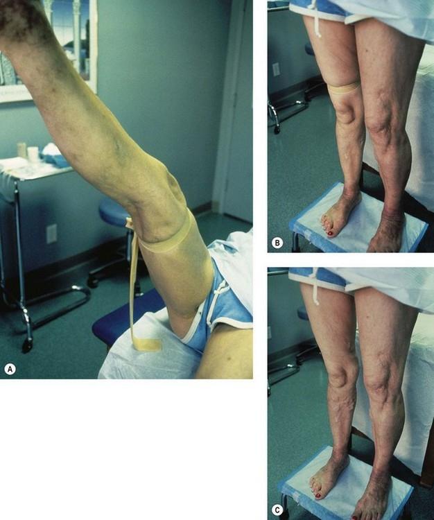
6 Blood Pressure Always check blood pressure on both arms Normally, there is a difference in pressure of 5 mmhg up to 10 mmhg Pressure difference of more than mmhg occurs in subclavian steal syndrome, aortic dissection. YES!
7 Inspection On inspection the physician looks for signs of: trauma previous surgery (scars) muscle wasting/muscle asymmetry edema (swelling) erythema (redness) ulcers arterial ulcers tend to be on the borders / sides of the foot, neuropathic ulcers on the plantar surface of the foot, venous ulcers tend on be on the medial aspect of the leg superior to the medial malleolus. hair hair is absent in peripheral vascular disease (PVD) shiny skin seen in PVD Haemosiderin deposits Lipodermatosclerosis


8 Inspection When you examine the extremities, always note: Their size and symmetry; And ALWAYS compare left and right side
9 Palpation Temperature cool suggest poor circulation,both sides should be compared Pitting edema should be tested for in dependent locations dorsum of foot, if present then on the shins. If the patient has been in bed for a longer period of time one should check the sacrum. Capillary refill the time taken for color to return to a nail after pressure is applied to cause blanching Normal: 2 sec or less Bordeline: 3 sec Ischemia: more than 5 sec

10 5 P's signs of acute ischemia Pallor Pain Pulsless Parasthesia Paralyse Poikylothermia

11 Pitting Edema Example of pitting edema in a patient with liver failure
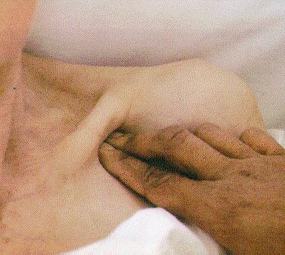
12 Arterial Pulses Always compare both sides Dorsalis pedis artery pulse on dorsal surface of the foot, running lateral to the tendon of the first toe Posterior tibial artery pulse posterior and inferior to the medial malleolus Popliteal artery pulse behind the knee, typically done with both hands Femoral artery pulse in the femoral triangle / halfway between the ASIS and pubic tubercle


13 Dorsalis Pedis Posterior Tibialis


14 Femoral artery Popliteal Pulse
15 Arterial Pulses Always compare both sides Radial artery pulse on the flexor surface of the wrist laterally. Brachial artery pulse flex the elbow slightly, and palpate artery just medial to the biceps tendon at the antecubital crease. It can also be felt higher up in the groove between biceps and triceps muscles. Carotid artery plulse Subclavian artery pulse

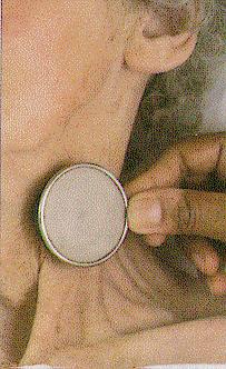
16 Carotid Artery Exam
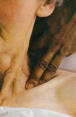
17 Subclavian Artery Exam
18 Ausculatation for Bruits Palpate carotid upstroke, and auscultate for bruits. Auscultate for aortic, renal, and femoral bruits; palpate aorta and determine maxmimal diameter.

19 Aortic Aneurysm Examination - palplate aorta and detemine maximal diameter
20 Special maneuvers Buerger's Test Assesment of arterial sufficiency With the patient supine, note the colour of the feet soles. They should be pink. Then elevate both legs to 45 degrees for more than 1 minute. Observe the soles. If there is marked pallor whiteness, ischemia should be suspected. Next check for rubor of dependency. Sit the patient upright and observe the feet. In normal patients, the feet quickly turn pink. If, more slowly, they turn red like a cooked lobster, suspect ischemia.
21 Special maneuvers Venous Refill Venous refill with dependency (should be less than 30 seconds) the vein should bulge outward within 30 seconds of limb elevation for one minute.
22 Special maneuvers Brodie-Trendelenburg Test assessment of valvular competence if varicose veins are present: One leg at a time. With the patient supine, empty the superficial veins by 'milking' the leg in the distal to proximal direction. Now press with your thumb over the saphenofemoral junction (2.5 cm below and 2.5 cm lateral to the pubic tubercle) and ask the patient to stand while you maintain pressure. If the leg veins now refill rapidly, the incompentence is located below the saphenofemoral junction, and vice versa. This test can be repeated using pressure at any point along the leg until the incompetence has been mapped out.
23 Brodie-Trendelenburg Test
24 Perthes Test The Perthes test is a clinical test for assessing the patency of the deep femoral vein prior to varicose vein surgery. The limb is elevated and an elastic bandage is applied firmly from the toes to the upper 1/3 of the thigh to obliterate the superficial veins only. Modified: The test is done by applying a tourniquet at the level of the sapheno-femoral junction to occlude the superficial pathway, and then the patient is asked to move in situ. If the deep veins are occluded, the dilated veins increase in prominence. This is a more objective test as it does not depend on patient's pain threshold.
25 Pratts Test The Pratt Test is a simple test to check for deep vein thrombosis in the leg. It involves having the patient lie supine with the leg bent at the knee, grasping the calf with both hands and pressing on the popliteal vein in the proximal calf. If the patient feels pain, it is a sign that a deep vein thrombosis exists.
26 Special maneuvers Allen Test The patient should rest with arms in lap, palms up. Ask the patient to make a tight fist with one hand; Then compress both radial and ulnar arteries firmly between your thumbs and fingers. Next, ask the patient to open the hand into a relaxed, slightly flexed position. The palm is pale. Release the pressure over ulnar artery. If ulnar artery is patent, the palm flushes within about 3 5 seconds. You can du the same with radial artery while still compressing ulnar artery. It is also a good test to use before taking blood samples from the radial artery.

27 Special maneuvers Allen Test
28 Bedside Examination Ankle-brachial Index (ABI) assesses peripheral vascular disease and find the cutpoint. It may however be unreliable in patients with calcified arteries in the calf (often diabetic patients) or those with extensive oedema. In which case Toebrachial index (TBI) should be performed to aid in the diagnosis. Physiology: higher pressure in ankle ABI: 1,2mmHg Pathology: ABI: < 0,9mmHG
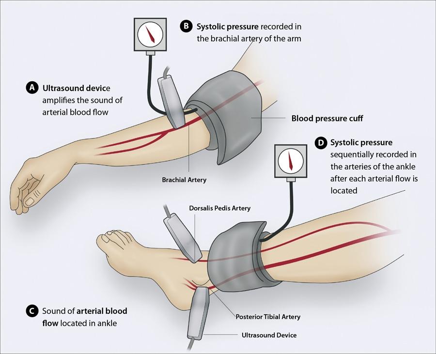
29 ABI
:- 8MHz.")
30 Bedside Examination Doppler Examination Continuous Wave Doppler Assessment (Hand held Doppler):- 8MHz.
31 Vascular Physical Exam Examination Instrument Student Worksheet -- Specific Observations 1. Pulses -- should note quality (-, +, ++) right left Superficial Temporal Common Carotid Brachial Radial Aorta Common Femoral Popliteal Dorsalis Pedis Posterior Tibialis 2. Aneurysms (yes, no) right left Aorta Common Femoral Popliteal left Common Carotid Aorta Common Femoral 3. Bruit (yes, no) right 4. Ischemic Signs -- signs of arterial disease. (normal, abnormal; yes, no; If yes, location) right left Color Temperature Capillary Refill Ulceration Eschar Location 5. Venous Signs -- signs of venous disease. (yes, no. If yes, location) Brawny Color Varicose Veins Ulceration Edema Location
32 Vascular Physical Exam Assessment Instrument Did the student examine and record the following? YES NO 17 pulses 5 aneurysms 5 bruit 6 sings of arterial disease 5 signs of venous disease explain examination procedure position patient correctly uncover the skin of the part to be examined. inspect auscultate with stethoscope on skin touch skin (no through clothing or dressing) palpate for aortic aneurysm between umbilicus and xyphoid palpate for popliteal pulse or aneurysm with two hands stand at foot of patient while palpating dorsalis pedis and posterior compress ankle to assess edema feel toes to asses temperature press toes to asses refill Totals: tibial
33 Sources BOOK: Bates' Guide to physical examination and history taking, 11 th ed. By Lynn S. Bickley INTERNET: Wikipedia: on SEMINAR
34 THANK YOU!
Peripheral Vascular Examination. Dr. Gary Mumaugh Western Physical Assessment
 Peripheral Vascular Examination Dr. Gary Mumaugh Western Physical Assessment Competencies 1. Inspection of upper extremity for: size symmetry swelling venous pattern color Texture nail beds Competencies
BATES VISUAL GUIDE TO PHYSICAL EXAMINATION. Vol. 11: Peripheral Vascular System
 BATES VISUAL GUIDE TO PHYSICAL EXAMINATION Vol. 11: Peripheral Vascular System Hello, Mrs. Roth, welcome to our clinic. Thank you. Your learning objectives for mastering the examination of the Peripheral
The Peripheral Vascular System
 The Peripheral Vascular System Anatomy and Physiology Arteries Arteries contain 3 concentric layers of tissue: - the intima - the media - the adventitia The intima The endothelium of the intima has metabolic
Artery 1 Head and Thoracic Arteries. Arrange the parts in the order blood flows through them.
 Artery 1 Head and Thoracic Arteries 1. Given the following parts of the aorta: 1. abdominal aorta 2. aortic arch 3. ascending aorta 4. thoracic aorta Arrange the parts in the order blood flows through
Surgical Short Case Scripts Last updated 23/4/14 Nigel Fong
 Surgical Short Case Scripts Last updated 23/4/14 Nigel Fong The surgical short case benefits from a running commentary. The purpose of the commentary is twofold first to explain to the examiner what you
Peripheral Arterial Disease Extremity
 Peripheral Arterial Disease Lower Extremity 05 Contributor Dr Steven Chong Advisors Dr Ashish Anil Dr Tay Jam Chin Introduction Risk Factors Clinical Presentation Classification History PHYSICAL examination
VASCULAR DISEASE: THREE THINGS YOU SHOULD KNOW JAMES A.M. SMITH, D.O. KANSAS VASCULAR MEDICINE, P.A. WICHITA, KANSAS
 VASCULAR DISEASE: THREE THINGS YOU SHOULD KNOW JAMES A.M. SMITH, D.O. KANSAS VASCULAR MEDICINE, P.A. WICHITA, KANSAS KANSAS ASSOCIATION OF OSTEOPATHIC MEDICINE ANNUAL CME CONVENTION APRIL 13, 2018 THREE
Lab CT scan. Murad Kharabsheh Yaman Alali
 Lab CT scan Murad Kharabsheh Yaman Alali Some rules to read The CT Scan : 1. Remember that it s a transverse section across the body and we are looking at the inferior part of the section (not the superior),
Leg ulcer assessment and management
 Leg ulceration The views expressed in this presentation are solely those of the presenter and do not necessarily represent the views of Smith & Nephew. Smith & Nephew does not guarantee the accuracy or
Where should you palpate the pulse of different arteries in the lower limb?
 Where should you palpate the pulse of different arteries in the lower limb? The femoral artery In the femoral triangle, its pulse is easily felt just inferior to the inguinal ligament midway between the
Address: Left Leg. other: Nails: thick yellow brittle fungus abnormal thick yellow brittle fungus abnormal
 South West Regional Wound Care Toolkit: Interdisciplinary Lower Leg Assessment Form Instructions for use: Competent/ Proficient/ Expert level HCP to complete if lower leg ulcer present or risk of ulcer
Leicester Medical School
 Leicester Medical School THE CARDIOVASCULAR SYSTEM PHYSICAL EXAMINATION Overview The cardiovascular examination should include the following: - General inspection from the end of the bed. - General examination
Arterial & Venous Ulcers. A Comprehensive Review Assessment & Management
 Arterial & Venous Ulcers A Comprehensive Review Assessment & Management 1 Objectives Understand Arterial & Venous disease Understand the etiology of lower extremities ulcers Understand assessment of lower
BIOE221. Session 02. Skin, Mucous Membrane and Periphery Assessment. Bioscience Department. Endeavour College of Natural Health endeavour.edu.
 BIOE221 Session 02 Skin, Mucous Membrane and Periphery Assessment Bioscience Department Session Objectives Understand the physiology of blood pressure and how to measure blood pressure. Understand the
Misc Anatomy. Upper Limb! 2. Lower Limb! 5. Venous Drainage! Head & neck! 8
 Misc Anatomy Upper Limb! 2 Arteries!... 2 Veins!... 2 Spaces!... 4 Lower Limb! 5 Arteries!... 5 Venous Drainage!... 6 Spaces!... 7 Head & neck! 8 Artery!... 8 Ultrasound View for IJ CVL!... 8 Arteries
Human Cardiovascular Physiology: Blood Pressure and Pulse Determinations
 ighapmlre33apg269_274 5/12/04 3:10 PM Page 269 impos03 302:bjighapmL:ighapmLrevshts:layouts: NAME Human Cardiovascular Physiology: Blood Pressure and Pulse Determinations LAB TIME/DATE REVIEW SHEET exercise
Non-invasive examination
 Non-invasive examination Segmental pressure and Ankle-Brachial Index (ABI) The segmental blood pressure (SBP) examination is a simple, noninvasive method for diagnosing and localizing arterial disease.
3 Circulatory Pathways
 40 Chapter 3 Circulatory Pathways Systemic Arteries -Arteries carry blood away from the heart to the various organs of the body. -The aorta is the longest artery in the body; it branches to give rise to
VASCULAR WOUNDS PATHOPHYSIOLOGY AND MANAGEMENT
 VASCULAR WOUNDS PATHOPHYSIOLOGY AND MANAGEMENT Lucy Stopher, A/CNS Vascular Surgery ...it is best to think of a wound not as a disease, but rather as a manifestation of disease. Joe McCulloch In order
RN(EC) ENC(C) GNC(C) MN ACNP *** MECHANISM OF INJURY.. MOST IMPORTANT ***
 ENC(C) GNC(C) MN ACNP *** MECHANISM OF INJURY.. MOST IMPORTANT ***") HISTORY *** MECHANISM OF INJURY.. MOST IMPORTANT *** Age of patient - Certain conditions are more prevalent in particular age groups (Hip pain in children may refer to the knee from Legg-Calve-Perthes
HISTORY *** MECHANISM OF INJURY.. MOST IMPORTANT *** Age of patient - Certain conditions are more prevalent in particular age groups (Hip pain in children may refer to the knee from Legg-Calve-Perthes
Venous Insufficiency Ulcers. Patient Assessment: Superficial varicosities. Evidence of healed ulcers. Dermatitis. Normal ABI.
 Venous Insufficiency Ulcers Patient Assessment: Superficial varicosities Evidence of healed ulcers Dermatitis Normal ABI Edema Eczematous skin changes 1. Scaling 2. Pruritus 3. Erythema 4. Vesicles Lipodermatosclerosis
Venous drainage of the lower limb
 Venous drainage of the lower limb INTRODUCTION It is of immense clinical and surgical importance. The venous blood against gravity. FACTORS HELPING THE VENOUS DRAINAGE OF THE LOWER LIMB The contraction
Which Artery am I? I am one of two smaller arteries that arise from the brachial. I supply blood to the medial aspect of the forearm.
 I am one of two smaller arteries that arise from the brachial. I supply blood to the medial aspect of the forearm. A. I supply blood to the head and neck. I am large and will branch into two smaller arteries.
The Diabetic Foot Latest Statistics
 The Diabetic Foot Latest Statistics There are 2.6 million people with diagnosed diabetes in the UK. There are predicted to be 500,000 who have the condition but are unaware of it. There are 11,859 in TH
Anatomy MCQs Week 13
 Anatomy MCQs Week 13 1. Posterior to the medial malleolus of the ankle: The neurovascular bundle lies between Tibialis Posterior and Flexor Digitorum Longus The tendon of Tibialis Posterior inserts into
Year 2004 Paper one: Questions supplied by Megan
 QUESTION 47 A 58yo man is noted to have a right foot drop three days following a right total hip replacement. On examination there is weakness of right ankle dorsiflexion and toe extension (grade 4/5).
Musculoskeletal Examination Benchmarks
 Musculoskeletal Examination Benchmarks _ The approach to examining the musculoskeletal system is the same no matter what joint or limb is being examined. The affected and contralateral region should both
Appendix D: Leg Ulcer Assessment Form
 Nursing Best Practice Guideline Appendix D: Ulcer Assessment Form Person Completing Assessment: Date: Client Name: Caf # CM# VON ID #: District CCAC ID # Address Telephone Home: Work: Date of Birth Y/M/D:
NCVH. Ultrasongraphy: State of the Art Vein Forum 2015 A Multidisciplinary Approach to Otptimizing Venous Circulation From Wounds to WOW
 Ultrasongraphy: State of the Art 2015 NCVH New Cardiovascular Horizons Vein Forum 2015 A Multidisciplinary Approach to Otptimizing Venous Circulation From Wounds to WOW Anil K. Chagarlamudi, M.D. Cardiovascular
Venous Reflux Duplex Exam
 Venous Reflux Duplex Exam GWENDOLYN CARMEL, RVT PHYSIOLOGIST, DEPARTMENT OF VASCULAR SURGERY NEW JERSEY VETERANS HEALTHCARE CENTER EAST ORANGE, NJ PURPOSE: To identify patterns of incompetence and which
All WALES LYMPHOEDEMA GUIDANCE:
 All WALES LYMPHOEDEMA GUIDANCE: Lymphoedema Vascular Assessment Policy (Toe Brachial Pressure Index / TBPI) April 2013 Created by the All Wales Lymphoedema Service Leads 1 Background The presence of peripheral
Person s Name: ID Number: Date:
 South West Regional Wound Care Program Person s Name: ID Number: Interdisciplinary Diabetic/Neuropathic Foot Assessment Form MEDICAL HISTORY: Question Year diabetes diagnosed: Characteristics of onset
GUIDELINES FOR THE MEASUREMENT OF ANKLE BRACHIAL PRESSURE INDEX USING DOPPLER ULTRASOUND
 GUIDELINES FOR THE MEASUREMENT OF ANKLE BRACHIAL PRESSURE INDEX USING DOPPLER ULTRASOUND AIM To provide evidence based principles for the measurement of Ankle Brachial Pressure Index (ABPI) using a BACKGROUND/EVIDENCE
British Columbia Provincial Nursing Skin and Wound Committee
 Developed by the BC Provincial Nursing Skin and Wound Care Committee in collaboration with Wound Clinicians from: / Education Module Ankle Brachial Index(ABI) Procedure in Adults for Handheld Doppler &
Essential intervention No. 3 Oedema control KEY OBJECTIVES. Danger
 Essential intervention No. 3 Oedema control KEY OBJECTIVES To know what causes oedema. To know which kind of oedema needs to be referred for emergency surgery and why. To know the effects of oedema on
Bio& 242, Unit 3/ Lab 4 Blood Vessels, Lymphatic System and Blood Pressure G. Blevins/ G. Brady Summer 2009
 Bio& 242, Unit 3/ Lab 4 Blood Vessels, Lymphatic System and Blood Pressure G. Blevins/ G. Brady Summer 2009 Major Arteries and for arteries and veins with common names your answer must include either artery
Due to Perimed s commitment to continuous improvement of our products, all specifications are subject to change without notice.
 A summary Disclaimer The information contained in this document is intended to provide general information only. It is not intended to be, nor does it constitute, medical advice. Under no circumstances
REVIEW SHEET Anatomy of Blood Vessels
 REVIEW SHEET Anatomy of Blood Vessels Name LabTime/Date Microscopic Structure of the Blood Vessels 1. Cross-sectional views of an aftery of a vein are shown here. ldentify each; on the lines to the sides,
RN(EC) ENC(C) GNC(C) MN ACNP *** MECHANISM OF INJURY.. MOST IMPORTANT *** - Useful in determining mechanism of injury / overuse
 ENC(C) GNC(C) MN ACNP *** MECHANISM OF INJURY.. MOST IMPORTANT *** - Useful in determining mechanism of injury / overuse") HISTORY *** MECHANISM OF INJURY.. MOST IMPORTANT *** Age of patient Sport / Occupation - Certain conditions are more prevalent in particular age groups (Osgood Schlaters in youth / Degenerative Joint Disease
HISTORY *** MECHANISM OF INJURY.. MOST IMPORTANT *** Age of patient Sport / Occupation - Certain conditions are more prevalent in particular age groups (Osgood Schlaters in youth / Degenerative Joint Disease
Schedule of Benefits. for Professional Fees Vascular Procedures
 Schedule of Benefits for Professional Fees 2018 Vascular Procedures ANASTOMOSIS RULES 820 Arteriovenous anastomosis in arm 1453 Arteriovenous anastomosis, open by basilic vein transposition 1465 Splenorenal
Static Flexibility/Stretching
 Static Flexibility/Stretching Points of Emphasis Always stretch before and after workouts. Stretching post-exercise will prevent soreness and accelerate recovery. Always perform a general warm-up prior
MUSCULOSKELETAL LOWER LIMB
 MUSCULOSKELETAL LOWER LIMB Spinal Cord Lumbar and Sacral Regions Spinal cord Dorsal root ganglion Conus medullaris Cauda equina Dorsal root ganglion of the fifth lumbar nerve End of subarachnoid space
Topic: Baseline Vitals and Sample History Company Drill
 Baseline Vitals and Sample History Company Drill Instructor Guide Session Reference: 1 Topic: Baseline Vitals and Sample History Company Drill Level of Instruction: 2 Time Required: Three Hours Materials
VESSELS: GROSS ANATOMY
 ACTIVITY 10: VESSELS AND CIRCULATION OBJECTIVES: 1) How to get ready: Read Chapter 23, McKinley et al., Human Anatomy, 4e. All text references are for this textbook. 2) Observe and sketch histology slide
General Procedure and Rules
 General Procedure and Rules PROCEDURE Description: This assessment is a measure of upper extremity (UE) and lower extremity (LE) motor and sensory impairment. Equipment: A chair, bedside table, reflex
YOU MUST BRING GLOVES FOR THIS ACTIVITY
 ACTIVITY 10: VESSELS AND CIRCULATION OBJECTIVES: 1) How to get ready: Read Chapter 23, McKinley et al., Human Anatomy, 5e. All text references are for this textbook. 2) Observe and sketch histology slide
LEARNING OUTCOME The students will be able to elicit vital signs correctly on human volunteers/patients
 Vital signs (pulse, blood pressure, temperature, respiratory rate, pain) are physiological parameters that a healthcare professional requires when dealing with patients. Accurate measurement of vital signs
Injuries to the Hands and Feet
 Injuries to the Hands and Feet Chapter 26 Injuries to the Hands and Feet Introduction Combat injuries to the hands and feet differ from those of the arms and legs in terms of mortality and morbidity. Death
Chapter 12 - Vital_Signs_and_Monitoring_Devices
 Introduction to Emergency Medical Care 1 OBJECTIVES 12.1 Define key terms introduced in this chapter. Slides 13 15, 17, 21 22, 26, 28, 30, 32 33, 35, 44, 47 48, 50, 55, 60 12.2 Identify the vital signs
Physical Sense Activation Programme
 Flexion extension exercises for neck and upper back Sitting on stool Arms hanging by side Bend neck and upper back Breathe out Extend your neck and upper back Lift chest to ceiling Squeeze shoulder blades
Neuro Exam Workshop. AAO Convocation, 2018 Drew Lewis, DO, FAAO, FAOCPMR Associate Professor, OMM Department Des Moines University
 Neuro Exam Workshop AAO Convocation, 2018 Drew Lewis, DO, FAAO, FAOCPMR Associate Professor, OMM Department Des Moines University Table of Contents I. Neuro Exam Screen... 2 A. Inspection... 2 B. Reflexes...
Abdominal Examination Benchmarks
 Abdominal Examination Benchmarks Preparation and Positioning: Stand on the right side of the patient. The patient should be supine and double draped so only the abdomen is exposed o To relax the abdominal
Functional Movement Screen (Cook, 2001)
") Functional Movement Screen (Cook, 2001) TEST 1 DEEP SQUAT Purpose - The Deep Squat is used to assess bilateral, symmetrical, mobility of the hips, knees, and ankles. The dowel held overhead assesses bilateral,
Functional Movement Screen (Cook, 2001) TEST 1 DEEP SQUAT Purpose - The Deep Squat is used to assess bilateral, symmetrical, mobility of the hips, knees, and ankles. The dowel held overhead assesses bilateral,
An Illustrated Guide For Peripheral Nerve Examination. Bedside Teaching for 2 nd year medical Students
 An Illustrated Guide For Peripheral Nerve Examination Bedside Teaching for 2 nd year medical Students Prepared by: Dr. Farid Ghalli Clinical Teacher (Hon) November 2016 Before Examination : Wash hands
Lower Extremity Venous Insufficiency Evaluation
 VASCULAR TECHNOLOGY PROFESSIONAL PERFORMANCE GUIDELINES Lower Extremity Venous Insufficiency Evaluation This Protocol was prepared by members of the Society for Vascular Ultrasound (SVU) as a template
Lower Limb Nerves. Clinical Anatomy
 Lower Limb Nerves Clinical Anatomy Lumbar Plexus Ventral rami L1 L4 Supplies: Abdominal wall External genitalia Anteromedial thigh Major nerves.. Lumbar Plexus Nerves relation to psoas m. : Obturator n.
Protocols for the evaluation of lower extremity venous reflux: supine, sitting, or standing?
 Protocols for the evaluation of lower extremity venous reflux: supine, sitting, or standing? Susan Whitelaw RVT, RDMS PURPOSE Duplex imaging of the lower extremity veins is performed to assess the deep
Leg. Dr. Heba Kalbouneh Associate Professor of Anatomy and Histology
 Leg Dr. Heba Kalbouneh Associate Professor of Anatomy and Histology Skin of the Leg Cutaneous Nerves Medially: The saphenous nerve, a branch of the femoral nerve supplies the skin on the medial surface
Stretching - At the Workstation Why is stretching important?
 Stretching - At the Workstation Why is stretching important? No matter how well a workstation is designed, problems may arise if attention is not paid to the way the work is done. Working at a computer
Will it heal? How to assess the probability of wound healing
 Will it heal? How to assess the probability of wound healing Richard F. Neville, M.D. Professor of Surgery Chief, Division of Vascular Surgery George Washington University Limb center case 69 yr old male
SMALL GROUP SESSION 16 January 8 th or 10 th Shoulder pain case/ Touch workshop/ Upper and Lower Extremity Examination
 SMALL GROUP SESSION 16 January 8 th or 10 th Shoulder pain case/ Touch workshop/ Upper and Lower Extremity Examination Suggested Readings: Opatrny L. The Healing Touch. Ann Int Med 2002; 137:1003. http://www.annals.org/cgi/reprint/137/12/1003.pdf
Lower Leg Ulceration. Wendy McInnes Vascular Nurse Practitioner; Northern Adelaide Local Health Network;
 Lower Leg Ulceration Wendy McInnes Vascular Nurse Practitioner; Northern Adelaide Local Health Network; wendy.mcinnes@sa.gov.au 0447 051 036 1 Lower Leg Ulceration A manifestation of underlying pathology/disease
A A U
 PVD Venous AUC Rating Sheet 2nd Round 1 2 3 4 5 6 7 8 9 10 11 12 13 14 15 Median I NI MADM Rating Agree Disagree Upper Extremity Venous Evaluation Table 1. Venous Duplex of the Upper Extremities for Patency
Physical Examination of the Shoulder
 General setup Patient will be examined in both the seated and supine position so exam table needed 360 degree access to patient Expose neck and both shoulders (for comparison); female in gown or sports
Examination of the Knee
 Examination of the Knee Wash your hands & Introduce the exam to the patient Positioning & Draping With the patient supine, make sure both legs are exposed in order to compare each side be sure to use draping
Musculoskeletal Examination
 Musculoskeletal Examination Statement of Goals Know how to perform a complete musculoskeletal examination. Learning Objectives A. Describe the anatomy of the musculoskeletal system including the bony structures,
Segmental GSV reflux
 Segmental GSV reflux History of presentation A 43 year old female presented with right lower extremity varicose veins and swelling. She had symptoms of aching, heaviness and tiredness in the right leg.
COMPLETION PROJECT POSITIONING THE PATIENT IN THE OR Source- Alexander s Care of the Patient in Surgery
 COMPLETION PROJECT POSITIONING THE PATIENT IN THE OR Source- Alexander s Care of the Patient in Surgery Name Date 1. The systems involved with anesthesia, positioning and operative procedures are: a. b.
Role of free tissue transfer in management of chronic venous ulcer
 Original Article Role of free tissue transfer in management of chronic venous ulcer K. Murali Mohan Reddy, D. Mukunda Reddy Department of Plastic Surgery, Nizams Institute of Medical Sciences, India. Address
5.1 Identify, describe the attachments of and deduce the actions of the muscles of the thigh:
 5.1 Identify, describe the attachments of and deduce the actions of the muscles of the thigh: Anterior group Proximal attachment Distal attachment Sartorius ASIS» Upper part of shaft tibia (middle surface)»
EVALUATION OF THE VASCULAR STATUS OF DIABETIC WOUNDS Travis Littman, MD NorthWest Surgical Specialists
 EVALUATION OF THE VASCULAR STATUS OF DIABETIC WOUNDS Travis Littman, MD NorthWest Surgical Specialists Nothing To Disclosure DISCLOSURES I have no outside conflicts of interest, financial incentives, or
Social History. Retired internist 2 scotches a day 50 pack-year history, stopped in 2005
 April 17, 2008 HPI 78 year old internist complains of 10 days of tingling and discomfort in left toes Unable to walk or sleep due to severe pain Pain worse with movement Redness in left toes Bilateral
PUT YOUR BEST FOOT FORWARD
 PUT YOUR BEST FOOT FORWARD Bala Ramanan, MBBS 1 st year vascular surgery fellow Introduction The epidemic of diabetes and ageing of our population ensures critical limb ischemia will continue to grow.
How to manage leg ulcers in the elderly
 How to manage leg ulcers in the elderly David Riding Clinical Research Fellow / Specialty Registrar in Vascular Surgery University of Manchester / MFT British Geriatric Society Trainees Meeting 2018 Objectives
Determine the patients relative risk of thrombosis. Be confident that you have had a meaningful discussion with the patient.
 Patient Assessment :Venous History, Examination and Introduction to Doppler and PPG Dr Louis Loizou The 11 th Annual Scientific Meeting and Workshops of the Australasian College of Phlebology Tuesday 18
AN INTRODUCTION TO DOPPLER. Sarah Gardner, Clinical lead, Tissue viability service. Oxford Health NHS Foundation Trust.
 AN INTRODUCTION TO DOPPLER Sarah Gardner, Clinical lead, Tissue viability service. Oxford Health NHS Foundation Trust. THE DOPPLER EFFECT The Doppler Principle was described by Physicist and mathematician
THE VESSELS OF BLOOD CIRCULATION
 THE VESSELS OF BLOOD CIRCULATION scientistcindy.com /the-vessels-of-blood-circulation.html NOTE: You should familiarize yourself with the anatomy of the heart and have a good understanding of the flow
SOCM Physical Exam of the Cardiovascular and Peripheral Vascular Systems PFN: SOMPYL0P. Terminal Learning Objective. References. Hours: 1.
 SOCM Physical Exam of the Cardiovascular and Peripheral Vascular Systems PFN: SOMPYL0P Hours: 1.5 Slide 1 Terminal Learning Objective Action: Communicate knowledge of Physical Exam of the Cardiovascular
Copyright 2004, Yoshiyuki Shiratori. All right reserved.
 Ankle and Leg Evaluation 1. History Chief Complaint: A. What happened? B. Is it a sharp or dull pain? C. How long have you had the pain? D. Can you pinpoint the pain? E. Do you have any numbness or tingling?
1 Ankle Brachial Pressure Index
 1 Ankle Brachial Pressure Index Stuart Wind Video Time 3 mins 42 s Overview The Ankle-Brachial Pressure Index (ABPI) is a very useful non-invasive test which can quickly identify the presence and severity
Parkland Health & Hospital System Women & Infant Specialty Health
 Parkland Health & Hospital System Women & Infant Specialty Health NS 1700.04 Nursery Services Procedure Manual Arterial Puncture Practice Statement Upon the written order of the provider, the credentialled
SMALL GROUP SESSION 19 January 30 th or February 1st. Groups 1-12: Cardiac Case and Cardiac Exam Workshop
 SMALL GROUP SESSION 19 January 30 th or February 1st Groups 1-12: Cardiac Case and Cardiac Exam Workshop Readings: Complete the cardiac examination tutorial on the POM1 web site. Optional: http://medicine.ucsd.edu/clinicalmed/heart.htm
Gross Anatomy Coloring Book Series. Lower Extremity Arteries
 Gross Anatomy Coloring Book Series Lower Extremity Arteries 1 Femoral Artery and Associated Branches For the life of the flesh is in the blood. Leviticus 17:11 Femoral Artery and Associated Branches After
The Language of Anatomy. (Anatomical Terminology)
") The Language of Anatomy (Anatomical Terminology) Terms of Position The anatomical position is a fixed position of the body (cadaver) taken as if the body is standing (erect) looking forward with the upper
The Language of Anatomy (Anatomical Terminology) Terms of Position The anatomical position is a fixed position of the body (cadaver) taken as if the body is standing (erect) looking forward with the upper
Body Organizations Flashcards
 1. What are the two main regions of the body? 2. What three structures are in the Axial Region? 1. Axial Region (Goes down midline of the body) 2. Appendicular Region (limbs) 3. Axial Region (Goes down
Strength and Balance Exercises
 Strength and Balance Exercises LEG EXTENSIONS Purpose: Strengthen the thigh and leg muscles. Starting Position: Sit tall with your feet flat on floor, shoulderwidth apart. You may hold onto the sides of
Steven Hadesman, MD Chief Medical Officer, MeridianRx Internal Medicine Physician, St. John Hospital
 Steven Hadesman, MD Chief Medical Officer, MeridianRx Internal Medicine Physician, St. John Hospital Deep Venous Thrombosis Varicose Veins Venous insufficiency Phlebitis Lymphedema Elephantiasis nostras
Compartment Syndrome
 Compartment Syndrome Chapter 34 Compartment Syndrome Introduction Compartment syndrome may occur with an injury to any fascial compartment. The fascial defect caused by the injury may not be adequate to
Chapter 10: Flexibility
 Chapter 10: Flexibility Lesson 10.1: Flexibility Facts Self-Assessment 10: Arm, Leg, and Trunk Flexibility Lesson Objectives: Describe the characteristics of flexibility. Explain how you benefit from good
Indications: following: embolization. artery that has diseases 5. The evaluation. of suspected. such entities. a cold hand. biopsy
 Peripheral Arterial Ultrasound Protocol Using Color and Spectral Doppler Reviewed by: Mark Yuhasz, MD Last Review Date: January 2015 Contact: (866) 761 4200, Option 1 Indications: The indications for peripheral
Interactive Learning Session
 Chronic Venous Disease - Part I Interactive Learning Session 2011 Ali Sabbour Prof of Vascular Surgery http://mic.shams.edu.eg/moodle6 Login as a guest Surgery 2 Ali Sabbour - Chronic Venous Disease Intended
Country Health SA Medical Imaging
 Country Health SA Medical Imaging REMOTE OPERATORS POSITIONING GUIDE Contents Image Evaluation Page 4 Positioning Guides Section 1 - THORAX 1.1 Chest Page 5 1.2 Bedside Chest Page 7 1.3 Ribs Page 8 Section
Leg Ulcer Case Study
 Leg Ulcer Case Study Wound Healing Community Outreach Service Mrs Ivy Hurtzalot, a 71-year-old lady, presents to her general practitioner with an ulcer on her right medial malleolus. Ivy reveals that the
Candidate s instructions Look at this cross-section taken at the level of C5. Answer the following questions.
 Section 1 Anatomy Chapter 1. Trachea 1 Candidate s instructions Look at this cross-section taken at the level of C5. Answer the following questions. Pretracheal fascia 1 2 5 3 4 Questions 1. Label the
Hip Flexor Stretch. Glute Stretch. Hamstring stretch
 STATIC FLEXIBILITY STRETCHES Hip Flexor Stretch If the iliopsoas is tight, back pain may be present. Extended periods of the seated position can harbor tight hip flexors. Be sure to have a mat or towel
Introduction to Peripheral Arterial Disease. Stacey Clegg, MD Interventional Cardiology August
 Introduction to Peripheral Arterial Disease Stacey Clegg, MD Interventional Cardiology August 20 2014 Outline (and for the ABIM board exam * ** ***) Prevalence* Definitions Lower Extremity: Aorta*** Claudication***
Clinical/Duplex Evaluation of Varicose Veins: Who to Treat?
 Clinical/Duplex Evaluation of Varicose Veins: Who to Treat? Sanjoy Kundu MD, FASA, FCIRSE, FSIR The Vein Institute of Toronto Scarborough Vascular Group Scarborough Vascular Ultrasound Scarborough Vascular
LOWER EXTREMITY VENOUS COMPRESSION ULTRASOUND. CPT Stacey Good, DO Emergency Medicine Ultrasound Fellow Madigan Army Medical Center
 LOWER EXTREMITY VENOUS COMPRESSION ULTRASOUND CPT Stacey Good, DO Emergency Medicine Ultrasound Fellow Madigan Army Medical Center Learning Objectives Setup and patient positioning for optimizing success
Neurological Examination
 Neurological Examination Charles University in Prague 1st Medical Faculty and General University Hospital Neurological examination: Why important? clinical history taking and bedside examination: classical
Checklist for Physical Examination of the Knee Muscuoskeletal Block -- Chris McGrew MD, Andrew Ashbaugh DO
 Checklist for Physical Examination of the Knee Muscuoskeletal Block -- Chris McGrew MD, Andrew Ashbaugh DO This handout is for use as a rough guide and study aid. Your instructor may perform certain maneuvers