RHS 221 Manual Muscle Testing Theory 1 hour practical 2 hours Dr. Ali Aldali, MS, PT Tel# Department of Physical Therapy King Saud University
|
|
|
- Simon McCormick
- 5 years ago
- Views:
Transcription


1 1
2 RHS 221 Manual Muscle Testing Theory 1 hour practical 2 hours Dr. Ali Aldali, MS, PT Tel# Department of Physical Therapy King Saud University 2

3 The scapulae lie against the thorax approximately between the levels of the second and seventh ribs.
4 Observation of the scapulae, both at rest and during active and passive shoulder flexion, is a routine part of the test. Examine the patient in short sitting position with hands in lap. Palpate the vertebral borders of both scapulae with the thumbs; place the web of the thumb below the inferior angle; the fingers extend around the axillary borders. Specific Elements: 1. Position and symmetry of scapula: determine the position of the scapulae at rest and whether the two sides are symmetrical. 2. Scapular range of motion: within the total arc of 180 o of shoulder forward flexion, 120 o is glenohumeral motion, and 60 o is scapular motion. 4
5 5
6 Includes: G-H joint A-C joint S-T articulation S-C joint Subacromial space 6
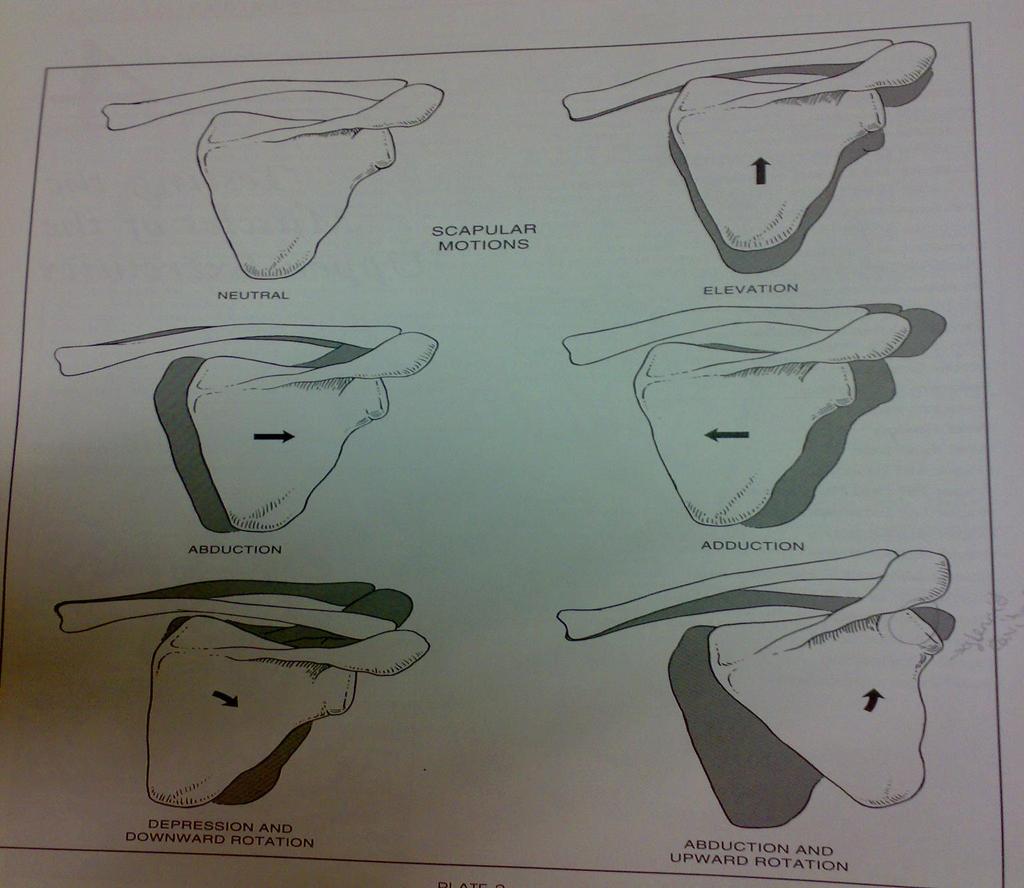
7 1. Scapula Abduction and Upward Rotation 2. Scapula Elevation 3. Scapula Adduction 4. Scapula Depression and Adduction 5. Scapula Adduction and Downward Rotation 7

8 1. Prime mover/agonist: origin insertion Serratus anterior ribs 1-8 intercostal fascia Scapula (ventral surface of vertebral border) 2. Synergist/ Accessory muscles: Pectoralis minor. 3. Nerve supply: Long thoracic n. C5-C7 4. Range of motion: measure the distance between spine process and medial border of scapula. 8
9 5. Fixation: a. in strong scapular abduction, by pull of obliqus externus abdominis on same side. b. By weight of thorax. 6. Effect of weakness ancontracture:-video? Effect of weakness result in: - Winging of Scapula (due to paralysis of serratus anterior) - In ability to raise the arm overhead. 7. Factor limited range of motion: a. Tension of trapezoid ligament. b. Tension of trapezius and rhomboid major and rhomboid minor muscles. 8. Substitution: None. 9
10 9. Procedures: a- observation for scapula position: At Rest and with motion. b- patient position (pt): c- Therapist Position: inner hand: Outer hand: Direction of Resistance : Instruction to patient: c- grading system: Normal(5), Good(4), Fair(3), Poor(2), Trace(1), Zero(0) make sure patient tolerates maximal resistance plus hold 3 sec. e. Palpation site: 10
 and Dorsal Scapular n.")
11 1. Prime mover/agonist: origin insertion Trapezius Occiput C7 Vert.SP. Clavicle (post. border) (superior fibers) Ligamentum nuchae Levator scapulae C1-C4 vert tp. Scapula (vert. border superior angle and root of spine 2. Synergist/ Accessory muscles: Rhomboid major and minor. 3. Nerve supply: XI Accessory(C3-C4) and Dorsal Scapular n.(c5) for Levator scapulae, XI Accessory(C3-C4) n. for Trapizius (superior fibers) 4. Range of motion: measure the distance between top of the shoulder and loops of ear. 11
12 5. Fixation: a. By flexor muscles of cervical spine.. b. By weight of head. 6. Effect of weakness/contracture/shortening: Effect of weakness result in: effect of weakness result in: In ability to raise shoulder upwards; either bilateral or unilateral weakness. 7. Factor limited range of motion: a. Tension of costoclavicular ligament. b. Tension of scapular depression muscle and clavicle: pectoralis minor, subclavius, and trapezius (lower fibers) muscles. 8. Substitution: by Rhomboids (scapula adduction and downward rotation) inferior angle will move medially. 12
13 9. Procedures: a- observation for scapula position: At Rest and with motion. b- patient position (pt): c- Therapist Position: inner hand: Outer hand: Direction of Resistance : Instruction to patient: c- grading system: Normal(5), Good(4), Fair(3), Poor(2), Trace(1), Zero(0) make sure patient tolerates maximal resistance plus hold 3 sec. e. Palpation site: 13
14 1. Prime mover/agonist: origin insertion Trapezius T1-T5 vert.sp Scapula (superior lip of spine) (middle fibers) 2. Synergist/ Accessory muscles: Rhomboid major and minor, and Trapezius (upper and lower fibers) 3. Nerve supply: XI Accessory(C3-C4) n. 4. Range of motion: measure the distance between transverse process and medial border of scapula 14
15 5. Fixation: a. By weight of thorax. 6. Effect of weakness/contracture/shortening: effect of weakness result in: In ability to adduct the scapula. 7. Factor limited range of motion: a. Tension of conoid ligament. b. Tension of pectoralis major and minor, and serratus anterior muscles. c. Contact of vertebral border of scapula with spinal musculature. 8. Substitution: by Rhomboids (scapula adduction and downward rotation) inferior angle will move medially. By the post. Deltoid if the scapular muscles are absent (horizontal adduction of shoulder will occur). 15
16 9. Procedures: a- observation for scapula position: At Rest and with motion. b- patient position (pt): c- Therapist Position: inner hand: Outer hand: Direction of Resistance : Instruction to patient: c- grading system: Normal(5), Good(4), Fair(3), Poor(2), Trace(1), Zero(0) make sure patient tolerates maximal resistance plus hold 3 sec. e. Palpation site: 16
17 1. Prime mover/agonist: origin insertion Trapezius T1-T5 vert.sp Scapula (spine) (lower fibers) 2. Synergist/ Accessory muscles: latissimus dorsi. 3. Nerve supply: XI Accessory(C3-C4) n. 4. Range of motion: measure the distance between spine process and inferior angle of scapula 17
18 5. Fixation: a. By contraction of spinal extensor muscles. b. By weight of thorax. 6. Effect of weakness/contracture/shortening: Effect of weakness result in: - In ability to raise the arm overhead. 7. Factor limited range of motion: a. Tension of inter clavicular ligament and articular disk of sternoclavicular joint. b. Tension of trapezius muscle. 8. Substitution: None. 18
19 9. Procedures: a- observation for scapula position: At Rest and with motion. b- patient position (pt): c- Therapist Position: inner hand: Outer hand: Direction of Resistance : Instruction to patient: c- grading system: Normal(5), Good(4), Fair(3), Poor(2), Trace(1), Zero(0) make sure patient tolerates maximal resistance plus hold 3 sec. e. Palpation site: 19
 Rhomboid minor C7-T1 vert sp Scapula (vert border at root of spine ) 2. Synergist/ Accessory muscles: Levator scapulae. 3.")
20 1. Prime mover/agonist: origin insertion Rhomboid major T2-T5 vert. sp Scapula (vertebral border between root of spine and inferior angle) Rhomboid minor C7-T1 vert sp Scapula (vert border at root of spine ) 2. Synergist/ Accessory muscles: Levator scapulae. 3. Nerve supply: dorsal scapular n. C5 4. Range of motion: measure the distance between the vertebral border of scapula and vert sp. 20
21 5. Fixation: a. By weight of thorax. 6. Effect of weakness/contracture/shortening: effect of weakness result in: - The scapula will abduct and the inferior angle rotate outward. - In ability to adduct & downward rotate the scapula. 7. Factor limited range of motion: a. Tension of conoid ligament. b. Tension of pectoralis major and minor, serratus anterior muscles. c. Contact of vertebral border of scapula with spinal musculature. 8. Substitution: by middle trapezius (only adduct the scapula without downward rotation). 21
22 9. Procedures: a- observation for scapula position: At Rest and with motion. b- patient position (pt): c- Therapist Position: inner hand: Outer hand: Direction of Resistance : Instruction to patient: c- grading system: Normal(5), Good(4), Fair(3), Poor(2), Trace(1), Zero(0) make sure patient tolerates maximal resistance plus hold 3 sec. e. Palpation site: 22
23 Thank You 23
Muscle Action Origin Insertion Nerve Innervation Chapter Page. Deltoid. Trapezius. Latissimus Dorsi
 Muscle Action Origin Insertion Nerve Innervation Chapter Page All Fibers Abduct the shoulder (glenohumeral joint) Deltoid Anterior Fibers Flex the shoulder (G/H joint) Horizontally adduct the shoulder
Upper Limb Muscles Muscles of Axilla & Arm
 Done By : Saleh Salahat Upper Limb Muscles Muscles of Axilla & Arm 1) Muscles around the axilla A- Muscles connecting the upper to thoracic wall (4) 1- pectoralis major Origin:- from the medial half of
Shoulder: Clinical Anatomy, Kinematics & Biomechanics
 Shoulder: Clinical Anatomy, Kinematics & Biomechanics Dr. Alex K C Poon Department of Orthopaedics & Traumatology Pamela Youde Nethersole Eastern Hospital Clinical Anatomy the application of anatomy to
Anatomy of the Shoulder Girdle. Prof Oluwadiya Kehinde FMCS (Orthop)
") Anatomy of the Shoulder Girdle Prof Oluwadiya Kehinde FMCS (Orthop) www.oluwadiya.com Bony Anatomy Shoulder Complex: Sternum(manubrium) Clavicle Scapula Proximal humerus Manubrium Sterni Upper part of
Anatomy of the Shoulder Girdle Prof Oluwadiya Kehinde FMCS (Orthop) www.oluwadiya.com Bony Anatomy Shoulder Complex: Sternum(manubrium) Clavicle Scapula Proximal humerus Manubrium Sterni Upper part of
MUSCLE MECHANICS AND CONTROL
 MUSCLE MECHANICS AND CONTROL STRUCTURE OF A SKELETAL MUSCLE NEUROVASCULAR BUNDLE - CONTAINS THE BLOOD VESSELS AND THE NERVES TO A MUSCLE SKELETAL MUSCLE - LONGITUDINAL SECTION MUSCLE ATTACHMENTS: TENDONS
Scapular and Deltoid Regions
 M1 Gross and Developmental Anatomy Scapular and Deltoid Regions Dr. Peters 1 Outline I. Skeleton of the Shoulder and Attachment of the Upper Extremity to Trunk II. Positions and Movements of the Scapula
Main Menu. Shoulder Girdle click here. The Power is in Your Hands. 1:07:11 PM]
![Main Menu. Shoulder Girdle click here. The Power is in Your Hands. 1:07:11 PM]](/thumbs/86/94418253.jpg "Main Menu. Shoulder Girdle click here. The Power is in Your Hands. 1:07:11 PM]") 1 The Shoulder Girdle click here Main Menu K.2 http://www.handsonlineeducation.com/classes//k2entry.htm[3/23/18, 1:07:11 PM] Bones Scapula and Clavicle Move as a unit Clavicle s articulation with sternum
1 The Shoulder Girdle click here Main Menu K.2 http://www.handsonlineeducation.com/classes//k2entry.htm[3/23/18, 1:07:11 PM] Bones Scapula and Clavicle Move as a unit Clavicle s articulation with sternum
Scapular Dyskinesis. Orthopaedic Update 2018 April 15, Peter Tang, MD, MPH, FAOA
 Scapular Dyskinesis Orthopaedic Update 2018 April 15, 2018 Peter Tang, MD, MPH, FAOA Director Center for Brachial Plexus and Nerve Injury Program Director Hand, Upper Extremity & Microvascular Surgery
3 Mohammad Al-Mohtasib Areej Mosleh
 3 Mohammad Al-Mohtasib Areej Mosleh ***Muscles Connecting the Upper Limb to the Vertebral Column 1.Trapezius Muscle ***The first muscle on the back is trapezius muscle, it s called so according
Joint G*H. Joint S*C. Joint A*C. Labrum. Humerus. Sternum. Scapula. Clavicle. Thorax. Articulation. Scapulo- Thoracic
 A*C Joint Scapulo- Thoracic Articulation Thorax Sternum Clavicle Scapula Humerus S*C Joint G*H Joint Labrum AC Ligaments SC Ligaments SC JOINT AC Coracoacromial GH GH Ligament Complex Coracoclavicular
Clinical examination of the shoulder girdle
 Clinical of the shoulder girdle CHAPTER CONTENTS Symptoms referred to the shoulder girdle........ e72 Symptoms referred from the shoulder girdle...... e72 History........................... e72 Inspection.........................
Pectoral region. Lecture 2
 Pectoral region Lecture 2 Muscle Action Each muscle has: Origin Beginning. Insertion End. Body (belly). Law: When a muscle performs its action, its insertion, moves towards its origin. Spring 2016 Dr.
Region of upper limb attachment to the trunk Proximal segment of limb overlaps parts of the trunk (thorax and back) and lower lateral neck.
 and lower lateral neck.") Region of upper limb attachment to the trunk Proximal segment of limb overlaps parts of the trunk (thorax and back) and lower lateral neck. includes Pectoral Scapular Deltoid regions of the upper limb
Region of upper limb attachment to the trunk Proximal segment of limb overlaps parts of the trunk (thorax and back) and lower lateral neck. includes Pectoral Scapular Deltoid regions of the upper limb
Returning the Shoulder Back to Optimal Function. Scapula. Clavicle. Humerus. Bones of the Shoulder (Osteology) Joints of the Shoulder (Arthrology)
 Joints of the Shoulder (Arthrology)") Returning the Shoulder Back to Optimal Function Sternum Clavicle Ribs Scapula Humerus Bones of the Shoulder (Osteology) By Rick Kaselj Clavicle Scapula Medial Left Anterior Clavicle Inferior View 20 degree
Returning the Shoulder Back to Optimal Function Sternum Clavicle Ribs Scapula Humerus Bones of the Shoulder (Osteology) By Rick Kaselj Clavicle Scapula Medial Left Anterior Clavicle Inferior View 20 degree
Pectoral region. Lecture 2
 Pectoral region Lecture 2 Muscle Action Each muscle has: Origin Beginning. Insertion End. Body (belly). Law: When a muscle performs its action, its insertion, moves towards its origin. Spring 2016 Dr.
Lab Workbook. ANATOMY Manual Muscle Testing Lower Trapezius Patient: prone
 ANATOMY Manual Muscle Testing Lower Trapezius Patient: prone Lab Workbook Fixation: place on hand below the scapula on the opposite side Test: adduction and depression of the scapula with lateral rotation
The pectoral region. University of Babylon College of Medicine Dr.HaythemAli Alsayigh M.B.CH.B.-F.I.M.B.S. Surgical Clinical Anatomy
 The pectoral region University of Babylon College of Medicine Dr.HaythemAli Alsayigh M.B.CH.B.-F.I.M.B.S. Surgical Clinical Anatomy Objective Study the Bones and Joints A. Clavicle (collarbone) B. Scapula
The Upper Limb II. Anatomy RHS 241 Lecture 11 Dr. Einas Al-Eisa
 The Upper Limb II Anatomy RHS 241 Lecture 11 Dr. Einas Al-Eisa Sternoclavicular joint Double joint.? Each side separated by intercalating articular disc Grasp the mid-portion of your clavicle on one side
MUSCLES OF SHOULDER REGION
 Dr Jamila EL Medany OBJECTIVES At the end of the lecture, students should: List the name of muscles of the shoulder region. Describe the anatomy of muscles of shoulder region regarding: attachments of
Upper limb Pectoral region & Axilla
 Upper limb Pectoral region & Axilla 黃敏銓 mchuang@ntu.edu.tw 1 Pectoral region Intercostal nerve Anterior branch of lateral cutaneous branch Lateral cutaneous branch Anterior cutaneous branch Anterior cutaneous
Upper limb Arm & Cubital region 黃敏銓
 Upper limb Arm & Cubital region 黃敏銓 1 Arm Lateral intermuscular septum Anterior (flexor) compartment: stronger Medial intermuscular septum Posterior (extensor) compartment 2 Coracobrachialis Origin: coracoid
Treatment of the Shoulder Girdle for Functional Outcomes. Postural Alignment and it s Effect on the Shoulder Girdle. Left Anterior Rotation of Pelvis
 Treatment of the Shoulder Girdle for Functional Outcomes Gail Ritchie, OTR/L Postural Alignment and it s Effect on the Shoulder Girdle Floating system Relies on the alignment of the axial skeleton Left
This figure (of humerus) is from Dr. Maher's newest slides. -Its added here just for consideration-
 is from Dr. Maher's newest slides. -Its added here just for consideration-") This figure (of humerus) is from Dr. Maher's newest slides. -Its added here just for consideration- Slides of Anatomy Please note : These slides are Dr. Maher Hadidi s slides of spring 2016 and were edited
This figure (of humerus) is from Dr. Maher's newest slides. -Its added here just for consideration- Slides of Anatomy Please note : These slides are Dr. Maher Hadidi s slides of spring 2016 and were edited
Continuing Education: Shoulder Stability
 Continuing Education: Shoulder Stability Anatomy & Kinesiology: The GHJ consists of the articulation of three bones: the scapula, clavicle and humerus. The scapula has three protrusions: the coracoid,
Connects arm to thorax 3 joints. Glenohumeral joint Acromioclavicular joint Sternoclavicular joint
 Connects arm to thorax 3 joints Glenohumeral joint Acromioclavicular joint Sternoclavicular joint Scapula Elevation Depression Protraction (abduction) Retraction (adduction) Downward Rotation Upward Rotation
بسم هللا الرحمن الرحيم
 بسم هللا الرحمن الرحيم Laboratory RHS 221 Manual Muscle Testing Theory 1 hour practical 2 hours Dr. Ali Aldali, MS, PT Department of Physical Therapy King Saud University Talocrural and Subtalar Joint
Sports Medicine Part II : ANATOMY OF THE SPINE, ABDOMEN AND SHOULDER COMPLEX
 Sports Medicine 25 1.1 Part II : ANATOMY OF THE SPINE, ABDOMEN AND SHOULDER COMPLEX c.w.p. Wagner High School, Sports Medicine, A. Morgan, T. Morgan & A. Eastlake, 2008 Muscles of the Upper Limbs In this
CLINICAL EXAMINATION OF THE SHOULDER JOINT 대한신경근골격연구회 분당제생병원재활의학과 박준성
 CLINICAL EXAMINATION OF THE SHOULDER JOINT 대한신경근골격연구회 분당제생병원재활의학과 박준성 Clinical Examination of the Shoulder Good history, full clinical examination Detailed knowledge of the anatomy solve the majority of
SHOULDER TO SHOULDER The Range Of Possibilities
 mouse click to advance the slides SHOULDER TO SHOULDER The Range Of Possibilities HATHA YOGA Hatha yoga asanas Take the shoulders through every possible range of motion in both weight-bearing and relatively
3 Movements of the Trunk. Flexion Rotation Extension
 3 Movements of the Trunk Flexion Rotation Extension 1 TRUNK FLEXION 2 TRUNK FLEXION: Rectus Abdominalis O: Crest of Pubis & ligaments covering front of symphysis pubis. I: By «3 portions into cartilages
Muscles in the Shoulder, Chest, Arm, Stomach, and Back
 Muscles in the Shoulder, Chest, Arm, Stomach, and Back Shoulder Muscles Deltoid Supraspinatus Infraspinatus Teres Major Teres Minor Subscapularis Deltoid (Delts) Function: Raises the upper arm Origin:
Prime movers provide the major force for producing a specific movement Antagonists oppose or reverse a particular movement Synergists
 Dr. Gary Mumaugh Prime movers provide the major force for producing a specific movement Antagonists oppose or reverse a particular movement Synergists Add force to a movement Reduce undesirable or unnecessary
MLT Muscle(s) Patient Position Therapist position Stabilization Limb Position Picture Put biceps on slack by bending elbow.
 Patient Position Therapist position Stabilization Limb Position Picture Put biceps on slack by bending elbow.") MLT Muscle(s) Patient Position Therapist position Stabilization Limb Position Picture Put biceps on slack by bending elbow. Pectoralis Minor Supine, arm at side, elbows extended, supinated Head of Table
MLT Muscle(s) Patient Position Therapist position Stabilization Limb Position Picture Put biceps on slack by bending elbow. Pectoralis Minor Supine, arm at side, elbows extended, supinated Head of Table
Shoulder Biomechanics
 Shoulder Biomechanics Lecture originally developed by Bryan Morrison, Ph.D. candidate Arizona State University Fall 2000 1 Outline Anatomy Biomechanics Problems 2 Shoulder Complex Greatest Greatest Predisposition
The Clavicle Right clavicle Deltoid tubercle: Conoid tubercle, conoid ligamen Impression for the
 The Clavicle Muscle Attachment Sites in the Upper Limb Pectoralis major Right clavicle Smooth superior surface of the shaft, under the platysma muscle tubercle: attachment of the deltoid Acromial facet
Structure and Function of the Bones and Joints of the Shoulder Girdle
 Structure and Function of the Bones and Joints of the Shoulder Girdle LEARNING OBJECTIVES: At the end of this laboratory exercise the student will be able to: Palpate the important skeletal landmarks of
Anatomy and Physiology II. Review Shoulder Girdle New Material Upper Extremities - Bones
 Anatomy and Physiology II Review Shoulder Girdle New Material Upper Extremities - Bones Anatomy and Physiology II Shoulder Girdle Review Questions From Last Lecture Can you identify the following muscles?
medial half of clavicle; Sternum; upper six costal cartilages External surfaces of ribs 3-5
 MUSCLE ORIGIN INSERTION ACTION NERVE Pectoralis Major medial half of clavicle; Sternum; upper six costal cartilages Lateral lip of intertubercular groove of horizontal adduction Medial and lateral pectoral
G24: Shoulder and Axilla
 G24: Shoulder and Axilla Syllabus - Pg. 2 ANAT 6010- Medical Gross Anatomy David A. Morton, Ph.D. Objectives Upper limb Systemically: Bones (joints) Muscles Nerves Vessels (arteries/veins) Fascial compartments
BLUE SKY SCHOOL OF PROFESSIONAL MASSAGE AND THERAPEUTIC BODYWORK. Musculoskeletal Anatomy & Kinesiology II REVIEW
 BLUE SKY SCHOOL OF PROFESSIONAL MASSAGE AND THERAPEUTIC BODYWORK Musculoskeletal Anatomy & Kinesiology II REVIEW MSAK101-II Session 4 LEARNING OBJECTIVES: By the end of this session, the student will be
Thoracolumbar Anatomy Eric Shamus Catherine Patla Objectives
 1 2 Thoracolumbar Anatomy Eric Shamus Catherine Patla Objectives List the muscular and ligamentous attachments of the thoracic and lumbar spine Describe how the muscles affect the spine and upper extremity
FUNCTIONAL ANATOMY OF SHOULDER JOINT
 FUNCTIONAL ANATOMY OF SHOULDER JOINT ARTICULATION Articulation is between: The rounded head of the Glenoid cavity humerus and The shallow, pear-shaped glenoid cavity of the scapula. 2 The articular surfaces
Muscles of the Upper Limb
 Muscles of the Upper Limb anterior surface of ribs 3 5 coracoid process Pectoralis minor pectoral nerves protracts / depresses scapula Serratus anterior Subclavius ribs 1-8 long thoracic nerve rib 1 ----------------
Gross Anatomy Faculty: Gross Anatomy Faculty: Gross Anatomy Faculty: Dr. Melissa McGinn. Welcome to Gross and Developmental Anatomy
 Welcome to Gross and Developmental Anatomy M1 Anatomy Gross Anatomy Faculty: Dr. Richard Krieg Dr. Milton Sholley Dr. David Simpson 1 2 Gross Anatomy Faculty: Gross Anatomy Faculty: Dr. Steve Gudas Dr.
Lab Activity 11: Group I
 Lab Activity 11: Group I Muscles Martini Chapter 11 Portland Community College BI 231 Origin and Insertion Origin: The place where the fixed end attaches to a bone, cartilage, or connective tissue. Insertion:
Anatomy and Physiology II. Spine
 Anatomy and Physiology II Spine Bones and Other Structures Vertibrae Contains Cervical, Thoracic, Lumbar, Sacral and Coccygeal regions We use Capital letters to refer to these (C, T, L, S, and Co) and
Physical Examination of the Shoulder
 General setup Patient will be examined in both the seated and supine position so exam table needed 360 degree access to patient Expose neck and both shoulders (for comparison); female in gown or sports
SHOULDER JOINT ANATOMY AND KINESIOLOGY
 SHOULDER JOINT ANATOMY AND KINESIOLOGY SHOULDER JOINT ANATOMY AND KINESIOLOGY The shoulder joint, also called the glenohumeral joint, consists of the scapula and humerus. The motions of the shoulder joint
WEEKEND THREE HOMEWORK
 WEEKEND THREE HOMEWORK READING ASSIGNMENTS Salvo Massage Therapy Principles and Practice 4 th Edition Muscolino The Muscular System Manual Muscolino The Muscle and Bone Palpation Manual Ch. 19 Skeletal
INSTRUCTION MANUAL FOR THE FLEXTEND AC Exercise System for The Acromioclavicular (AC) / Shoulder Joint
 / Shoulder Joint") INSTRUCTION MANUAL FOR THE FLEXTEND AC Exercise System for The Acromioclavicular (AC) / Shoulder Joint FLEXTEND -AC: Congratulations! You have chosen to use the FLEXTEND -AC Upper Extremity Training System,
INSTRUCTION MANUAL FOR THE FLEXTEND AC Exercise System for The Acromioclavicular (AC) / Shoulder Joint FLEXTEND -AC: Congratulations! You have chosen to use the FLEXTEND -AC Upper Extremity Training System,
Anatomical Considerations/ Pathophysiology The shoulder is the most mobile joint in the body. : Three bones:
 Introduction Musculoskeletal training is generally underrepresented in medical training and residency curriculums. There is a general deficit in musculoskeletal knowledge amongst current medical students,
STEP #1: Anatomy STEP #2: Awareness STEP #3: Action
 Stabilize the Scapula in Three Easy Steps by NFPT Idea World 2016: Session 611 Saturday July 16th 7:30-9:20am Beverly Hosford, MA STEP #1: Anatomy STEP #2: Awareness STEP #3: Action 1. Anatomy *Memorize
Axilla and Brachial Region
 L 4 A B O R A T O R Y Axilla and Brachial Region BRACHIAL PLEXUS 5 Roots/Rami (ventral rami C5 T1) 3 Trunks Superior (C5, C6) Middle (C7) Inferior (C8, T1) 3 Cords Lateral Cord (Anterior Superior and Anterior
Synergist Muscles. Shoulder (glenohumeral joint) Flexion Deltoid (anterior fibers) Pectoralis major (upper fibers) Biceps Brachii Coracobrachialis
 Flexion Deltoid (anterior fibers) Pectoralis major (upper fibers) Biceps Brachii Coracobrachialis") Synergist Muscles Dr Gene Desepoli DrGeneLMT@gmail.com Shoulder (glenohumeral joint) Deltoid (anterior fibers) Pectoralis major (upper fibers) Biceps Brachii Coracobrachialis Deltoid (posterior fibers)
Synergist Muscles Dr Gene Desepoli DrGeneLMT@gmail.com Shoulder (glenohumeral joint) Deltoid (anterior fibers) Pectoralis major (upper fibers) Biceps Brachii Coracobrachialis Deltoid (posterior fibers)
9/4/10. James J. Lehman, DC, MBA, DABCO. Why is posture important to you, the chiropractic physician?
 James J. Lehman, DC, MBA, DABCO The posture of homo sapiens is a complex biomechanical continuum, which involves the function of muscles, ligaments, fascia, nerves, osseous structures, neuromuscular control,
Secrets and Staples of Training the Athletic Shoulder
 Secrets and Staples of Training the Athletic Shoulder Eric Beard Corrective Exercise Specialist Athletic Performance Enhancement Specialist EricBeard.com AthleticShoulder.com Presentation Overview Rationale
UPPERTONE Exercise Manual
 UPPERTONE Exercise Manual GPK Inc., 535 Floyd Smith Dr., El Cajon, CA 92020, 800-468-8679, 619-593-7381, Fax: 619-593-7514, www.gpk.com Index Introduction...... 3 The Major Muscles... 4 Definitions of
213: HUMAN FUNCTIONAL ANATOMY: PRACTICAL CLASS 1: Proximal bones, plexuses and patterns
 213: HUMAN FUNCTIONAL ANATOMY: PRACTICAL CLASS 1: Proximal bones, plexuses and patterns CLAVICLE Examine an isolated clavicle and compare it with a clavicle on an articulated skeleton. Viewed from above,
Muscles of the Upper Limb that are dissected in the Back Region Muscle Origin Insertion Action Innervation Artery Notes
 Muscles of Upper Limb that are dissected in Back Region Muscle Origin Insertion Action Innervation Artery Notes floor of thoraco thoraco inserting spines from intertubercular arm nerve (C7,8) a. tendon
Overuse Injuries. Overuse injury defined. Overuse Injuries
 Overuse Injuries Lisa DeStefano, DO Associate Professor and Chair Department of Osteopathic Manipulative Medicine College of Osteopathic Medicine Michigan State University Overuse injury defined Overuse
*Agonists are the main muscles responsible for the action. *Antagonists oppose the agonists and can help neutralize actions. Since many muscles have
 1 *Agonists are the main muscles responsible for the action. *Antagonists oppose the agonists and can help neutralize actions. Since many muscles have more than 1 action sometimes a muscle has to neutralize
Improving swimming through Pilates
 Improving swimming through Pilates Cleia Ramsey 10 Mar 2017 2016 BASI Training Class Herndon, VA Introduction As a mother of two swimmers, I spend many hours at the pool deck and have learned a lot about
Functional Anatomy. CHAPTER 5 Functional Anatomy of the Upper Extremity. CHAPTER 6 Functional Anatomy of the Lower Extremity
 Hamill_ch05_137-186.qxd 11/2/07 3:55 PM Page 137 S E C T I O N II Functional Anatomy CHAPTER 5 Functional Anatomy of the Upper Extremity CHAPTER 6 Functional Anatomy of the Lower Extremity CHAPTER 7 Functional
Mercer County Community College Donna Doulong March 8, 2013
 Mercer County Community College Donna Doulong March 8, 2013 Why strengthen the scapular muscles? The scapular thoracic articulation is the true core of the upper extremity. (Shankman & Manske, 2011, p.
Current Concepts in the Management of Patients with Shoulder Pain
 Current Concepts in the Management of Patients with Shoulder Pain CAD Meeting Education Topics Low Back Pain Alternative Medicine Legal Issues NDT Shoulder Pain Aquatics Wound Care Marketing Your Practice
The Back. Anatomy RHS 241 Lecture 9 Dr. Einas Al-Eisa
 The Back Anatomy RHS 241 Lecture 9 Dr. Einas Al-Eisa The spine has to meet 2 functions Strength Mobility Stability of the vertebral column is provided by: Deep intrinsic muscles of the back Ligaments
THE SHOULDER JOINT T H E G L E N O H U M E R A L ( G H ) J O I N T
 J O I N T") THE SHOULDER JOINT T H E G L E N O H U M E R A L ( G H ) J O I N T CLARIFICATION OF TERMS Shoulder girdle = scapula and clavicle Shoulder joint (glenohumeral joint) = scapula and humerus Lippert, p115
THE SHOULDER JOINT T H E G L E N O H U M E R A L ( G H ) J O I N T CLARIFICATION OF TERMS Shoulder girdle = scapula and clavicle Shoulder joint (glenohumeral joint) = scapula and humerus Lippert, p115
Gateway to the upper limb. An area of transition between the neck and the arm.
 Gateway to the upper limb An area of transition between the neck and the arm. Pyramidal space inferior to shoulder @ junction of arm & thorax Distribution center for the neurovascular structures that serve
Chapter 2 Examination of the Shoulder
 Chapter 2 Examination of the Shoulder The shoulder girdle consists of three joints and one articulation namely: 1. The sternoclavicular joint 2. The acromioclavicular joint 3. The glenohumeral or shoulder
Pectoral girdle, SUPERIEUR ARM AND HAND. Danil Hammoudi.MD
 Pectoral girdle, SUPERIEUR ARM AND HAND Danil Hammoudi.MD The pectoral girdle is the set of bones which connect the upper limb to the axial skeleton on each side. It consists of the clavicle scapula in
Describe methods to evaluate for scapular. Perform a scapular dyskinesis examination. With humeral elevation, the scapula:
 Describe methods to evaluate for scapular dyskinesis Perform a scapular dyskinesis examination Lori Michener, PhD, PT, ATC Virginia Commonwealth University Richmond, VA Ant / Post Tilting Internal / External
Yoga Straps for Scapular Strength
 ACE Pro Source Yoga Straps for Scapular Strength By Elizabeth R. Kovar M.A. In a well-rounded yoga practice, it is important to demonstrate scapular or shoulder blade strength. For some people, however,
1TRUNK: BODY WALL AND SPINE
 TRUNK: BODY WALL AND SPINE SURFACE ANATOMY SKELETON JOINTS & LIGAMENTS MUSCLES VASCULATURE NERVES SPINAL CORD & VERTEBRAL CANAL ANTERIOR BODY WALL & MAMMARY GLAND LATERAL BODY WALL INGUINAL REGION SUPERFICIAL
Prater Chiropractic Wellness Center 903 W. South St. Kalamazoo, MI PH: (269)
") Purpose of Program After an injury or surgery, an exercise conditioning program will help you return to daily activities and enjoy a more active, healthy lifestyle. Following a well-structured conditioning
Purpose of Program After an injury or surgery, an exercise conditioning program will help you return to daily activities and enjoy a more active, healthy lifestyle. Following a well-structured conditioning
Certified Personal Trainer Re-Certification Manual
 Certified Personal Trainer Re-Certification Manual Section II 1 Anatomy & Physiology Terms Anatomy and physiology are closely related fields of study: anatomy is the study of form, and physiology is the
Unit 3 -- Relieve the Burden of Shoulder Dysfunction. Upper Torso & Shoulder Unit Study Guide
 Unit 3 -- Relieve the Burden of Shoulder Dysfunction Upper Torso & Shoulder Unit Study Guide This unit identifies problems of the shoulder joint and thoracic regions. Module 1 Module 2 Module 3 Module
The Shoulder. Anatomy and Injuries PSK 4U Unit 3, Day 4
 The Shoulder Anatomy and Injuries PSK 4U Unit 3, Day 4 Shoulder Girdle Shoulder Complex is the most mobile joint in the body. Scapula Clavicle Sternum Humerus Rib cage/thorax Shoulder Girdle It also includes
Human Anatomy Biology 351
 1 Human Anatomy Biology 351 Upper Limb Exam Please place your name on the back of the last page of this exam. You must answer all questions on this exam. Because statistics demonstrate that, on average,
Chapter 3: Applied Kinesiology. ACE Personal Trainer Manual Third Edition
 Chapter 3: Applied Kinesiology ACE Personal Trainer Manual Third Edition Introduction Kinesiology is the study of the body s infinite number of movements, positions, and postures and is grounded in the
7/31/2012 THE SHOULDER JOINT CLARIFICATION OF TERMS OSTEOLOGY OF THE GH JOINT(BONES)
") THE SHOULDER JOINT T H E G L E N O H U M E R AL ( G H ) J O I N T CLARIFICATION OF TERMS Shoulder girdle = scapula and clavicle Shoulder joint (glenohumerual joint) = scapula and Lippert, p115 OSTEOLOGY
THE SHOULDER JOINT T H E G L E N O H U M E R AL ( G H ) J O I N T CLARIFICATION OF TERMS Shoulder girdle = scapula and clavicle Shoulder joint (glenohumerual joint) = scapula and Lippert, p115 OSTEOLOGY
Anatomy & Physiology B. Chapter 6: Muscles
 Anatomy & Physiology B Chapter 6: Muscles Warm-up What are the three types of muscle tissue? Where are each located? Which are voluntary and which are involuntary? Which are striated which are unstriated?
CLINICAL SUMMARY AND RECOMMENDATIONS 378
 Shoulder 9 CLINICAL SUMMARY AND RECOMMENDATIONS 378 Anatomy 379 Osteology 379 Arthrology 380 Scapulohumeral Rhythm 381 Ligaments 382 Muscles 384 Nerves 387 Patient History 389 Initial Hypotheses Based
Throwing Athlete Rehabilitation. Brett Schulz LAT/CMSS Sport and Spine Physical Therapy
 Throwing Athlete Rehabilitation Brett Schulz LAT/CMSS Sport and Spine Physical Therapy Disclosure No conflicts to disclose Throwing Athlete Dilemma The shoulder must have enough range of motion to allow
Glenohumeral. Laura Leonetti Genna Moak Taylor Hansen
 Glenohumeral Laura Leonetti Genna Moak Taylor Hansen Surface anatomy Genna Anterior axillary fold Clavicle Clavicular head of Pectoralis major Clavipectoral triangle Acromial part of Deltoid Manubrium
Introduction. Rarely does a single muscle act in isolation at the shoulder complex.
 Shoulder complex 1 Introduction Our study of the upper limb begins with the shoulder complex, a set of four articulations involving the sternum, clavicle, ribs, scapula, and humerus. Rarely does a single
Muscles Built on the Maniken
 Muscles Built on the Maniken Facial Muscle Group 1. Temporalis O temporal fossa I anterior border of the ramus of the mandible A elevates the mandible (bite muscle) and holds jaw while at rest 2. Procerus
The Upper Limb III. The Brachial Plexus. Anatomy RHS 241 Lecture 12 Dr. Einas Al-Eisa
 The Upper Limb III The Brachial Plexus Anatomy RHS 241 Lecture 12 Dr. Einas Al-Eisa Brachial plexus Network of nerves supplying the upper limb Compression of the plexus results in motor & sensory changes
Anatomy and Physiology II. Review Spine and Neck
 Anatomy and Physiology II Review Spine and Neck Spine regions How many cervical vertibrae are there? 7 The curvature is the cervical region posterior? Concave posterior How many thoracic? And curvature?
Module 6 - The Muscular System Introduction to the Muscular System and Muscles of the Head, Neck and Shoulder
 Module 6 - The Muscular System Introduction to the Muscular System and Muscles of the Head, Neck and Shoulder There will be three modules to cover the muscle anatomy of the body. The first module will
30b Passive Stretches:! Technique Demo and Practice - Upper Body
 30b Passive Stretches:! Technique Demo and Practice - Upper Body 30b Passive Stretches:! Technique Demo and Practice - Upper Body! Class Outline" 5 minutes" "Attendance, Breath of Arrival, and Reminders
SUPERIEUR ARM AND HAND
 Pectoral girdle, SUPERIEUR ARM AND HAND Danil Hammoudi.MD The pectoral girdle is the set of bones which connect the upper limb to the axial skeleton on each side. It consists of the clavicle scapula in
Rotator Cuff and Shoulder Conditioning Program
 Rotator Cuff and Shoulder Conditioning Program Purpose of Program After an injury or surgery, an exercise conditioning program will help you return to daily activities and enjoy a more active, healthy
Structure and Function of the Vertebral Column
 Structure and Function of the Vertebral Column Posture Vertebral Alignment Does it really matter? Yes it does! Postural Curves The vertebral column has a series of counterbalancing curves posterior anterior
Muscles Of The Back. Musculoskeletal block- Anatomy-lecture 3. Editing file
 Muscles Of The Back Musculoskeletal block- Anatomy-lecture 3 Editing file Objectives At the end of the lecture, students should be able to: Distinguish between the different groups of back muscles. Compare
Thoracic Muscles Origin Insertion Action Innervation
 MUSCLES OF THE THORAX, BACK & ABDOMEN Muscles of the Thorax Thoracic Muscles Origin Insertion Action Innervation M. pectoralis major pars clavicularis clavicula (medial ½ ) M. pectoralis major pars sternocostalis
Thoracic and Lumbar Spine Anatomy.
 Thoracic and Lumbar Spine Anatomy www.fisiokinesiterapia.biz Thoracic Vertebrae Bodies Pedicles Laminae Spinous Processes Transverse Processes Inferior & Superior Facets Distinguishing Feature Costal Fovea
Chest cavity, vertebral column and back muscles. Respiratory muscles. Sándor Katz M.D., Ph.D.
 Chest cavity, vertebral column and back muscles. Respiratory muscles. Sándor Katz M.D., Ph.D. Chest cavity - bony structures Chest cavity- bony structures Sternum Ribs True ribs: first seven pairs connect
Active-Assisted Stretches
 1 Active-Assisted Stretches Adequate flexibility is fundamental to a functional musculoskeletal system which represents the foundation of movement efficiency. Therefore a commitment toward appropriate
Muscular Nomenclature and Kinesiology - One
 Chapter 16 Muscular Nomenclature and Kinesiology - One Lessons 1-3 (with lesson 4) 1 Introduction 122 major muscles covered in this chapter Chapter divided into nine lessons Kinesiology study of human
Cervico-Thoracic Management Exercise and Manual Therapy. Deep Neck Flexor Training. Deep Neck Flexor Training. FPTA Spring 2011 Eric Chaconas 1
 Cervico-Thoracic Management Exercise and Manual Therapy Eric Chaconas PT, DPT, CSCS, FAAOMPT Deep Neck Flexor Training Evidence of dysfunction in the longus coli and longus capitus. Chronic Neck Pain Idiopathic