3/16/2018. Ultrasound Biomicroscopy in Glaucoma By Ahmed Salah Abdel Rehim. Prof. of Ophthalmology Al-Azhar University
|
|
|
- David Stone
- 5 years ago
- Views:
Transcription

1 Ultrasound Biomicroscopy in Glaucoma By Ahmed Salah Abdel Rehim Prof. of Ophthalmology Al-Azhar University 1
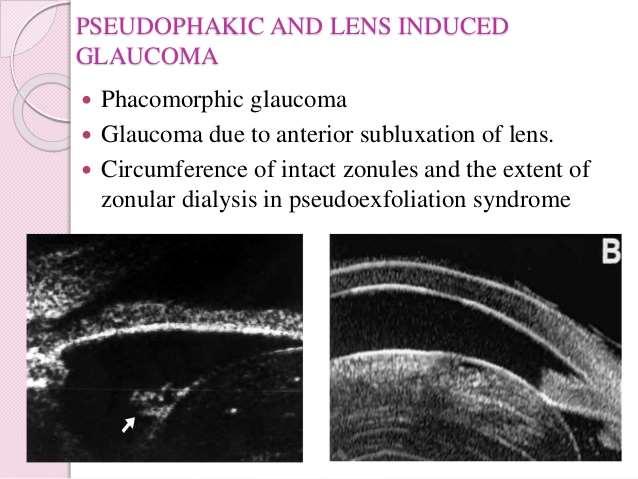
2 Ultrasound biomicroscopy (UBM) is a recent technique to visualize anterior segment with the help of high frequency ultrasound transducer. UBM is capable to show the structures of anterior segment that are relevant to glaucoma and provides a system of measurements. UBM has many advantages including easy, non-invasive, inexpensive and reproducible. Glaucoma is an eye disorder in which the optic nerve suffers damage. It is often, but not always, associated with increased IOP. 2


3 There are four major types of glaucoma each of them is subdivided into primary and secondary categories; angle closure glaucoma, open angle glaucoma, combined-mechanism glaucoma and developmental glaucoma. examination procedures: The patient is usually examined in a supine position looking up at the ceiling. Series of eye cups have been designed; they have a smooth flanged inferior margin that fits between the eyelids and hold them open. 3
4 Fluid is required to produce a coupling medium between the transducer and the eye. The main rule for making fine probe movements is "if the image is getting better, keep going; if it is getting worse, go to the opposite way". The best images are obtained in any ultrasound examination when the ultrasound beam is perpendicular to the structures being examined. the front of the transducer corresponds to the top of the screen. There is a mark on the probe that indicates the plane of the scanning motion. 4

5 This mark coincides with the left side of the screen. Thus if the probe is held with the mark oriented toward the temporal aspect of the eye, the left side of the screen would display pathology located in the temporal side of the globe. Examination Conventions Radial Section Of The Globe: Radial sections of the globe are performed with the probe marker on the scleral side 5

6 Transverse Section Of The Globe: Transverse sections of the globe are performed with the probe marker on the counterclockwise side The UBM images were evaluated for; central corneal thickness, anterior chamber depth, anterior chamber angle parameters, iris, ciliary body, visibility of a route under the scleral flap, reflectivity inside the bleb and formation of a cavernous fluid filled space inside the bleb. 6


7 The Cornea: The superficial location of the cornea permits the use of higher frequency transducers. Higher frequency transducers allow a better definition of small distances such as epithelial thickness. The Corneoscleral junction: The corneoscleral junction can be differentiated because of the lower internal reflectivity of the cornea compared to the sclera. The inner junction is generally referred to as schwalbe's line. 7

8 The corneoscleral junction and scleral spur can be distinguished consistently with ultrasound biomicroscopy. The scleral spur is a useful landmark presenting a constant reference point for measurement in the angle region. Anterior Chamber: The axial distance is measured from the internal corneal surface to the lens surface. The average axial anterior chamber depth measurement is 3128 ± 372 um. 8
: is a distance between a point on the internal ocular wall 500 um anterior to the scleral spur perpendicular to the plane of")
9 The Angle: The angle can be visualized by orienting the probe in a radial fashion above the limbal region. How to measure the angle width: angle opening distance (AOD): is a distance between a point on the internal ocular wall 500 um anterior to the scleral spur perpendicular to the plane of the trabecular meshwork, to the opposing iris. Normally, it measures 347 ± 181 um. 9

10 Trabecular-iris angle (θ1): The angle formed with the apex at the iris recess, and the arms passing through the point on the meshwork 500 µm from the scleral spur and the point on the iris perpendicularly opposite. 10


11 The Iris: The iris shows variations in thickness. there are variations in thickness depending on the presence of crypts and the state of dilation or constriction of the pupil. The iris epithelium forms a constant highly reflective layer on the posterior iris surface. 11
12 How to measure the iris thickness Trabecular-ciliary process distance (TCPD): A line is run from the point on the trabecular meshwork 500 um from the scleral spur, perpendicularly through the iris, extended to the ciliary process (average distance = 1122 ± 232 um) How to measure the iris thickness ID1: is the Iris thickness along TCPD line (average thickness = 372 ± 58 um). ID2: is measured 2 mm from the iris root (average thickness = 457 ± 80 um). ID3: is measured near the pupil (average thickness = 645 ± 103 um). 12
13 The zone of iris-lens contact distance (ILCD) is 1388 ± 370 um, normally. The angle at which the iris leaves the lens named θ2 (average = 12 ± 3 ). The angle the iris makes to a tangent to the scleral surface named θ3 (average of 30±7 ). The angle the ciliary processes make to the scleral surface can also be defined θ4 (average = 52 ± 18 ). 13
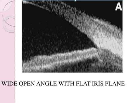
14 Normal anatomical angle UBM in Glaucoma Open angle glaucoma 14
15 15

16 Closed angle glaucoma 16

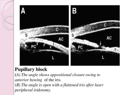
17 Before After 17
18 18

19 Secondary glaucoma 19
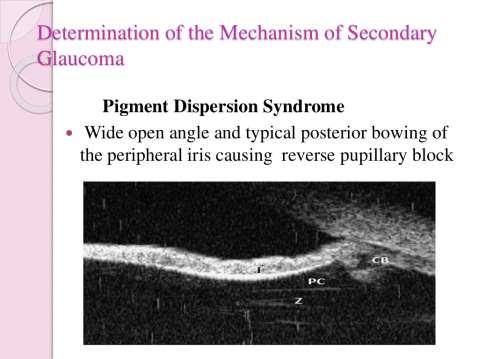
20 20
21 21
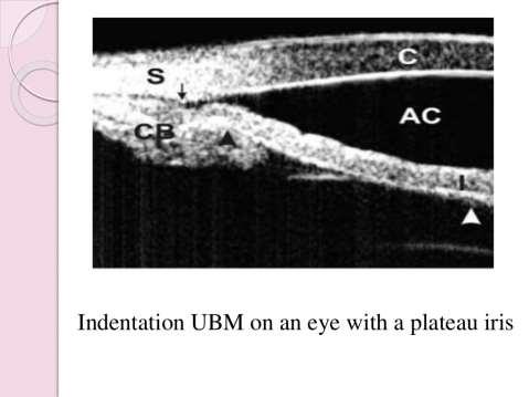
22 22
23 UBM after trabeculectomy 23
24 The UBM images of eyes with good IOP control are characterized by better visibility of the route under the scleral flap and a low reflectivity inside the bleb, the internal structure displayed variable internal reflectivity and a clear fluid space was also sometimes found. The UBM images of eyes with poor IOP control are characterized by lesser visibility of the route under the scleral flap, higher reflectivity inside the bleb or the formation of a cavernous fluid filled space surrounded by a bleb wall of high reflectivity. 24


25 AOD/ Ɵ1 Iris-lens angle (Ɵ2) a. preoperative (19.1 D). b. postoperative(13.1 D). 25
26 Ostium Types of Blebs 26


27 a b c Types of bleb a, b.good.c.fair.d.poor d 27
28 28
29 29


30 30
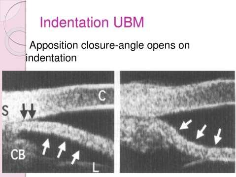
_ Assessment of the anterior chamber. Review of anatomy of the angle
 Assessment of the anterior chamber Dr Simon Barnard PhD BSc FCOptom FAAO DCLP Department of Optometry & Visual Science City University London, UK Review of anatomy of the angle Figure 1. Anatomical section
Ultrasound Biomicroscopic Changes of the Angle after Laser Iridotomy and Primary Trabeculectomy in Primary Angle Closure Glaucoma Patients
 Med. J. Cairo Univ., Vol. 79, No. 2, December: 113-118, 2011 www.medicaljournalofcairouniversity.com Ultrasound Biomicroscopic Changes of the Angle after Laser Iridotomy and Primary Trabeculectomy in Primary
Management of Angle Closure Glaucoma Hospital Authority Convention 18 May 2015
 Management of Angle Closure Glaucoma Hospital Authority Convention 18 May 2015 Jimmy Lai Clinical Professor Department of Ophthalmology The University of Hong Kong 1 Primary Angle Closure Glaucoma PACG
Are traditional assessments a waste of time? NZAO 2015
 Are traditional assessments a waste of time? NZAO 2015 Disclosures No financial interests other than Optometry Practice owner Full time optometrist Not a glaucoma prescriber ODOB Board Chair Previously
Systems for Anterior Chamber Angle Evaluation 長庚紀念醫院青光眼科吳秀琛
 Systems for Anterior Chamber Angle Evaluation 長庚紀念醫院青光眼科吳秀琛 Clinical Techniques for Assessing Angle Width A light from the side showing physiological iris bombe Slit lamp-grading of peripheral AC depth
5/18/2014. Fundamentals of Gonioscopy Workshop Aaron McNulty, OD, FAAO Walt Whitley, OD, MBA, FAAO
 1 Fundamentals of Gonioscopy Workshop Aaron McNulty, OD, FAAO Walt Whitley, OD, MBA, FAAO 2 3 4 5 6 Optometry s Meeting 2014 The Most Valuable Glaucoma Tool Glaucoma Diagnosis Gonioscopy Central corneal
Relationship between limbal incisions. angle. and the structures of the anterior chamber
 Brit. _7. Ophthal. (I 973) 57, 722 Relationship between limbal incisions and the structures of the anterior chamber angle MOHAMED I. AYOUB AND AHMED H. SAID Department of Ophthalmology, Faculty of Medicine,
SOCT Copernicus REVO. * - Currently import and overlay are avaibale in manual mode only
 SOCT Copernicus REVO Easy Operation (Full auto & Auto mode) Auto alignment (Z-position, C-gate, Focus, Tomogram) Voice guide (support patient through examination) Powerful analysis tools Enhanced tomograms
Corporate Medical Policy
 Corporate Medical Policy Optical Coherence Tomography (OCT) Anterior Segment of the Eye File Name: Origination: Last CAP Review: Next CAP Review: Last Review: optical_coherence_tomography_(oct)_anterior_segment_of_the_eye
The Anterior Segment & Glaucoma Visual Recognition & Interpretation of Clinical Signs
 The Anterior Segment & Glaucoma Visual Recognition & Interpretation of Clinical Signs Quiz created by Jane Macnaughton MCOptom & Peter Chapman BSc MCOptom FBDO CET Accreditation C19095 2 CET Points (General)
he Role of UBM and Anterior Segment OCT in Anterior Segment Imaging
 Ophthalmic Instrumentation T he Role of UBM and Anterior Segment OCT in Anterior Segment Imaging M. Chockalingam DNB FRCS PGDHM N. V. Arulmozhi Varman MS Since its development and usage, Ultrasound biomicroscopy
Routine OCT and UBM of the anterior segment
 Reprinted from No. 160 december 2012 Volume 17 Routine OCT and UBM of the anterior segment Michel Puech ISSN : 1274-5243 R e p r i n t e d w i t h t h e s u p p o r t o f t h e L a b o r a t o r y Q u
THE CHRONIC GLAUCOMAS
 THE CHRONIC GLAUCOMAS WHAT IS GLAUCOMA People with glaucoma have lost some of their field of all round vision. It is often the edge or periphery that is lost. That is why the condition can be missed until
Wallace L.M. Alward, M.D. Frederick C. Blodi Chair Department of Ophthalmology
 5/13/17 Gonioscopy Still State of the Art After 100 Years Principles of Gonioscopy I have no conflicts to report regarding this lecture. Wallace L.M. Alward, M.D. Frederick C. Blodi Chair Wallace L.M.
Ultrasound Biomicroscopy Noel Moniz, MS L.F. Hospital and Reserach Centre, Angamaly
 Ultrasound Biomicroscopy Noel Moniz, MS L.F. Hospital and Reserach Centre, Angamaly F O C U S The ultrasound biomicroscope works on the principle of an ultrasound but at a higher frequency. The normal
Chronicity. Narrow Minded. Course Outline. Acute angle closure. Subacute angle closure. Classification of Angle Closure 5/19/2014
 Chronicity Narrow Minded The management of narrow angles in the optometric practice Acute Subacute Chronic Aaron McNulty, OD, FAAO Course Outline Classification of Angle Closure Evaluation of narrow angles
03/04/2015. LOC Talk Anterior Chamber & Gonioscopy 1st April Methods of Assessing Anterior Chamber Depth (and angle width) Outline
 Outline") LOC Talk Anterior & 1st April 2015 Mr. Areeb Moosavi MBBS BSc FRCOphth Glaucoma Consultant Milton Keynes University Hospital NHS Foundation Trust Methods of Assessing Anterior Open Versus Closed angle
LOC Talk Anterior & 1st April 2015 Mr. Areeb Moosavi MBBS BSc FRCOphth Glaucoma Consultant Milton Keynes University Hospital NHS Foundation Trust Methods of Assessing Anterior Open Versus Closed angle
THE CHRONIC GLAUCOMAS
 THE CHRONIC GLAUCOMAS WHAT IS GLAUCOMA? People with glaucoma have lost some of their field of all round vision. It is often the edge or periphery that is lost. That is why the condition can be missed until
A LITTLE ANATOMY. three layers of eye: 1. outer: corneosclera. 2. middle - uvea. anterior - iris,ciliary body. posterior - choroid
 GLAUCOMA A LITTLE ANATOMY three layers of eye: 1. outer: corneosclera 2. middle - uvea anterior - iris,ciliary body posterior - choroid connection at the pars plana between post and ant uvea 3. retina
02/03/2014. Average Length: 23mm (Infant ~16mm) Approximately the size of a quarter Volume: ~5mL
 Approximately the size of a quarter Volume: ~5mL") Identify the anatomy of the eye. Explain the basic physiology of the parts of the eye. Briefly discuss various surgeries related to different parts of the anatomy. Average Length: 23mm (Infant ~16mm) Approximately
Identify the anatomy of the eye. Explain the basic physiology of the parts of the eye. Briefly discuss various surgeries related to different parts of the anatomy. Average Length: 23mm (Infant ~16mm) Approximately
Differential diagnosis of the red eye. Carol Slight Nurse Practitioner Ophthalmology
 Differential diagnosis of the red eye Carol Slight Nurse Practitioner Ophthalmology The red eye Conjunctivitis HSV Keratitis Acute angle closure glaucoma Anterior Uveitis Red eye Scleritis Subconjunctival
Intraoperative Gonioscopy: A Key to Angle Surgery
 Intraoperative Gonioscopy: A Key to Angle Surgery Shakeel Shareef, MD Associate Professor Flaum Eye Institute Univ. of Rochester School of Med. Rochester, NY Phaco vs. MIGS PHACO MIGS Viewing Full corneal
Ultrasound Biomicroscopy Study of Anterior Segment Measurements in Normal Eyes in a Tertiary Care Rural Hospital of SPRR Nellore District
 www.jmscr.igmpublication.org Impact Factor 3.79 ISSN (e)-2347-176x Ultrasound Biomicroscopy Study of Anterior Segment Measurements in Normal Eyes in a Tertiary Care Rural Hospital of SPRR Nellore District
HISTOPATHOLOGIC FEATURES OF TRABECULECTOMY SURGERY
 HISTOPATHOLOGIC FEATURES OF TRABECULECTOMY SURGERY BY Anthony C. Castelbuono MD* AND W. Richard Green MD ABSTRACT Purpose: Trabeculectomy surgery is the most common operative procedure for the treatment
Clinical and Anterior Segment Anatomical Features in Primary Angle Closure Subgroups Based on Configurations of Iris Root Insertion
 pissn: 1011-8942 eissn: 2092-9382 Korean J Ophthalmol 2016;30(3):206-213 http://dx.doi.org/10.3341/kjo.2016.30.3.206 Original Article Clinical and Anterior Segment Anatomical Features in Primary Angle
Written by Administrator Wednesday, 13 January :27 - Last Updated Thursday, 21 January :34
 angle closure glaucoma A type of glaucoma caused by a sudden and severe rise in eye pressure. Occurs when the pupil enlarges too much or too quickly, and the outer edge of the iris blocks the eye s drainage
Primary Angle Closure Glaucoma
 www.eyesurgeonlondon.co.uk Primary Angle Closure Glaucoma What is Glaucoma? Glaucoma is a condition in which there is damage to the optic nerve. This nerve carries visual signals from the eye to the brain.
ANTERIOR SEGMENT EXAMINATION TECHNIQUES
 ANTERIOR SEGMENT EXAMINATION TECHNIQUES GERS October 2017 Amanda Harding - Principal Optometrist, MREH MSc., MCOptom, Dip Glauc., Dip TP (IP). Anterior Segment Examination Depth of anterior chamber Central
Anterior Segment Morphology in Primary Angle Closure Glaucoma using Ultrasound Biomicroscopy
 Tarannum Mansoori, Nagalla Balakrishna ORIGINAL ARTICLE 10.5005/jp-journals-10028-1230 Anterior Segment Morphology in Primary Angle Closure Glaucoma using Ultrasound Biomicroscopy 1 Tarannum Mansoori,
Glaucoma Glaucoma is a complication which has only recently been confirmed as a feature of
 1.2.4 OPHTHALMOLOGICAL ABNORMALITIES Ocular abnormalities are well documented in patients with NPS 6 62 81 95. 1.2.4.1 Glaucoma Glaucoma is a complication which has only recently been confirmed as a feature
Ibopamine is a poorly selective dopaminergic drug that is
 Comparative Study of the Effects of 2% Ibopamine, 10% Phenylephrine, and 1% Tropicamide on the Anterior Segment Giorgio Marchini, Silvia Babighian, Roberto Tosi, Sergio Perfetti, and Luciano Bonomi PURPOSE.
The Pathology and Pathogenesis of Acute Glaucoma in Dogs. Richard R Dubielzig
 The Pathology and Pathogenesis of Acute Glaucoma in Dogs Richard R Dubielzig Overview of Glaucoma Intraocular Pressure too High to Support a Healthy Optic Nerve Terminology used in the classification of
Glaucoma. Cornea. Iris
 Glaucoma Introduction Glaucoma is a group of eye diseases that can lead to blindness if not treated. Openangle glaucoma, the most common form of glaucoma, affects about 3 million Americans. Half of those
Populations Interventions Comparators Outcomes Individuals: Who are being evaluated for angleclosure
 Protocol Optical Coherence Tomography of the Anterior Eye Segment (90318) Medical Benefit Effective Date: 07/01/14 Next Review Date: 05/18 Preauthorization No Review Dates: 07/11, 07/12, 07/13, 05/14,
Sinus trabeculectomy. Preliminary results of IOO operations
 Brit. J. Ophthal. (I 972) 56, 833 Sinus trabeculectomy Preliminary results of IOO operations A. P. NESTEROV, N. V. FEDEROVA, AND Y. E. BATMANOV Department of Ophthalmology, Kazan Medical Institute, Kazan,
INTRODUCTION J. DAWCZYNSKI, E. KOENIGSDOERFFER, R. AUGSTEN, J. STROBEL. Department of Ophthalmology, University Hospital Jena, Jena - Germany
 European Journal of Ophthalmology / Vol. 17 no. 3, 2007 / pp. 363-367 Anterior segment optical coherence tomography for evaluation of changes in anterior chamber angle and depth after intraocular lens
Histology of the Eye
 Histology of the Eye Objectives By the end of this lecture, the student should be able to describe: The general structure of the eye. The microscopic structure of:»cornea.»retina. EYE BULB Three coats
Probe Selection A high frequency (7-12 MHz) linear array transducer should be used to visualize superficial structures (Image 1).
 linear array transducer should be used to visualize superficial structures (Image 1).") ! Teresa S. Wu, MD, FACEP Director, Emergency Ultrasound Program & Fellowships Co-Director, Women s Imaging Fellowship Maricopa Medical Center Associate Professor, Emergency Medicine Director, Simulation
! Teresa S. Wu, MD, FACEP Director, Emergency Ultrasound Program & Fellowships Co-Director, Women s Imaging Fellowship Maricopa Medical Center Associate Professor, Emergency Medicine Director, Simulation
Clinical Study Age and Positional Effect on the Anterior Chamber Angle: Assessment by Ultrasound Biomicroscopy
 Hindawi Publishing Corporation ISRN Ophthalmology Volume 2013, Article ID 706201, 5 pages http://dx.doi.org/10.1155/2013/706201 Clinical Study Age and Positional Effect on the Anterior Chamber Angle: Assessment
Around The Globe in 60 Minutes
 Around The Globe in 60 Minutes Around the GLOBE in Sixty Minutes Basic Ocular Anatomy, Examination, and Diagnostic Techniques Introduction Focusing on canine and feline ocular anatomy and basic examination
OPHTHALMOLOGY AND ULTRASOUND
 Vet Times The website for the veterinary profession https://www.vettimes.co.uk OPHTHALMOLOGY AND ULTRASOUND Author : JAMES OLIVER Categories : Vets Date : April 28, 2008 JAMES OLIVER discusses why ultrasound
Downloaded from:
 Philippin, H; Shah, P; Burton, M (2012) The next step: Detailed assessment of an adult glaucoma patient. Community eye health / International Centre for Eye Health, 25 (79-80). pp. 50-53. ISSN 0953-6833
Gonioscopy and Slit Lamp Exam for the Glaucoma Suspect. Disclosure GONIOSCOPY: Gonioscopy Why?? What should I look for? GONIOSCOPY
 Gonioscopy and Slit Lamp Exam for the Glaucoma Suspect Disclosure Michael Chaglasian has the following disclosures:» 1. Advisory Board: Alcon, Allergan, Bausch+Lomb, Carl Zeiss Meditec, Merck, Sucampo»
Gonioscopy is currently regarded as the reference standard
 Glaucoma Qualitative Assessment of Ultrasound Biomicroscopic Images Using Standard Photographs: The Liwan Eye Study Yuzhen Jiang, 1 Mingguang He, 1,2 Wenyong Huang, 1 Qunxiao Huang, 1 Jian Zhang, 1 and
Glaucoma. How is Glaucoma Diagnosed? Glaucoma Testing
 Glaucoma How is Glaucoma Diagnosed? Glaucoma Testing There is no single test for glaucoma. The diagnosis is made by evaluating the patient from a number of perspectives, using specialized instruments.
Increased iris thickness and association with primary angle closure glaucoma
 1 Singapore Eye Research Institute and Singapore National Eye Center, Singapore 2 Department of Ophthalmology, Beijing Shijitan Hospital, Beijing, China 3 State Key Laboratory of Ophthalmology, Zhongshan
Basic microsurgical suturing techniques for beginners
 ESCRS 2014 Basic microsurgical suturing techniques for beginners Trauma, sclera, trabeculectomy B.O. Bachmann Dept. of Ophthalmology, University of Cologne, Germany Financial interests: none Investigating
THE EYE: RETINA AND GLOBE
 Neuroanatomy Suzanne Stensaas February 24, 2011, 10:00-12:00 p.m. Reading: Waxman Ch. 15. Your histology and gross anatomy books should be useful. Reading: Histology of the Eye from any histology book
Structural changes of the anterior chamber following cataract surgery during infancy
 Structural changes of the anterior chamber following cataract surgery during infancy Matthew Nguyen, Emory University Marla Shainberg, Emory University Allen Beck, Emory University Scott Lambert, Emory
Cornea/Anterior Segment OCT. User Experience
 Cornea/Anterior Segment OCT User Experience User Experience Case#1 Post Penetrating Keratoplasty Tokyo Medical University / Kohsei Chuo General Hospital Hideki Mori MD, PhD Almost eight years have passed
The second most common causes of blindness worldwide. ( after cataract) The commonest cause of irreversible blindness in the world Estimated that 3%
 The commonest cause of irreversible blindness in the world Estimated that 3%") The second most common causes of blindness worldwide. ( after cataract) The commonest cause of irreversible blindness in the world Estimated that 3% of our population age > 40 have glaucoma In the past:
The second most common causes of blindness worldwide. ( after cataract) The commonest cause of irreversible blindness in the world Estimated that 3% of our population age > 40 have glaucoma In the past:
XUE HUI Department of Histology& Embryology, Basic Medicine College of Jilin University
 SENSE ORGAN XUE HUI Department of Histology& Embryology, Basic Medicine College of Jilin University EYE fibrous globe lens photosensitive cells a system of cells and nerves concentric layers the sclera
GLAUCOMA SUMMARY BENCHMARKS FOR PREFERRED PRACTICE PATTERN GUIDELINES
 SUMMARY BENCHMARKS FOR PREFERRED PRACTICE PATTERN GUIDELINES Introduction These are summary benchmarks for the Academy s Preferred Practice Pattern (PPP) guidelines. The Preferred Practice Pattern series
NEW YORK UNIVERSITY SCHOOL OF MEDICINE DEPARTMENT OF OPHTHALMOLOGY EDUCATIONAL OBJECTIVES AND GOALS
 NEW YORK UNIVERSITY SCHOOL OF MEDICINE DEPARTMENT OF OPHTHALMOLOGY EDUCATIONAL OBJECTIVES AND GOALS Revision Date: 6/30/06 Distribution Date: 7/6/06 The Department of Ophthalmology at the NYU Medical Center
Technicians & Nurses Program
 ASCRS ASOA Symposium & Congress Technicians & Nurses Program April 17-21, 2015 San Diego, California Optical Coherence Tomography: Essentials in Anterior and Posterior Segment Imaging Michael Stewart,
Ocular Anatomy for the Paraoptometric
 Ocular Anatomy for the Paraoptometric Minnesota Optometric Association Paraoptometric CE Friday September 30, 2016 Lindsay A. Sicks, OD, FAAO Assistant Professor, Illinois College of Optometry lsicks@ico.edu
EXAMINATIONN WITH B SCAN ULTRASONOGRAPHY
 CLINICAL OPHTHALMIC ULTRASOUND PROFESSOR OF OPTHALMOLGY FACULTY OF MEDICINE TANTA UNIVERSITY MEMBER OF INTERNATIONAL SOCIETY FOR OPHTHALMIC ULTRASOUND (SIDUO) EXAMINATIONN WITH B SCAN ULTRASONOGRAPHY THREE
Understanding Angle Closure
 Case Understanding Angle Closure Dominick L. Opitz, OD, FAAO Associate Professor Illinois College of Optometry 56 year old Caucasian Male Primary Eye Exam BCVA: 20/25 OD with+1.25 DS 20/25 OS with +1.75
Outline. Brief history and principles of ophthalmic ultrasound. Types of ocular ultrasound. Examination techniques. Types of Ultrasound
 Ultrasound and Intraocular Tumors 2015 Ophthalmic Photographers' Society Mid-Year Program Cagri G. Besirli MD, PhD Kellogg Eye Center University of Michigan Outline Brief history and principles of ophthalmic
Role of Lens Extraction in Primary Angle Closure Disease
 10.5005/jp-journals-10008-1125 Anubha Rathi et al REVIEW ARTICLE Role of Lens Extraction in Primary Angle Closure Disease Anubha Rathi, Reetika Sharma, Bhaskar Jha, Shibal Bhartiya, Anita Panda Glaucoma
Clinical Study Application of Anterior Segment Optical Coherence Tomography in Pediatric Ophthalmology
 Hindawi Publishing Corporation Journal of Ophthalmology Volume 2012, Article ID 313120, 6 pages doi:10.1155/2012/313120 Clinical Study Application of Anterior Segment Optical Coherence Tomography in Pediatric
Overview of Ultrasound Biomicroscopy
 10.5005/jp-journals-10008-1105 IMAGING Mingguang He, Dandan Wang, Yuzheng Jiang Zhongshan Ophthalmic Center, Sun Yat-sen University, Guangzhou, China Correspondence: Mingguang He, State Key Laboratory
2/26/2017. Sameh Galal. M.D, FRCS Glasgow. Lecturer of Ophthalmology Research Institute of Ophthalmology
 Sameh Galal M.D, FRCS Glasgow Lecturer of Ophthalmology Research Institute of Ophthalmology No financial interest in the subject presented 1 Managing cataracts in children remains a challenge. Treatment
Diffuse infiltrating retinoblastoma
 Brit. 1. Ophthal. (I 971) 55, 6oo Diffuse infiltrating retinoblastoma GWYN MORGAN Department of Pathology, Institute of Ophthalmology, University of London The term "diffuse infiltrating retinoblastoma"
Training Checking Vision Tonometry
 Training 101-2 Checking Vision Tonometry Checking Vision The classic example of an eye chart is the Snellen eye chart, in general they show 11 rows of capital letters. The top row contains one letter (usually
STAB INCISION GLAUCOMA SURGERY (SIGS)
") STAB INCISION GLAUCOMA SURGERY (SIGS) Dr. Soosan Jacob, MS, FRCS, DNB Senior Consultant Ophthalmologist, Dr. Agarwal's Eye Hospital, Chennai, India dr_soosanj@hotmail.com Videos available in Youtube channel:
STAB INCISION GLAUCOMA SURGERY (SIGS) Dr. Soosan Jacob, MS, FRCS, DNB Senior Consultant Ophthalmologist, Dr. Agarwal's Eye Hospital, Chennai, India dr_soosanj@hotmail.com Videos available in Youtube channel:
Anterior segment imaging
 Article Date: 11/1/2016 Anterior segment imaging AS OCT vs. UBM vs. endoscope; case based approaches BY BENJAMIN BERT, MD, FACS AND BRIAN FRANCIS, MD, MS Currently, numerous imaging modalities are available
Vision I. Steven McLoon Department of Neuroscience University of Minnesota
 Vision I Steven McLoon Department of Neuroscience University of Minnesota 1 Eye Cornea Sclera Conjunctiva 2 Eye The conjunctiva lines the inner surface of the eyelids and outer surface of the sclera. 3
CHARACTERIZING IRIS SURFACE FEATURES AND THEIR ASSOCIATION WITH ANGLE CLOSURE RELATED TRAITS IN ASIAN EYES. ELIZABETH SIDHARTHA (B.Sc. (Hons.
 CHARACTERIZING IRIS SURFACE FEATURES AND THEIR ASSOCIATION WITH ANGLE CLOSURE RELATED TRAITS IN ASIAN EYES ELIZABETH SIDHARTHA (B.Sc. (Hons.)), NUS A THESIS SUBMITTED FOR THE DEGREE OF MASTER OF SCIENCE
Eye Movements. Geometry of the Orbit. Extraocular Muscles
 Eye Movements Geometry of the Orbit The eye (oculus) is located in the anterior aspect of the orbit: the equator of the eye (defined by a coronal plane passing through its middle) lies at the margin of
Anterior Chamber Angle: Assessment and Anomalies
 Anterior Chamber Angle: Assessment and Anomalies Dave Hicks, OD, FAAO Please silence all mobile devices and remove items from chairs so others can sit. Unauthorized recording of this session is prohibited.
UBM and glaucoma: diagnosis and follow-up of plateau iris. M. Puech
 ophtalmologiques UBM and glaucoma: diagnosis and follow-up of plateau iris M. Puech Extrait du mensuel Réalités Ophtalmologiques N 204 Juin 2013 Cahier 1 Editeur : Performances Médicales 91, avenue de
Unit VIII Problem 8 Anatomy: Orbit and Eyeball
 Unit VIII Problem 8 Anatomy: Orbit and Eyeball - The bony orbit: it is protecting our eyeball and resembling a pyramid: With a base directed: anterolaterally. And an apex directed: posteromedially. Notes:
CLASS-y Laser Treats Glaucoma
 Article # 404 Comments About the Author Released: Author: Category: March 12th, 2014 Issue #0314 Ehud Assia Feature S S S S S CLASS-y Laser Treats Glaucoma Transforming complex, invasive and risky glaucoma
MORE ON COMBINING OR NOT COMBINING...
 MORE ON COMBINING OR NOT COMBINING... A. GALAND* At the XVIII Congress of the European Society of Cataract and Refractive Surgeons (ESCRS) in Brussels, September 2 nd -6 th 2000, I was in charge of organizing
LEUKAEMIA*t INFILTRATION OF THE IRIS IN CHRONIC LYMPHATIC. pattemn * Received for pubiication November io, i967.
 Brit. J. Ophthal. (1968) 52, 781 INFILTRATION OF THE IRIS IN CHRONIC LYMPHATIC LEUKAEMIA*t BY BRIAN MARTIN The General Infirmary, Leeds OCULAR involvement is common in the leukaemias though the anterior
Glaucoma. Glaucoma. Optic Disc Cupping
 Glaucoma What is Glaucoma? Bruce James A group of diseases in which damage to the optic nerve occurs as a result of intraocualar pressure being above the physiological norm for that eye Stoke Mandeville
3/23/2016. Diagnostic Services Taylor Pannell CRA, OCT-C. Services Available. Important info for the Tech to know. Visual Fields
 Services Available Diagnostic Services Taylor Pannell CRA, OCT-C Static and Kinetic Visual Fields Pachymetry Anterior and Posterior Segment OCT Fundus Photos FAF,FA,ICG Slit Lamp Photography Confocal HRT
Glaucoma Clinical Update. Barry Emara MD FRCS(C) Giovanni Caboto Club October 3, 2012
 Giovanni Caboto Club October 3, 2012") Glaucoma Clinical Update Barry Emara MD FRCS(C) Giovanni Caboto Club October 3, 2012 Objectives Understand the different categories of glaucoma Recognize the symptoms and signs of open angle and angle-closure
Glaucoma Clinical Update Barry Emara MD FRCS(C) Giovanni Caboto Club October 3, 2012 Objectives Understand the different categories of glaucoma Recognize the symptoms and signs of open angle and angle-closure
INFANTILE NEPHROPATHIC cystinosis
 CLINICAL SCIENCES Ultrasound Biomicroscopy of the Eye in Cystinosis Nils Mungan, MD, FRCSC; Ken K. Nischal, FRCOphth; Elise Héon, MD, FRCSC; Leslie MacKeen, CRA; J. Williamson Balfe, MD, FRCPC; Alex V.
Gonioscopy and 3-Mirror Retinal Evaluation Workshop Edeline Lu, O.D., FAAO Benedicte Gonzalez, O.D., MPH, FAAO Tina Zheng, O.D.
 Gonioscopy and 3-Mirror Retinal Evaluation Workshop Edeline Lu, O.D., FAAO Benedicte Gonzalez, O.D., MPH, FAAO Tina Zheng, O.D., FAAO Please silence all mobile devices and remove items from chairs so others
EFFICACY AND SAFETY OF CANALOPLASTY IN SAUDI PATIENTS WITH UNCONTROLLED OPEN ANGLE GLAUCOMA
 EFFICACY AND SAFETY OF CANALOPLASTY IN SAUDI PATIENTS WITH UNCONTROLLED OPEN ANGLE GLAUCOMA DR.FAISAL ALMOBARAK ASSISTANT PROFESSOR AND CONSULTANT DEPARTMENT OF OPHTHALMOLOGY COLLEGE OF MEDICINE AND KING
Correlation between presence of primary irisand cilliary body cysts and intraocular pressure
 European Review for Medical and Pharmacological Sciences 2017; 21: 3985-3989 Correlation between presence of primary irisand cilliary body cysts and intraocular pressure R. ZHU, L. CHENG, D.-M. WANG Institute
Medical Policy. MP Optical Coherence Tomography of the Anterior Eye Segment
 Medical Policy MP 9.03.18 BCBSA Ref. Policy: 9.03.18 Last Review: 03/29/2018 Effective Date: 03/29/2018 Section: Other Related Policies 9.03.05 Corneal Topography/Computer-Assisted Corneal Topography/Photokeratoscopy
Closed Angle Glaucoma Or Narrow Angle Glaucoma. What s is a closed angle type of glaucoma,
 Closed Angle Glaucoma Or Narrow Angle Glaucoma What s is a closed angle type of glaucoma, This is where the iris is found to be blocking the drainage of the eye through the trabecular meshwork. The eye
A. Incorrect! Acetazolamide is a carbonic anhydrase inhibitor given orally or by intravenous injection.
 Pharmacology - Problem Drill 20: Drugs that Treat Glaucoma Question No. 1 of 10 1. is a topical carbonic anhydrase inhibitor. Question #01 (A) Acetazolamide (B) Clonidine (C) Dorzolamide (D) Apraclonidine
Romanian Journal of Ophthalmology, Volume 59, Issue 3, July-September pp:
 Romanian Journal of Ophthalmology, Volume 59, Issue 3, July-September 2015. pp:177-183 CASE REPORT A RARER FORM OF ANGLE-CLOSURE GLAUCOMA - DIAGNOSIS AND TREATMENT Zemba Mihail*,**, Stamate Alina-Cristina*,
4/22/16. Eye. External Anatomy of Eye. Accessory Structures. Bio 40B Dr. Kandula
 Eye Bio 40B Dr. Kandula External Anatomy of Eye Accessory Structures l Eyebrows l Levator Palpebrae Superioris - opens eye l Eyelashes l Ciliary glands modified sweat glands l Small sebaceous glands l
Clinical Study Age-Related Changes in Trabecular Meshwork Imaging
 BioMed Research International Volume 2013, Article ID 295204, 6 pages http://dx.doi.org/10.1155/2013/295204 Clinical Study Age-Related Changes in Trabecular Meshwork Imaging Mark E. Gold, 1 Seema Kansara,
Over the past decade, new anterior chamber (AC) imaging
 imaging") Glaucoma Biometric Evaluation of Anterior Chamber Changes after Physiologic Pupil Dilation Using Pentacam and Anterior Segment Optical Coherence Tomography Florent Aptel, 1,2 Christophe Chiquet, 1,2 Sylvain
Glaucoma Biometric Evaluation of Anterior Chamber Changes after Physiologic Pupil Dilation Using Pentacam and Anterior Segment Optical Coherence Tomography Florent Aptel, 1,2 Christophe Chiquet, 1,2 Sylvain
Optical Coherence Tomography (OCT) of the Anterior Eye Segment
 of the Anterior Eye Segment") Optical Coherence Tomography (OCT) of the Anterior Eye Segment Policy Number: 9.03.18 Last Review: 2/2019 Origination: 1/2008 Next Review: 8/2019 Policy Blue Cross and Blue Shield of Kansas City (Blue
Optical Coherence Tomography (OCT) of the Anterior Eye Segment Policy Number: 9.03.18 Last Review: 2/2019 Origination: 1/2008 Next Review: 8/2019 Policy Blue Cross and Blue Shield of Kansas City (Blue
Distinction layer by layer. HRT II Rostock Cornea Module
 Distinction layer by layer HRT II Rostock Cornea Module Homogenously illuminated, undistorted images Movie capture Manual Pachymetry Epithelial and intra-corneal pachymetry Full corneal thickness Post-LASIK
Surgery for COEXISTING CATARACT AND GLAUCOMA:
 Surgery for COEXISTING CATARACT AND GLAUCOMA: UNEASY RELATIONSHIP Session: 20-107 Monday, April 20, 2015 Time: 8:00 AM-9:30 AM Room 7B (San Diego Convention Center) Course Instructors Ahmad K Khalil Alan
SAFE, PERMANENT EYE-COLOR CHANGE
 SAFE, PERMANENT EYE-COLOR CHANGE Prepared by Gregg Homer JSD (PhD) February 1, 2012 THE PIGMENTARY GLAUCOMA ISSUE Glaucoma Defined Glaucoma is currently defined as a disturbance of the structural or functional
FACING YOUR FUNDIC FEARS: EXAMINATION OF THE OCULAR FUNDUS J. Seth Eaton, VMD, DACVO Cornell University Veterinary Specialists
 FACING YOUR FUNDIC FEARS: EXAMINATION OF THE OCULAR FUNDUS J. Seth Eaton, VMD, DACVO Cornell University Veterinary Specialists The goal of a thorough fundus examination is to clinically evaluate the structures
Sclerokeratoplasty David S. Chu, M.D. Cases
 Sclerokeratoplasty David S. Chu, M.D. Cases Case 1 40 year-old female from Peru presented to our Service with inflamed OS for 2 months duration. Her symptoms began as red painful OS, which progressively
Ultrasound Biomicroscopy Dark Room Provocative Testing: A Quantitative Method for Estimating Anterior Chamber Angle Width
 Ultrasound Biomicroscopy Dark Room Provocative Testing: A Quantitative Method for Estimating Anterior Chamber Angle Width Hiroshi Ishikawa,* Koji Esaki,* Jeffrey M. Liebmann, Yukitaka Uji* and Robert Ritch
TISSUE* ABERRANT INTRA-OCULAR LACRIMAL GLAND. of the left eye of a full-term infant of 2 weeks; the tumour was globular and
 Brit. J. Ophthal. (1960) 44, 619. ABERRANT INTRA-OCULAR LACRIMAL GLAND TISSUE* BY W. S. HUNTERt Department of Pathology, Institute of Ophthalmology, University oflondon THE occurrence of aberrant lacrimal
of Ciliary body, Iris and Irido-corneal angle in Adult Marwari Goat (Capra hircus) R. K. Barhaiyaa *, Malsawmkima, D. M. Bhayani and Y. L.
 R. K. Barhaiyaa *, Malsawmkima, D. M. Bhayani and Y. L.") DHR International Journal Of Biomedical and Life Sciences (DHR-IJBLS) ISSN 2278-8301, Vol.5(3), 2014 Available online http://www.doublehelixresearch.com/dhrijbls Double Helix Research Original Article
DHR International Journal Of Biomedical and Life Sciences (DHR-IJBLS) ISSN 2278-8301, Vol.5(3), 2014 Available online http://www.doublehelixresearch.com/dhrijbls Double Helix Research Original Article