Technicians & Nurses Program
|
|
|
- Kathleen Cummings
- 5 years ago
- Views:
Transcription

1 ASCRS ASOA Symposium & Congress Technicians & Nurses Program April 17-21, 2015 San Diego, California


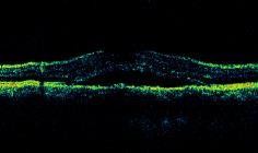
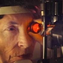
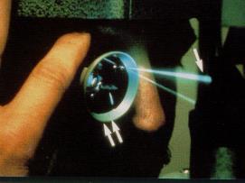
2 Optical Coherence Tomography: Essentials in Anterior and Posterior Segment Imaging Michael Stewart, M.D. Disclosure Allergan: Advisory Board, Research Support Boehringer-Ingelheim: Consultant Regeneron: Advisory Board, Research Support I will refer to off-label use of approved medications OCT What is this? Optical Coherence Tomography Laser Cross section Angle-closure Glaucoma ( Structural Levels) Do you need an anterior segment OCT To adequately image the anterior segment? Pupillary Block Plateau Iris VISANTE OCT Phacomorphic Glaucoma Malignant Glaucoma SL-OCT Ritch R. Liebmann JM, Lowe RF The history of angle-closure glaucomas: Philadelphia, Pa: Lippincott-Raven; 1997:


 and")
 were")


3 CASIA OCT Irido-Trabecular Angle imaging Pre-LPI Schlemm's canal Schwalbe's line Post- 1 week Scleral spur Post- 1 month 8 Quantitative Analysis Capabilities - Anterior chamber diameter, volume and pupillary diameter Cirrus HD-OCT Schwalbe's line Schlemm's canal Sectoral scan AS-OCT New parameters - Trabecular-iris contact length (TICL) and trabecular-iris space area (TISA-500 and 750) were introduced to describe the angle appearance. Radhakrishnan et al. Arch Ophthalmol. 2005; 123: Collector Channels Anterior segment imaging with spectral-domain / swept source OCT CC TM * SC Episcleral plexus RTVUE CIRRUS HD-OCT CASIA OCT IVP CC SC Ciliary body TM TM=Trabecular Meshwork *SC=Schlemm s canal CC=Collector channel CB=Ciliary body Dvorak-Theobald G

 B.")
 CE, choroidal effusion; CB, ciliary body; AC, anterior")

 using SL-OCT color image function CE, choroidal")


4 Ocular Trauma: Choroidal Haemorrhage Iris, Ciliary Body Trauma Choroidal Effusion Cyclodialysis Cleft A. UBM image showing choroidal effusion with no ciliary body detachment at 6-o clock (arrow) B. SL-OCT image showing choroidal effusion with no ciliary body detachment at the same position (arrow) CE, choroidal effusion; CB, ciliary body; AC, anterior chamber A. UBM image showing the cyclodialysis cleft at 3-o clock (arrow) B. SL-OCT image showing the cyclodialysis cleft at the same position (arrow) C. Well defined cleft (arrow) using SL-OCT color image function CE, choroidal effusion; CB, ciliary body; S, sclera; AC, anterior chamber Filtering Blebs ASOCT-Trabeculectomy Blebs Structures visualized: Bleb height and thickness of the conjunctiva in the bleb wall Cystic spaces within the bleb wall Scleral flap apposition and underlying sclera Patency of internal ostiumoutflow channel Some difficulty in visualizing the forniceal edge of the bleb hidden beneath the upper lid Singh M et al, Imaging of Trabeculectomy Blebs Using Anterior Segment Optical Coherence Tomography. Ophthalmology Jan; 114(1): Epub 2006 Oct 27. Miura M et al, Three-dimensional anterior segment optical coherence tomography of filtering blebs after trabeculectomy. Glaucoma Apr-May;17(3): Vertical AS-OCT image showing bleb micro-cysts and a slightly thinner cornea underlying the bleb in superior periphery (black arrow) AC: Anterior chamber; I: Iris


 SD-OCT")
 Jhanji,")

5 Glaucoma implants Anterior Segment Tumors-Iris Tumors Long term follow-up images of tube insertion in the sulcus in different patients: (A) Slit-lamp picture showing tube tip in pupillary area, well positioned between iris and lens (B) Tube positioned parallel to iris and lens visualized by ultrasound biomicroscopy (C) SL-OCT image confirming location of the tube in posterior chamber sulcus Post DSAEK- Visante Enhanced Depth Imaging OCT TD-OCT SD-OCT (Cirrus) SD-OCT (Spectralis) EDI SD-OCT (Spectralis) Jhanji, Greenrod, Vajpayee. Donor dislocation after DSAEK. BJO 2009 * Spaide RF et al. Am J Ophthalmol. 2008;146:496. Lamina Cribrosa 4


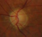
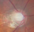
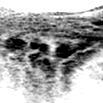
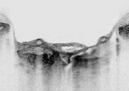
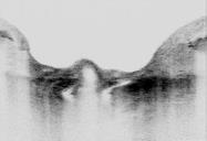
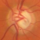
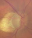

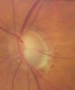
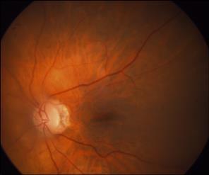
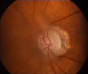
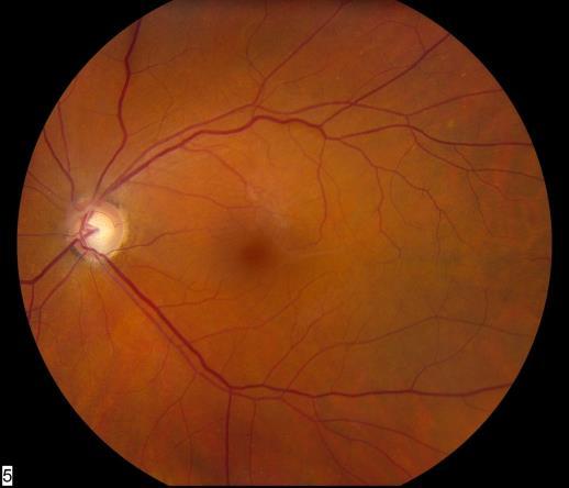
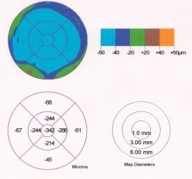
6 Focal Laminar Defect Lamina Cribrosa Depth OD max IOP 28 OS max IOP 20 Posterior Laminar Surface Lamina Cribrosa Pore?????? Central Retinal Vessels Short Posterior Ciliary Artery Artery Short posterior ciliary artery Vein Artery & veins Cilioretinal artery 5
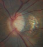
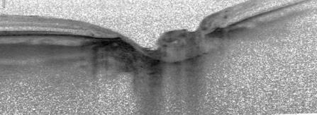
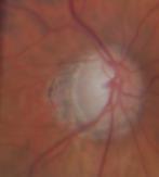
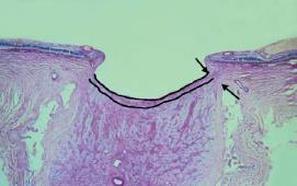
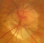
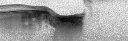
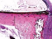
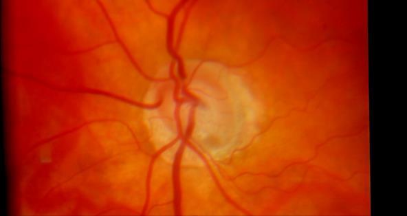
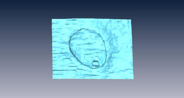
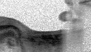
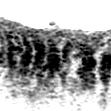
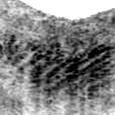
7 Parapapillary Choroid Subarachnoid Space Jonas JB, et al. IOVS 2003;44:5189. Optic Nerve Complex Examination of deep ONC structures - Lamina cribrosa - Vascular structures - Peripapillary choroid/sclera - Subarachnoid space - Lamina cribrosa - Vascular structures - Peripapillary choroid/sclera - Subarachnoid space 6


8 Examine angles What is this? Normal cornea Pre-op Visante Visante OCT Epithelium Bowman s layer Stroma Descemet s membrane 300 μm SD UHR OCT Eye with an anterior corneal scar 7

 43 Visante POD #11 OCT Imaging =")
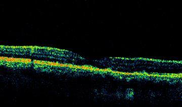
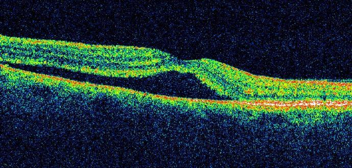
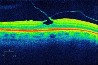
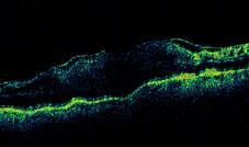
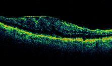
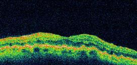
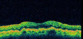
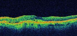
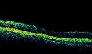
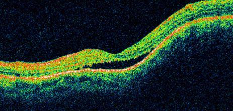
 OCT B-Scan Normal Macular")


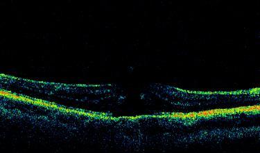
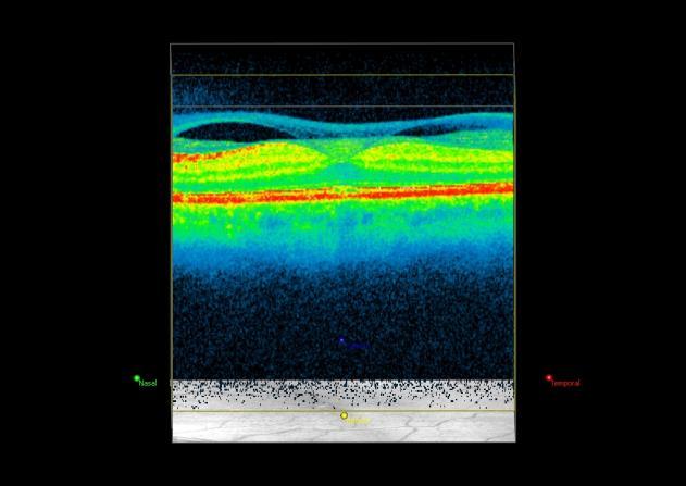
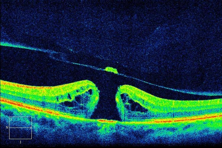
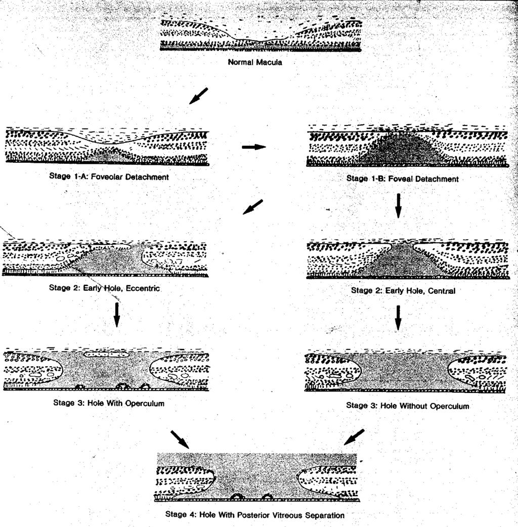
9 Pre-op UHR OCT Visante POD #11 Graft: 221 um Residual scar: 107 um Central Corneal thickness: 590 um ( um) 43 Visante POD #11 OCT Imaging = Optical Biopsy Normal Macular Anatomy Graft: 221 um Residual scar: 107 um Central Corneal thickness: 590 um ( um) OCT B-Scan Normal Macular Anatomy Pre-operative macular hole NFL GCL IPL OPL ONL ELM IS/OS RPE Choroid Post-operative Modified from: Retina 5 th Ed, Chapter 3, Optical Coherence Tomography 8


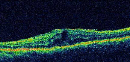
10 Previous vitrectomy with silicone oil 20 minutes later Aqueous SDOCT Imaging of Macula Qualitative assessment of retinal anatomy: SDOCT Imaging of Macula Segmentation of retinal layers necessary for quantitative assessments: ILM segmentation RPE segmentation Subretinal Fluid PED Subretinal Fluid Retinal thickness SDOCT Imaging of Wet AMD Segmentation algorithms result in reliable retinal thickness maps: Retinal thickness VA: 20/100 SDOCT Imaging of Wet AMD: Anti-VEGF Therapy Retinal thickness map ILM segmentation RPE segmentation VA: 20/40 Anti-VEGF Therapy: 1 month post-injection RPE segmentation 9


")

")


:1373-9.")


11 SDOCT Imaging of Dry AMD Segmentation algorithms result in reliable RPE elevation (drusen) maps (Cirrus, software 6.0): SDOCT Imaging of Wet AMD Segmentation algorithms result in reliable RPE elevation (PED) maps (Cirrus, software 6.0): RPE RPE floor RPE RPE floor Vol: 0.58mm 3 RPE segmentation RPE Elevation map Gregori G, Wang F, Rosenfeld PJ, et al. Spectral domain optical coherence tomography imaging of drusen in nonexudative age-related macular degeneration. Ophthalmology. 2011;118(7): RPE segmentation Retinal thickness RPE Elevation map Macular edema RPED SDOCT Imaging of Wet AMD Vitreomacular interface abnormalities Epiretinal membrane 10

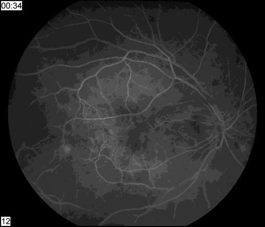
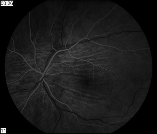
12 11

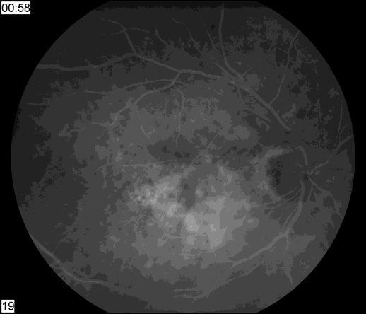
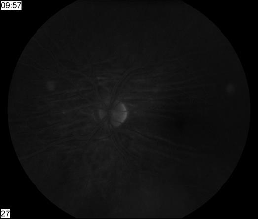
13 12


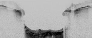
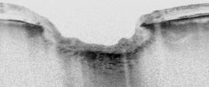
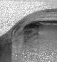
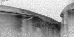
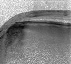
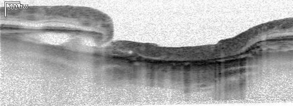
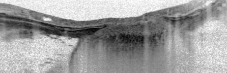
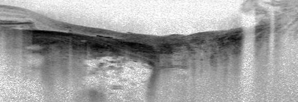
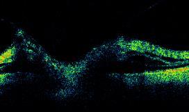
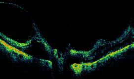
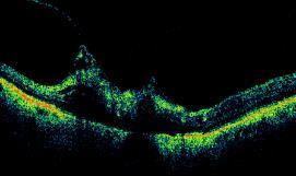
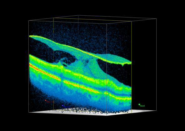
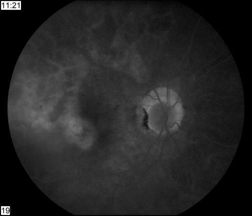
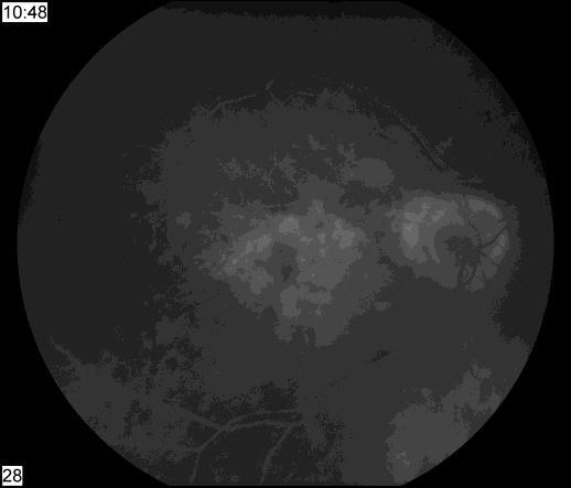
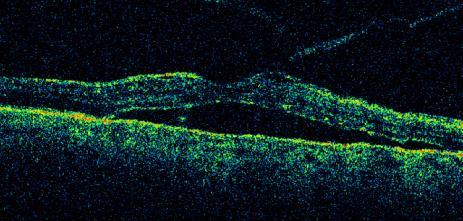
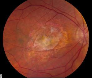
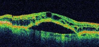
14 Why this shadow? Why this shadow? Gas bubble 1945µ 2819µ 13
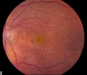
15 14


16 2 weeks later 15


17 OS 20/40 Stage 2 OD 20/25 Stage 1 Lamellar hole Cyst? Lamellar hole Epiretinal Membrane 16


18 20/40 20/30 Pre-Op Post-Op 17
19 18

20 9/29/ /31/ /30/2005 1/11/


21 Subretinal fluid RPED Case 71 yo man with wet AMD OS Multiple injections for the last 12 months Complains of worsening in VA after treatment Wet AMD Dry AMD Do anti-vegf injections cause geographic atrophy of the RPE? Case 74 y.o. man decreased vision OD (20/200) Case 20/200 Ranibizumab #1 After 1 month 20/80 Ranibizumab #2 20


 July 2007 20/50 Case 4:")


22 VA: 20/40 Ranibizumab #1 Case Monthly Injections 73 y.o. woman with neovascular AMD Case 3 65 y/o woman with a vascularized, hemorrhagic PED VA: 20/20 Ranibizumab #2 Vol: 0.73 mm 3 Vol: 0.63 mm 3 VA: 20/20 Ranibizumab #3 Vol: 0.56 mm 3 Color photo Early FA Late FA Baseline Anti-VEGF #1, Anti-VEGF #2 4w post Inj. #2 Observed 6w post Inj. #2 Observed Volume: 6.9 mm 3 Volume: 2.6 mm 3 66 year old woman 10 injections: Bev X6, Ran X4 (~q5wks) July /50 Case 4: Dosing Every 2 Weeks FA-early FA-late 8w post Inj. #2 Anti-VEGF #3 Volume: 3.2 mm 3 1 year later 20/200 4w post Inj. #3 Observed Volume: 3.9 mm 3 Volume: 1.6 mm 3 Modified from: Stewart MW, et al. Pharmacokinetic Rationale For Dosing Every 2 Weeks Versus 4 Weeks With Intravitreal Ranibizumab, Bevacizumab, And Aflibercept (Vascular Endothelial Growth Factor Trap-eye). Retina Dec 18. OCT Baseline 20/200 Ran Inj. #1 2 w post Inj. #1 20/40 Bev Inj. #2 2 w post Inj. #2 20/30 Ran Inj. #3 2 w post Inj.#3 20/30 Bev Inj. #4 21


23 OD BDR OS Diagnosis? IOP = 4 OD 1 month later OS Dense cataract Pre-op Baseline Phaco + IVT 1 day Post-op Following steroid injection 6 months VA: 20/20 22


24 c/o superior field defect Acute retinal artery occlusion Inner retina thickening Retinal thinning 23


25 Treatment options: 1. External beam irradiation 2. Systemic chemotherapy Treatment 1.Observation 2.Laser photocoagulation 24


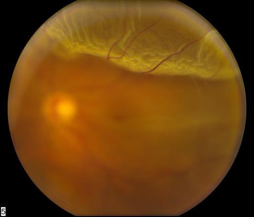
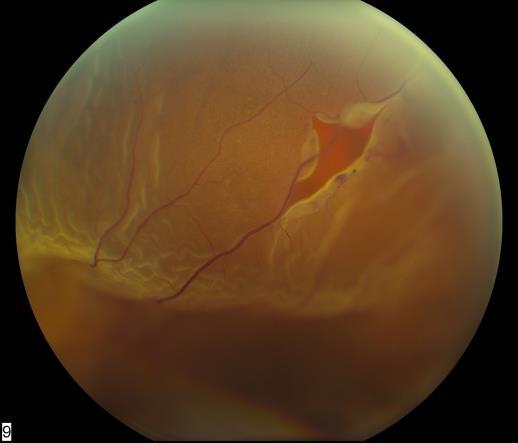
26 NFL Lines in nasal periphery 65 y.o. s/p scleral buckle for RD Now with VA of 20/100 for 6 months. 2 nd opinion: 68 y.o s/p renal transplant with iritis and VA of 20/40 OU 25


27 Conclusions 1. OCT has revolutionized the management of chorioretinal vascular diseases. 2. OCT has added greatly to the management of glaucoma. 3. Improved techniques and scanners are advancing the imaging of anterior segment structures. 26
Optical Coherence Tomography: Pearls for the Anterior Segment Surgeon Basic Science Michael Stewart, M.D.
 Optical Coherence Tomography: Pearls for the Anterior Segment Surgeon Basic Science Michael Stewart, M.D. Disclosure OCT Optical Coherence Tomography No relevant financial relationships I will refer to
Optical Coherence Tomography in Diabetic Retinopathy. Mrs Samantha Mann Consultant Ophthalmologist Clinical Lead of SEL-DESP
 Optical Coherence Tomography in Diabetic Retinopathy Mrs Samantha Mann Consultant Ophthalmologist Clinical Lead of SEL-DESP Content OCT imaging Retinal layers OCT features in Diabetes Some NON DR features
When optical coherence tomography (OCT)
") Macular Imaging: SD-OCT in nterior Segment Surgical Practice Many pathologic processes of the macula can be visualized or quantified only with this modality. y Steven G. Safran, MD When optical coherence
Macular Imaging: SD-OCT in nterior Segment Surgical Practice Many pathologic processes of the macula can be visualized or quantified only with this modality. y Steven G. Safran, MD When optical coherence
Mark Dunbar: Disclosure
 Important Things to Understand About OCT Mark T. Dunbar, O.D., F.A.A.O. Bascom Palmer Eye Institute University of Miami, School of Medicine Mark Dunbar: Disclosure Optometry Advisory Board for: Allergan
OCT Interpretation in Retinal Disease
 OCT Interpretation in Retinal Disease Jay M. Haynie, OD, FAAO Financial Disclosure I have received honoraria or am on the advisory board for the following companies: Carl Zeiss Meditec Advanced Ocular
5/18/2014. Fundamentals of Gonioscopy Workshop Aaron McNulty, OD, FAAO Walt Whitley, OD, MBA, FAAO
 1 Fundamentals of Gonioscopy Workshop Aaron McNulty, OD, FAAO Walt Whitley, OD, MBA, FAAO 2 3 4 5 6 Optometry s Meeting 2014 The Most Valuable Glaucoma Tool Glaucoma Diagnosis Gonioscopy Central corneal
3/16/2018. Ultrasound Biomicroscopy in Glaucoma By Ahmed Salah Abdel Rehim. Prof. of Ophthalmology Al-Azhar University
 Ultrasound Biomicroscopy in Glaucoma By Ahmed Salah Abdel Rehim Prof. of Ophthalmology Al-Azhar University 1 Ultrasound biomicroscopy (UBM) is a recent technique to visualize anterior segment with the
Corporate Medical Policy
 Corporate Medical Policy Optical Coherence Tomography (OCT) Anterior Segment of the Eye File Name: Origination: Last CAP Review: Next CAP Review: Last Review: optical_coherence_tomography_(oct)_anterior_segment_of_the_eye
SOCT Copernicus REVO. * - Currently import and overlay are avaibale in manual mode only
 SOCT Copernicus REVO Easy Operation (Full auto & Auto mode) Auto alignment (Z-position, C-gate, Focus, Tomogram) Voice guide (support patient through examination) Powerful analysis tools Enhanced tomograms
Course # Getting to Know Your OCT
 Course # 140 Getting to Know Your OCT Course Title: Lecturer: Getting to Know Your OCT Brad Sutton, OD, FAAO IU School of Optometry Financial Disclosures No financial disclosures Optical Coherence Tomography-OCT
Advances in OCT Murray Fingeret, OD
 Disclosures Advances in OCT Murray Fingeret, OD Consultant Alcon, Allergan, Bausch & Lomb, Carl Zeiss Meditec, Diopsys, Heidelberg Engineering, Reichert, Topcon Currently Approved OCT Devices OCT Devices
Anterior segment imaging
 Article Date: 11/1/2016 Anterior segment imaging AS OCT vs. UBM vs. endoscope; case based approaches BY BENJAMIN BERT, MD, FACS AND BRIAN FRANCIS, MD, MS Currently, numerous imaging modalities are available
The Quick Guide to OCT Mastery 50 Real Cases with Expert Analysis
 OPTICAL COHERENCE TOMOGRAPHY The Quick Guide to OCT Mastery 50 Real Cases with Expert Analysis VOL 1 Sanjay Sharma, MD, FRCS, MSc (Epid), MBA Ophthalmologist, Epidemiologist Queen s University, Canada
THE CHRONIC GLAUCOMAS
 THE CHRONIC GLAUCOMAS WHAT IS GLAUCOMA? People with glaucoma have lost some of their field of all round vision. It is often the edge or periphery that is lost. That is why the condition can be missed until
Glaucoma Past, Present and Future
 Glaucoma Past, Present and Future Paul A. Weber, M.D. Professor Emeritus Department of Ophthalmology and Visual Science- Havener Eye Institute The Ohio State University Before we talk about glaucoma, I
OCT Interpretation. Financial Disclosure. Jay M. Haynie, OD, FAAO. OCT Image Layers 7/21/2014
 OCT Interpretation Jay M. Haynie, OD, FAAO Financial Disclosure I have received honoraria or am on the advisory board for the following companies: Olympia Tacoma Renton Kennewick - Washington Carl Zeiss
Gonioscopy and Slit Lamp Exam for the Glaucoma Suspect. Disclosure GONIOSCOPY: Gonioscopy Why?? What should I look for? GONIOSCOPY
 Gonioscopy and Slit Lamp Exam for the Glaucoma Suspect Disclosure Michael Chaglasian has the following disclosures:» 1. Advisory Board: Alcon, Allergan, Bausch+Lomb, Carl Zeiss Meditec, Merck, Sucampo»
OCT Angiography The Next Frontier
 Choroid Retina avascular 5/13/2017 OCT Angiography The Next Frontier Pierce Kenworthy OD, FAAO June 9, 2017 OCT Angiography (OCTA) 2016 Non-invasive, motion contrast imaging Represents erythrocyte movement
What Is O.C.T. and Why Should I Give A Rip? OCT & Me How Optical Coherence Tomography Changed the Life of a Small Town Optometrist 5/19/2014
 OCT & Me How Optical Coherence Tomography Changed the Life of a Small Town Optometrist Email: myoder@wcoil.com Mark A. Yoder, O.D. 107 N. Main Street PO Box 123 Bluffton, OH 45817 @yoderod 115.02 Histoplasma
Retina Conference. Janelle Fassbender, MD, PhD University of Louisville Department of Ophthalmology and Visual Sciences 09/04/2014
 Retina Conference Janelle Fassbender, MD, PhD University of Louisville Department of Ophthalmology and Visual Sciences 09/04/2014 Subjective CC/HPI: 64 year old Caucasian female referred by outside ophthalmologist
OCCLUSIVE VASCULAR DISORDERS OF THE RETINA
 OCCLUSIVE VASCULAR DISORDERS OF THE RETINA Learning outcomes By the end of this lecture the students would be able to Classify occlusive vascular disorders (OVD) of the retina. Correlate the clinical features
Ganglion cell analysis by optical coherence tomography (OCT) Jonathan A. Micieli, MD Valérie Biousse, MD
 Jonathan A. Micieli, MD Valérie Biousse, MD") Ganglion cell analysis by optical coherence tomography (OCT) Jonathan A. Micieli, MD Valérie Biousse, MD Figure 1. Normal OCT of the macula (cross section through the line indicated on the fundus photo)
Ganglion cell analysis by optical coherence tomography (OCT) Jonathan A. Micieli, MD Valérie Biousse, MD Figure 1. Normal OCT of the macula (cross section through the line indicated on the fundus photo)
Optical Coherence Tomography (OCT) in Uveitis Piergiorgio Neri, BMedSc, MD, PhD Head Ocular Immunology Unit
 in Uveitis Piergiorgio Neri, BMedSc, MD, PhD Head Ocular Immunology Unit") The Eye Clinic Polytechnic University of Marche Head: Prof Alfonso Giovannini November, 1991 Optical Coherence Tomography (OCT) in Uveitis Piergiorgio Neri, BMedSc, MD, PhD Head Ocular Immunology Unit
The Eye Clinic Polytechnic University of Marche Head: Prof Alfonso Giovannini November, 1991 Optical Coherence Tomography (OCT) in Uveitis Piergiorgio Neri, BMedSc, MD, PhD Head Ocular Immunology Unit
The Human Eye. Cornea Iris. Pupil. Lens. Retina
 The Retina Thin layer of light-sensitive tissue at the back of the eye (the film of the camera). Light rays are focused on the retina then transmitted to the brain. The macula is the very small area in
Management of Angle Closure Glaucoma Hospital Authority Convention 18 May 2015
 Management of Angle Closure Glaucoma Hospital Authority Convention 18 May 2015 Jimmy Lai Clinical Professor Department of Ophthalmology The University of Hong Kong 1 Primary Angle Closure Glaucoma PACG
 measure of your overall performance. An isolated glucose test is helpful to let you know what your sugar level is at one moment, but it doesn t tell you whether or not your diabetes is under adequate control
Cirrus TM HD-OCT. Details defi ne your decisions
 Cirrus TM HD-OCT Details defi ne your decisions 2 With high-defi nition OCT Carl Zeiss Meditec takes you beyond standard spectral domain Built on 10 years experience at the vanguard of innovation, Carl
Routine OCT and UBM of the anterior segment
 Reprinted from No. 160 december 2012 Volume 17 Routine OCT and UBM of the anterior segment Michel Puech ISSN : 1274-5243 R e p r i n t e d w i t h t h e s u p p o r t o f t h e L a b o r a t o r y Q u
Understanding Angle Closure
 Case Understanding Angle Closure Dominick L. Opitz, OD, FAAO Associate Professor Illinois College of Optometry 56 year old Caucasian Male Primary Eye Exam BCVA: 20/25 OD with+1.25 DS 20/25 OS with +1.75
Revitalization of the Anterior Segment: Corneal Transplantation and Secondary Lens Repair
 Revitalization of the Anterior Segment: Corneal Transplantation and Secondary Lens Repair CATHERINE REPPA, MD CORNEA SPECIALIST, ASSISTANT PROFESSOR TTUHSC DEPARTMENT OF OPHTHALMOLOGY AND VISUAL SCIENCES
The second most common causes of blindness worldwide. ( after cataract) The commonest cause of irreversible blindness in the world Estimated that 3%
 The commonest cause of irreversible blindness in the world Estimated that 3%") The second most common causes of blindness worldwide. ( after cataract) The commonest cause of irreversible blindness in the world Estimated that 3% of our population age > 40 have glaucoma In the past:
The second most common causes of blindness worldwide. ( after cataract) The commonest cause of irreversible blindness in the world Estimated that 3% of our population age > 40 have glaucoma In the past:
EXP11677SK. Financial Disclosure. None to be Declared EXP11677SK
 Financial Disclosure None to be Declared Presentation overview Glaucoma Surgical History Complications of trabeculectomy Express Device Specifications Surgical Steps Clinical advantages, indications and
Ophthalmology Macular Pathways
 Ophthalmology Macular Pathways Age related Macular Degeneration Diabetic Macular Oedema Macular Oedema secondary to Central Retinal Macular Oedema secondary to Branch Retinal CNV associated with pathological
Overview. Macular OCT Artifact Study
 Imaging Artifacts Sarah Moyer, CRA, OCT-C Director, Ophthalmic Imaging Kittner Eye Center University of North Carolina Chapel Hill, NC Disclose financial interest now Overview Sarah s Thoughts on Artifacts
DOME SHAPED MACULOPATHY. Ιωάννης Ν. Βαγγελόπουλος Χειρ. Οφθαλμίατρος - Βόλος
 DOME SHAPED MACULOPATHY Ιωάννης Ν. Βαγγελόπουλος Χειρ. Οφθαλμίατρος - Βόλος DOME SHAPED MACULOPATHY-DEFINITIONS The entity Dome Shaped Macula ( DSM ) was first described by Gaucher and associates in 2008
OPTIC DISC PIT Pathogenesis and Management OPTIC DISC PIT
 OPTIC DISC PIT Pathogenesis and Management Abdel-Latif Siam Ain Shams University Cairo Egypt OPTIC DISC PIT Congenital pit is an atypical coloboma usually located on the temporal edge of the disc, associated
Applications of Sustained Release Delivery Systems in Ocular Disease
 Applications of Sustained Release Delivery Systems in Ocular Disease Formulation and Delivery Systems For Peptide and Protein 2012 December 5, 2012 Christopher A. Rhodes, Ph.D. Principal, Christopher A
The Foundation WHAT IS THE RETINA?
 Age-Related Macular Degeneration (AMD) is a deterioration of the retina and choroid that leads to a substantial loss in visual acuity (sharpness of vision). AMD is the leading cause of significant visual
THE EYE: RETINA AND GLOBE
 Neuroanatomy Suzanne Stensaas February 24, 2011, 10:00-12:00 p.m. Reading: Waxman Ch. 15. Your histology and gross anatomy books should be useful. Reading: Histology of the Eye from any histology book
International Journal of Health Sciences and Research ISSN:
 International Journal of Health Sciences and Research www.ijhsr.org ISSN: 2249-9571 Original Research Article A Multivariate Analysis of Intravitreal Injection of Anti-VEGF Bevacizumab in the Treatment
Structural examina.on: Imaging
 ManaMa: Glaucoma Structural examina.on: Imaging Luís Abegão Pinto, MD, PhD Department of Ophthalmology CHLC Lisbon Faculty of Medicine, Lisbon University 1 11-10- 2013 Structural changes Qualitative changes
Diagnosis and Therapy in Ophthalmology Hand-held optical coherence tomography imaging in children with anterior segment dysgenesis
 Diagnosis and Therapy in Ophthalmology Hand-held optical coherence tomography imaging in children with anterior segment dysgenesis Anastasia V. Pilat, 1,2 Viral Sheth, 1,2 Ravi Purohit, 1,2 Frank A. Proudlock,
Local Coverage Determination (LCD): Scanning Computerized Ophthalmic Diagnostic Imaging (SCODI) (L34431)
: Scanning Computerized Ophthalmic Diagnostic Imaging (SCODI) (L34431)") Local Coverage Determination (LCD): Scanning Computerized Ophthalmic Diagnostic Imaging (SCODI) (L34431) Links in PDF documents are not guaranteed to work. To follow a web link, please use the MCD Website.
Local Coverage Determination (LCD): Scanning Computerized Ophthalmic Diagnostic Imaging (SCODI) (L34431) Links in PDF documents are not guaranteed to work. To follow a web link, please use the MCD Website.
The Anterior Segment & Glaucoma Visual Recognition & Interpretation of Clinical Signs
 The Anterior Segment & Glaucoma Visual Recognition & Interpretation of Clinical Signs Quiz created by Jane Macnaughton MCOptom & Peter Chapman BSc MCOptom FBDO CET Accreditation C19095 2 CET Points (General)
OCT Image Analysis System for Grading and Diagnosis of Retinal Diseases and its Integration in i-hospital
 Progress Report for1 st Quarter, May-July 2017 OCT Image Analysis System for Grading and Diagnosis of Retinal Diseases and its Integration in i-hospital Milestone 1: Designing Annotation tool extraction
02/03/2014. Average Length: 23mm (Infant ~16mm) Approximately the size of a quarter Volume: ~5mL
 Approximately the size of a quarter Volume: ~5mL") Identify the anatomy of the eye. Explain the basic physiology of the parts of the eye. Briefly discuss various surgeries related to different parts of the anatomy. Average Length: 23mm (Infant ~16mm) Approximately
Identify the anatomy of the eye. Explain the basic physiology of the parts of the eye. Briefly discuss various surgeries related to different parts of the anatomy. Average Length: 23mm (Infant ~16mm) Approximately
RETINAL CONDITIONS RETINAL CONDITIONS
 GENERAL INFORMATION RETINAL CONDITIONS RETINAL CONDITIONS WHAT ARE RETINAL CONDITIONS? Retinal conditions affect the light-sensitive tissue at the back of eye known as the retina. They include diseases
Optical Coherence Tomograpic Features in Idiopathic Retinitis, Vasculitis, Aneurysms and Neuroretinitis (IRVAN)
") Columbia International Publishing Journal of Ophthalmic Research (2014) Research Article Optical Coherence Tomograpic Features in Idiopathic Retinitis, Vasculitis, Aneurysms and Neuroretinitis (IRVAN)
Columbia International Publishing Journal of Ophthalmic Research (2014) Research Article Optical Coherence Tomograpic Features in Idiopathic Retinitis, Vasculitis, Aneurysms and Neuroretinitis (IRVAN)
Past ocular history. DME Case 1. Patient presents blurred VA. Hemoglobin A1c 11.5% -- patient states sugars have not been in good control
 Past ocular history Patient presents blurred VA DME Case 1 Hemoglobin A1c 11.5% -- patient states sugars have not been in good control PDR with macular edema OU Rishi Singh MD Cleveland Clinic OD OS 1
EFFICACY OF ANTI-VASCULAR ENDOTHELIAL GROWTH FACTOR AGENTS IN RETINAL DISORDER FOR BETTER VISUAL ACUITY
 EFFICACY OF ANTI-VASCULAR ENDOTHELIAL GROWTH FACTOR AGENTS IN RETINAL DISORDER FOR BETTER VISUAL ACUITY Diwakar chaudhary *1, 2, Hu shuqiong, Long Yuan and Xiong kun 1 Yangtze University, 1 Nanhuan Road
Recurrent intraocular hemorrhage secondary to cataract wound neovascularization (Swan Syndrome)
") Recurrent intraocular hemorrhage secondary to cataract wound neovascularization (Swan Syndrome) John J. Chen MD, PhD; Young H. Kwon MD, PhD August 6, 2012 Chief complaint: Recurrent vitreous hemorrhage,
Recurrent intraocular hemorrhage secondary to cataract wound neovascularization (Swan Syndrome) John J. Chen MD, PhD; Young H. Kwon MD, PhD August 6, 2012 Chief complaint: Recurrent vitreous hemorrhage,
Ophthalmology. Juliette Stenz, MD
 Ophthalmology Juliette Stenz, MD Required Slide Disclosures NO SIGNIFICANT FINANCIAL, GENERAL, OR OBLIGATION INTERESTS TO REPORT Required Slide At the end of this session, students will be able to: 1.
_ Assessment of the anterior chamber. Review of anatomy of the angle
 Assessment of the anterior chamber Dr Simon Barnard PhD BSc FCOptom FAAO DCLP Department of Optometry & Visual Science City University London, UK Review of anatomy of the angle Figure 1. Anatomical section
Hong Kong College of Surgical Nursing
 Hong Kong College of Surgical Nursing Surgical Nursing Training: Part B Specialty - Ophthalmological Nursing Curriculum 1 TABLE OF CONTENTS No. Contents Page 1. Introduction 3 2. Aims 3 3. Programme Intended
Cornea/Anterior Segment OCT. User Experience
 Cornea/Anterior Segment OCT User Experience User Experience Case#1 Post Penetrating Keratoplasty Tokyo Medical University / Kohsei Chuo General Hospital Hideki Mori MD, PhD Almost eight years have passed
Cirrus TM HD-OCT. Details define your decisions
 Cirrus TM HD-OCT Details define your decisions 2 With high-definition OCT Carl Zeiss Meditec takes you beyond standard spectral domain Built on 10 years experience at the vanguard of innovation, Carl Zeiss
Diagnosis in AMD. Managing your AMD Patients
 Managing your AMD Patients Robert W. Dunphy, O.D., F.A.A.O. Diagnosis in AMD Have suspicion Identify relative risk Conduct surveillance Biometry Utilize technology to facilitate detection of change / stability
Objectives. Tubes, Ties and Videotape: Financial Disclosure. Five Year TVT Results IOP Similar
 Tubes, Ties and Videotape: Surgical Video of Glaucoma Implants and Financial Disclosure I have no financial interests or relationships to disclose. Herbert P. Fechter MD, PE Eye Physicians and Surgeons
Why Is Imaging Critical in My Uveitis Practice?
 Why Is Imaging Critical in My Uveitis Practice? Dilraj S. Grewal, MD Developed in collaboration Imaging Is the Backbone of Uveitis Workup and Monitoring Treatment Response FP FAF B- scan Multimodal Imaging
TRABECULECTOMY. Dr. Sandra M. Johnson, MD
 TRABECULECTOMY Dr. Sandra M. Johnson, MD FILTRATION OPTIONS Trabeculotomy, Schlemn s canal, internal Deep Non-penetrating Sclerectomy filtering to a scleral lake, or viscocanulostomy Trabeculectomy shunting
Choroidal Mapping; a Novel Approach for Evaluating Choroidal Thickness and Volume
 Imaging Technique Choroidal Mapping; a Novel Approach for Evaluating Choroidal Thickness and Volume Jila Noori 1, MD; Mohammad Riazi Esfahani 1,2, MD Fedra Hajizadeh 2, MD; Mohammad-Mehdi Zaferani 1, MD
Clinically Significant Macular Edema (CSME)
") Clinically Significant Macular Edema (CSME) 1 Clinically Significant Macular Edema (CSME) Sadrina T. Shaw OMT I Student July 26, 2014 Advisor: Dr. Uwaydat Clinically Significant Macular Edema (CSME) 2
Clinically Significant Macular Edema (CSME) 1 Clinically Significant Macular Edema (CSME) Sadrina T. Shaw OMT I Student July 26, 2014 Advisor: Dr. Uwaydat Clinically Significant Macular Edema (CSME) 2
VMA at the macula resulting in VMT
 Ocriplasmina for pharmacologic treatment in VMT Teresio Avitabile 1 Introduction PVD is a normal, physiologic process that occurs with aging; however, in some cases, PVD is incomplete Incomplete PVD localized
Clinical Case Presentation. Branch Retinal Vein Occlusion. Sarita M. Registered Nurse Whangarei Base Hospital
 Clinical Case Presentation on Branch Retinal Vein Occlusion Sarita M. Registered Nurse Whangarei Base Hospital Introduction Case Study Pathogenesis Clinical Features Investigations Treatment Follow-up
ACTIVATED OR NOT? RETINAL CASE PRESENTATION Shorye Payne, MD Medical Retinal Specialist Robley Rex VA Eye Clinic
 ACTIVATED OR NOT? RETINAL CASE PRESENTATION Shorye Payne, MD Medical Retinal Specialist Robley Rex VA Eye Clinic C We anticipate that the future management of posterior uveal melanoma (PUM) will focus
Intraocular Radiation Therapy for Age-Related Macular Degeneration
 Medical Policy Manual Medicine, Policy No. 134 Intraocular Radiation Therapy for Age-Related Macular Degeneration Next Review: April 2019 Last Review: June 2018 Effective: August 1, 2018 IMPORTANT REMINDER
From Outdated to Updated: A Review of Important Clinical Trials in Ocular Disease from 2014
 From Outdated to Updated: A Review of Important Clinical Trials in Ocular Disease from 2014 1. This course is designed to review the important ophthalmic literature that was released between October 2013
Anterior Chamber Angle: Assessment and Anomalies
 Anterior Chamber Angle: Assessment and Anomalies Dave Hicks, OD, FAAO Please silence all mobile devices and remove items from chairs so others can sit. Unauthorized recording of this session is prohibited.
Practical Care of the Cataract Patient with Retinal Disease
 Practical Care of the Cataract Patient with Retinal Disease Brooks R. Alldredge, OD, FAAO Kelly L. Cyr, OD, FAAO The Retina Center Eye Associates of New Mexico 4411 The 25 Way NE, Suite 325 Albuquerque,
Diagnosis and treatment of diabetic retinopathy. Blake Cooper MD Ophthalmologist Vitreoretinal Surgeon Retina Associates Kansas City
 Diagnosis and treatment of diabetic retinopathy Blake Cooper MD Ophthalmologist Vitreoretinal Surgeon Retina Associates Kansas City Disclosures Consulted for Novo Nordisk 2017,2018. Will be discussing
3/23/2016. Diagnostic Services Taylor Pannell CRA, OCT-C. Services Available. Important info for the Tech to know. Visual Fields
 Services Available Diagnostic Services Taylor Pannell CRA, OCT-C Static and Kinetic Visual Fields Pachymetry Anterior and Posterior Segment OCT Fundus Photos FAF,FA,ICG Slit Lamp Photography Confocal HRT
Collaboration in the care of glaucoma patients and glaucoma suspects. Barry Emara MD FRCS(C) Nico Ristorante November 29, 2012
 Nico Ristorante November 29, 2012") Collaboration in the care of glaucoma patients and glaucoma suspects Barry Emara MD FRCS(C) Nico Ristorante November 29, 2012 Goals of Collaboration Patient-centred and evidence based approach Timely access
Collaboration in the care of glaucoma patients and glaucoma suspects Barry Emara MD FRCS(C) Nico Ristorante November 29, 2012 Goals of Collaboration Patient-centred and evidence based approach Timely access
Chronicity. Narrow Minded. Course Outline. Acute angle closure. Subacute angle closure. Classification of Angle Closure 5/19/2014
 Chronicity Narrow Minded The management of narrow angles in the optometric practice Acute Subacute Chronic Aaron McNulty, OD, FAAO Course Outline Classification of Angle Closure Evaluation of narrow angles
Goals. Glaucoma PARA PEARL TO DO. Vision Loss with Glaucoma
 Glaucoma Janet R. Fett, OD Drs. Kincaid, Fett and Tharp So Sioux City, NE eyewear21@hotmail.com Goals Understand Glaucoma Disease process Understand how your data (objective and subjective) assists in
Royal Berkshire Hospital Dunedin Hospital. Prince Charles Eye Unit Pi Princess Margaret Hospital
 Vitreoretinal Surgery Mr Vaughan Tanner www.tanner-eyes.co.uk eyes Reading Royal Berkshire Hospital Dunedin Hospital Windsor Prince Charles Eye Unit Pi Princess Margaret Hospital Success rates VR surgery
PRIMUS 200 from ZEISS The essential OCT
 EN 00_00I The contents of the brochure may differ from the current status of approval of the product in your country. Please contact your regional representative for more information. Subject to change
Diabetic Retinopathy
 Diabetic Retinopathy Diabetes can be classified into type 1 diabetes mellitus and type 2 diabetes mellitus, formerly known as insulin-dependent diabetes mellitus, and non-insulin diabetes mellitus, respectively.
Moving forward with a different perspective
 Moving forward with a different perspective The Leader In Vision Diagnostics Offers A New Perspective Marco has served the eyecare community by offering exceptional lane products and automated high tech
R&M Solutions
 Mohamed Hosny El-Bradey, MD., Assistant Professor of Ophthalmology, Tanta University. Wael El Haig, MD., Professor of Ophthalmology. Zagazeeg University. 1 Myopic CNV is considered the most common vision
Mild NPDR. Moderate NPDR. Severe NPDR
 Diabetic retinopathy Diabetic retinopathy is the most common cause of blindness in adults aged 35-65 years-old. Hyperglycaemia is thought to cause increased retinal blood flow and abnormal metabolism in
GLAUCOMA SURGERY: FROM THEN TIL NOW (COPE Course ID: GL)
") MIGS: How did we get here? GLAUCOMA SURGERY: FROM THEN TIL NOW (COPE Course ID: 55570-GL) John Gelvin, O.D., F.A.A.O. MOYES EYE CENTER Kansas City, Missouri jgelvin@moyeseye.com 1. Tube vs Trabeculectomy?
MIGS: How did we get here? GLAUCOMA SURGERY: FROM THEN TIL NOW (COPE Course ID: 55570-GL) John Gelvin, O.D., F.A.A.O. MOYES EYE CENTER Kansas City, Missouri jgelvin@moyeseye.com 1. Tube vs Trabeculectomy?
Retinal Complications of Obstructive Sleep Apnea A Growing Concern!
 Retinal Complications of Obstructive Sleep Apnea A Growing Concern! Jay M. Haynie, OD, FAAO Financial Disclosure I have received honoraria or am on the advisory board for the following companies: Carl
Vision loss in elderly. Erica Weir, April 2015
 Vision loss in elderly Erica Weir, April 2015 1 Burden Enter nursing homes 3 years earlier Twice the risk of falling 4x the risk of hip fracture Independent risk factor for delirium What are the leading
Vision loss in elderly
 Vision loss in elderly Erica Weir, February 2016 1 Burden Enter nursing homes 3 years earlier Twice the risk of falling 4x the risk of hip fracture Independent risk factor for delirium What are the leading
OCT Angiography in Primary Eye Care
 OCT Angiography in Primary Eye Care An Image Interpretation Primer Julie Rodman, OD, MS, FAAO and Nadia Waheed, MD, MPH Table of Contents Diabetic Retinopathy 3-6 Choroidal Neovascularization 7-9 Central
EFFICACY AND SAFETY OF CANALOPLASTY IN SAUDI PATIENTS WITH UNCONTROLLED OPEN ANGLE GLAUCOMA
 EFFICACY AND SAFETY OF CANALOPLASTY IN SAUDI PATIENTS WITH UNCONTROLLED OPEN ANGLE GLAUCOMA DR.FAISAL ALMOBARAK ASSISTANT PROFESSOR AND CONSULTANT DEPARTMENT OF OPHTHALMOLOGY COLLEGE OF MEDICINE AND KING
Systems for Anterior Chamber Angle Evaluation 長庚紀念醫院青光眼科吳秀琛
 Systems for Anterior Chamber Angle Evaluation 長庚紀念醫院青光眼科吳秀琛 Clinical Techniques for Assessing Angle Width A light from the side showing physiological iris bombe Slit lamp-grading of peripheral AC depth
WORKSHOP B Ophthalmic Imaging: All Hands on Tech! COPE Course PS
 WORKSHOP B Ophthalmic Imaging: All Hands on Tech! COPE Course 44334-PS Ophthalmic Imaging: All Hands on Tech! Southern College of Optometry April 17, 2015 COPE #44334-PS Faculty Dr. Michael Gerstner Dr.
THE CHRONIC GLAUCOMAS
 THE CHRONIC GLAUCOMAS WHAT IS GLAUCOMA People with glaucoma have lost some of their field of all round vision. It is often the edge or periphery that is lost. That is why the condition can be missed until
Written by Administrator Wednesday, 13 January :27 - Last Updated Thursday, 21 January :34
 angle closure glaucoma A type of glaucoma caused by a sudden and severe rise in eye pressure. Occurs when the pupil enlarges too much or too quickly, and the outer edge of the iris blocks the eye s drainage
An A to Z guide on Epiretinal Membranes (ERMs) Paris Tranos PhD,ICO,FRCS OPHTHALMICA Vitreoretinal & Uveitis Department
 Paris Tranos PhD,ICO,FRCS OPHTHALMICA Vitreoretinal & Uveitis Department") An A to Z guide on Epiretinal Membranes (ERMs) Paris Tranos PhD,ICO,FRCS OPHTHALMICA Vitreoretinal & Uveitis Department Types of ERM Natural history OCT prognostic factors ERM with co-existing pathology
An A to Z guide on Epiretinal Membranes (ERMs) Paris Tranos PhD,ICO,FRCS OPHTHALMICA Vitreoretinal & Uveitis Department Types of ERM Natural history OCT prognostic factors ERM with co-existing pathology
Optometric Postoperative Cataract Surgery Management
 Financial Disclosures Optometric Postoperative Cataract Surgery Management David Dinh, OD Oak Cliff Eye Clinic Dallas Eye Consultants March 10, 2015 Comanagement Joint cooperation between two or more specialists
Case report 12/10/2014. Delphine Lam ; Dr Mayer Srour Service d ophtalmologie Professeur E.Souied Université Paris Est
 Case report 12/10/2014 Delphine Lam ; Dr Mayer Srour Service d ophtalmologie Professeur E.Souied Medical history Man, 75 years old Complaint: Vision loss in left eye in June 2014 Past ophthalmologic history:
A Patient s Guide to Diabetic Retinopathy
 Diabetic Retinopathy A Patient s Guide to Diabetic Retinopathy 840 Walnut Street, Philadelphia PA 19107 www.willseye.org Diabetic Retinopathy 1. Definition Diabetic retinopathy is a complication of diabetes
11/29/2016 MACULAR MALADIES: TYPICAL & ATYPICAL CASES
 MACULAR MALADIES: TYPICAL & ATYPICAL CASES Dawn Pewitt, OD, FAAO Triad Eye Institute, Grove, OK Dpewitt@triadeye.com Disclosure Statement: No financial disclosures COPE 51218-PS Please silence all mobile
HHS Public Access Author manuscript Ophthalmic Surg Lasers Imaging Retina. Author manuscript; available in PMC 2016 January 14.
 High-Speed Ultrahigh-Resolution OCT of Bruch s Membrane in Membranoproliferative Glomerulonephritis Type 2 Mehreen Adhi, MD, Sarah P. Read, MD, PhD, Jonathan J. Liu, PhD, James G. Fujimoto, PhD, and Jay
TRABECULECTOMY THE BEST AND WORST CANDIDATES
 TRABECULECTOMY THE BEST AND WORST CANDIDATES MICHAEL F. OATS, MD OPHTHALMIC CONSULTANTS OF BOSTON ASCRS 2014 FINANCIAL DISCLOSURES None TRABECULECTOMY Performed for over 100 years Most commonly performed
Management of Neovascular AMD
 Kapusta AMD Part 1 Management of Neovascular AMD Dr. Michael A. Kapusta, MD, FRCSC Ophthalmologist in Chief Jewish General Hospital Vitreoretinal Surgeon 1 FINANCIAL DISCLOSURES Consulting honoraria Bayer,
MIGS Rapid Fire Outline 1 st talk: Goniotomy, Lisa Young, OD, FAAO
 Rapid Fire MIGS A Modern Solution to a Complex Problem Course Description: Minimally (or Micro-) Invasive Glaucoma Surgeries, or MIGS, are an increasingly popular treatment modality in the management of