Sonographic findings of immunoglobulin G4-related sc. Author(s) Akihiro; Nakamaru, Yuji; Hatanaka, Kanako C.; Shimiz
|
|
|
- Egbert Evans
- 5 years ago
- Views:
Transcription
1 Title Sonographic findings of immunoglobulin G4-related sc Omotehara, Satomi; Nishida, Mutsumi; Satoh, Megumi; Author(s) Akihiro; Nakamaru, Yuji; Hatanaka, Kanako C.; Shimiz CitationJournal of medical ultrasonics, 43(2): Issue Date Doc URL Rights The final publication is available at link.springer. Type article (author version) File Information JMedUltrason43_257.pdf Instructions for use Hokkaido University Collection of Scholarly and Aca
2 Sonographic findings of immunoglobulin G4-related sclerosing sialadenitis Satomi Omotehara 1 Satomi Omotehara 1), 2), Mutsumi Nishida 1), 2), Megumi Satoh 2), 3), Mamiko Inoue 1), 2), Yusuke Kudoh 1), 2), Tatsunori Horie 2), 3), Akihiro Homma 4), Yuji Nakamaru 4), Kanako C Hatanaka 5), Chikara Shimizu 1) 1. Division of Laboratory and Transfusion Medicine, Hokkaido University Hospital. 2. Diagnostic Center for Sonography, Hokkaido University Hospital. 3. Department of Radiological Technology, Hokkaido University Hospital. 4. Department of Otolaryngology, Hokkaido University Hospital. 5. Department of Surgical Pathology, Hokkaido University Hospital. Corresponding author: Satomi Omotehara Address: Division of Laboratory and Transfusion Medicine, Hokkaido University Hospital. N14 W5, Kita-ku, Sapporo , Japan TEL: +81-(0) FAX: +81-(0) s-omote@med.hokudai.ac.jp
3 Satomi Omotehara 2 Abstract Purpose We evaluated the sonographic findings of immunoglobulin G4-related sclerosing sialadenitis (IgG4-SS). Methods Nineteen patients with IgG4-SS and 12 healthy volunteers (controls) were enrolled. The following sonographic features were evaluated: 1) enlargement of the submandibular gland by measurement of the longitudinal diameter and thickness; 2) the contour texture of the submandibular gland (smooth or rough); 3) the internal echo texture, categorized into three sonographic patterns (homogeneous, multiple hypoechoic nodule, and diffuse hypoechoic); and 4) quantitative color Doppler signaling. Results The longitudinal diameter and the thickness (mean ± SD) of the submandibular gland were significantly greater in patients than in controls (p=0.005 and p<0.001, respectively). Contour roughness was seen in 62.9% and 8.3% of patients and controls (p<0.001), respectively. Homogeneous echo textures alone were seen in controls, whereas multiple hypoechoic nodule patterns were seen in 60% of patients, and diffuse hypoechoic patterns were seen in 40%. Color Doppler signaling (mean ± SD) was significantly higher in patients as compared with controls (p<0.001). Conclusion Patients could be distinguished from healthy volunteers using four distinctive sonographic findings, suggesting that ultrasonography would be a useful diagnostic tool for IgG4-SS. Key words: ultrasonography, immunoglobulin G4-related sclerosing sialadenitis, submandibular gland, diagnosis, Doppler signaling
4 Satomi Omotehara 3 Introduction Immunoglobulin (Ig) G4-related sclerosing disease is a systemic disease that is characterized by elevated serum IgG4 levels, IgG4-positive plasma cells, and lymphocyte infiltration of various organs [1, 2]. In 2004, Yamamoto et al. confirmed the infiltration of IgG4-positive cells in Mikulicz s disease [3]. In addition, Kitagawa et al. reported that Küttner tumor was an IgG4-related disease [4]. Although Mikulicz s disease and Küttner tumor were formerly thought of as different diseases, serological and histopathological similarities between them have been reported [5]. Both diseases have common clinical features including elevated serum IgG4 levels and histopathologic infiltration of IgG4-positive plasma cells. At present, these diseases are regarded as IgG4-related lesions of the salivary gland [6-9]. There have been some reports of the sonographic features of Mikulicz s disease and Küttner tumors [10, 11]; however, the sonographic features of IgG4-related sialadenitis (IgG4-SS) are not well established. In addition, IgG4-related disease is often difficult to distinguish from malignant tumors of the submandibular gland. Therefore, we evaluated the sonographic features of IgG4-SS. Material and Methods Subjects Between December 2007 and October 2013, 35 glands in 19 patients (11 men, mean age 63 ± 10 years) underwent an ultrasonography examination (US). The diagnosis of IgG4-SS was made when elevated serum IgG4 levels (>135 mg/dl) and diffuse or localized swelling of the submandibular gland were detected, and when marked lymphocyte and plasmacyte infiltration and fibrosis, and infiltration of IgG4-positive plasma cells were seen in histopathologic examination [12]. All of patients were examined by US before administration of prednisolone. In addition, 24 glands in 12 healthy volunteers (controls) including nine men with a mean age of 57 ± 7 years were assessed by US. We analyzed levels of serum immunoglobulin G and immunoglobulin G4 in all study cases. Procedures US was performed using a Toshiba Aplio TM XG (with PLT-1204BT and PLT-805AT; Toshiba Medical Systems Corp., Tochigi, Japan), a Logiq 7 (with M12L and 9L; GE Healthcare, Milwaukee, WI, USA) and a Logiq E9 (ML6-15 and 9L; GE Healthcare). We used one of these units with a Toshiba Aplio TM XG for scanning healthy volunteers. The following features were evaluated: 1) the size of the submandibular gland by measurement of the longitudinal diameter (mm) and thickness (mm); 2) the contour texture of the submandibular gland (smooth or rough); 3) the internal echo texture, categorized into three sonographic patterns including homogeneous, multiple hypoechoic nodule, and diffuse hypoechoic (Fig. 1); and 4) quantitative color Doppler signaling. We defined the criteria for the three internal echo textures patterns as follows. The homogeneous pattern had no definite hypoechoic area. The multiple hypoechoic nodule pattern showed apparent hypoechoic lesions, regardless of nodule size. If the majority of the gland showed a hypoechoic
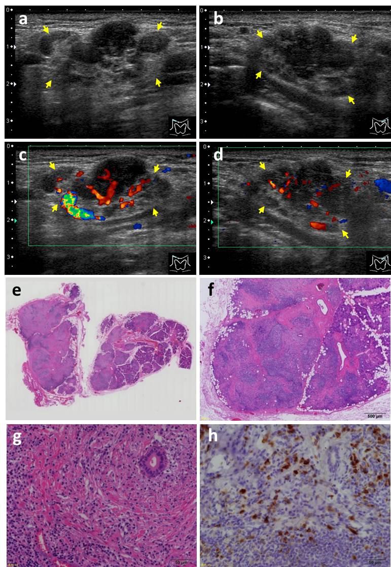
5 Satomi Omotehara 4 pattern, it was classified as the diffuse hypoechoic pattern. If a gland showed both multiple hypoechoic nodule and the diffuse pattern, the dominant pattern was selected. We used a DICOM viewer to analyze the contour and the internal echo texture. The contour texture and the internal echo textures were reviewed retrospectively in a blinded manner by two sonographers with 5 and 25 years of experience of evaluating superficial organs. In cases of disagreement, the texture pattern was decided by consensus between the sonographers. The quantitative assessment of color Doppler signaling was undertaken in 40 glands using the Toshiba Aplio TM XG connected to an 8-MHz linear transducer. The pulse repetition frequency was fixed to avoid parameter differences. The color Doppler gain was maximized until disappearance of noise. Color Doppler images were converted into black and white using Adobe Photoshop 5.0J (Adobe Systems Inc, CA, USA), and the ratio of color Doppler signaling to the parenchyma was calculated (mean ± SD, ratios were expressed as percentages). Statistical analyses Statistical analyses were performed using the Mann-Whitney U test to compare lesion sizes and color Doppler ratios, and the Chi-square test was used to compare internal echo texture patterns between patients and controls. All statistical analyses were performed using IBM SPSS software version A p-value of <0.05 was considered to be statistically significant. Results Clinical and US features of the study participants are shown in Table 1. Among patients with IgG4-SS, the serum IgG4 level exceeded the measurement limit (1500 mg/dl) in one patient. Therefore, data from that patient were excluded when determining the mean IgG4 levels. The longitudinal diameter and thickness of the submandibular gland were significantly greater in patients with IgG4-SS than in controls (p=0.005 and p<0.001, respectively). The majority of patients with IgG4-SS had a rough contour texture, whereas the majority of controls had a smooth contour texture (p<0.001). Patients with IgG4-SS showed diffuse hypoechoic or multiple hypoechoic nodule internal echo patterns, whereas all controls had a homogeneous pattern. The mean Doppler signaling ratio was significantly higher in patients with IgG4-SS than in controls (p<0.001). Typical ultrasonograms of the submandibular glands in a control subject are shown in Figure 2. The serum IgG and IgG4 levels in this subject were both within the normal range at 1277 mg/dl and 32.1 mg/dl, respectively. Typical ultrasonograms from a patient with IgG4-SS are shown in Figure 3. The serum IgG and IgG4 levels in this patient were elevated at 2326 mg/dl and 534 mg/dl, respectively. In addition, the ratio of color Doppler signaling was significantly increased in both submandibular glands. A pathological specimen was obtained from the patient by excisional biopsy of the left submandibular gland (Figure 3E H). Pathological analysis revealed marked fibrosis around acinar structures and obliterative phlebitis. Inflammatory lymphocytes and plasma cells were also seen. The ratio of IgG4-positive to IgG-positive cells was 72%. The region of
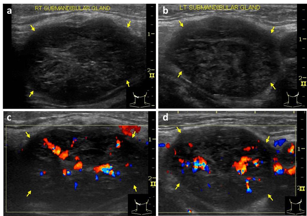
6 Satomi Omotehara 5 lymphoplasmacytic infiltration mostly corresponded to the hypoechoic region on the ultrasonogram. Ultrasonograms of a patient with IgG4-SS showing submandibular glands exhibiting the diffuse hypoechoic pattern are shown in Figure 4. The serum IgG and IgG4 levels in the patient were both elevated at 2761 mg/dl and 143 mg/dl, respectively. Discussion In this study, both the longitudinal diameter and thickness of the submandibular glands were significantly greater in patients with IgG4-SS than in controls, and the increase in the thickness was especially remarkable. The typical sonographic features of the submandibular glands in patients with IgG4-SS were rough contours, multiple hypoechoic nodule, or a diffuse hypoechoic pattern, and increased color Doppler signaling ratios. These findings are thought to represent the characteristic appearance of IgG4-SS. The histological characteristics of IgG4-SS have been reported as lymphoplasmacytic infiltration and fibrosis around ducts and lobules with preservation of lobular architecture [13-15], which would be consistent with our IgG4-SS findings in the present study. The contour roughness and high-echoic borders of the multiple hypoechoic nodules might reflect marked fibrosis around lobules and ducts, whereas the diffuse hypoechoic pattern would be consistent with lymphoplasmacytic infiltration. In some of our cases, distinguishing internal echo patterns was difficult, especially between the diffuse hypoechoic and multiple hypoechoic nodule patterns. Disagreements were recorded in the assessments of 9 out of 35 glands, and we needed to reach consensus. For example, in Figure 3B, although we defined the left submandibular gland as having a diffuse hypoechoic pattern, the gland could easily have been categorized as having the multiple hypoechoic nodule pattern. Because the hypoechoic area in the gland would increase according to the degree of lymphoplasmacytic infiltration, eventually an intermediate pattern would likely exist. However, all submandibular glands in controls showed a homogenous internal echo pattern, so it was still possible to distinguish between IgG4-SS and controls by US. The increased color Doppler signaling ratio detected in patients with IgG4-SS in the present study was thought to correspond with increased blood flow via existing blood vessels, rather than represent any changes caused by tumor angiogenesis, because blood flow was detected along existing blood vessels, none of which were tortuous or had an aberrant structure. However, increased color Doppler ratios could also be indicative of inflammation. Asai et al. classified IgG4-SS-related sonography findings into two patterns based on the shape, contour texture, and internal echo texture [16]. Glands that had an almost lobular shape and uneven contours represented the hypoechoic pattern and were considered the tumor-forming type, whereas the diffuse focal type had a preserved shape and normal contours with multiple hypoechoic foci. They reported that, according to histopathological analysis of the specimen, the region of fibrosis was the only difference between the two IgG4-SS types. These sonographic features were similar to those uncovered in the present study. Enlargement of submandibular glands is also seen in sialolithiasis and malignant tumors, which can make the differential diagnosis of IgG4-SS by US problematic. However, sialolithiasis shows stones
7 Satomi Omotehara 6 with acoustic shadows in the dilated ducts. Malignant lymphomas, especially mucosa-associated lymphoid tissue lymphoma (MALT lymphoma), show hypoechoic areas with rough or dense linear echogenic strands and a lobulated shape with a markedly hypoechoic internal echo texture in the thyroid and salivary glands [17, 18], making them especially difficult to distinguish from IgG4-SS. However, Asai et al. reported that the sonographic findings of MALT lymphoma included a tortoiseshell pattern with an increased hypoechoic internal echo texture compared with IgG4-related disease [19]. Therefore, it is important to bear in mind that, when making a diagnosis of IgG4-SS, a combination of serological testing and biopsy would be needed for a definitive diagnosis. A limitation of the present study was that we did not examine any patients with MALT lymphoma during the study period and therefore could not compare the sonographic features of IgG4-SS and MALT lymphoma. Another limitation was that we focused on the amount of color Doppler signaling, but a detailed evaluation of the vascular structure might have been more useful for making a differential diagnosis. Conclusion The sonographic features evaluated in the present study could differentiate IgG4-SS from healthy submandibular glands. Sonographic submandibular gland features such as enlargement, a rough contour texture, a diffuse hypoechoic or multiple hypoechoic nodule pattern, and increased color Doppler signaling were all useful indicators for a diagnosis of IgG4-SS. Conflict of interest: Satomi Omotehara, Mutsumi Nishida, Megumi Satoh, Mamiko Inoue, Yusuke Kudoh, Tatsunori Horie, Akihiro Homma, Yuji Nakamaru, Kanako C Hatanaka, and Chikara Shimizu declare that they have no conflict of interest. Statement of Ethics: All procedures followed were in accordance with the ethical standards of the responsible committee on human experimentation (institutional and national) and with the Helsinki Declaration of 1964 and later versions. Informed consent was obtained from all subjects included in the study.
8 Satomi Omotehara 7 References [1] Kamisawa T, Okamoto A. IgG4-related sclerosing disease. World J Gastroenterol. 2008;14: [2] Stone JH, Zen Y, Deshpande V. IgG4-related disease. N Engl J Med. 2012;366: [3] Yamamoto M, Ohara M, Suzuki C, et al. Elevated IgG4 concentrations in serum of patients with Mikulicz s disease. Scand J Rheumatol. 2004;33: [4] Kitagawa S, Zen Y, Harada K, et al. Abundant IgG4-positive plasma cell infiltration characterizes chronic sclerosing sialadenitis (Küttner s tumor). Am J Surg Pathol. 2005;29: [5] Takano K, Yamamoto M, Takahashi H, et al. Clinicopathologic similarities between Mikulicz disease and Küttner tumor. Am J Otolaryngol. 2010;31: [6] Himi T, Takano K, Yamamoto M, et al. A novel concept of Mikulicz s disease as IgG4-related disease. Auris Nasus Larynx. 2012;39:9 17. [7] Yamamoto M, Takahashi H,Ohara M,et al. A new conceptualization for Mikulicz s disease as an IgG4-related plasmacytic disease. Mod Rheumatol. 2006;16: [8] Geyer JT, Deshpande V. IgG4-associated sialadenitis. Curr Opin Rheumatol. 2011;23: [9] Kamisawa T, Zen Y, Pillai S, et al. IgG4-related disease. Lancet.2015;385: [10] Shimizu M, Moriyama M, Okazaki K, et al. Sonographic diagnosis for Mikulicz disease. Oral Surg Oral Med Pathol Oral Radiol Endod. 2009;108: [11] Ahuja AT, Richards PS, Wong KT, et al. Kuttner tumour (chronic sclerosing sialadenitis) of the submandibular gland: sonographic appearances. Ultrasound Med Biol. 2003;29:913 9.[12] Umehara H, Okazaki K, Masaki Y, et al. Comprehensive diagnostic criteria for IgG4-related disease (IgG4-RD), Mod Rheumatol. 2012;22: [13] Takagi Y, Nakamura H, Origuchi T, et al. IgG4-related Mikulicz z disease: ultrasonography of the salivary and lacrimal glands for monitoring the efficacy of corticosteroid therapy. Clin Exp Rheumatol. 2013;31: [14] Ohta N, Kurakami K,Ishida A, et al. Clinical and pathological characteristics of IgG4-related sclerosing sialadenitis. Laryngoscope. 2012;122: [15] Laco J, Ryska A, Celakovsky P, et al. Chronic sclerosing sialadenitis as one of the immunoglobulin G4-related diseases: a clinicopathological study of six cases from Central Europe. Histopathology. 2011;58: [16] Asai S, Okami K, Nakamura, et al. Localized or diffuse lesions of the submandibular glands in immunoglobulin g4-related disease in association with differential organ involvement. J Ultrasound Med. 2013;32: [17] Orita Y, Sato Y, Kimura N, et al. Characteristic ultrasound features of mucosa-associated lymphoid tissue lymphoma of the salivary and thyroid gland. Acta Otolaryngol. 2014;134: [18] Bahn YE, Lee SK, Kwon SY, et al. Sonographic appearances of mucosa-associated lymphoid tissue lymphoma of the submandibular gland confirmed with sonographically guided core needle biopsy. J Clin Ultrasound. 2011;39: [19] Asai S, Okami K, Nakamura N, et al. Sonographic appearance of the submandibular glands in patients with immunoglobulin G4-related disease. J Ultrasound Med. 2012;31:
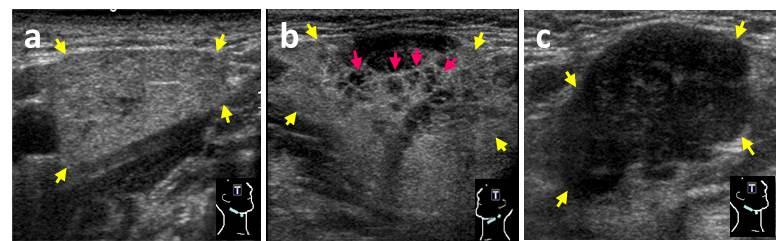
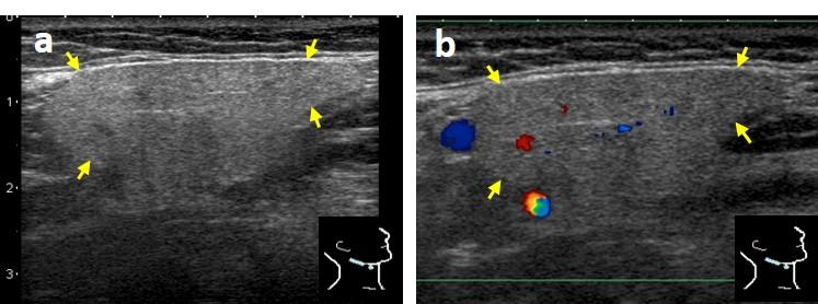
9 Figure legends Satomi Omotehara 8 Figure 1. Classification of the internal echo texture of the submandibular gland. (A) Homogeneous pattern. (B) Multiple hypoechoic nodule pattern. (C) Diffuse hypoechoic pattern. Yellow arrows indicate the borders of the submandibular gland. Pink arrows indicate multiple hypoechoic nodules. Figure 2. Ultrasonograms of a normal submandibular gland. The longitudinal diameter and the thickness of the right submandibular gland are 35.4 mm and 15.6 mm, respectively. (A) The contour is smooth and the internal echo texture shows the homogeneous pattern. (B) The ratio of color Doppler signaling is 0.8 %. Yellow arrows indicate the borders of the submandibular gland. Figure 3. A case of typical IgG4-SS. Both submandibular glands are swollen. The longitudinal diameter and the thickness of the right and left submandibular glands are 33.3 mm and 19.1 mm, and 32.2 mm and 17.3 mm, respectively. The contour texture is rough on both sides. (A) The right submandibular gland shows the multiple hypoechoic nodule pattern. (B) The left submandibular gland shows the diffuse hypoechoic pattern. (C) The color Doppler signaling ratio in the right submandibular gland is 9.7%, whereas that in the left submandibular gland is 8.3% (D). Yellow arrows indicate the borders of the submandibular gland. Histopathological analysis of an excisional biopsy of the left submandibular gland (E H). (E) Macroscopic hematoxylin-eosin staining. (F, G) Marked fibrosis can be seen around acinar structures and obliterative phlebitis is apparent. (F) The majority of inflammatory cells were lymphocytes or plasma cells (hematoxylin-eosin; F, magnification 40; G, magnification 400). (H) Infiltration of IgG4-positive cells is apparent (immunohistochemical staining with an anti-igg4 antibody, magnification 400). Figure 4. Ultrasonograms of submandibular glands showing the diffuse hypoechoic pattern in a patient with IgG4-SS. The longitudinal diameter and the thickness of the right and left submandibular glands are 35.6 mm and 22.1 mm, and 29.8 mm and 20.3 mm, respectively. Both glands show a smooth contour texture. (A) The right submandibular gland. (B) The left submandibular gland. (C, D) Increased color Doppler signaling can be seen in both glands. Yellow arrows indicate the borders of the submandibular gland.
10
11
12
13
14 Table 1. Clinical and ultrasound feature of the study participants IgG4-SS Healthy Volunteers n Age, years a 63±10 57±7 Existence of other organ lesions, n 15 - Glands, n IgG, mg/dl a ±1375.0** ±271.5 IgG4, mg/dl a 743.3±656.4** 61.0±36.6 Longitudinal diameter, mm a 37.0±6.2* 32.4±4.0 Thickness, mm a 19.3±4.2** 15.1±2.2 Contour, % (n) smooth 37.1 (13) 91.7 (22) rough 62.9 (22) 8.3 (2) Internal echo texture, % (n) homogeneous 0 (0) 100 (24) multiple hypoechoic nodule 60 (21) 0 (0) diffuse hypoechoic 40 (14) 0 (0) Color Doppler signaling ratio, % a (n) 9.5±5.3 (16)** 1.6±1.5 (24) *:p=0.005 **:p<0.001 a Values are mean±sd.
Chronic Sclerosing Dacryoadenitis
 The Korean Journal of Pathology 2008; 42: 118-22 Chronic Sclerosing Dacryoadenitis - Report of 2 Cases - Ji Eun Kwon Sang Kyum Kim Sang-Ryul Lee 1 Woo-Ick Yang Haeryoung Kim 2 Department of Pathology and
Immunoglobulin G4-Related Disease with Several Inflammatory Foci
 CASE REPORT Immunoglobulin G4-Related Disease with Several Inflammatory Foci Akira Sakamaki 1, Kenya Kamimura 1, Kazuhiko Shioji 1, Junko Sakurada 2, Takeshi Nakatsue 3, Yoko Wada 3, Michitaka Imai 1,
Comparison of multidetector-row computed tomography findings of IgG4-related sclerosing cholangitis and cholangiocarcinoma
 Comparison of multidetector-row computed tomography findings of IgG4-related sclerosing cholangitis and cholangiocarcinoma Poster No.: C-0245 Congress: ECR 2014 Type: Scientific Exhibit Authors: M. Yata,
Case Report An IgG4-Related Salivary Gland Disorder: A Case Series Presenting with a Different Clinical Setting
 Case Reports in Immunology Volume 2011, Article ID 236079, 4 pages doi:10.1155/2011/236079 Case Report An IgG4-Related Salivary Gland Disorder: A Case Series Presenting with a Different Clinical Setting
Among the benign intraepithelial melanocytic proliferations, Inflamed Conjunctival Nevi. Histopathological Criteria. Resident Short Reviews
 Resident Short Reviews Inflamed conjunctival nevi (ICN) may suggest malignancy because of their rapid growth and atypical histology. The objective of this study was to characterize the diagnostic features
Unusual Involvement of IgG4-Related Sclerosing Disease in Lacrimal and Submandibular Glands and Extraocular Muscles
 pissn: 1011-8942 eissn: 2092-9382 Korean J Ophthalmol 2012;26(3):216-221 http://dx.doi.org/10.3341/kjo.2012.26.3.216 Case Report Unusual Involvement of IgG4-Related Sclerosing Disease in Lacrimal and Submandibular
IgG4-Negative Autoimmune Pancreatitis with Sclerosing Cholangitis and Colitis: Possible Association with Primary Sclerosing Cholangitis?
 CASE REPORT IgG4-Negative Autoimmune Pancreatitis with Sclerosing Cholangitis and Colitis: Possible Association with Primary Sclerosing Cholangitis? Keita Saeki 1, Shigenari Hozawa 1, Naoteru Miyata 1,
Clinical outcomes and pathological characteristics of immunoglobulin G4-related ophthalmic disease versus orbital inflammatory pseudotumor
 ORIGINAL ARTICLE Korean J Intern Med 2019;34:220-226 Clinical outcomes and pathological characteristics of immunoglobulin G4-related ophthalmic disease versus orbital inflammatory pseudotumor Hong Ki Min
Mikulicz s Disease with Progressively Transformed Germinal Centers-type Immunoglobulin G4-related Lymphadenopathy Mimicking Sjögren s Syndrome
 Journal of Rheumatic Diseases Vol. 22, No. 6, December, 2015 http://dx.doi.org/10.4078/jrd.2015.22.6.395 Case Report Mikulicz s Disease with Progressively Transformed Germinal Centers-type Immunoglobulin
IgG4-related sclerosing disease
 IgG4-related sclerosing disease TERUMI KAMISAWA, KENSUKE TAKUMA, NAOTO EGAWA Department of Internal Medicine Tokyo Metropolitan Komagome Hospital 3-18-22 Honkomagome, Bunkyo-ku, Tokyo 113-8677, Japan JAPAN
IgG4 ᛶ ᝈ䛸 ᶫᮏ ᕫච ᛶ ⅖ ḷᒣ Ꮫ య Ꮫ ぬ㐨
 IgG4 Autoimmune pancreatitis Histopathological features: Diffuse lymphoplasmacytic infiltration Stromal fibrosis Acinar atrophy Obliterative phlebitis 1. Hamano et al reported that serum IgG4 levels were
Case year old Chinese female. Radiological echo-distortion in the right breast at o clock. Core biopsy of the o clock lesion.
 Case 3 64 year old Chinese female. Radiological echo-distortion in the right breast at 10-12 o clock. Core biopsy of the 11-12 o clock lesion. Division of Pathology Courtesty of Dr Lester Leong ill-defined,
Case Report IgG4-Related Nasal Pseudotumor
 Case Reports in Otolaryngology Volume 2015, Article ID 749890, 4 pages http://dx.doi.org/10.1155/2015/749890 Case Report IgG4-Related Nasal Pseudotumor L. K. Døsen, 1 P. Jebsen, 2 B. Dingsør, 3 and R.
Clinical Features of Patients with Basedow s Disease and High Serum IgG4 Levels
 ORIGINAL ARTICLE Clinical Features of Patients with Basedow s Disease and High Serum IgG4 Levels Keiichi Torimoto, Yosuke Okada, Akira Kurozumi, Manabu Narisawa, Tadashi Arao and Yoshiya Tanaka Abstract
Diagnostic benefits of ultrasound-guided. CNB) versus mammograph-guided biopsy for suspicious microcalcifications. without definite breast mass
 versus mammograph-guided biopsy for suspicious microcalcifications. without definite breast mass") Volume 118 No. 19 2018, 531-543 ISSN: 1311-8080 (printed version); ISSN: 1314-3395 (on-line version) url: http://www.ijpam.eu ijpam.eu Diagnostic benefits of ultrasound-guided biopsy versus mammography-guided
Volume 118 No. 19 2018, 531-543 ISSN: 1311-8080 (printed version); ISSN: 1314-3395 (on-line version) url: http://www.ijpam.eu ijpam.eu Diagnostic benefits of ultrasound-guided biopsy versus mammography-guided
Key words: diagnosis, immunoglobulin G4, immunoglobulin G4-related diseases, immunohistochemistry, pseudolymphoma. CASE HISTORY
 doi: 10.1111/1346-8138.12301 Journal of Dermatology 2013; 40: 998 1003 ORIGINAL ARTICLE Case of immunoglobulin G4-related skin disease: Possible immunoglobulin G4-related skin disease cases in cutaneous
CASE 01 LA Path Slide Seminar 13 March, 08. Deepti Dhall, MD Department of Pathology and Laboratory Medicine Cedars-Sinai Medical Center
 CASE 01 LA Path Slide Seminar 13 March, 08 Deepti Dhall, MD Department of Pathology and Laboratory Medicine Cedars-Sinai Medical Center Clinical History 60 year old male presented with obstructive jaundice
Ultrasonographic Evaluation of Cervical Lymphadenopathy with Cytological Correlation
 Original Article Print ISSN: 2321-6379 Online ISSN: 2321-595X DOI: 10.17354/ijss/2017/74 Ultrasonographic Evaluation of Cervical Lymphadenopathy with Cytological Correlation Suresh Kumar 1, Sonjjay Pande
Overview of the Immunoglobulin G4-related Disease Spectrum
 Review Article The Korean Journal of Pancreas and Biliary Tract 2015;20:124-129 http://dx.doi.org/10.15279/kpba.2015.20.3.124 pissn 1976-3573 eissn 2288-0941 면역글로불린 G4 연관질환의개요 1 한림대학교의과대학한림대학교성심병원내과, 2
A-005 US DIAGNOSIS OF NONPALPABLE BREAST LESIONS
 A-005 US DIAGNOSIS OF NONPALPABLE BREAST LESIONS Hideaki Shirai M.D., M. Sakurai M.D., K. Yoshida M.D., N. Usuda M.D., H. Masuoka M.D., I. Shimokawara M.D, K. Asaishi M.D. Sapporo Kotoni Breast Clinic,
CASE REPORT. Abstract. Introduction. Case Report
 CASE REPORT Branch Duct Intraductal Papillary Mucinous Neoplasms of the Pancreas Involving Type 1 Localized Autoimmune Pancreatitis with Normal Serum IgG4 Levels Successfully Diagnosed by Endoscopic Ultrasound-guided
J of Evolution of Med and Dent Sci/ eissn , pissn / Vol. 4/ Issue 39/ May 14, 2015 Page 6787
 ROLE OF HIGH RESOLUTION SONOGRAPHY IN CHARACTERIZATION OF SOLID SALIVARY GLAND TUMORS Sheetal Singh 1, Amlendu Nagar 2, Pramod Sakhi 3, Sachin Kataria 4, Kumud Julka 5, Anup Gupta 6 HOW TO CITE THIS ARTICLE:
Clinical Study Evaluation and Clinical Validity of a New Questionnaire for Mikulicz s Disease
 International Rheumatology Volume 2, Article ID 283459, pages doi:1.1155/2/283459 Clinical Study Evaluation and Clinical Validity of a New Questionnaire for Mikulicz s Disease Motohisa Yamamoto, 1 Hiroki
Shimizu et al. Arthritis Research & Therapy (2015) 17:223 DOI /s x
 17:223 DOI /s x") Shimizu et al. Arthritis Research & Therapy (2015) 17:223 DOI 10.1186/s13075-015-0751-x RESEARCH ARTICLE Open Access Effectiveness of imaging modalities for screening IgG4-related dacryoadenitis and sialadenitis
Shimizu et al. Arthritis Research & Therapy (2015) 17:223 DOI 10.1186/s13075-015-0751-x RESEARCH ARTICLE Open Access Effectiveness of imaging modalities for screening IgG4-related dacryoadenitis and sialadenitis
Title. CitationNeurology and Clinical Neuroscience, 5(1): Issue Date Doc URL. Rights. Type. Additional There Information
: Issue Date Doc URL. Rights. Type. Additional There Information") Title Pseudodystonia in sarcoid myopathy Uwatoko, Hisashi; Yabe, Ichiro; Shirai, Shinichi; Ta Author(s) Hidenao CitationNeurology and Clinical Neuroscience, 5(1): 34-35 Issue Date 2017-01 Doc URL http://hdl.handle.net/2115/68037
Title Pseudodystonia in sarcoid myopathy Uwatoko, Hisashi; Yabe, Ichiro; Shirai, Shinichi; Ta Author(s) Hidenao CitationNeurology and Clinical Neuroscience, 5(1): 34-35 Issue Date 2017-01 Doc URL http://hdl.handle.net/2115/68037
Review Article Orbital IgG4-Related Disease: Clinical Features and Diagnosis
 International Scholarly Research Network Volume 2012, Article ID 412896, 5 pages doi:10.5402/2012/412896 Review Article Orbital IgG4-Related Disease: Clinical Features and Diagnosis Toshinobu Kubota 1
Title. CitationHepato-Gastroenterology, 61(135): Issue Date Doc URL. Type. File Information. IgG4-Related Sclerosing Cholangitis
: Issue Date Doc URL. Type. File Information. IgG4-Related Sclerosing Cholangitis") Title Difference from Bile Duct Cancer and Relationship be IgG4-Related Sclerosing Cholangitis Kuwatani, Masaki; Kawakami, Hiroshi; Zen, Yoh; Kawak Author(s) Sakamoto, Naoya CitationHepato-Gastroenterology,
Title Difference from Bile Duct Cancer and Relationship be IgG4-Related Sclerosing Cholangitis Kuwatani, Masaki; Kawakami, Hiroshi; Zen, Yoh; Kawak Author(s) Sakamoto, Naoya CitationHepato-Gastroenterology,
IgG4-related Kidney Disease in Which the Urinalysis, Kidney Function and Imaging Findings Were Normal
 CASE REPORT IgG4-related Kidney Disease in Which the Urinalysis, Kidney Function and Imaging Findings Were Normal Miho Otani 1, Masahiro Morinaga 1, Yoshihiko Nakajima 1, Hiromi Tomioka 2, Michiko Nishii
A Case of Multiple Giant Coronary Aneurysms and Abdominal Aortic Aneurysm Coexisting with IgG4-related Disease
 CASE REPORT A Case of Multiple Giant Coronary Aneurysms and Abdominal Aortic Aneurysm Coexisting with IgG4-related Disease Hirofumi Takei 1, Hayato Nagasawa 1, Ryota Sakai 1, Koji Nishimura 1, Takahiko
Churg-Strauss Syndrome Concomitant with Chronic Symmetrical Dacryoadenitis Suggesting Mikulicz s Disease
 CASE REPORT Churg-Strauss Syndrome Concomitant with Chronic Symmetrical Dacryoadenitis Suggesting Mikulicz s Disease Yusuke Hanioka 1, Keiko Yamagami 1, Katsunobu Yoshioka 2, Tomomi Nakamura 1, Masatsugu
IgG4-related disease: features and treatment response in a multi-ethnic cohort in Singapore
 IgG4-related disease: features and treatment response in a multi-ethnic cohort in Singapore W. Fong 1,2,3, I. Liew 1, D. Tan 2,3,4, K.H. Lim 5, A. Low 6, Y.Y. Leung 1,2 1 Department of Rheumatology and
IgG4 Disease. General Principles of IgG4-related disease. EL Cluvar, AC Bateman
 IgG4 Disease General Principles of IgG4-related disease. EL Cluvar, AC Bateman Diagnostic Guidelines for IgG4-related disease with a focus on histopathological criteria. V Deshpande, A Khosroshahi Diagnostic
Immunoglobulin G4-Related Sclerosing Disease Involving the Urethra: Case Report
 Case Report http://dx.doi.org/10.3348/kjr.2012.13.6.803 pissn 1229-6929 eissn 2005-8330 Korean J Radiol 2012;13(6):803-807 Immunoglobulin G4-Related Sclerosing Disease Involving the Urethra: Case Report
Clinicopathological characteristics of immunoglobulin G4-related sialadenitis
 Li et al. Arthritis Research & Therapy (2015) 17:186 DOI 10.1186/s13075-015-0698-y RESEARCH ARTICLE Open Access Clinicopathological characteristics of immunoglobulin G4-related sialadenitis Wei Li 1, Yan
IgG4-related Sclerosing Disease of the Lung without Pancreas Involvement: Presentation on 18F-FDG PET/CT
 J Radiol Sci 2013; 38: 129-133 IgG4-related Sclerosing Disease of the Lung without Pancreas Involvement: Presentation on 18F-FDG PET/CT Han-Jui Lee 1 Yi-Chen Yeh 2,3 Chun-Ku Chen 1,3 Rheun-Chuan Lee 1,3
Original Article CT features and pathologic characteristics of IgG4-related systemic disease of submandibular gland
 Int J Clin Exp Pathol 2015;8(12):16111-16116 www.ijcep.com /ISSN:1936-2625/IJCEP0014889 Original Article CT features and pathologic characteristics of IgG4-related systemic disease of submandibular gland
Evaluation of Diffuse Liver Diseases Using Conventional Ultrasound
 IOSR Journal of Dental and Medical Sciences (IOSR-JDMS) e-issn: 2279-0853, p-issn: 2279-0861.Volume 16, Issue 6 Ver. VII (June. 2017), PP 70-74 www.iosrjournals.org Evaluation of Diffuse Liver Diseases
Case Report. A Surgical Case of Venous Aneurysm of the Cephalic Vein. with Unique Clinicopathological Findings for Venous Dissection
 Case Report A Surgical Case of Venous Aneurysm of the Cephalic Vein with Unique Clinicopathological Findings for Venous Dissection Takashi Kobata, 1 Sohsuke Yamada, 2,3* Ken-ichi Mizutani, 2 Nozomu Kurose,
IgG4-Related Disease: Dataset of 235 Consecutive Patients
 IgG-Related Disease: Dataset of 235 Consecutive Patients Dai Inoue, MD, PhD, Kotaro Yoshida, MD, PhD, Norihide Yoneda, MD, PhD, Kumi Ozaki, MD, PhD, Takashi Matsubara, MD, PhD, Keiichi Nagai, MD, PhD,
Chronic Sclerosing Sialadenitis (Küttner s tumour) of the Parotid Gland
 of the Parotid Gland") Case Report Chronic Sclerosing Sialadenitis (Küttner s tumour) of the Parotid Gland Güçlü Kaan Beriat 1, Sefik Halit Akmansu 1, Sinan Kocatürk 1, Ömür Ataoğlu 2 Submitted: 4 Dec 2009 Accepted: 18 Mar 2010
Case Report Chronic Sclerosing Sialadenitis (Küttner s tumour) of the Parotid Gland Güçlü Kaan Beriat 1, Sefik Halit Akmansu 1, Sinan Kocatürk 1, Ömür Ataoğlu 2 Submitted: 4 Dec 2009 Accepted: 18 Mar 2010
OPTO-ACOUSTIC BREAST IMAGING
 OPTO-ACOUSTIC BREAST IMAGING A Novel Fusion of Functional and Morphologic Imaging Reni S. Butler, MD A. Thomas Stavros, MD F. Lee Tucker, MD Michael J. Ulissey, MD PURPOSE 1. Explain opto-acoustic (OA)
Acta Med. Okayama Vol. 70, No. 2. Iwamuro et al.
 140 Iwamuro et al. cta Med. Okayama Vol. 70, No. 2 emission tomography (PET) scanning showed tracer uptake in the spleen and iliac bone as well as in the swollen lymph nodes. There were no abnormalities
A case of marginal zone B cell lymphoma mimicking IgG4-related dacryoadenitis and sialoadenitis
 Ohta et al. World Journal of Surgical Oncology (2015) 13:67 DOI 10.1186/s12957-015-0459-z WORLD JOURNAL OF SURGICAL ONCOLOGY CASE REPORT Open Access A case of marginal zone B cell lymphoma mimicking IgG4-related
Evolution of diagnostic ultrasound systems Current achievements in breast ultrasound
 Evolution of diagnostic ultrasound systems Current achievements in breast ultrasound Dr. Ayumi Izumori, M. D. Department of Breast Surgery, Takamatsu Heiwa Hospital Tokushima Breast Care Clinic, Japan
A case of retroperitoneal fibrosis responding to steroid therapy
 Challenging Clinical Cases Vol. 43 (6): 1185-1189, November - December, 2017 doi: 10.1590/S1677-5538.IBJU.2016.0520 A case of retroperitoneal fibrosis responding to steroid therapy Ryuta Watanabe 1, Akira
FOR PUBLIC CONSULTATION ONLY. Evidence Review: Rituximab for immunoglobulin G4-related disease (IgG4-RD)
") Evidence Review: Rituximab for immunoglobulin G4-related disease (IgG4-RD) NHS England FOR PUBLIC CONSULTATION ONLY Evidence Review: Rituximab for immunoglobulin G4-related disease (IgG4- RD) First published:
Evidence Review: Rituximab for immunoglobulin G4-related disease (IgG4-RD) NHS England FOR PUBLIC CONSULTATION ONLY Evidence Review: Rituximab for immunoglobulin G4-related disease (IgG4- RD) First published:
Cystic Hypersecretory Carcinoma of the Breast:
 J Korean Soc Radiol 2010;62:287-294 Cystic Hypersecretory Carcinoma of the Breast: Sonographic Features with a Histological Correlation 1 Sang Yu Nam, M.D., Boo-Kyung Han, M.D., Jung Hee Shin, M.D., Eun
Shadow because the air
 Thyroid Ultrasound Thyroid US examination needs: 1. high frequency transducer 2. extended patient's neck 3. check all the neck area because the swelling could be in areas other than the thyroid such as
B-330 Cervical lymph node ultrasonography in HIV infected children
 B-330 Cervical lymph node ultrasonography in HIV infected children Scientific Paper B-330 Cervical lymph node ultrasonography in HIV infected children A. Butnaru (Cluj Napoca/RO) S. D. Bolboaca (Cluj Napoca/RO)
Review Article The Utility of Serum IgG4 Concentrations as a Biomarker
 International Rheumatology Volume 2012, Article ID 198314, 4 pages doi:10.1155/2012/198314 Review Article The Utility of Serum IgG4 Concentrations as a Biomarker Shigeyuki Kawa, 1 Tetsuya Ito, 2 Takayuki
Case Report The Challenging Diagnosis of Pancreatic Masses: Not All Tumors Are Cancers
 Case Reports in Medicine Volume 2015, Article ID 832463, 4 pages http://dx.doi.org/10.1155/2015/832463 Case Report The Challenging Diagnosis of Pancreatic Masses: Not All Tumors Are Cancers Alessandro
Jpn J Med Ultrasonics Vol 32 No
 Jpn J Med Ultrasonics Vol 32 No 6 2005 589 590 Jpn J Med Ultrasonics Vol 32 No 6 2005 Jpn J Med Ultrasonics Vol 32 No 6 2005 591 Terminology and Diagnostic Criteria Committee of The Japan Society of Ultrasonics
The UGent Institutional Repository is the electronic archiving and dissemination platform for
 biblio.ugent.be The UGent Institutional Repository is the electronic archiving and dissemination platform for all UGent research publications. Ghent University has implemented a mandate stipulating that
Role of ultrasonography in recognition of malignant potential of thyroid nodules on the basis of their internal composition
 Role of ultrasonography in recognition of malignant potential of thyroid nodules on the basis of their internal composition Nodular thyroid is a common clinical entity. All patients were evaluated by grey
Thyroid in a Nutshell Dublin Catherine Kirkpatrick Consultant Sonographer ULHT
 Thyroid in a Nutshell Dublin 2017 Catherine Kirkpatrick Consultant Sonographer ULHT Acknowledgements Dr. Steve Colley Dr. Rhodri Evans Dr. Rhian Rhys Dr. Andrew McQueen Aims Anatomy & Physiology Incidence
J of Evolution of Med and Dent Sci/ eissn , pissn / Vol. 4/ Issue 09/Jan 29, 2015 Page 1533
 EVALUATION OF CERVICAL LYMPHNODES BY ULTRASONOGRAPHY IN CORRELATION WITH FNAC Aaditya Kumar Singh 1, Purnima Hegde 2, Anil Kumar Sakalecha 3, T. N. Suresh 4, P. N. Sreeramulu 5 HOW TO CITE THIS ARTICLE:
Title. CitationJournal of medical ultrasonics, 44(4): Issue Date Doc URL. Rights. Type. File Information.
: Issue Date Doc URL. Rights. Type. File Information.") Title Altered oscillation of Doppler-derived renal and ren patients Kudo, Yusuke; Mikami, Taisei; Nishida, Mutsumi; Okad Author(s) Satomi; Shibuya, Hitoshi; Kahata, Kaoru; Shimizu, Ch CitationJournal of
Title Altered oscillation of Doppler-derived renal and ren patients Kudo, Yusuke; Mikami, Taisei; Nishida, Mutsumi; Okad Author(s) Satomi; Shibuya, Hitoshi; Kahata, Kaoru; Shimizu, Ch CitationJournal of
Autoimmune Pancreatitis, Pancreatic and Extrapancreatic Imaging Findings
 Autoimmune Pancreatitis, Pancreatic and Extrapancreatic Imaging Findings Poster No.: R-0074 Congress: RANZCR-AOCR 2012 Type: Educational Exhibit Authors: J. Stegeman, A. Borsaru; Clayton/AU Keywords: Education
A subset of ocular adnexal marginal zone lymphomas may arise in association with
 A subset of ocular adnexal marginal zone lymphomas may arise in association with IgG4-related disease 1)*, Yasuharu Sato 1) 2)*, Koh-ichi Ohshima 3), Katsuyoshi Takata 1), Tomoko Miyata-Takata 1), Mai
glands: a correlation in postmortem subjects
 J. clin. Path., 1970, 23, 690-694 Lymphocytic sialadenitis in the major and minor glands: a correlation in postmortem subjects D. M. CHISHOLM, J. P. WATERHOUSE, AND D. K. MASON From the Department of Oral
High Resolution Ultrasound of the Submandibular Gland
 DOI: 10.7860/IJARS/2017/26094:2276 Radiology Section Review Article High Resolution Ultrasound of the Submandibular Gland Prashant Madhukarrao Onkar, Chetana Ramesh Ratnparkhi, Kajal Mitra ABSTRACT Submandibular
Sonographic Differentiation of Thyroid Nodules With Eggshell Calcifications
 Article Sonographic Differentiation of Thyroid Nodules With Eggshell Calcifications Byung Moon Kim, MD, Min Jung Kim, MD, Eun-Kyung Kim, MD, Jin Young Kwak, MD, Soon Won Hong, MD, Eun Ju Son, MD, Ki Hwang
A 44-Year-Old Man With Chronic Cough, Weakness, and a Mediastinum Mass
 [ Pulmonary, Critical Care, and Sleep Pearls ] A 44-Year-Old Man With Chronic Cough, Weakness, and a Mediastinum Mass Dimitrios Theofilos, MD ; Christina Triantafillidou, MD, PhD ; Athanasios Zetos, MD
2017 ATA Victoria Advanced Thyroid US
 2017 ATA Victoria Advanced Thyroid US DIFFUSE THYROID CONDITIONS Stephanie L. Lee, M.D., Ph.D. Director of the BMC Thyroid Nodule and Cancer Center Section of Endocrinology, Diabetes and Nutrition Boston
Case Report Usefulness of minor salivary gland biopsy in the diagnosis of IgG4-related disease: a case report
 Int J Clin Exp Pathol 2014;7(5):2673-2677 www.ijcep.com /ISSN:1936-2625/IJCEP0000175 Case Report Usefulness of minor salivary gland biopsy in the diagnosis of IgG4-related disease: a case report Kentaro
Salivary ultrasound. Dr T J Beale Royal National Throat Nose & Ear and UCLH Hospitals London UK
 Salivary ultrasound Dr T J Beale Royal National Throat Nose & Ear and UCLH Hospitals London UK Two main groups of patients with presenting symptoms of: Obstructive or chronic inflammatory symptoms (salivary
Chronic sclerosing sialadenitis of the sublingual gland: case report and literature review
 Thomopoulos et al. Stomatological Dis Sci 2018;2:8 DOI: 10.20517/2573-0002.2017.22 Stomatological Disease and Science Case Report Open Access Chronic sclerosing sialadenitis of the sublingual gland: case
Diffuse Sclerosing Variant of Papillary Thyroid Carcinoma
 PITORL ESSY iffuse Sclerosing Variant of Papillary Thyroid arcinoma Sonography and Specimen Radiography Hyun Kyung Jung, M, Soon Won Hong, M, Eun-Kyung Kim, M, Jung Hyun Yoon, M, Jin Young Kwak, M The
ISSN X (Print) Research Article. *Corresponding author Dr. Amlendu Nagar
 Research Article. *Corresponding author Dr. Amlendu Nagar") Scholars Journal of Applied Medical Sciences (SJAMS) Sch. J. App. Med. Sci., 2015; 3(3A):1069-1073 Scholars Academic and Scientific Publisher (An International Publisher for Academic and Scientific Resources)
Scholars Journal of Applied Medical Sciences (SJAMS) Sch. J. App. Med. Sci., 2015; 3(3A):1069-1073 Scholars Academic and Scientific Publisher (An International Publisher for Academic and Scientific Resources)
Characteristic feautures of cholangitis with serum IgG4 elevation compared with primary sclerosing cholangitis
 Characteristic feautures of cholangitis with serum IgG4 elevation compared with primary sclerosing cholangitis Poster No.: C-2005 Congress: ECR 2011 Type: Scientific Paper Authors: T. Takeda, T. Ueda,
Cervical Lymph Nodes
 Cervical Lymph Nodes Diana Gaitini, MD Unit of Ultrasound, Department of Medical Imaging Rambam Medical Center and Faculty of Medicine Technion, Israel Institute of Technology Haifa, Israel Learning Targets
Autoimmune Pancreatitis: A Great Imitator
 Massachusetts General Hospital Harvard Medical School Autoimmune Pancreatitis: A Great Imitator Dushyant V Sahani MD dsahani@partners.org Autoimmune Pancreatitis: Learning Objectives Clinical manifestations
ShearWave elastography in lymph nodes
 ShearWave elastography in lymph nodes Poster No.: B-0158 Congress: ECR 2015 Type: Authors: Keywords: DOI: Scientific Paper F. Houari, O. Lucidarme, J. Gabarre, F. Charlotte, C. Pellot- Barakat, M. Lefort,
Intracystic papillary carcinoma of the breast
 Intracystic papillary carcinoma of the breast Poster No.: C-1932 Congress: ECR 2011 Type: Educational Exhibit Authors: V. Dimarelos, F. TZIKOS, N. Kotziamani, G. Rodokalakis, 1 2 3 1 1 1 2 T. MALKOTSI
Value of Serum IgG4 in the Diagnosis of Autoimmune Pancreatitis and in Distinguishing it from Acute and Chronic Pancreatitis of Other Etiology
 94 Jul 2017 Vol 10 No.3 North American Journal of Medicine and Science Original Research Value of Serum IgG4 in the Diagnosis of Autoimmune Pancreatitis and in Distinguishing it from Acute and Chronic
Scrotum Kacey Morrison Amanda Baxter Sabrina Tucker July 18, 2006 SCROTUM
 Scrotum Kacey Morrison Amanda Baxter Sabrina Tucker July 18, 2006 SCROTUM 1) Other Names: Scrotum None Testicles Testes (Curry Tempkin, p. 236, 2/3/2) Ductus deferens spermatic cord (Tempkin, p. 279, Anatomy
IgG4-Related Disease
 IgG4-related disease (IgG4-RD) is a systemic autoimmune fibroinflammatory disease that produces sclerotic, tumefactive masses containing dense lymphoplasmacytic infiltrates rich in immunoglobulin (Ig)
Title. CitationInternational Cancer Conference Journal, 4(1): Issue Date Doc URL. Rights. Type. File Information
: Issue Date Doc URL. Rights. Type. File Information") Title Lymph node metastasis in the suprasternal space from Homma, Akihiro; Hatakeyama, Hiromitsu; Mizumachi, Ta Author(s) Tomohiro; Fukuda, Satoshi CitationInternational Cancer Conference Journal, 4(1):
Title Lymph node metastasis in the suprasternal space from Homma, Akihiro; Hatakeyama, Hiromitsu; Mizumachi, Ta Author(s) Tomohiro; Fukuda, Satoshi CitationInternational Cancer Conference Journal, 4(1):
Lymph Node Hilus. Gray Scale and Power Doppler Sonography of Cervical Nodes. Article
 Article Lymph Node Hilus Gray Scale and Power Doppler Sonography of Cervical Nodes Anil Ahuja, FRCR, Michael Ying, MPhil, Ann King, FRCR, Hok Yuen Yuen, FRCR Objective. To investigate the difference in
Clinical Study Is IgG4-Related Disease a Cause of Xerostomia? A Cohort Study of 60 Patients
 International Rheumatology Volume 2012, Article ID 303506, 6 pages doi:10.1155/2012/303506 Clinical Study Is IgG4-Related Disease a Cause of Xerostomia? A Cohort Study of 60 Patients M. Hermet, 1 M. André,
THYROID NODULES: THE ROLE OF ULTRASOUND
 THYROID NODULES: THE ROLE OF ULTRASOUND NOVEMBER 2017 DR. DEAN DURANT DEFINITION Thyroid nodule: Focal area within the thyroid gland with echogenicity different from surrounding parenchyma. THYROID NODULES
Nozomu Kurose. Key Words: IgG4-related disease, comprehensive diagnostic criteria, fibrosis grade, IL-6. J Kanazawa Med Univ , 2016
 J Kanazawa Med Univ 41 76 85, 2016 Clinicopathological Analysis between IgG4-related and Non-IgG4-related Diseases Occurring in Various Organs and Tissues: Re-evaluation of Comprehensive Diagnostic Criteria
Dermatopathology. Dr. Rafael Botella Estrada. Hospital La Fe de Valencia
 Dermatopathology Dr. Rafael Botella Estrada. Hospital La Fe de Valencia DERMATOPATHOLOGY CASE CHALLENGE: RECOGNIZING MIMIS AND MASQUERADERS Rosalie Elenitsas. University of Pennsylvania Spectrum Lupus
of Thyroid Lesions Comet Tail Crystals
 2 Ultrasound Features of Thyroid Lesions There are many different features indicating a certain benign or malignant tumor type, but many of these are overlapping signs. Combining several features is considered
A CASE OF A Huge Submandibular Pleomorphic Adenoma
 ISPUB.COM The Internet Journal of Head and Neck Surgery Volume 4 Number 2 S VERMA Citation S VERMA.. The Internet Journal of Head and Neck Surgery. 2009 Volume 4 Number 2. Abstract Pleomorphic adenoma
Autoimmune retinopathy associated with colonic adeno. The original publication is available at Instructions for use
 Title Autoimmune retinopathy associated with colonic adeno Author(s)Saito, Wataru; Kase, Satoru; Ohguro, Hiroshi; Ishida CitationGraefe's Archive for Clinical and Experimental Ophth Issue Date 2013-05
Long-term Outcome of Autoimmune Pancreatitis after Oral Prednisolone Therapy
 ORIGINAL ARTICLE Long-term Outcome of Autoimmune Pancreatitis after Oral Prednisolone Therapy Takayoshi Nishino 1, Fumitake Toki 2,HiroyasuOyama 3, Kyoko Shimizu 1 and Keiko Shiratori 1 Abstract Objective
Diagnostic And Therapeutic Challenges in IgG4-Related disease in the Sphenoid Sinus
 Diagnostic And Therapeutic Challenges in IgG4-Related disease in the Sphenoid Sinus Omar Abu Suliman, MBBS, SB-ORL Senior Registrar, otolaryngology Head & neck surgery King Abdullah Medical City, Makkah,
Common things are common, but not always the answer
 Kevin Conroy, Joe Mackenzie, Stephen Cowie kevin.conroy@nhs.net Respiratory Dept, Darlington Memorial Hospital, Darlington, UK. Common things are common, but not always the answer Case report Cite as:
Gray Scale and Power Doppler Sonography in Cases of Kimura Disease
 AJNR Am J Neuroradiol :51 51, March 1 Case Report Gray Scale and Power Doppler Sonography in Cases of Kimura Disease Anil Ahuja, Michael Ying, J.S.W. Mok, and Constantine Metreweli Anil Summary: Kimura
Contents. Basic Ultrasound Principles and Terminology. Ultrasound Nodule Characteristics
 Contents Basic Ultrasound Principles and Terminology Basic Ultrasound Principles... 1 Ultrasound System... 2 Linear Transducer for Superficial Images and Ultrasound-Guided FNA... 3 Scanning Planes... 4
Original Report. Mucocele-Like Tumors of the Breast: Mammographic and Sonographic Appearances. Katrina Glazebrook 1 Carol Reynolds 2
 Katrina Glazebrook 1 Carol Reynolds 2 Received January 2, 2002; accepted after revision August 28, 2002. 1 Department of Radiology, Mayo Clinic, 200 First St. S.W., Rochester, MN 55905. Address correspondence
Immunoglobulin G4 Non-Related Sclerosing Disease with Intracardiac Mass Mimicking Mitral Stenosis: Case Report
 SE REPORT ardiovascular isorders J Korean Med Sci 2013; 28: 1830-1834 Immunoglobulin G4 Non-Related Sclerosing isease with Intracardiac Mass Mimicking Mitral Stenosis: ase Report Ji-won Hwang, 1 * Sung-Ji
Title: An ulcerated gastric ulcer and pseudotumour with pancreatic affectation associated with immunoglobulin G4-
 Title: An ulcerated gastric ulcer and pseudotumour with pancreatic affectation associated with immunoglobulin G4- related disease: a case report and literature review Authors: María Isabel Ortuño Moreno,
Research Article Papillary Thyroid Carcinoma Arising in Children and Adolescent Hashimoto s Thyroiditis: Ultrasonographic and Pathologic Findings
 International Endocrinology Volume 2016, Article ID 2397690, 6 pages http://dx.doi.org/10.1155/2016/2397690 Research Article Papillary Thyroid Carcinoma Arising in Children and Adolescent Hashimoto s Thyroiditis:
Thyroid Nodules: US Risk Stratification and FNA Guidelines
 Thyroid Nodules: US Risk Stratification and FNA Guidelines Mark A. Lupo, MD, FACE, ECNU Thyroid & Endocrine Center of Florida Assistant Clinical Professor of Medicine Florida State University, College
Index terms: Thyroid Ultrasonography Pathology Cancer. DOI: /kjr
 Histopathologic Findings Related to the Indeterminate or Inadequate Results of Fine-Needle Aspiration Biopsy and Correlation with Ultrasonographic Findings in Papillary Thyroid Carcinomas So Lyung Jung,
Role of ultrasound in the assessment of benignity and malignancy of parotid masses
 (2012) 41, 131 135 2012 The British Institute of Radiology http://dmfr.birjournals.org RESEARCH Role of ultrasound in the assessment of benignity and malignancy of parotid masses S Wu*, G Liu, R Chen and
Small Liver Nodule Detection With a High-Frequency Transducer in Patients With Chronic Liver Disease
 SE SERIES Small Liver Nodule Detection With a High-Frequency Transducer in Patients With hronic Liver Disease Report of 3 cases nnemarie uadu, MD, Monique. Meyer, MD We report 3 cases in which small liver