SOME ESSENTIAL FACTORS IN THE PATHOLOGY AND TREATMENT OF CANCER OF THE SKIN LOUIS H. JORSTAD, M.D.
|
|
|
- Shawn Grant
- 5 years ago
- Views:
Transcription
 The stimulus for this study on the pathology and treatment of cancer of the skin was twofold: first the gross and microscopic examination of a large series of laboratory animals in")
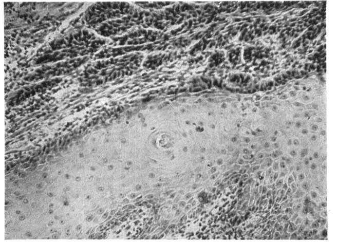
1 SOME ESSENTIAL FACTORS IN THE PATHOLOGY AND TREATMENT OF CANCER OF THE SKIN LOUIS H. JORSTAD, M.D. (From the Department of Pathology, the Barnard Free Skin and Cancer Hospital, St. Louis, Missouri) The stimulus for this study on the pathology and treatment of cancer of the skin was twofold: first the gross and microscopic examination of a large series of laboratory animals in which a small area of skin was subjected to different dosages of x-ray (1); second, the microscopic diagnosis and clinical study of 200 cases of carcinoma of the skin. Basal-cell carcinomata of the skin were at one time called epitheliomata, the term carcinoma being used to indicate squamous-cell carcinoma. This nomenclature placed too much emphasis on the identity of the two forms. There are a certain number of tumors which are a mixture of basal-cell carcinoma and squamouscell carcinoma. This is not a new finding, but is one that cannot be over-emphasized. Since Krompecher (2) discovered this mixed form years ago it has too often been forgotten. The use of such terms as epithelioma, spinous carcinoma, and acanthoma should, I believe, be discontinued. These terms are confusing. A rearrangement of the classification of the carcinomata which we find in the skin would seem advisable. We have first the typical basal-cell carcinoma (Fig. I). In tumors of this type the basement layer of cells undergoes division but the basement membrane remains intact. Metastasis does not occur. The tumors may be multiple. The cystic basal-cell carcinomata form a second group (Fig. 2). These tumors are characteristically multiple. A third group of basal-cell carcinomata I would call the adenoid type (Fig. 3). This type is closely allied to hair follicle carcinoma and in many instances the histology of the two forms is similar. These adenoid basal-cell carcinomata have been called adenocarcinomata. From the small series which I have had opportunity 177
.")
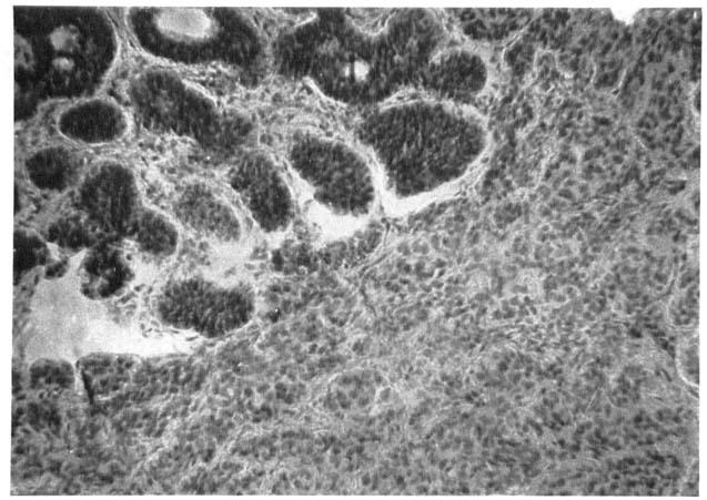
2 178 LOUIS H. JORSTAD to review it would seem that this adenoid type is more malignant than other basal-cell carcinomata. The invasion is deeper, and a certain percentage of these growths metastasize. One may group tumors of this type with the adenocarcinomata of 'sweat gland, mucous gland, and hair follicle origin (Fig. 4). A fourth group consists of the combined basal-cell and squamous-cell carcinomata. This type should be called basal-squamous or baso-squamous, as Montgomery (4) has suggested. In the cases which I have studied I have distinguished two varieties; in one the basal-cell and squamous-cell areas occur side by side on one microscopic field (Figs. 5 and 6), while in the other variety the squamous-cell areas occur within and among the basal-cell areas (Fig. 7). It is not unusual to see a tumor change in type from basal-cell carcinoma to squamous-cell carcinoma as a result of stimulation during the course of insufficient and prolonged treatment. In an insufficiently radiated or in a radio-resistant basal-cell carcinoma such a change begins with squamous anaplasia. From the standpoint of Broders' work on grading it is interesting to note that the squamous changes which I have seen in basal-cell carcinomata have usually been Grade I. It is unusual to find metastasis from Grade


3 Fro. 2. CYSTIC BASAL-CELL CARCINOMA
 has reviewed certain pathological features regarding the classification of carcinomata.")
4 180 LOUIS H. JORSTAD I carcinoma, and it is also unusual to find metastasis from these anaplastic basal-cell carcinomata. Squamous-cell carcinoma constitutes a final group in a classification of skin carcinomata. Recently May Owen (5) has reviewed certain pathological features regarding the classification of carcinomata. The stimulus for her work came from reports that primary basal-cell carcinomata of the mucous membranes do occur. It was her experience, however, that these cases were highly malignant squamous-cell carcinomata. They resembled basal-cell carcinoma histologically, but on careful study of a number of sections taken from each tumor, the squamous-cell structure could be made out. In her review Owen cites the work of Lunford and Taussig, of Martzloff, and of Vinson, each of whom has reported a large series of carcinomata of the mucous membranes, including none of the basal-cell type. Krompecher, on the other hand, is cited as finding about one-half of the malignant tumors of the nose and larynx corresponding to the basal-cell type. The more recent studies of New and Broders mention no basal-cell carcinomata of the internal cavities of the head and neck, and Broders states that he has never seen one originating on mucous membrane. In my classification I have not in-
5

6 182 LOUIS H. JORSTAD cluded among the basal-cell carcinomata those highly malignant squamous-cell carcinomata occurring in the cervix and in the mucous membrane of the mouth mentioned by Owen as being confused with basal-cell carcinoma. It is characteristic of the basal-cell carcinoma that it is made up of small polyhedral or spindle-shaped cells which contain a large amount of nuclear material that stains deeply with a basic stain such as hematoxylin. The squamous cell has one nucleolus in contrast to the many nucleoli of the basal cell. Carcinoma developing upon keratosis is interesting from the standpoint of classification. This may be either basal-cell or squamous-cell in type, depending upon the degree of keratinization. The relationship of keratosis and hyalinization to carcinoma was clearly brought out in a study made by the author, of the reaction of the skin of animals to roentgen irradiation. In a number of patients with marked destruction of skin following irradiation the removal of these areas for therapeutic benefit has given considerable material for study. If keratosis of the superficial layer of the epithelium is present, there is a hyperplasia of the lower layers. If the keratosis is of the proper degree, atavism occurs, which may lead to basal-cell or squamous-cell carcinoma. With still deeper destruction of tissue, hair follicle carcinoma may develop. The hair follicle

7 being more cellular, it is stimulated to activity when located in the zone of connective tissue which has undergone hyalinization. It is usually the rule to see a more malignant carcinoma following a keratosis in a moderately young individual. Some have regarded this fact as demonstrating the development of an increasing immunity to carcinoma with advancing age. The keratosis above described is the most clearly proved precancerous lesion. To conclude, I am convinced that all skin carcinomata should be treated as if they were malignant. It cannot be determined clinically whether a tumor is a typical basal-cell, a basal-squamouscell, or a squamous-cell carcinoma. One never knows when a basal-squamous-cell carcinoma will change into a squamous carcinoma. Some clinicians apparently are not cognizant of this fact. If these factors were kept in mind when dealing with skin carcinoma, I feel certain that the percentage of recurrences and bad results would be greatly diminished. 1. JORSTAD, L. H., AND LANE, C. W.: Proc. Soc. Exper. Biol. & Med. 24: 886, 1927; Arch. Derm. & Syph. 19: 954, KROMPECHER, E.: Beitr. z. path. Anat. u. z. allg. Path. 28: 1, KROMPECHER, E.: Beitr. z. path. Anat. u. a. allg. Path. 44: 88, MONTGOMERY, H.: Arch. Derm. & Syph. 18: 50, OWEN, M.: Arch. Path. 10: 386, 1930.
Epithelial tumors. Dr. F.F. Khuzin, PhD Dr. M.O. Mavlikeev
 Epithelial tumors Dr. F.F. Khuzin, PhD Dr. M.O. Mavlikeev Epithelial tumors Tumors from the epithelium are the most frequent among tumors. There are 2 group features of these tumors: The presence in most
SARCOMA FOLLOWING X-RAY THERAPY FOR GRAVES' DISEASE
 SARCOMA FOLLOWING X-RAY THERAPY FOR GRAVES' DISEASE By P. H. JAYES, M.B., B.S., F.R.C.S., and R. H. DALE, M.B., B.Chir., F.R.C.S.Ed. From the Plastic Surgery and Jaw Injuries Centre, East Grinstead IT
THE PROBLEMS OF HISTOLOGICAL DIAGNOSIS IN
 J. clin. Path. (1959), 12, 73. THE PROBLEMS OF HISTOLOGICAL DIAGNOSIS IN BASO-SQUAMOUS CELL CARCINOMA OF THE SKIN BY J. BURSTON* AND R. D. CLAY From the Portsmouth and I.O.W. Area Pathological Service
Dysplasia, Mimics and Other Controversies
 Dysplasia, Mimics and Other Controversies Mary S. Richardson, MD Dept. of Pathology Medical University of South Carolina Charleston, SC Notice of Faculty Disclosure In accordance with ACGME guidelines,
NEOPLASIA-I CANCER. Nam Deuk Kim, Ph.D.
 NEOPLASIA-I CANCER Nam Deuk Kim, Ph.D. 1 2 Tumor in the hieroglyphics of the Edwin Smith papyrus (1,600 B.C., Breasted s translation 1930) 3 War on Cancer (National Cancer Act, 1971) 4 Cancer Acts in Korea
Neoplasia 2018 Lecture 2. Dr Heyam Awad MD, FRCPath
 Neoplasia 2018 Lecture 2 Dr Heyam Awad MD, FRCPath ILOS 1. List the differences between benign and malignant tumors. 2. Recognize the histological features of malignancy. 3. Define dysplasia and understand
What is ACC? (Adenoid Cystic Carcinoma)
") What is ACC? (Adenoid Cystic Carcinoma) 10-9-10 Where ACC Occurs ACC (Adenoid Cystic Carcinoma) is a rare and unique form of cancer that is known to be unpredictable in nature, with a typical growth pattern
What is ACC? (Adenoid Cystic Carcinoma) 10-9-10 Where ACC Occurs ACC (Adenoid Cystic Carcinoma) is a rare and unique form of cancer that is known to be unpredictable in nature, with a typical growth pattern
Neoplasia literally means "new growth.
 NEOPLASIA Neoplasia literally means "new growth. A neoplasm, defined as "an abnormal mass of tissue the growth of which exceeds and is uncoordinated with that of the normal tissues and persists in the
Histopathology: Cervical HPV and neoplasia
 Histopathology: Cervical HPV and neoplasia These presentations are to help you identify basic histopathological features. They do not contain the additional factual information that you need to learn about
04/09/2018. Squamous Cell Neoplasia and Precursor Lesions. Agenda. Squamous Dysplasia. Squamo-proliferative lesions. Architectural features
 Squamous Cell Neoplasia and Precursor Lesions Jennifer L. Hunt, MD, MEd Aubrey J. Hough Jr, MD, Endowed Professor of Pathology Chair of Pathology and Laboratory Medicine University of Arkansas for Medical
Head & Neck Squamous Carcinoma: Artifacts, Challenges, and Controversies. Agenda
 Head & Neck Squamous Carcinoma: Artifacts, Challenges, and Controversies Jennifer L. Hunt, MD, MEd Aubrey J. Hough Jr, MD, Endowed Professor of Pathology Chair of Pathology and Laboratory Medicine University
Pathology of the skin. 2nd Department of Pathology, Semmelweis University
 Pathology of the skin 2nd Department of Pathology, Semmelweis University Histology of the skin Epidermis: Stratum corneum Stratum granulosum Stratum spinosum Stratum basale Dermis: papillary and reticular
Glossary of Terms Primary Urethral Cancer
 Patient Information English Glossary of Terms Primary Urethral Cancer Advanced cancer A tumour that grows into deeper layers of tissue, adjacent organs, or surrounding muscles. Anaesthesia (general, spinal,
Maligna Melanoma and Atypical Fibroxanthoma: An Unusual Collision Tumour G Türkcü 1, A Keleş 1, U Alabalık 1, D Uçmak 2, H Büyükbayram 1 ABSTRACT
 Maligna Melanoma and Atypical Fibroxanthoma: An Unusual Collision Tumour G Türkcü 1, A Keleş 1, U Alabalık 1, D Uçmak 2, H Büyükbayram 1 ABSTRACT Two different neoplasia in the same biopsy material called
Cerebral Parenchymal Lesions: I. Metastatic Neoplasms
 Chapter 4 Cerebral Parenchymal Lesions: I. Metastatic Neoplasms After one has reasonably ruled out the possibility of a nonneoplastic diagnosis (see Chap. 3), one is left with considering a diagnosis of
Muco-epidermoid tumours of the anal canal
 J. clin. Path. (1963), 16, 200 Muco-epidermoid tumours of the anal canal B. C. MORSON AND H. VOLKSTADT From the Research Department, St. Mark's Hospital, London SYNOPSIS The pathology of 21 cases of muco-epidermoid
THE SIGNIFICANCE OF CELL TYPE IN CERVICAL CANCER'
 THE SIGNIFICANCE OF CELL TYPE IN CERVICAL CANCER' DANIEL G. MORTON, M.D. Assistant Professor of Obstetrics and Gynecology, University of California Medical School, San Francisco Among the factors which
*with blood clot. Microscopically, the tumor was made up of papillary. Krompecher,2 Bloodgood,8 and Hazen I also have clinical and histologic
 THE HISTOGENESIS OF CLEAR CELL PAPILLARY CARCINOMA OF THE SKIN * Y. Lru, M.D. (From the Department of Pathology, Peiping Union Medical Colege, Peiping, China) Clear cell papillary carcinoma, so designated
Squamous Cell Carcinoma of the Head and Neck (SCCHN)
") Squamous Cell Carcinoma of the Head and Neck (SCCHN) Part 1 Bruce M. Wenig, M.D. Dept. of Pathology & Laboratory Medicine Continuum Health Partners New York, NY College of American Pathologists 2004. Materials
Squamous Cell Carcinoma of the Head and Neck (SCCHN) Part 1 Bruce M. Wenig, M.D. Dept. of Pathology & Laboratory Medicine Continuum Health Partners New York, NY College of American Pathologists 2004. Materials
Human Papillomavirus and Head and Neck Cancer. Ed Stelow, MD
 Human Papillomavirus and Head and Neck Cancer Ed Stelow, MD No conflict of interest Declaration Cancer 1974 Lancet Oncol 2016; 17: e477-8 JAMA 1984; 252: 1857 JAMA 1988;259(13):1943-1944 Clin Cancer Res
Carcinoma of Unknown Primary site (CUP) in HEAD & NECK SURGERY
 in HEAD & NECK SURGERY") Carcinoma of Unknown Primary site (CUP) in HEAD & NECK SURGERY SEARCHING FOR THE PRIMARY? P r o f J P P r e t o r i u s H e a d : C l i n i c a l U n i t C r i t i c a l C a r e U n i v e r s i t y O f
Carcinoma of Unknown Primary site (CUP) in HEAD & NECK SURGERY SEARCHING FOR THE PRIMARY? P r o f J P P r e t o r i u s H e a d : C l i n i c a l U n i t C r i t i c a l C a r e U n i v e r s i t y O f
Tissues. Tissues - Overview. Bio 101 Laboratory 3. Epithelial Tissues and Integument
 Bio 101 Laboratory 3 Epithelial Tissues and Integument 1 Tissues Tissues to be examined under the microscope Epithelial Tissue Integument Connective Tissue **We will be doing muscle and nervous tissues
Benign and malignant epithelial lesions: Seborrheic keratosis: A common benign pigmented epidermal tumor occur in middle-aged or older persons more
 Benign and malignant epithelial lesions: Seborrheic keratosis: A common benign pigmented epidermal tumor occur in middle-aged or older persons more common on the trunk; but extremities, head and neck are
SOLITARY BASAL CELL NEVUS STAGE OF BASAL CELL EPITHELIOMA
 SKIN RESEARCH Dec. 1968 Vol.10 No.5 679-686 SOLITARY BASAL CELL NEVUS PREINVASIVE STAGE OF BASAL CELL EPITHELIOMA YOSHIHARU MIKI, M.D. Department of Dermatology, University of Osaka, School of Medicine,
Rare Presentation Of Adenoidcystic Carcinoma Of External Auditory Canal With Subcutaneous Metastasis In Temporal Region
 ISPUB.COM The Internet Journal of Otorhinolaryngology Volume 13 Number 2 Rare Presentation Of Adenoidcystic Carcinoma Of External Auditory Canal With Subcutaneous Metastasis In Temporal Region S Kaushik,
(CYLINDROMA) ATLAS OF HEAD AND NECK PATHOLOGY ADENOID CYSTIC CARCINOMA
 ATLAS OF HEAD AND NECK PATHOLOGY ADENOID CYSTIC CARCINOMA") (CYLINDROMA) This malignant tumor is poorly encapsulated and while seemingly well defined within the affected gland, there is usually infiltration of surrounding tissue on closer examination. The cut surface
(CYLINDROMA) This malignant tumor is poorly encapsulated and while seemingly well defined within the affected gland, there is usually infiltration of surrounding tissue on closer examination. The cut surface
Malignant tumors of melanocytes: Part 1. Deba P Sarma, MD., Omaha
 Malignant tumors of melanocytes: Part 1 Deba P Sarma, MD., Omaha The melanocytic tumor is one of the most difficult and confusing areas in Dematopathology. It is true that most (95%) of such lesions are
DERMATITIS CHRONICA HELICIS
 J. clin. Path. (1957), 10, 46. THE HISTOLOGICAL APPEARANCES OF CHONDRO- DERMATITIS CHRONICA HELICIS BY E. M. McCONNELL From the Department of Pathology, Liverpool Radium Institute, Liverpool (RECEIVED
SCOPE OF PRACTICE PGY-5
 Recognize normal cytomorphology of cells derived from the respiratory, gastrointestinal, and genitourinary tracts, and body fluid (Cerebrospinal fluid, pleural and peritoneal fluid) Recognize normal cytomorphology
Malignant transformation in benign cystic teratomas, dermoids of the ovary
 European JournalofObstetrics& Gynecology andreproductivebiology, 29 (1988) 197-206 197 Elsevier EJO 00716 Malignant transformation in benign cystic teratomas, dermoids of the ovary S. Chadha 1 and A. Schaberg
A neoplasm is defined as "an abnormal tissue proliferation, which exceeds that of adjacent normal tissue. This proliferation continues even after
 NEOPLASIA Neoplasia is a very important topic in pathology because neoplasms are both common and serious diseases. A neoplasm literally means a new growth, and this term is used interchangeably with a
Skin Cancer - Non-Melanoma
 Skin Cancer - Non-Melanoma Introduction Each year, millions of people find out that they have skin cancer. Skin cancer is almost 100% curable if found early and treated right away. It is possible to prevent
Histopathology: skin pathology
 Histopathology: skin pathology These presentations are to help you identify, and to test yourself on identifying, basic histopathological features. They do not contain the additional factual information
(a), in a discussion of Paget s disease of the nipple, has expressed
, in a discussion of Paget s disease of the nipple, has expressed") SKIN INVOLVEMENT IN BREAST CANCER WITH REFERENCE TO ITS BEARING ON THE INTERPRE- TATION OF APPEARANCES OF TRANSITION BE- TWEEN NORMAL EPITHELIUM AND CANCER ALSON R. KILGORE From tlw Surgiral Pathology
SKIN INVOLVEMENT IN BREAST CANCER WITH REFERENCE TO ITS BEARING ON THE INTERPRE- TATION OF APPEARANCES OF TRANSITION BE- TWEEN NORMAL EPITHELIUM AND CANCER ALSON R. KILGORE From tlw Surgiral Pathology
In the third part of the present study tumours which previous were described as basal cell tumours but now have been reclassified as trichoblastomas
 170 6. SUMMARY Immunhistochemical investigations for identifying the histogenesis of basaloid neoplasias and hyperplasias in the mamma parenchyma of the bitch, for the use of the human nuclear protein
Dr. Issraa Ali Hussein
 CLINICAL 09888888;rCYTOLOGY Dr. Issraa Ali Hussein objectives Define diagnostic cytology (clinical cytology). Explain the differences between histopathology and cytopathology. Recognize the methods for
number Done by Corrected by Doctor Maha Shomaf
 number 16 Done by Waseem Abo-Obeida Corrected by Zeina Assaf Doctor Maha Shomaf MALIGNANT NEOPLASMS The four fundamental features by which benign and malignant tumors can be distinguished are: 1- differentiation
Trichofolliculoma of the Guinea Pig 1,2
 Trichofolliculoma of the Guinea Pig 1,2 Raymond D. Ediger, Garrett S. Dill, Jr., and Robert M. Kovatch, Aerobiology and Evaluation Laboratories and Medical Sciences Laboratories, Fort Detrick, Frederick,
Lecture Overview. Chapter 4 Epithelial Tissues Lecture 9. Introduction to Tissues. Epithelial Tissues. Glandular Epithelium
 Visual Anatomy & Physiology First Edition Martini & Ober Chapter 4 Lecture 9 Lecture Overview Introduction to Tissues Location General characteristics Functions Classification Glandular Epithelium 2 Where
Neoplasia part I. Dr. Mohsen Dashti. Clinical Medicine & Pathology nd Lecture
 Neoplasia part I By Dr. Mohsen Dashti Clinical Medicine & Pathology 316 2 nd Lecture Lecture outline Review of structure & function. Basic definitions. Classification of neoplasms. Morphologic features.
Chapter 4 :Organization & Regulation of Body Systems
 Chapter 4 :Organization & Regulation of Body Systems 4.1 Types of tissues What is a tissue? A collection of cells of the same type that perform a common function There are 4 major tissue types in the body:
Received, June 29, 1904; accepted for publication
 THE AMEBICAN JOURNAL OF CLINICAL PATHOLOGY Copyright 1964 by The Williams & Wilkins Co. Vol. 42, No. 0 Printed in U.S.A. CARCINOMA IN SITU OF THE ENDOMETRIUM ISABELLE A. BUEHL, M.D., PRANK VELLIOS, M.D.,
Premalignant lesions may expose to a promoting. factor & may be induced to undergo malignant. Carcinoma in situ displays the cytologic features of
 بسم رلاهللا Def. Premalignant lesions may expose to a promoting factor & may be induced to undergo malignant transformation. Carcinoma in situ displays the cytologic features of malignancy without invasion
Case history: Figure 1. H&E, 5x. Figure 2. H&E, 20x.
 1 Case history: A 49 year-old female presented with a 5 year history of chronic anal fissure. The patient s past medical history is otherwise unremarkable. On digital rectal examination there was a very
APOCRINE SWEAT GLAND CARCINOMA OF THE VULVA* JOHN R. McDONALD, M.D. Section on Surgical Pathology, The Mayo Clinic, Rochester, Minnesota
 APOCRINE SWEAT GLAND CARCINOMA OF THE VULVA* JOHN R. McDONALD, M.D. Section on Surgical Pathology, The Mayo Clinic, Rochester, Minnesota The wide variety of neoplasms, both benign and malignant, originates
Update in Salivary Gland Pathology. Benjamin L. Witt University of Utah/ARUP Laboratories February 9, 2016
 Update in Salivary Gland Pathology Benjamin L. Witt University of Utah/ARUP Laboratories February 9, 2016 Objectives Review the different appearances of a selection of salivary gland tumor types Establish
Lecture Overview. Marieb s Human Anatomy and Physiology. Chapter 4 Tissues: The Living Fabric Epithelial Tissues Lecture 9. Introduction to Tissues
 Marieb s Human Anatomy and Physiology Marieb Hoehn Chapter 4 Tissues: The Living Fabric Epithelial Tissues Lecture 9 Lecture Overview Introduction to Tissues Epithelial Tissues Location General characteristics
Policy #: 127 Latest Review Date: June 2011
 Name of Policy: Mohs Micrographic Surgery Policy #: 127 Latest Review Date: June 2011 Category: Surgery Policy Grade: Active Policy but no longer scheduled for regular literature reviews and updates. Background/Definitions:
أملس عضلي غرن = Leiomyosarcoma. Leiomyosarcoma 1 / 5
 Leiomyosarcoma 1 / 5 EPIDEMIOLOGY Exact incidence is unknown, but older studies suggest that leiomyosarcomas comprise approximately 3 percent of soft-tissue sarcomas. Superficial leiomyosarcoma occurs
OSCaR UPDATE. Manager s Update Donald Shipley, MS. Oregon State Cancer Registry
 Oregon State Cancer Registry OSCaR UPDATE VOLUME 8, QUARTER 4 W INTER 2008 Manager s Update Donald Shipley, MS Since the Fall issue of OSCaR Update, the registry staff has completed several significant
Cutaneous Adnexal Tumors
 Cutaneous Adnexal Tumors Lesions with Predominant Follicular Differentiation Special Emphasis on Basal Cell Carcinoma 2014-04-01 Prof. Dr. med. Katharina Glatz Pathologie Cutaneous Adnexal Tumors Hair
Mitosis Models 3-5. Chromosome. #1 Prophase. #2 Prophase. 2n = 4 4 Chromosomes 8 Chromatids. 2n = 4
 MITOSIS Mitosis Models 3-5 Chromosome #1 Prophase 2n = 4 4 Chromosomes 8 Chromatids #2 Prophase 2n = 4 4 Chromosomes 8 Chromatids Mitosis Models 3-5 Astral Rays Chromosomes Chromosome Chromosome Spindle
THE CLASSIFICATION OF BLADDER TUMOURS
 41 THE CLASSIFICATION OF BLADDER TUMOURS T. J. DEELEY AND V. J. DESMET* From the Radiotherapy Department, Hammersmith Hospital, Du Cane Road, London, IF7.12, and the Department of Pathology, Louvain University,
Subject Index. Dry desquamation, see Skin reactions, radiotherapy
 Subject Index Actinic keratosis disseminated disease 42 surgical excision 42 AIDS, see Kaposi s sarcoma Amifostine, skin reaction prophylaxis 111 Basal cell carcinoma, superficial X-ray therapy Bowen s
Squamous Cell Neoplasia and Precursor Lesions
 Squamous Cell Neoplasia and Precursor Lesions Jennifer L. Hunt, MD, MEd Aubrey J. Hough Jr, MD, Endowed Professor of Pathology Chair of Pathology and Laboratory Medicine University of Arkansas for Medical
SKIN. 3. How is the skin structured around the finger joints to allow for flexible movement of the fingers?
 SKIN Objectives for Exam #1: 1. List various skin structures and describe their functions. 2. Describe skin responses to increases and decreases in body temperature. 3. Provide examples of various skin
Case Report A Rare Cutaneous Adnexal Tumor: Malignant Proliferating Trichilemmal Tumor
 Case Reports in Medicine Volume 2015, Article ID 742920, 4 pages http://dx.doi.org/10.1155/2015/742920 Case Report A Rare Cutaneous Adnexal Tumor: Malignant Proliferating Trichilemmal Tumor Omer Alici,
2 to 3% of All New Visceral Cancers Peak Incidence is 6th Decade M:F = 2:1 Grossly is a Bright Yellow, Necrotic Mass with a Pseudocapsule
 GENITOURINARY PATHOLOGY Kathleen M. O Toole, M.D. Renal Cell Carcinoma 2 to 3% of All New Visceral Cancers Peak Incidence is 6th Decade M:F = 2:1 Grossly is a Bright Yellow Necrotic Mass Grossly is a Bright
ONCOLOGY. Csaba Bödör. Department of Pathology and Experimental Cancer Research november 19., ÁOK, III.
 ONCOLOGY Csaba Bödör Department of Pathology and Experimental Cancer Research 2018. november 19., ÁOK, III. bodor.csaba1@med.semmelweis-univ.hu ONCOLOGY Characteristics of Benign and Malignant Neoplasms
Basal cell carcinoma diagnosed on Fine-Needle Aspiration Cytology A. Pathological Case Report
 Basal cell carcinoma diagnosed on Fine-Needle Aspiration Cytology A Abstract Dr. Madhuri S.Kate 1, Dr. Preeti Jain 2, Dr. Shailesh S. Patne 3 Introduction: Basal cell carcinoma (BCC) is a locally invasive
Salivary Glands 3/7/2017
 Salivary Glands 3/7/2017 Goals and objectives Focus on the entities unique to H&N Common board type facts Information for your future practice Salivary Glands Salivary Glands Major gland. Paratid. Submandibular.
THE SURGICAL TREATMENT OF POST-RADIATION RECURRENT BASAL-CELL CARCINOMA OF THE FACE AND SCALP
 THE SURGICAL TREATMENT OF POST-RADIATION RECURRENT BASAL-CELL CARCINOMA OF THE FACE AND SCALP By RANDELL CHAMPION, M.B.E., F.R.C.S.Ed., and ROBERT GIBB, M.B., Ch.B., D.M.R.T.(Eng.) From Christie Hospital
Prepared By Jocelyn Palao and Layla Faqih
 Prepared By Jocelyn Palao and Layla Faqih The structure of the suspected atypical cell should always be compared to the structure of other similar, benign, cells which are present in the smears. The diagnosis
Skin and Body Membranes
 Essentials of Human Anatomy & Physiology Elaine N. Marieb Seventh Edition Chapter 4 Skin and Body Membranes Slides 4.1 4.32 Lecture Slides in PowerPoint by Jerry L. Cook Skin and Body Membranes Function
MALIGNANT POROMA SYNONYM: POROCARCINOMA ECCRINE POROMA MALIGNANT Divvya B 1, M. Valluvan 2, Rehana Tippoo 3, P. Viswanathan 4, R.
 MALIGNANT POROMA SYNONYM: POROCARCINOMA ECCRINE POROMA MALIGNANT Divvya B 1, M. Valluvan 2, Rehana Tippoo 3, P. Viswanathan 4, R. Ramesh 5 HOW TO CITE THIS ARTICLE: Divvya B, M. Valluvan, Rehana Tippoo,
AGGRESSIVE VARIANTS OF PAPILLARY THYROID CARCINOMA DIAGNOSIS AND PROGNOSIS
 AGGRESSIVE VARIANTS OF PAPILLARY THYROID CARCINOMA DIAGNOSIS AND PROGNOSIS PAPILLARY THYROID CARCINOMA Clinical Any age Microscopic to large Female: Male= 2-4:1 Radiation history Lymph nodes Prognosis
General information about skin cancer
 Skin Cancer General information about skin cancer Key points Skin cancer is a disease in which malignant (cancer) cells form in the tissues of the skin. There are different types of cancer that start in
LARYNGEAL DYSPLASIA. Tomas Fernandez M; 3 rd year ENT resident, Son Espases University Hospital
 LARYNGEAL DYSPLASIA Tomas Fernandez M; 3 rd year ENT resident, Son Espases University Hospital INTRODUCTION Laryngeal cancer constitutes 1-2% of all malignancies diagnosed worldwide Survival is related
ATLAS OF HEAD AND NECK PATHOLOGY METAPLASIA
 Metaplasia is the conversion of one adult differentiated cell type to another. Generally it is the result of persistent cellular trauma and serves as a protective mechanism. Thus anteriorly along the nasal
Maram Abdaljaleel, MD Dermatopathologist and Neuropathologist University of Jordan, School of Medicine
 Maram Abdaljaleel, MD Dermatopathologist and Neuropathologist University of Jordan, School of Medicine The most common non-skin malignancy of women 2 nd most common cause of cancer deaths in women, following
See the latest estimates for new cases of salivary gland cancers in the US and what research is currently being done.
 About Salivary Gland Cancer Overview and Types If you have been diagnosed with salivary gland cancer or are worried about it, you likely have a lot of questions. Learning some basics is a good place to
(formalin fixed) 6 non-neoplastic spots (6 spots) Corresponding normal tissues with cancers: Yes Diameter: 1. 0 mm
 6 non-neoplastic spots (6 spots) Corresponding normal tissues with cancers: Yes Diameter: 1. 0 mm") CBA729-Test slide, Head and neck cancer tissues (formalin fixed) For research use only Specifications: No. of cases: 6 Tissue type: Test slide, Head and neck cancer tissues No. of spots: 6 spots from each
CBA729-Test slide, Head and neck cancer tissues (formalin fixed) For research use only Specifications: No. of cases: 6 Tissue type: Test slide, Head and neck cancer tissues No. of spots: 6 spots from each
Diseases of the breast (1 of 2)
") Diseases of the breast (1 of 2) Introduction A histology introduction Normal ducts and lobules of the breast are lined by two layers of cells a layer of luminal cells overlying a second layer of myoepithelial
Diseases of the breast (1 of 2) Introduction A histology introduction Normal ducts and lobules of the breast are lined by two layers of cells a layer of luminal cells overlying a second layer of myoepithelial
Integumentary System. Integumentary System
 1. General aspects a. The integumentary system consists of several organs major organ of the system is the skin other organs are relatively small and they can be considered as specialized structures of
Epithelial Lecture Test Questions
 Epithelial Lecture Test Questions 1. Which of the following free surfaces lack(s) epithelia: a. lung alveoli (air sacs) b. hard palate c. joint cavities d. abdominal cavity e. salivary gland ducts 2. Which
Case Presentation Protocol 2018 Hot Spots in Dermatology
 Metastatic 1 Running Head: METASTATIC BASAL CELL CARCINOMA Case Presentation Protocol 2018 Hot Spots in Dermatology A Case Study of Metastatic BCC Marianna F. Karewicz, NP Mentor: Dr. Roman W. Glamb, MD
Diagnostic difficulties with lesions of the oral mucosa
 BDIAP London, November 2010 School of Clinical Dentistry University of Sheffield Diagnostic difficulties with lesions of the oral mucosa Paul M Speight Dept Oral & Maxillofacial Pathology University of
Technicians & Nurses Program
 ASCRS ASOA Symposium & Congress Technicians & Nurses Program May 6-10, 2016 New Orleans Evaluation and Treatment of Eyelid Malignancies Richard C. Allen MD PhD FACS Professor Section of Ophthalmology Dept.
Los Angeles Society Of Pathologists Dr. Shobha Castelino Prabhu
 Los Angeles Society Of Pathologists Dr. Shobha Castelino Prabhu Loma Linda University Medical Center June 12, 2007 CASE 1 76 year-old gentleman Status post right parotidectomy 1 year ago for a rare tumor
GI Histology Lab 1. Prepared by: Zeina Kalaji
 GI Histology Lab 1 Prepared by: Zeina Kalaji Lip ORAL MUCOSA -Arrow shows labial salivary glands in the submucosa. VERMILLION transitional zone. SKIN Stratified Squamous epithelium, keratinized -Arrow
The International Federation of Head and Neck Oncologic Societies. Current Concepts in Head and Neck Surgery and Oncology
 The International Federation of Head and Neck Oncologic Societies Current Concepts in Head and Neck Surgery and Oncology www.ifhnos.net The International Federation of Head and Neck Oncologic Societies
My Journey into the World of Salivary Gland Sebaceous Neoplasms
 My Journey into the World of Salivary Gland Sebaceous Neoplasms Douglas R. Gnepp Warren Alpert Medical School at Brown University Rhode Island Hospital Pathology Department Providence RI Asked to present
Changes in the normal anatomy. In some sections a fairly normal. (entropion). Obliteration of glands and occasionally a cyst were
. Obliteration of glands and occasionally a cyst were") 234 THE BRITISH JOURNAL OF OPHTHALMOLOGY PAGET'S DISEASE OF THE EYELID ASSOCIATED WITH CARCINOMA BY DR. A. HAGEDOORN (UNIVERSITY EYE HOSPITAL, AMSTERDAM) IN September, 1929, B. P., a woman aged 56 years
234 THE BRITISH JOURNAL OF OPHTHALMOLOGY PAGET'S DISEASE OF THE EYELID ASSOCIATED WITH CARCINOMA BY DR. A. HAGEDOORN (UNIVERSITY EYE HOSPITAL, AMSTERDAM) IN September, 1929, B. P., a woman aged 56 years
Prostate Pathology: Prostate Carcinoma, variants and Gleason Grading (Part 1)
") Prostate Pathology: Prostate Carcinoma, variants and Gleason Grading (Part 1) Jae Y. Ro, MD, PhD June 7, 2012 Ten Leading Cancer Types for the Estimated New Cancer Cases and Deaths By Sex, United States,
Prostate Pathology: Prostate Carcinoma, variants and Gleason Grading (Part 1) Jae Y. Ro, MD, PhD June 7, 2012 Ten Leading Cancer Types for the Estimated New Cancer Cases and Deaths By Sex, United States,
Anatomy Ch 6: Integumentary System
 Anatomy Ch 6: Integumentary System Introduction: A. Organs are body structures composed of two or more different tissues. B. The skin and its accessory organs make up the integumentary system. Types of
On 180 Biopsies of Oral Carcinomas in Our Department of Pathology. Yasuyuki AWAZAWA * and Itaru MORO * Introduction
 On 180 Biopsies of Oral Carcinomas in Our Department of Pathology by Yasuyuki AWAZAWA * and Itaru MORO * Introduction Carcinomas in the oral region, like those found in other regions of human body, have
Skin and Body Membranes Body Membranes Function of body membranes Cover body surfaces Line body cavities Form protective sheets around organs
 Skin and Body Membranes Body Membranes Function of body membranes Cover body surfaces Line body cavities Form protective sheets around organs Classification of Body Membranes Epithelial membranes Cutaneous
Synonyms. Nephrogenic metaplasia Mesonephric adenoma
 Nephrogenic Adenoma Synonyms Nephrogenic metaplasia Mesonephric adenoma Definition Benign epithelial lesion of urinary tract with tubular, glandular, papillary growth pattern Most frequently in the urinary
THE PROGNOSTIC VALUE OF THE MITOSIS COUNT IN BIOPSIES OF LYMPHOSARCOMA
 THE PROGNOSTIC VALUE OF THE MITOSIS COUNT IN BIOPSIES OF LYMPHOSARCOMA ALBERT E. CASEY (From the Departments of Pathology at the University of Virginia and at St. Louis University) 1 For the past two years
A adipose cells. B capillary. C epithelium
 EPITHELIA Objective The objective of this class is to observe how different epithelia vary in terms of cell shape, size and number of cell layers enabling them to be well adapted for functions in different
Central Poorly Differentiated Adenocarcinoma of the Maxilla: Report of a Case
 Kobe J. Med. Sci., Vol. 49, No. 2, pp. 45-49, 2003 Central Poorly Differentiated Adenocarcinoma of the Maxilla: Report of a Case MASAHIRO UMEDA 1), SATOSHI YOKOO 1), YASUYUKI SHIBUYA 1), TAKAHIDE KOMORI
Acantholytic Anaplastic Extramammary Paget s Disease: A Case Report and Review of the Literature
 Ann Dermatol Vol. 23, Suppl. 2, 2011 http://dx.doi.org/10.5021/ad.2011.23.s2.s226 CASE REPORT Acantholytic Anaplastic Extramammary Paget s Disease: A Case Report and Review of the Literature Yu-Jin Oh,
HOW TO DIAGNOSE VULVAR DISEASES:
 HOW TO DIAGNOSE VULVAR DISEASES: Diagnostic approach. Normal findings. Maria Sol Peremateu, MD A wide spectrum of benign, premalignant, and malignant lesions may occur on the vulva. Some of the disorders
Differential Diagnosis of Oral Masses. Palatal Lesions
 Differential Diagnosis of Oral Masses Palatal Lesions Palatal Masses Periapical Abscess Torus Palatinus Mucocele Lymphoid Hyperplasia Adenomatous Hyperplasia Benign Salivary Neoplasms Malignant Salivary
Management guideline for patients with differentiated thyroid cancer. Teeraporn Ratanaanekchai ENT, KKU 17 October 2007
 Management guideline for patients with differentiated thyroid Teeraporn Ratanaanekchai ENT, KKU 17 October 2007 Incidence (Srinagarind Hospital, 2005, both sex) Site (all) cases % 1. Liver 1178 27 2. Lung
IT S FUNDAMENTAL MY DEAR WATSON! A SHERLOCKIAN APPROACH TO DERMATOLOGY
 IT S FUNDAMENTAL MY DEAR WATSON! A SHERLOCKIAN APPROACH TO DERMATOLOGY Skin, Bones, and other Private Parts Symposium Dermatology Lectures by Debra Shelby, PhD, DNP, FNP-BC, FADNP, FAANP Debra Shelby,
2. Occupancy rate of beds in the hospital: Occupancy rate of at least 60%
 Appendix A Training Centre Accreditation Checklist A. Accreditation of the HOSPITAL 1. Total number of beds in the hospital : Minimum 500 beds 2. Occupancy rate of beds in the hospital: Occupancy rate
Review and Updates of Immunohistochemistry in Selected Salivary Gland and Head and Neck Tumors
 Review and Updates of Immunohistochemistry in Selected Salivary Gland and Head and Neck Tumors. Monophasic tumors : myoepithelioma, acinic cell carcinoma, and salivary duct carcinoma. Biphasic tumors includes
Basaloid carcinoma of the anal canal
 J. clin. Path. (1967), 0, 18 Basaloid carcinoma of the anal canal LILLIAN S. C. PANG AND B. C. MORSON From the Research Department, St. Mark's Hospital, London SYNOPSIS The pathology and results of treatment