POST EXAMINATION REPORT DATE
|
|
|
- Arron Osborne
- 5 years ago
- Views:
Transcription
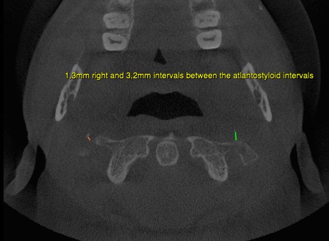
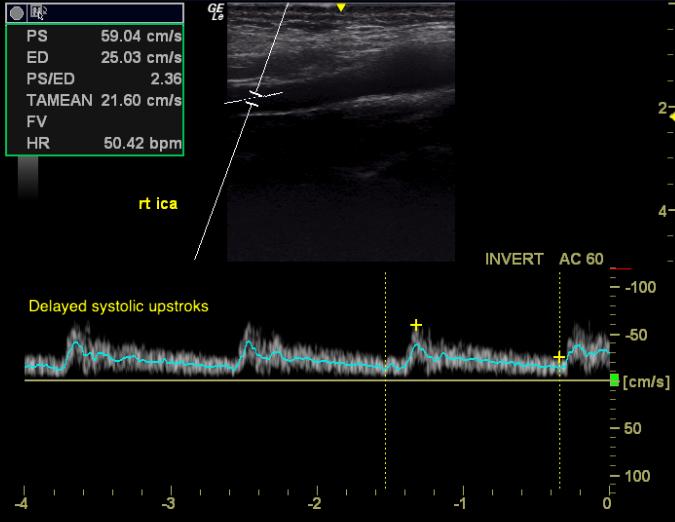
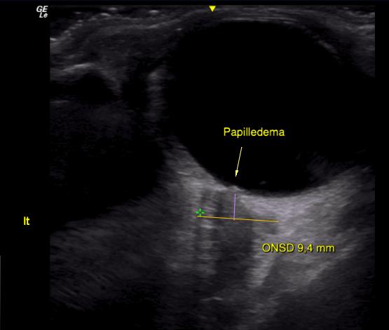
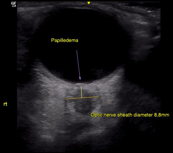
1 POST EXAMINATION REPORT DATE Regarding xxxx (DOB: xxxx). The patient has been struggling with severe fatigue, dyspnea, dysphagia, neck and temporomandibular pain for many years. He is also plagued by visual impairment, brachialgia, chest pain and other nagging symptoms. The patient states that his symptoms of fatigue have been reducing his quality of life since late adolescence, and throughout his adulthood, indicating longstanding underlying problems. Consistently, upon examination of the patient, it is my impression that this is a complex, multifactorial problem. The patient has several cranial nerve deficits revealed during neurological examination. He is light and sound sensitive, has nystagmus and diplopia toward the right. Periodic difficulty swallowing. There is atrophy of the masseter and temporalis muscles with poor contraction when biting (this may also be a TMD sequela). Morley s test and Pemberton s tests are positive, indicating thoracic outlet syndrome with subsequent cerebroarterial hyper perfusion (a consequence of distal subclavian compression) and intracranial hypertension. There is brachial weakness, most pronounced on the right side, where weakness of shoulder abduction (C5-6), wrist flexion (C6-7), elbow extension (C7-8), 1 st (C56) & 5 th (C8-T1) finger opposition and abduction are all weak, again compatible with TOS. Common symptoms of TOS are arm and chest pain, periscapular pain, dyspnea and tightness of the throat. Examination of the patient s CTCB images reveal severe narrowing of the atlantostyloid interval (ASI), likely obstructing the internal jugular veins bilaterally. There is 1mm and 3mm right/left intervals, respectively, which is very narrow (Gweon 2011). Mechanical obstruction of the IJVs at the ASI is a common occurrence (Jayaraman 2012), but little-known. It is mainly caused by poor cervical posture, where the patient hinges back at the upper neck, causing narrowing of the C1 transverse process and cranial styloid process, impinging the CN9-11 and internal jugular veins between them. Further, TOS (arterial hyperperfusion) in tandem with obstructed cerebrovenous drainage, is very unfortunate, and may lead to severe increase in intracranial pressures, likely explaining the patient s cranial nerve deficits and diffuse symptoms. Common symptoms of ICH are headache, fatigue, cranial nerve deficits such as diplopia, visual impairment, dysphagia, etc (Higgins 2017, Higgins 2015, Kincaid 2006, Jensen 2016, Mollan 2014, Larsen 2018c). In search of further evidence in support of raised intracranial pressure, a transorbital ultrasound examination was performed. In this regard, approximately 9 mm dilation of the optic nerve sheaths was demonstrated, with adherent papilledema. This is extremely high, and highly compatible with the patient s symptoms, and neurological examination. Research shows that an optic nerve sheath diameter > 5,8mm has high correlation with pathological increases in ICP (80% (Geeraerts 2008)). Doppler examination of the extracranial vessels was also performed. The systolic peaks of the internal carotid arteries are delayed, dampened and rounded, and the diastolic declination is faster than normal. Similar waveforms are seen in the vertebral arteries. There are thus abnormal waveforms of the carotid and vertebral arteries, compatible with cerebrovascular saturation (congestion) (Larsen, 2018c). Interestingly, the patient s waveforms are clearly abnormal despite a very low heart rate, indicating severe congestion. He frequently went below 50 bpm during the examination, consistent with bradycardia. It is known that patients with intracranial hypertension develop compensatory bradycardia in severe circumstances (Agrawal 2008), as this reduces arteriocerebral pressure, and I thus consider this finding to be a secondary effect of longstanding ICH. Based on the above, it is my impression that xxxx suffers from primary mechanical jugular stenosis and thoracic outlet syndrome, leading to secondary intracranial hypertension, myalgic encephalomyelitis. We have commenced conservative treatment of these problems, mainly aiming to broaden the atlantostyloid interval and to rehabilitate the scalenus muscles TRENING OG REHAB - Eikenga OSLO Post@treningogrehab.no

2 which are compressing his brachial plexus and subclavian vessels, causing TOS. With regards to the patient s jaw pain, he is already involved with several dentists and treatment of this problem. The patient has been prescribed strengthening exercises for the pterygoid muscles on my behalf, to aid in decompression of the condyle and glenoid fossa. Finally, in light of the patient s extensive medical history and symptoms, I find it peculiar and somewhat worrisome that proper imaging has not been done. It is my opinion that MR images of the cervical spine and cerebrum should be obtained. With regards Kjetil Larsen Trainingandrehabilitation.com 1. Larsen K. Occult intracranial hypertension as a sequela of biomechanical internal jugular vein stenosis: A case report. Anaesth Pain & Intensive Care 2018;22(2): Geeraerts T, Merceron S, Benhamou D, Vigue B, Duranteau J. Noninvasive assessment of intracranial pressure using ocular sonography in neurocritical care patients. Crit Care. 2008;12(Suppl 2):P Higgins N, Trivedi R, Greenwood R, Pickard J. Brain slump caused by jugular venous stenoses treated by stenting: a hypothesis to link spontaneous intracranial hypotension with idiopathic intracranial hypertension. J Neurol Surg Rep Jul;76(1):e188 e193. [PubMed] [Free Full Text] doi: /s Jayaraman MV, Boxerman JL, David LM, Haas RA, Rogg JM. Incidence of Extrinsic Compression of the Internal Jugular Vein in Unselected Patients Undergoing CT Angiography. AJNR Am J Neuroradiol Aug;33(7): doi: /ajnr.A2953. Epub 2012 Feb Higgins JNP, Pickard JD, Lever AML. Chronic fatigue syndrome and idiopathic intracranial hypertension: Different manifestations of the same disorder of intracranial pressure? Med Hypotheses Aug;105:6-9. doi: /j.mehy Epub 2017 Jun Kincaid O, Rowin J. Intracranial hypertension causing polyradiculopathy and late or absent F-waves. J Neurol Neurosurg Psychiatry. 2006;77(12): Jensen RH, Radojicic A, Yri H. The diagnosis and management of idiopathic intracranial hypertension and the associated headache. Ther Adv Neurol Disord. 2016;9(4): Mollan SP, Ali F, Hassan-Smith G, Botfield H, Friedman DI, Sinclair AJ. Evolving evidence in adult idiopathic intracranial hypertension: pathophysiology and management. J Neurol Neurosurg Psychiatry. 2016;87:
3 3
4 4
5 5
Occult intracranial hypertension as a sequela of biomechanical internal jugular vein stenosis: A case report
 CASE REPORT ANAESTHESIA, PAIN & INTENSIVE CARE www.apicareonline.com Occult intracranial hypertension as a sequela of biomechanical internal jugular vein stenosis: A case report Kjetil Larsen, CES* *Corrective
Upper Extremity Venous Duplex. Michigan Sonographers Society Fall Ultrasound Symposium October 15, 2016
 Upper Extremity Venous Duplex Michigan Sonographers Society Fall Ultrasound Symposium October 15, 2016 Patricia A. (Tish) Poe, BA RVT FSVU Director of Quality Assurance Navix Diagnostix Patricia A. Poe
THORACIC OUTLET SYNDROME (T.O.S.) Prof J van Marle
 Prof J van Marle") THORACIC OUTLET SYNDROME (T.O.S.) Prof J van Marle 1. Definition Clinical syndrome caused by compression of the neurovascular bundle as it passes through the thoracic outlet, a narrow space bordered by
THORACIC OUTLET SYNDROME (T.O.S.) Prof J van Marle 1. Definition Clinical syndrome caused by compression of the neurovascular bundle as it passes through the thoracic outlet, a narrow space bordered by
Thoracic Outlet Syndrome
 Thoracic Outlet Syndrome Part 1: The Scalene Triangle TOS: Vascular Symptom Presentation Venous persistent/intermittent edema heaviness and fatigue deep pain in neck/shoulder increased pain at night warm
The Upper Limb III. The Brachial Plexus. Anatomy RHS 241 Lecture 12 Dr. Einas Al-Eisa
 The Upper Limb III The Brachial Plexus Anatomy RHS 241 Lecture 12 Dr. Einas Al-Eisa Brachial plexus Network of nerves supplying the upper limb Compression of the plexus results in motor & sensory changes
THORACIC OUTLET SYNDROME: A FREQUENT CAUSE OF NON-DISCOGENIC BRACHIALGIA
 THORACIC OUTLET SYNDROME: A FREQUENT CAUSE OF NON-DISCOGENIC BRACHIALGIA Debora Garozzo Brachial Plexus and Peripheral Nerve Surgery Unit Neurospinal Hospital Dubai, United Arab Emirates THE THORACIC OUTLET
PTA 106 Unit 1 Lecture 3
 PTA 106 Unit 1 Lecture 3 The Basics Arteries: Carry blood away from the heart toward tissues. They typically have thicker vessels walls to handle increased pressure. Contain internal and external elastic
Richard Dobrusin DO FACOFP
 Richard Dobrusin DO FACOFP Define Thoracic Outlet Syndrome (TOS) Describe the Mechanisms of Dysfunction List Diagnostic tests for (TOS) Understand (TOS) referral patterns Discuss Treatment Options Definition:
University Journal of Medicine and Medical Specialities
 ISSN 2455-2852 2018, Vol. 4(5) Imaging Assessment of Vascular Thoracic Outlet Syndrome BALAJI A AYYAMPERUMAL Department of Radio Diagnosis,MADRAS MEDICAL COLLEGE AND GOVERNMENT GENERAL HOSPITAL Abstract
Vascular Technology Examination Content Outline
 Vascular Technology Examination Content Outline (Outline Summary) # Domain Subdomain Percentage 1 Normal Anatomy, Perfusion, and Function Evaluate normal anatomy, perfusion, function 2 Pathology, Perfusion,
Pseudothrombosis of the Subclavian Vein
 416507JDMXXX10.1177/8756479311416507Wash ko et al.journal of Diagnostic Medical Sonography Pseudothrombosis of the Subclavian Vein Journal of Diagnostic Medical Sonography 27(5) 231 235 The Author(s) 2011
D E L L O N I N S T I T U T E S F O R P E R I P H E R A L N E R V E S U R G E R Y
 Thoracic Outlet (TOS), Winged Scapula, Brachial Plexus Compression 12 D E L L O N I N S T I T U T E S F O R P E R I P H E R A L N E R V E S U R G E R Y 1122 KENILWORTH DRIVE, SUITE 18, TOWSON, MARYLAND
Subclavian artery Stenting
 Subclavian artery Stenting Etiology Atherosclerosis Takayasu s arteritis Fibromuscular dysplasia Giant Cell Arteritis Radiation-induced Vascular Injury Thoracic Outlet Syndrome Neurofibromatosis Incidence
Post-op Carotid Complications A Nursing Perspective of What to Watch Out for
 Post-op Carotid Complications A Nursing Perspective of What to Watch Out for By Kariss Peterson, ARNP Swedish Medical Center Inpatient Neurology Team 1 Post-op Carotid Management Objectives Review the
DISCLOSURE TEST YOUR WAVEFORM IQ. Partial volume artifact. 86 yo female with right arm swelling, picc line. AVF on left? Dx?
 Deborah Rubens University of Rochester Rochester, NY DISCLOSURE Neither I nor my immediate family have a financial relationship with a commercial organization that may have a direct or indirect interest
Sometimes symptomatic intracranial venous. Styloidogenic Jugular Venous Compression Syndrome: Diagnosis and Treatment: Case Report CASE REPORT TOPIC
 TOPIC CASE REPORT CASE REPORT Shervin R. Dashti, MD, PhD* Peter Nakaji, MD Yin C. Hu, MD Don F. Frei, MD Adib A. Abla, MD Tom Yao, MD* David Fiorella, MD, PhD *Norton Neuroscience Center, Norton Healthcare,
 Nerve Compression Syndromes Median Nerve Carpal Tunnel Syndrome Pronator Syndrome Ulnar Nerve Cubital Tunnel Syndrome Ulnar Tunnel Syndrome TOS www.fisiokinesiterapia.biz Carpal Tunnel Syndrome (CTS) Definition
The Brachial Plexus and Thoracic Outlet Syndrome
 The Brachial Plexus and Thoracic Outlet Syndrome Understanding Signs and Symptoms By Joseph E. Muscolino, DC The brachial plexus of nerves and the subclavian/axillary artery and vein comprise a neurovascular
Symptoms and Referred Pain from Myofascial Trigger Points in the Anterior Scalene Muscle or Scalenus Anterior
 Symptoms and Referred Pain from Myofascial Trigger Points in the Anterior Scalene Muscle or Scalenus Anterior picture Symptoms and signs Aching or throbbing in the lateral forearm extending to thumb and
Stroke & Neurovascular Center of New Jersey. Jawad F. Kirmani, MD Director, Stroke and Neurovascular Center
 Stroke & Neurovascular Center of New Jersey Jawad F. Kirmani, MD Director, Stroke and Neurovascular Center Past, present and future Past, present and future Cerebral Blood Flow Past, present and future
Unit #3: Dry Lab A. David A. Morton, Ph.D.
 Unit #3: Dry Lab A David A. Morton, Ph.D. Skull Intracranial Hemorrhage Pg. 26 Epidural Hematoma Pg. 26 Skull Pg. 26 Subdural Hematoma Pg. 26 Subdural Hematoma Pg. 26 Subarachnoid Hemorrhage Pg. 26 Subarachnoid
Case Report: CASE REPORT OF FACET ARTHROPATHY INDUCED NERVE ROOT COMPRESSION RESULTING IN MOTOR WEAKNESS AND PAIN
 Cox Technic Case Report #100 published at www.coxtechnic.com (sent October 2011 on 10/11/11 ) 1 Case Report: CASE REPORT OF FACET ARTHROPATHY INDUCED NERVE ROOT COMPRESSION RESULTING IN MOTOR WEAKNESS
LABETTE COMMUNITY COLLEGE BRIEF SYLLABUS. Please check with the LCC bookstore for the required texts for this class.
 LABETTE COMMUNITY COLLEGE BRIEF SYLLABUS SPECIAL NOTE: This brief syllabus is not intended to be a legal contract. A full syllabus will be distributed to students at the first class session. TEXT AND SUPPLEMENTARY
TECHNOLOGY AND HOW WE USE IT TO DAMAGE OURSELVES WILLIAM A. DELP, DO ASSISTANT PROFESSOR OF OMM GA PCOM
 TECHNOLOGY AND HOW WE USE IT TO DAMAGE OURSELVES WILLIAM A. DELP, DO ASSISTANT PROFESSOR OF OMM GA PCOM OBJECTIVES Understand how we interact with technology new and old Understand how injury occurs Texting
Carotid Abnormalities Coils, Kinks and Tortuosity David Lorelli M.D., RVT, FACS Michigan Vascular Association Conference Saturday, October 20, 2012
 Carotid Abnormalities Coils, Kinks and Tortuosity David Lorelli M.D., RVT, FACS Michigan Vascular Association Conference Saturday, October 20, 2012 Page 1 Table of Contents Carotid Anatomy Carotid Duplex
Ultrasound diagnostics of a spontaneous arteriovenous fistula of the head and neck
 Case report Cite as: Zakharkina MV, Chechetkin O, Krotenkova MV, Konovalov RN: Ultrasound diagnostics of a spontaneous arteriovenous fistula of the head and neck.. Submitted: 29.03.2017 ccepted: 24.05.2017
2015 ARDMS Physicians Vascular Interpretation Job Task Analysis Summary Report
 P a g e 1 2015 ARDMS Physicians Vascular Interpretation Job Task Analysis Summary Report American Registry for Diagnostic Medical Sonography (ARDMS) P a g e 2 Table of Contents ABOUT THE REPORT... 3 METHODOLOGY...
Physician s Vascular Interpretation Examination Content Outline
 Physician s Vascular Interpretation Examination Content Outline (Outline Summary) # Domain Subdomain Percentage 1 2 3 4 5 6 Cerebrovascular Abdominal Peripheral Arterial - Duplex Imaging Peripheral Arterial
Papilledema. Golnaz Javey, M.D. and Jeffrey J. Zuravleff, M.D.
 Papilledema Golnaz Javey, M.D. and Jeffrey J. Zuravleff, M.D. Papilledema specifically refers to optic nerve head swelling secondary to increased intracranial pressure (IICP). Optic nerve swelling from
For exam: VL DUPLEX EXTREMITY VEINS UNILAT LT
 For exam: VL DUPLEX EXTREMITY VEINS UNILAT LT - 8870390 METHOD/TECHNIQUE: The veins of the left upper extremity were studied at multiple For exam: VL DUPLEX EXTREMITY VEINS UNILAT RT - 8870400 METHOD/TECHNIQUE:
Diagnosis of Middle Cerebral Artery Occlusion with Transcranial Color-Coded Real-Time Sonography
 Diagnosis of Middle Cerebral Artery Occlusion with Transcranial Color-Coded Real-Time Sonography Kazumi Kimura, Yoichiro Hashimoto, Teruyuki Hirano, Makoto Uchino, and Masayuki Ando PURPOSE: To determine
Nerve Conduction Studies and EMG
 Nerve Conduction Studies and EMG Limitations of other methods of investigations of the neuromuscular system - Dr Rob Henderson, Neurologist Assessment of Weakness Thanks Peter Silburn PERIPHERAL NEUROPATHY
WHI Form Report of Cardiovascular Outcome Ver (For items 1-11, each question specifies mark one or mark all that apply.
 WHI Form - Report of Cardiovascular Outcome Ver. 6. COMMENTS To be completed by Physician Adjudicator Date Completed: - - (M/D/Y) Adjudicator Code: OMB# 095-044 Exp: 4/06 -Affix label here- Clinical Center/ID:
Sonography of Isolated Internal Jugular Vein Impingement and Thrombosis
 498935JDMXXX10.1177/8756479313498935Journal of Diagnostic Medical SonographyRodriguez research-article2013 Case Study Sonography of Isolated Internal Jugular Vein Impingement and Thrombosis Journal of
Carotid Duplex: Beyond Stenosis Ido Weinberg, MD Vascular Medicine Massachusetts General Hospital Assistant Professor of Medicine Harvard Medical
 Carotid Duplex: Beyond Stenosis Ido Weinberg, MD Vascular Medicine Massachusetts General Hospital Assistant Professor of Medicine Harvard Medical School Boston, Massachusetts Disclosures I do not have
NEURO QUIZ 45 EHLERS DANLOS SYNDROME
 NEURO QUIZ 45 EHLERS DANLOS SYNDROME Verghese Cherian, MD, FFARCSI Penn State Hershey Medical Center, Hershey Quiz Team Shobana Rajan, M.D Suneeta Gollapudy, M.D Angele Marie Theard, M.D START 1. Regarding
Oltre la terapia medica nelle dissezioni carotidee
 Oltre la terapia medica nelle dissezioni carotidee Rodolfo Pini Chirurgia Vascolare Università di bologna Alma Mater Studiorum Carotid and Vertebral Artery Dissection What we know from the literature Epidemiology
The Neck the lower margin of the mandible above the suprasternal notch and the upper border of the clavicle
 The Neck is the region of the body that lies between the lower margin of the mandible above and the suprasternal notch and the upper border of the clavicle below Nerves of the neck Cervical Plexus Is formed
Carotid Doppler: Doppler wave forms obtained from the common, external and internal carotid arteries. As well as the vertebral and subclavian
 Competency Carotid Doppler: Doppler wave forms obtained from the common, external and internal carotid arteries. As well as the vertebral and subclavian arteries. Preferred angle is 60 degrees or less.
Subclavian and Axillary Artery Aneurysms
 Subclavian and Axillary Artery Aneurysms April 2008 Francesco A Aiello, M.D. Assistant Professor of Surgery Division of Vascular Endovascular Surgery University of Massachusetts Medical School None DISCLOSURES
Cervical Spine in Baseball
 Cervical Spine in Baseball Robert G Watkins, IV, MD Co-Director, Marina Spine Center Marina del Rey, CA Vice Chief of Staff Cedars-Marina del Rey Hospital Disclosures n Pioneer / RTI Consulting, Royalties
Anatomy and Physiology II. Review Spine and Neck
 Anatomy and Physiology II Review Spine and Neck Spine regions How many cervical vertibrae are there? 7 The curvature is the cervical region posterior? Concave posterior How many thoracic? And curvature?
Thoracic Outlet Syndrome
 James J. Lehman, DC, MBA, FACO Associate Professor of Clinical Sciences Director Health Sciences Postgraduate EducaCon University of Bridgeport Diagnosis is the key to successful treatment James J. Lehman,
The Role of MR Imaging in the Diagnosis of CCSVI and in Pre-Treatment Planning and Monitoring Patient Outcomes. E.
 The Role of MR Imaging in the Diagnosis of CCSVI and in Pre-Treatment Planning and Monitoring Patient Outcomes E. Mark Haacke, PhD The MRI Institute for Biomedical Research Detroit, Michigan 48202 Wayne
Original Research Paper ISSN : e- ISSN , p-issn X
 OPTIC NERVE SHEATH DIAMETER () MEASUREMENT WITH OPTIC NERVE ULTRASOUND (ONUS) IN THE EVALUATION OF ELEVATED INTRACRANIAL PRESSURE: A COMPARATIVE STUDY WITH CT SCAN IN HEAD TRAUMA PATIENTS. 1 Shreyas Patel,
HD Scanning: Velocities and Volume Flow
 HD Scanning: Velocities and Volume Flow Non-Invasive Lab Symposium West Orange, NJ April 27, 2018 Volume Flow Cindy Sturt, MD, FACS, RVT 500,000 Americans on dialysis 20-25% annual mortality 65% 5 year
Official Definition. Carpal tunnel syndrome, the most common focal peripheral neuropathy, results from compression of the median nerve at the wrist.
 Mod 2 MMT Course Official Definition Carpal tunnel syndrome, the most common focal peripheral neuropathy, results from compression of the median nerve at the wrist. epidemiology Affects an estimated 3
Clinical Examination. of the. Cervicothoracic Region. Neck Disability Index. Serious Pathological Conditions. Medical Screening Questionnaire
 Clinical Examination Clinical Examination of the Cervicothoracic Region Screening for associated serious pathological conditions Neck disability index Physical Exam Serious Pathological Conditions Cervical
TO CATCH A THIEF: IMAGING OF SUBCLAVIAN STEAL
 October 2013 TO CATCH A THIEF: IMAGING OF SUBCLAVIAN STEAL Sumir Pandit, Harvard Medical School, Year III 1 AGENDA Introduction to our patient A.B. Anatomy review of aorta and branches CT imaging of our
FUNCTIONAL ANATOMY OF SHOULDER JOINT
 FUNCTIONAL ANATOMY OF SHOULDER JOINT ARTICULATION Articulation is between: The rounded head of the Glenoid cavity humerus and The shallow, pear-shaped glenoid cavity of the scapula. 2 The articular surfaces
Cervical Spine Exercise and Manual Therapy for the Autonomous Practitioner
 Cervical Spine Exercise and Manual Therapy for the Autonomous Practitioner Eric Chaconas PT, PhD, DPT, FAAOMPT Assistant Professor and Assistant Program Director Doctor of Physical Therapy Program Eric
82a Orthopedic Massage! Introduction - Thoracic Outlet"
 82a Orthopedic Massage! Introduction - Thoracic Outlet" 82a Orthopedic Massage! Introduction - Thoracic Outlet! Class Outline" 5 minutes" "Attendance, Breath of Arrival, and Reminders " 10 minutes "Lecture:"
imaging sequences obtained in brachial plexopathy with/without TOS MR Imaging Sequences Associated Anatomic Structures or Pathologic Conditions
 Brachial plexus imaging sequences obtained in brachial plexopathy with/without TOS MR Imaging Sequences Associated Anatomic Structures or Pathologic Conditions Sagittal TSE T2WI through cervical spine
Non-invasive examination
 Non-invasive examination Segmental pressure and Ankle-Brachial Index (ABI) The segmental blood pressure (SBP) examination is a simple, noninvasive method for diagnosing and localizing arterial disease.
Vascular Injuries in the Throwing Shoulder MLB Winter Meeting 2015
 Neurovascular Problems in the Throwing Athlete s Shoulder Professor, Department of Orthopedics Head, Section of Shoulder and Elbow Surgery Team Physician, Chicago White Sox and Bulls Chief Medical Editor,
Cryptogenic Enlargement Of Bilateral Superior Ophthalmic Veins
 ISPUB.COM The Internet Journal of Radiology Volume 18 Number 1 Cryptogenic Enlargement Of Bilateral Superior Ophthalmic Veins K Kragha Citation K Kragha. Cryptogenic Enlargement Of Bilateral Superior Ophthalmic
Evaluation & Management of Penetrating Wounds to the NECK
 Evaluation & Management of Penetrating Wounds to the NECK Goal Effectively identify patients with a high probability of injury requiring surgical intervention Define the role of diagnostic tests in assessing
NON-ATHEROSCLEROTIC PATHOLOGY OF THE CAROTID ARTERIES
 NON-ATHEROSCLEROTIC PATHOLOGY OF THE CAROTID ARTERIES Leslie M. Scoutt, MD, FACR Professor of Diagnostic Radiology & Surgery Vice Chair, Dept of Radiology & Biomedical Imaging Chief, Ultrasound Section
Problems of Carotid Doppler Scanning Which Can Be Overcome by Using Frequency Analysis
 Problems of Carotid Doppler Scanning Which Can Be Overcome by Using Frequency Analysis K. W. JOHNSTON, M.D., F.R.C.S.(C), F.A.C.S., P. M. BROWN, M.D., F.R.C.S.(C), AND M. KASSAM, M.A.SC. SUMMARY The value
British Journal of Rheumatology 1991; 30:
 British Journal of Rheumatology 1991; 30:468-470 CASE REPORT CARPAL TUNNEL SYNDROME COMPLICATED BY REFLEX SYMPATHETIC DYSTROPHY SYNDROME BY M.-A. FITZCHARLES AND J.M. ESDAILE Rheumatic Disease Unit, McGill
3. A plane is an imaginary line dividing the body into.
 CHAPTER 2 Multiple Choice Choose the one alternative that best completes the statement or answers the question. 1. Knowing the exact body region where pain is located can help a physician determine the.
Penetrating Neck Injuries. Jason Levine MD Lutheran Medical Center July 22, 2010
 Penetrating Neck Injuries Jason Levine MD Lutheran Medical Center July 22, 2010 CASE PRESENTATION 19 YO M 3 Stab Wounds Right zone I neck SW 2 SW anterior abdomen Left epigastrium anterior axillary line
2014 PHYSICIAN PROCEDURE CODE CHANGES
 Page 1 of 5 2014 PHYSICIAN PROCEDURE CODE CHANGES Effective for dates of service on or after 1/1/2014, refer to the New Codes listed below for billing. The discontinued codes are not valid for billing
Regional Review of Musculoskeletal System: Head, Neck, and Cervical Spine Presented by Michael L. Fink, PT, DSc, SCS, OCS Pre- Chapter Case Study
 Regional Review of Musculoskeletal System: Presented by Michael L. Fink, PT, DSc, SCS, OCS (20 minutes CEU Time) Subjective A 43-year-old male, reported a sudden onset of left-sided neck and upper extremity
A Patient s Guide to Ulnar Nerve Entrapment at the Wrist (Guyon s Canal Syndrome)
") A Patient s Guide to Ulnar Nerve Entrapment at the Wrist (Guyon s Canal Syndrome) Introduction The ulnar nerve is often called the funny bone at the elbow. However, there is little funny about injury to
A Patient s Guide to Ulnar Nerve Entrapment at the Wrist (Guyon s Canal Syndrome) Introduction The ulnar nerve is often called the funny bone at the elbow. However, there is little funny about injury to
Michigan Vascular Association 2012 Conference Case studies from Massachusetts General Hospital. Our lab
 Michigan Vascular Association 2012 Conference Case studies from Massachusetts General Hospital Kathleen Hannon, MS, RVT, RDMS khannon@partners.org Our lab #1 in the nation! 15 full time RVT s 11 MD s IAC
Neuroradiology MR Protocols
 Neuroradiology MR Protocols Brain protocols N 1: Brain MRI without contrast N 2: Pre- and post-contrast brain MRI N 3 is deleted N 4: Brain MRI without or pre-/post-contrast (seizure protocol) N 5: Pre-
Shadow because the air
 Thyroid Ultrasound Thyroid US examination needs: 1. high frequency transducer 2. extended patient's neck 3. check all the neck area because the swelling could be in areas other than the thyroid such as
Exercises for Thoracic Outlet Syndrome
 Exercises for Thoracic Outlet Syndrome Information for patients who have been diagnosed with Thoracic Outlet Syndrome Read this pamphlet to learn more about: Thoracic Outlet Syndrome Treatment options
UPSTATE Comprehensive Stroke Center. Neurosurgical Interventions Satish Krishnamurthy MD, MCh
 UPSTATE Comprehensive Stroke Center Neurosurgical Interventions Satish Krishnamurthy MD, MCh Regional cerebral blood flow is important Some essential facts Neurons are obligatory glucose users Under anerobic
TRAUMATIC CAROTID &VERTEBRAL ARTERY INJURIES
 TRAUMATIC CAROTID &VERTEBRAL ARTERY INJURIES ALBERTO MAUD, MD ASSOCIATE PROFESSOR TEXAS TECH UNIVERSITY HEALTH SCIENCES CENTER EL PASO PAUL L. FOSTER SCHOOL OF MEDICINE 18TH ANNUAL RIO GRANDE TRAUMA 2017
Codes Requiring Authorization from MedSolutions (MSI): Updated 3/2014
: Updated 3/2014") s Requiring Authorization from MedSolutions (): Updated 3/2014 0042T Cerebral Perfusion Analysis using CT with contrast 0159T CAD, including computer algorithm analysis, BREAST MRI 0195T prepare interspace,
s Requiring Authorization from MedSolutions (): Updated 3/2014 0042T Cerebral Perfusion Analysis using CT with contrast 0159T CAD, including computer algorithm analysis, BREAST MRI 0195T prepare interspace,
Carotid Artery Disease and What s Pertinent JOSEPH A PAULISIN DO
 Carotid Artery Disease and What s Pertinent JOSEPH A PAULISIN DO Goal of treatment of carotid disease Identify those at risk of developing symptoms Prevent patients at risk from developing symptoms Prevent
A Case of Stent Placement for Intracranial Hypertension Associated with Venous Sinus Stenosis
 DOI: 10.5797/jnet.cr.2016-0080 A Case of Stent Placement for Intracranial Hypertension Associated with Venous Sinus Stenosis Rei Yamaguchi, Koji Sato, Hiroya Fujimaki, and Ken Asakura Objective: We encountered
Mechanisms of Headache in Intracranial Hypotension
 Mechanisms of Headache in Intracranial Hypotension Stephen D Silberstein, MD Jefferson Headache Center Thomas Jefferson University Hospital Philadelphia, PA Stephen D. Silberstein, MD, FACP Director, Jefferson
2013 Coding Changes. Diagnostic Radiology. Nuclear Medicine
 2013 Coding Changes The principal coding changes affecting Radiologists in 2013 occur in the Interventional Radiology Section of the AMA/CPT Manual. As in the past, we continue to see the Relative Update
University Journal of Medicine and Medical Sciences
 ISSN 2455-2852 Volume 2 Issue 5 2016 Case report -Opalski's syndrome A rare variant of lateral medullary syndrome in TAKAYASUS ARTERITIS SHANKAR GANESH N NAINAR Department of Neurology, MADRAS MEDICAL
THORACIC OUTLET SYNDROME AND THE OVERHEAD ATHLETE LISA PIROPATO, PT, DPT, ATC NORTHEAST INDIANA SPORTS MEDICINE SYMPOSIUM MARCH 25, 2017
 THORACIC OUTLET SYNDROME AND THE OVERHEAD ATHLETE LISA PIROPATO, PT, DPT, ATC NORTHEAST INDIANA SPORTS MEDICINE SYMPOSIUM MARCH 25, 2017 LEARNING OBJECTIVES Explain the etiologies of thoracic outlet syndrome.
Carotid Revascularization
 Options for Carotid Disease Carotid Revascularization Wayne Causey, MD 2 nd Year Vascular Surgery Fellow Best medical therapy, Carotid Endarterectomy, and Carotid Stenting Who benefits from best medical
Medical Review Guidelines Magnetic Resonance Angiography
 Medical Review Guidelines Magnetic Resonance Angiography Medical Guideline Number: MRG2001-05 Effective Date: 2/13/01 Revised Date: 2/14/2006 OHCA Reference OAC 317:30-5-24. Radiology. (f) Magnetic Resonance
Carotid US: More than just a chart on the wall
 Carotid US: More than just a chart on the wall Leslie M. Scoutt, MD, FACR Professor of Diagnostic Radiology & Surgery Vice Chair, Dept of Radiology & Biomedical Imaging Chief, Ultrasound Section Medical
Dynamic Neuromobilization for the Treatment of Thoracic Outlet Syndrome Courtney Convey and Dr. Erickson
 Dynamic Neuromobilization for the Treatment of Thoracic Outlet Syndrome Courtney Convey and Dr. Erickson Abstract Title: Dynamic Neuromobilization for the Treatment of Thoracic Outlet Syndrome Background:
Spontaneous Intracranial Hypotension Diagnosis and Treatment
 Spontaneous Intracranial Hypotension Diagnosis and Treatment John W. Engstrom MD, Philip R. Weinstein MD, and William P. Dillon M.D. University of California, San Francisco Spontaneous Intracranial Hypotension
Veins of the Face and the Neck
 Veins of the Face and the Neck Facial Vein The facial vein is formed at the medial angle of the eye by the union of the supraorbital and supratrochlear veins. connected through the ophthalmic veins with
What is the mechanism of the audible carotid bruit? How does one calculate the velocity of blood flow?
 CASE 8 A 65-year-old man with a history of hypertension and coronary artery disease presents to the emergency center with complaints of left-sided facial numbness and weakness. His blood pressure is normal,
Clinician s Guide To Ordering NeuroImaging Studies
 Clinician s Guide To Ordering NeuroImaging Studies MRI CT South Jersey Radiology Associates The purpose of this general guide is to assist you in choosing the appropriate imaging test to best help your
Thoracic Outlet Syndrome
 Thoracic Outlet Syndrome Cristina Sola, BSN, RN-BC Nurse Manager Department of Cardiothoracic and Vascular Surgery McGovern Medical School The University of Texas Science Center at Houston Memorial Hermann
McHenry Western Lake County EMS System Paramedic, EMT-B and PHRN Optional Continuing Education 2019 #3 Penetrating Neck Trauma
 McHenry Western Lake County EMS System Paramedic, EMT-B and PHRN Optional Continuing Education 2019 #3 Penetrating Neck Trauma Penetrating neck injury (PNI) comprises 5 to 10 percent of traumatic injuries
CEREBROVASCULAR ACCIDENT (CVA)
") Manual: LifeLine Patient Care Protocols Section: Adult/Pediatrics Protocol #: AP5-002 Approval Date: 12/01/2017 Effective Date: 12/01/2017 Revision Due Date: 12/01/2018 CEREBROVASCULAR ACCIDENT (CVA) PURPOSE
Manual: LifeLine Patient Care Protocols Section: Adult/Pediatrics Protocol #: AP5-002 Approval Date: 12/01/2017 Effective Date: 12/01/2017 Revision Due Date: 12/01/2018 CEREBROVASCULAR ACCIDENT (CVA) PURPOSE
Carotid Artery Dissection Causing an Isolated Hypoglossal. Nerve Palsy
 Archives of Clinical and Medical Case Reports doi: 10.26502/acmcr.96550035 Volume 2, Issue 5 Case Report Carotid Artery Dissection Causing an Isolated Hypoglossal Muzzammil Ali*, Yatin Sardana Nerve Palsy
A Case of Carotid-Cavernous Fistula
 A Case of Carotid-Cavernous Fistula By : Mohamed Elkhawaga 2 nd Year Resident of Ophthalmology Alexandria University A 19 year old male patient came to our outpatient clinic, complaining of : -Severe conjunctival
A Patient s Guide to Diffuse Idiopathic Skeletal Hyperostosis (DISH)
") A Patient s Guide to Diffuse Idiopathic Skeletal Hyperostosis (DISH) 6565 Fannin Street Houston, TX 77030 Phone: 713-790-3333 DISCLAIMER: The information in this booklet is compiled from a variety of sources.
A Patient s Guide to Diffuse Idiopathic Skeletal Hyperostosis (DISH) 6565 Fannin Street Houston, TX 77030 Phone: 713-790-3333 DISCLAIMER: The information in this booklet is compiled from a variety of sources.
9/11/11. Temporal Arteritis. Background. Background. Richard E. Castillo, OD, DO NORTHEASTERN STATE UNIVERSITY Director, Ophthalmic Surgery Service
 Temporal Arteritis Richard E. Castillo, OD, DO NORTHEASTERN STATE UNIVERSITY Director, Ophthalmic Surgery Service 1 Background Giant Cell Arteritis Temporal Arteritis Cranial Arteritis Granulomatous Arteritis
Michael Horowitz, MD Pittsburgh, PA
 Michael Horowitz, MD Pittsburgh, PA Introduction Cervical Artery Dissection occurs by a rupture within the arterial wall leading to an intra-mural Hematoma. A possible consequence is an acute occlusion
SDAVFs are rare acquired vascular lesions predominantly
 CLINICAL REPORT W.J. van Rooij R.J. Nijenhuis J.P. Peluso M. Sluzewski G.N. Beute B. van der Pol Spinal Dural Fistulas without Swelling and Edema of the Cord as Incidental Findings SUMMARY: SDAVFs cause
What do lumbar puncture and jugular venoplasty say about a connection between chronic fatigue syndrome and idiopathic intracranial hypertension?
 The ejournal of the European Society of Minimally Invasive Neurological Therapy What do lumbar puncture and jugular venoplasty say about a connection Nicholas Higgins, John D Pickard, Andrew M Lever Abstract
Advanced Vascular Imaging: Pulsatile Tinnitus. Disclosures. Pulsatile Tinnitus: Differential Diagnosis. Pulsatile Tinnitus
 Advanced Vascular Imaging: Pulsatile Tinnitus Patrick Turski MD, Zach Clark MD, Tabby Kennedy MD The Objectives of this presentation are to: Review the differential diagnosis of pulsatile tinnitus Discuss
5 minutes: Attendance and Breath of Arrival. 50 minutes: Problem Solving Torso
 5 minutes: Attendance and Breath of Arrival 50 minutes: Problem Solving Torso Punctuality- everybody's time is precious: o o Be ready to learn by the start of class, we'll have you out of here on time
Candidate s instructions Look at this cross-section taken at the level of C5. Answer the following questions.
 Section 1 Anatomy Chapter 1. Trachea 1 Candidate s instructions Look at this cross-section taken at the level of C5. Answer the following questions. Pretracheal fascia 1 2 5 3 4 Questions 1. Label the
Case Report 1. CTA head. (c) Tele3D Advantage, LLC
 Tele3D Advantage, LLC") Case Report 1 CTA head 1 History 82 YEAR OLD woman with signs and symptoms of increased intra cranial pressure in setting of SAH. CT Brain was performed followed by CT Angiography of head. 2 CT brain Extensive
Case Report 1 CTA head 1 History 82 YEAR OLD woman with signs and symptoms of increased intra cranial pressure in setting of SAH. CT Brain was performed followed by CT Angiography of head. 2 CT brain Extensive