Integrated PET/CT systems State of the art and Clinical Applications
|
|
|
- Dennis Parrish
- 6 years ago
- Views:
Transcription
1 Integrated PET/CT systems State of the art and Clinical Applications V. Bettinardi Nuclear Medicine Dep. Scientific Institute San Raffaele Hospital Milan Italy
 has been reported in the Time Magazine as the Medical")

2 The announcement of a new Diagnostic Imaging System (Integrated PET/CT or Hybrid PET/CT system) has been reported in the Time Magazine as the Medical Invention of the year (2000) to go with the title of most outstanding basic science paper the team members received from the Journal of Nuclear Medicine

 uptake")
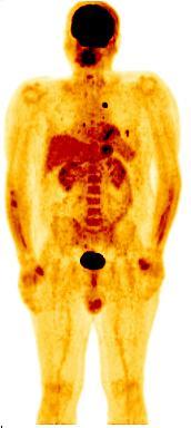
3 Why integrated PET/CT systems? 18 F-FDG The major success of PET in oncology is it s ability to detect tumours as increased levels of [ 18 F]-fluoro-2-deoxyglucose (FDG) uptake compared to normal tissues
 makes difficult the interpretation of PET images due to the lack of")

4 Why integrated PET/C systems? The poor spatial resolution of a PET system (4-5mm) makes difficult the interpretation of PET images due to the lack of identifiable anatomical structures. Localization of increased tracer uptake to a specific organ or structure is important when decisions affecting the diagnosis, staging and the treatment of the patient have to be taken. The need for an accurrate anatomical localization of the PET signal was the motivation for the PET/CT systems.
5 Integrated PET/CT systems A PET/CT system combines (in the same gantry) two of the most important (and complementary ) diagnostic imaging systems like CT and PET allowing to perform the PET/CT procedure, in a single study session, without moving the patient from the bed CT PET
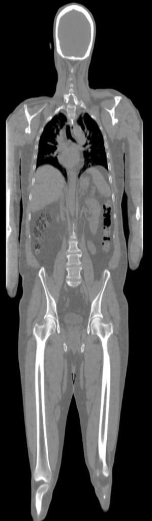
6 Integrated PET/CT systems The anatomical study : CT
7 Integrated PET/CT systems The functional study : Multiple bed positions PET
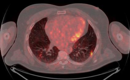
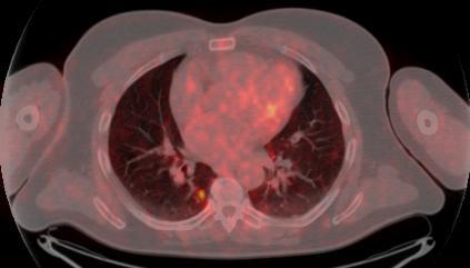
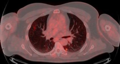
8 Integrated PET/CT systems The functional study : PET
9 Integrated PET/CT systems The functional study : PET
10 Integrated PET/CT systems The functional study : PET
11 Integrated PET/CT Systems In a PET/CT system both techniques use the same spatial reference system

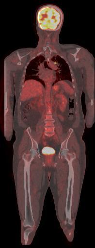
12 Integrated PET/CT systems Once reconstructed, the CT and PET images are spatially co-registered, allowing the physicians to fuse both the information and thus to localize the PET signals over the corresponding anatomy / anatomical structure. Lymph node metastasis Bone metastasis HSR Milano

 : 1) Much lower")
 3) Unbiased post injection CT scan")
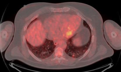
13 Integrated PET/CT systems CT images are also used for the Attenuation Correction of PET EM data CT based Attenuation Correction has significant advantages over more traditional methods (Measured Transmission AC - MAC ) : 1) Much lower statistical noise compared to MAC 2) Shorter acquisition time ( CT: 20-40sec Vs MAC: 20-30min) 3) Unbiased post injection CT scan PET Without AC CT PET With AC by CT
14 State of art of PET/CT systems Siemens mct General Electric (GE) Discovery-600 Discovery-690 Philips Gemini-GXL Gemini-TF An important characteristic of the PET/CT systems is to combine, in the same gantry, the state of the art of both technologies


15 Integrated PET/CT Helical Multi Slices CT scanners Helical CT scnner First models of PET/CT systems were equipped with : cm 2cm 4cm State of the art PET/CT system are equipped with :
16 MS-CT Position-sensitive detector (rotating) X-ray fan beam (rotating) + + Fast & UltraFast Ceramic Scintillator + Photodiodes X-ray source (rotating)

![a more specific tissue characterization : m(e) w (E) CT [HU] = ------------------- x](/docs-images/74/69939958/images/17-1.jpg "1000 w(e) 140 kvp image 80 kvp image Thorsten R.et al.")
17 State of the art and future of MS-CT Dual Sources Energy Discriminating Detectors kvp Switching 80keV 80keV 140keV 140keV Detector 1 Detector 2 Dual energy CT offers a more specific tissue characterization : m(e) w (E) CT [HU] = x 1000 w(e) 140 kvp image 80 kvp image Thorsten R.et al.n : Eur Radiol (2007) 17:


18 State of art of PET/CT systems Siemens mct GE Discovery-600 Discovery-690 Philips Gemini-GXL Gemini-TF PET Scanners
19 PET Scintillators BGO GSO LSO LYSO Density (g/cm 3 ) Effective Z keV (mm) Decay Time (ns) Relative Light Output % (to 100 for NaI(Tl) Energy Resolution % (@ kev) 10(511) 8.5(511) 10(511) 11(511) Wavelength (nm) Index of Refraction IDEAL CHARACTERISTICS high stopping power, to maximize the probability to stop the radiation good energy resolution, to reject scatter events good time resolution, for counting rate linearity, low random rate (PET), TOF to be inexpensive for mass production

20 PET Scintillators Conti Physica Medica 25;1-11, 2009

")


21 PET Detector Design General Electric PET detector block D-600 (BGO) matrix 8X6 4.7x6.3x30 mm 3 D-690 (LYSO) 4.2x6.3x25mm3 Siemens PET detector block mct (LSO) matrix 13x13 4x4x 20 mm 3 PMT PHILIPS PIXELAR Detector panels optically coupled to continuous light guide G-GXL (GSO) 4 x 6 x 30 mm 3 G-TF (LYSO) 4 x 4 x 22 mm 3
 1-2000 cm 2 1-100 mm")


22 Photo-Detectors PMT PS PMT APD and PS APD PMT APD SiPM Active area (mm2) cm mm mm 2 Gain Quantum efficiency 25% 60-80% <40% Temperature coefficient <1%/ C 2-3%/ C 3-5%/ C Magnetic suceptibility Very high No (up to 9.4T) No (up to 15T) Si PM

23 NON TOF Time-of-Flight LSO & LYSO Use time-of-flight to localize source along line of flight. Time of flight information reduces noise in images. Variance reduction given by: 2D / c t. TOF 500 ps timing resolution 7.5 cm localization 500 ps timing resolution 5 x reduction in variance! Reconstruction without TOF information assume that all the possible locations of the annihilation site along the LOR are equally likely. TOF information can be used in the Reconstruction of the PET data to constraint the possible locations of the annihilation site along the LOR SNR GAIN SENSITIVITY GAIN SNR TOF 2 D D = = SNR c t x t psec x cm D=20 cm D=35 cm D=20 cm D=35 cm
 Non-TOF similar contrast and scan time TOF")

24 TOF LESION PHANTOM (Diameter = 27 cm - Activity ratio Lesion / Background = 6:1 ) Non-TOF similar contrast and scan time TOF lower noise 60s 60s similar contrast and noise TOF shorter scan 60s 180s Slide courtesy Joel Karp,University of Pennsylvania
25 Extended Axial Field-Of-View PET/CT (mct) Standard Extended axial FOV Axial 16.2 cm 21.6 cm Starting from the standard configuration which consists in a 16.2 cm axial FOV scanner the user can order, as an option, the addition of one more ring of detectors extending the axial FOV up to 21.6cm The extension of the FOV results in an increase of the overall System Sesitivity of 77% The design of an extended axial FOV open the way to a modular design of the PET systems and represents the first step toward a very wide axial PET scanner.
26 Clinical Advantages of the Extended Axial Field-Of-View bed # Standard FOV: 5 15 min Extended FOV: 4 8 min Higher sensitivity => shorter imaging per bed (same dose, same counts) Higher sensitivity => lower dose (same acq. time, same counts) Higher sensitivity => higher SNR ( same dose, same acq. time)
27 PET/CT Clinical Applications Oncology (90%) Cardiology ( 5%) Neurology ( 5%)
28 Oncological Applications of PET/CT 18F-FDG WB-PET The primary clinical applications of PET/CT is oncology for: - staging, - restaging, - follow -up - monitoring the response to therapies.

29 Oncology PET/CT for Radiotherapy Applications The state of the art in the technology of the RT systems is the intensity modulated radiation therapy (IMRT), in which many small pencil beams are used, under computer control, to conform the irradiated volume to complex and irregular shape. IMRT allows the beams to be modulated to the required intentisy for delivering highly conformal doses of radiation to the targets, while sparing the adjacent normal tissue structures. IMRT allows the design of tailored hot-spots (which Tomotherapy Helical treatment delivery receives much higher local dose) tumour mass (Dose painting). within the overall PET/CT allows to improve the Target Volume Definition by using the: Anatomical CT images for the definitionof the Gross Tumor Volume ( GTV) Functional PET images for the definition of the Biological Tumor Volume (BTV) By combining GTV and BTV a more accurate TVD can be obtained Lung Simulation: Courtesy MD Anderson Huston
30 Tumour CT-based TT Lynphonodes Greater than 1cm CT TOMOTHERAPY TREATMENT PLAN CT-based TT HSR Milano


31 Tumour PET/CT-based TT Lynphonodes Greater than 1cm Tumour Lynphonodes No significant uptake of FDG CT PET/CT TOMOTHERAPY TREATMENT PLAN CT-based TT PET/CT-based TT HSR Milano

32 Oncology PET/CT for Radiotherapy Applications Respiratory gating PET/CT (4D PET/CT) Specific Patient s motion Information can be used for a more accurate TVD and to personalize the treatment planning by reducing the overall PTV, increasing the dose to the tumour while sparing healthy tissues.

33 PET/CT for Cardiological Applications The value of PET/CT in cardiac imaging has extended beyond the assessment of myocardial viability and perfusion. The combination of PET and CT allows a comprehensive anatomical and functional mapping / assessment of the Heart and has demonstrated its use in diagnosing and managing coronary artery disease. HSR-Milan
, to obtain")
 including the visualization of luminal")


34 Cardiological Application of PET/CT With the state of the art of MSCT scanner it is possible to image coronary arteries (non-invasively), to obtain important clinical information on the presence, severity, and characteristics of Coronary arthery disease (CAD) including the visualization of luminal obstruction and atherosclerosis plaques. The negative predictive value of CT is very high. LAD D1 LCX Case SM This suggest that CTA combined with the complementary information by PET (e.g. perfusion imaging) is likely to offer added value for the CAD evaluation. RCA CTA Image courtesy : Turku PET Centre Turku PET Centre, Finland


35 Cardiological Application of PET/CT Case SM O 15 -water parametric PET/CT hybrid images MBF 3 ml/g/min MBF 4 ml/g/min MBF 2 ml/g/min CTA+ Adenosine stress perfusion Courtesy:Turku PET Centre, Finland
 Epilepsy Movement Disorded")


36 Neurological Application of PET/CT Dementias (e.g. Alzheimer s) Epilepsy Movement Disorded (e.g. Parkinson, Huntingtons) Oncology Cerebravascular disease Tumour Tumour 18F-FDG Normal Brain 18F-FDG Oncology 11C-Choline Oncology 18F-FESP D2 dopamine receptors Movement disorders Parkinson
37 Prospective and future of the Hybrid PET/CT systems Nowadays Hybrid PET/CT is certainly on the most important cancer imaging modality. CT scanner Dual Energy CT Multispectral imaging Fastest Temporal Resolution (assessment of time-dependent parameters) Increased Axial FOV ( to capture whole organs in a single rotation ) Dose Reduction Techiques PET scanner New crystal scintillator / detector material Smaller crystal / detectors to improve spatial resolution Fastest crystal scintillator / detectors for TOF New Detector Desgin from PMT to APD to SiPMT APD could be used also for DOI correction Extended Axial FOV Improve System Sensitivity Dose Reduction
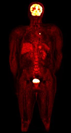
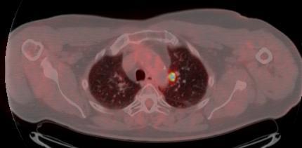
Typical PET Image. Elevated uptake of FDG (related to metabolism) Lung cancer example: But where exactly is it located?
 Lung cancer example: But where exactly is it located?") Typical PET Image Elevated uptake of FDG (related to metabolism) Lung cancer example: But where exactly is it located? PET/CT Oncology Imaging Anatometabolic fusion images are useful in the management
Typical PET Image Elevated uptake of FDG (related to metabolism) Lung cancer example: But where exactly is it located? PET/CT Oncology Imaging Anatometabolic fusion images are useful in the management
PET-CT for radiotherapy planning in lung cancer: current recommendations and future directions
 PET-CT for radiotherapy planning in lung cancer: current recommendations and future directions Gerry Hanna Centre for Cancer Research and Cell Biology Queen s University of Belfast @gerryhanna Talk Outline
A Snapshot on Nuclear Cardiac Imaging
 Editorial A Snapshot on Nuclear Cardiac Imaging Khalil, M. Department of Physics, Faculty of Science, Helwan University. There is no doubt that nuclear medicine scanning devices are essential tool in the
45 Hr PET Registry Review Course
 45 HR PET/CT REGISTRY REVIEW COURSE Course Control Document Timothy K. Marshel, MBA, R.T. (R), (N)(CT)(MR)(NCT)(PET)(CNMT) The PET/CT Training Institute, Inc. SNMMI-TS 028600-028632 45hr CEH s Voice Credits
4D PET: promises and limitations
 4D PET: promises and limitations Tinsu Pan, Ph.D. M.D. Anderson Cancer Center The University of Texas Background Outlines Gating techniques: Deep inspiration breath hold 4D PET/CT Non-gating techniques
Hybrid cardiac imaging Advantages, limitations, clinical scenarios and perspectives for the future
 Hybrid cardiac imaging Advantages, limitations, clinical scenarios and perspectives for the future Prof. Juhani Knuuti, MD, FESC Turku, Finland Disclosure: Juhani Knuuti, M.D. Juhani Knuuti, M.D. has financial
An Overview of Clinical PET/CT
 1 INVITED ARTICLE An Overview of Clinical PET/CT Arman Rahmim 1* PhD and Richard L. Wahl 2 MD Department of Radiology, School of Medicine Johns Hopkins University, Baltimore MD, USA (Received 10 November
CT Perfusion. U. Joseph Schoepf, MD, FAHA, FSCBT MR, FSCCT Professor of Radiology, Medicine, and Pediatrics Director of Cardiovascular Imaging
 CT Perfusion U. Joseph Schoepf, MD, FAHA, FSCBT MR, FSCCT Professor of Radiology, Medicine, and Pediatrics Director of Cardiovascular Imaging Disclosures Consultant for / research support from Bayer Bracco
Pitfalls and Remedies in PET/CT imaging for RT planning
 Pitfalls and Remedies in PET/CT imaging for RT planning Tinsu Pan, Ph.D. M.D. Anderson Cancer Center The University of Texas Outlines Background Average CT (< 1 msv) to reduce mis-alignment of PET and
Combined Anatomical and Functional Imaging with Revolution * CT
 GE Healthcare Case studies Combined Anatomical and Functional Imaging with Revolution * CT Jean-Louis Sablayrolles, M.D. Centre Cardiologique du Nord, Saint-Denis, France Case 1 Whole Brain Perfusion and
Future upcoming technologies and what audit needs to address
 Future upcoming technologies and what audit needs to address Dr R.I MacKay History of audit Absolute dose - Simple phantom standard dose measurement Point doses in beams - Phantoms of relatively simple
Multisclice CT in combination with functional imaging for CAD. Temporal Resolution. Spatial Resolution. Temporal resolution = ½ of the rotation time
 Multisclice CT in combination with functional imaging for CAD Prof. Juhani Knuuti, MD, FESC Turku University Hospital and University of Turku Turku, Finland MSCT and functional imaging for CAD Practical
GE Healthcare. Rad Rx. White Paper
 GE Healthcare Rad Rx White Paper Introduction This publication is part of a series of white papers aimed at communicating the importance of each component in the image chain of a PET/CT study. From data
CT Myocardial Perfusion: Is there Added Value to Coronary CT?
 CT Myocardial Perfusion: Is there Added Value to Coronary CT? U. Joseph Schoepf, MD, FAHA, FSCBT MR, FSCCT Professor of Radiology, Medicine, and Pediatrics Director of Cardiovascular Imaging Disclosures
Optimized. clinical pathway. propels high utilization of PET/MR at Pitié-Salpêtrière Hospital
 Optimized propels high utilization of PET/MR at Pitié-Salpêtrière Hospital clinical pathway As one of Europe s largest teaching hospitals, Pitié-Salpêtrière Hospital is renowned for its innovative research
Cardiac CT - Coronary Calcium Basics Workshop II (Basic)
") Cardiac CT - Coronary Calcium Basics Workshop II (Basic) J. Jeffrey Carr, MD, MSCE Dept. of Radiology & Public Health Sciences Wake Forest University School of Medicine Winston-Salem, NC USA No significant
Cardiac CT - Coronary Calcium Basics Workshop II (Basic) J. Jeffrey Carr, MD, MSCE Dept. of Radiology & Public Health Sciences Wake Forest University School of Medicine Winston-Salem, NC USA No significant
Cardiac Imaging Tests
 Cardiac Imaging Tests http://www.medpagetoday.com/upload/2010/11/15/23347.jpg Standard imaging tests include echocardiography, chest x-ray, CT, MRI, and various radionuclide techniques. Standard CT and
PET: quantification of perfusion and beyond
 PET: quantification of perfusion and beyond Juhani Knuuti Turku PET Centre University of Turku Turku, Finland Juhani.knuuti@utu.fi 27.8.2012 1 Disclosure: Juhani Knuuti, M.D. Juhani Knuuti, M.D. has financial
Dual Energy CT of the Heart: Perfusion and Beyond
 Dual Energy CT of the Heart: Perfusion and Beyond U. Joseph Schoepf, MD, FAHA, FSCBT MR, FSCCT Professor of Radiology, Medicine, and Pediatrics Director of Cardiovascular Imaging Disclosures Consultant
Simulations of Preclinical andclinical Scans in Emission Tomography, Transmission Tomography and Radiation Therapy. Using GATE
 GATE Simulations of Preclinical andclinical Scans in Emission Tomography, Transmission Tomography and Radiation Therapy Using GATE Quick tour & Highlights! GATE Training, INSTN-Saclay, October 2015 Albertine
IAEA RTC. PET/CT and Planning of Radiation Therapy 20/08/2014. Sarajevo (Bosnia & Hercegovina) Tuesday, June :40-12:20 a.
 Tuesday, June :40-12:20 a.") IAEA RTC PET/CT and Planning of Radiation Therapy Sarajevo (Bosnia & Hercegovina) Tuesday, June 17 2014 11:40-12:20 a.m María José García Velloso Servicio de Medicina Nuclear Clínica Universidad de Navarra
IAEA RTC PET/CT and Planning of Radiation Therapy Sarajevo (Bosnia & Hercegovina) Tuesday, June 17 2014 11:40-12:20 a.m María José García Velloso Servicio de Medicina Nuclear Clínica Universidad de Navarra
Identifying Image Artifacts, Their Causes and How to Fix Them: PET. Brad J Kemp, PhD Mayo Clinic, Rochester, MN
 Identifying Image Artifacts, Their Causes and How to Fix Them: PET Brad J Kemp, PhD Mayo Clinic, Rochester, MN Case 1 Can we scan with a defective block detector? Daily Quality Assurance Results Singles
Validation of CT Perfusion Imaging Against Invasive Angiography and FFR on a 320-MDCT Scanner
 Validation of CT Perfusion Imaging Against Invasive Angiography and FFR on a 320-MDCT Scanner Zhen Qian, Gustavo Vasquez, Sarah Rinehart, Parag Joshi, Eric Krivitsky, Anna Kalynych, Dimitri Karmpaliotis,
Dual-Energy CT: The Technological Approaches
 Dual-Energy CT: The Technological Approaches Dushyant Sahani, M.D Director of CT Associate Professor of Radiology Massachusetts General Hospital Harvard Medical School Email-dsahani@partners.org Disclosure
Introduction Pediatric malignancies Changing trends & Radiation burden Radiation exposure from PET/CT Image gently PET & CT modification - PET/CT
 Introduction Pediatric malignancies Changing trends & Radiation burden Radiation exposure from PET/CT Image gently PET & CT modification - PET/CT protocols Tips Leukaemia / lymphoma: ~ 35% acute lymphoblastic
X-Ray & CT Physics / Clinical CT
 Computed Tomography-Basic Principles and Good Practice X-Ray & CT Physics / Clinical CT INSTRUCTORS: Dane Franklin, MBA, RT (R) (CT) Office hours will be Tuesdays from 5pm to 6pm CLASSROOM: TIME: REQUIRED
Les Outils Cliniques de Demain en Scanner Cardiaque. Cardiaque Status en ECR 2018 From Diagnosis to Prognosis
 ECR 2018 From Diagnosis to Prognosis ECR 2018 From Diagnosis to Prognosis Thursday, March 1, 2018/08:30-10:00/Room N Les Outils Cliniques de Demain en Scanner Cardiaque Cardiaque Status en 2018 Rodrigo
MRI-PET: Oncologic Applications
 MRI-PET: Oncologic Applications Pablo R. Ros, MD University Hospitals Case Medical Center Case Western Reserve University SCBT-MR Boston, MA October, 2012 Pablo.Ros@UHhospitals.org Acknowledgement Osman
Photon Attenuation Correction in Misregistered Cardiac PET/CT
 Photon Attenuation Correction in Misregistered Cardiac PET/CT A. Martinez-Möller 1,2, N. Navab 2, M. Schwaiger 1, S. G. Nekolla 1 1 Nuklearmedizinische Klinik der TU München 2 Computer Assisted Medical
Ultrasound. Computed tomography. Case studies. Utility of IQon Spectral CT in. cardiac imaging
 Ultrasound Computed tomography Case studies Utility of IQon Spectral CT in cardiac imaging Cardiac imaging is a challenging procedure where it is necessary to image a motion-free heart. This requires a
POSITRON EMISSION TOMOGRAPHY PHANTOM STUDIES FOR RADIATION THERAPY TARGET DELINEATION MICHAEL VINTSON LAWRENCE
 POSITRON EMISSION TOMOGRAPHY PHANTOM STUDIES FOR RADIATION THERAPY TARGET DELINEATION BY MICHAEL VINTSON LAWRENCE A Dissertation Submitted to the Graduate Faculty of WAKE FOREST UNIVERSITY GRADUATE SCHOOL
Molecular Imaging: - SPECT agents under development - Imaging challenges
 Molecular Imaging: - SPECT agents under development - Imaging challenges Jody Garrard, CNMT Gamma Camera Product Manager Philips Nuclear Medicine jody.garrard@philips.com When you reach turning points
Y-PET versus 90 Y-Bremsstrahlung SPECT
 90 Y-PET versus 90 Y-Bremsstrahlung SPECT First 90 Y microspheres injected intra-arterially imaging 90 Y TOF PET/CT 198 Au rays scan 90 Y bremsstrahlung scan Lhommel et al. 2009 Simon and Feitelberg 1967
CT or PET/CT for coronary artery disease
 CT or PET/CT for coronary artery disease Rotterdam 2012 Juhani Knuuti, MD, PhD, FESC Turku PET Centre University of Turku Turku, Finland Juhani.knuuti@utu.fi Turku PET Centre University of Turku Åbo Akademi
Why is CT Dose of Interest?
 Why is CT Dose of Interest? CT usage has increased rapidly in the past decade Compared to other medical imaging CT produces a larger radiation dose. There is direct epidemiological evidence for a an increase
Usefulness of New CT Technologies for Interventional Cardiovascular Procedures
 Usefulness of New CT Technologies for Interventional Cardiovascular Procedures Ronen Rubinshtein, MD FACC FESC Department of Cardiovascular Medicine Lady Davis Carmel Medical Center & Technion Israel Institute
IMRT - the physician s eye-view. Cinzia Iotti Department of Radiation Oncology S.Maria Nuova Hospital Reggio Emilia
 IMRT - the physician s eye-view Cinzia Iotti Department of Radiation Oncology S.Maria Nuova Hospital Reggio Emilia The goals of cancer therapy Local control Survival Functional status Quality of life Causes
An Introduction to Dual Energy Computed Tomography
 An Introduction to Dual Energy Computed Tomography Michael Riedel University of Texas Health Science Center at San Antonio Introduction The idea of computed tomography (CT) was first introduced in the
TomoTherapy. Michelle Roach CNC Radiation Oncology Liverpool Hospital CNSA. May 2016
 TomoTherapy Michelle Roach CNC Radiation Oncology Liverpool Hospital CNSA May 2016 TomoTherapy The Facts Greek Tomo = slice Advanced form of IMRT 3D computerised tomography (CT) imaging immediately prior
CT Myocardial Perfusion
 1 CT Myocardial Perfusion Ting-Yim Lee, PhD, FCCPM, FCOMP Aaron So, PhD, FSCCT Gerald Wisenberg, MD, FRCPC Ali Islam, MD, FRCPC Lawson Health Research Institute Robarts research Institute The University
Cardiovascular Imaging
 Cardiovascular Imaging Cardiovascular Imaging Cardio and Vascular Imaging Vascularization / Angiogenesis Cardiovascular Imaging metabolic imaging of the heart myocardial perfusion imaging Cardiac CT Vascularization
I. Equipments for external beam radiotherapy
 I. Equipments for external beam radiotherapy 5 linear accelerators (LINACs): Varian TrueBeam 6, 10 & 18 MV photons, 6-18 MeV electrons, image-guided (IGRT) and intensity modulated radiotherapy (IMRT),
Introduction. Cardiac Imaging Modalities MRI. Overview. MRI (Continued) MRI (Continued) Arnaud Bistoquet 12/19/03
 MRI (Continued) Arnaud Bistoquet 12/19/03") Introduction Cardiac Imaging Modalities Arnaud Bistoquet 12/19/03 Coronary heart disease: the vessels that supply oxygen-carrying blood to the heart, become narrowed and unable to carry a normal amount
Introduction Cardiac Imaging Modalities Arnaud Bistoquet 12/19/03 Coronary heart disease: the vessels that supply oxygen-carrying blood to the heart, become narrowed and unable to carry a normal amount
Myocardial Perfusion: Positron Emission Tomography
 Myocardial Perfusion: Positron Emission Tomography TH. Schindler, MD University Hospitals of Geneva, Cardiovascular Center, Geneva, Switzerland ESC 2010 Stockholm Personal Disclosure Research Grant support
Radiation Detection and Measurement
 Radiation Detection and Measurement Range of charged particles (e.g.,!: µm; ": mm) Range of high energy photons (cm) Two main types of interactions of high energy photons Compton scatter Photoelectric
Defining Target Volumes and Organs at Risk: a common language
 Defining Target Volumes and Organs at Risk: a common language Eduardo Rosenblatt Section Head Applied Radiation Biology and Radiotherapy (ARBR) Section Division of Human Health IAEA Objective: To introduce
Title: TC simulation versus TC/PET simulation for radiotherapy in lung cancer: volumes comparison in two cases.
 Title: TC simulation versus TC/PET simulation for radiotherapy in lung cancer: volumes comparison in two cases. Authors: Franzone, P.; 1* Muni, A; 2 Cazzulo, E.; 3 Berretta, L.; 1 Pozzi, G. 1 ; Todisco,
ADVANCES IN RADIATION TECHNOLOGIES IN THE TREATMENT OF CANCER
 ADVANCES IN RADIATION TECHNOLOGIES IN THE TREATMENT OF CANCER Bro. Dr. Collie Miller IARC/WHO Based on trends in the incidence of cancer, the International Agency for Research on Cancer (IARC) and WHO
Fundamentals of Nuclear Cardiology. Terrence Ruddy, MD, FRCPC, FACC
 Fundamentals of Nuclear Cardiology Terrence Ruddy, MD, FRCPC, FACC Objectives To understand the Principles of Nuclear Cardiac Imaging Radiotracers Image acquisition and processing Stress protocols To appreciate
Page 1. Helical (Spiral) Tomotherapy. UW Helical Tomotherapy Unit. Helical (Spiral) Tomotherapy. MVCT of an Anesthetized Dog with a Sinus Tumor
 Tomotherapy. UW Helical Tomotherapy Unit. Helical (Spiral) Tomotherapy. MVCT of an Anesthetized Dog with a Sinus Tumor") Helical (Spiral) Tomotherapy Novel Clinical Applications of IMRT Linac Ring Gantry CT Detector X-Ray Fan Beam Binary Multileaf Collimator Binary MLC Leaves James S Welsh, MS, MD Department of Human Oncology
Helical (Spiral) Tomotherapy Novel Clinical Applications of IMRT Linac Ring Gantry CT Detector X-Ray Fan Beam Binary Multileaf Collimator Binary MLC Leaves James S Welsh, MS, MD Department of Human Oncology
Nuclear Medicine and PET. D. J. McMahon rev cewood
 Nuclear Medicine and PET D. J. McMahon 150504 rev cewood 2018-02-15 Key Points Nuclear Medicine and PET: Imaging: Understand how Nuc Med & PET differ from Radiography & CT by the source of radiation. Be
State-of-the-Art SPECT/CT: Cardiac Imaging
 State-of-the-Art SPECT/CT: Cardiac Imaging Ernest V Garcia*, PhD Endowed Professor in Cardiac Imaging Director, Nuclear Cardiology R&D Laboratory Disclosure: Dr. Garcia receives royalties from the sale
SPECT or PET for Cardiovascular Screening in High-Risk Patients
 SPECT or PET for Cardiovascular Screening in High-Risk Patients Paeng, Jin Chul MD PhD Department of Nuclear Medicine Seoul National University Hospital Contents Recent Development in SPECT and PET Technology
REVISITING ICRU VOLUME DEFINITIONS. Eduardo Rosenblatt Vienna, Austria
 REVISITING ICRU VOLUME DEFINITIONS Eduardo Rosenblatt Vienna, Austria Objective: To introduce target volumes and organ at risk concepts as defined by ICRU. 3D-CRT is the standard There was a need for a
Low-dose and High-resolution Cardiac Imaging with Revolution CT
 GE Healthcare Case study Low-dose and High-resolution Cardiac Imaging with Revolution CT Prof. Philipp A. Kaufmann, M.D. Ronny R. Buechel, M.D. Fran Mikulicic, M.D. Dominik C. Benz, M.D. University of
Molecular Imaging of Coronary Plaques
 Molecular Imaging of Coronary Plaques Daniel S. Berman, MD Director, Cardiac Imaging Cedars-Sinai Heart Institute CSMC 20113 Professor of Medicine David Geffen School of Medicine at UCLA Zahi A. Fayad,
Radionuclides in Medical Imaging. Danielle Wilson
 Radionuclides in Medical Imaging Danielle Wilson Outline Definitions History and development Radionuclide applications & techniques in imaging Conclusion Definition #1 : Radionuclide An unstable nucleus
Coronary Artery Imaging. Suvipaporn Siripornpitak, MD Inter-hospital Conference : Rajavithi Hospital
 Coronary Artery Imaging Suvipaporn Siripornpitak, MD Inter-hospital Conference : Rajavithi Hospital Larger array : cover scan area Detector size : spatial resolution Rotation speed : scan time Retrospective
SPECIFIC PRINCIPLES FOR DOSE REDUCTION IN HEAD CT IMAGING. Rajiv Gupta, MD, PhD Neuroradiology, Massachusetts General Hospital Harvard Medical School
 SPECIFIC PRINCIPLES FOR DOSE REDUCTION IN HEAD CT IMAGING Rajiv Gupta, MD, PhD Neuroradiology, Massachusetts General Hospital Harvard Medical School OUTLINE 1 st Presentation: Dose optimization strategies
PET/MR:Techniques, Indications and Applications
 PET/MR:Techniques, Indications and Applications Franz Wolfgang Hirsch Professor and Head of the Department of Pediatric Radiology University Hospital Leipzig / Germany Children s Hospital University Leipzig
Physics of MBI (~10 slides)
") Physics of MBI (~10 slides) Molecular Breast Imaging (MBI) physics and performance testing JW Hugg, BR Simrak, PD Smith, BE Patt Gamma Medica, Inc., Northridge, CA Molecular Breast Imaging (MBI) is an
Physics of MBI (~10 slides) Molecular Breast Imaging (MBI) physics and performance testing JW Hugg, BR Simrak, PD Smith, BE Patt Gamma Medica, Inc., Northridge, CA Molecular Breast Imaging (MBI) is an
Appendix 1: Regional Lymph Node Stations for Staging Esophageal Cancer
 Appendix 1: Regional Lymph Node Stations for Staging Esophageal Cancer Locoregional (N stage) disease was redefined in the seventh edition of the AJCC Cancer Staging Manual as any periesophageal lymph
First Clinical Experience with
 First Clinical Experience with Discovery MI Digital PET/CT Martin Huellner Department of Nuclear Medicine University Hospital Zurich / University of Zurich Switzerland Agenda BelNuc GE Symposium 1. Digital
CARDIAC PET PERFUSION IMAGING with RUBIDIUM-82
 CARDIAC PET PERFUSION IMAGING with RUBIDIUM-82 Pr Denis AGOSTINI Président du Groupe de Cardiologie Nucléaire et IRM CHU Caen Bordeaux 2006 Cardiac Perfusion-Metabolism Mismatch with PET Cumulative Survival
Bone PET/MRI : Diagnostic yield in bone metastases and malignant primitive bone tumors
 Bone PET/MRI : Diagnostic yield in bone metastases and malignant primitive bone tumors Lars Stegger, Benjamin Noto Department of Nuclear Medicine University Hospital Münster, Germany Content From PET to
Maximizing the Utility of Integrated PET/MRI in Clinical Applications
 Maximizing the Utility of Integrated PET/MRI in Clinical Applications Spencer Behr, MD Department of Nuclear Medicine & Abdominal Imaging University of California, San Francisco PET/MR at UCSF Device:
SPECT-CT: Τι πρέπει να γνωρίζει ο Καρδιολόγος
 SPECT-CT: Τι πρέπει να γνωρίζει ο Καρδιολόγος Δρ Αναστασία Κίτσιου Διευθύντρια, Καρδιολογική Κλινική, Σισμανόγλειο ΓΝΑ Chair, Education Committee, Section on Nuclear Cardiology & Cardiac CT, EACVI, ESC
Medical Imaging. Alex Elliott Western Infirmary Glasgow
 Medical Imaging Alex Elliott Western Infirmary Glasgow History of medical imaging X-rays - Roentgen, 1895 Nuclear medicine - Cassen, 1951 Ultrasound Donald, 1962 SPECT - Kuhl, Edwards, 1963 PET Ter-Pogossian,
Cardiac PET. John Buscombe
 Cardiac PET John Buscombe Why PET? Improved resolution-not really required in cardiology Improved sensitivity this may be important-financially as reduced acquisition time Improved attenuation correction-good
Pearls & Pitfalls in nuclear cardiology
 Pearls & Pitfalls in nuclear cardiology Maythinee Chantadisai, MD., NM physician Division of Nuclear Medicine, Department of radiology, KCMH Principle of myocardial perfusion imaging (MPI) Radiotracer
Verification of treatment planning system parameters in tomotherapy using EBT Radiochromic Film
 Verification of treatment planning system parameters in tomotherapy using EBT Radiochromic Film E.B.Rajmohan¹, Pratik Kumar¹, Bhudatt Paliwal,² David Westerly², N.Gopishankar³, R.K.Bisht³, D.Tewatia²,
Imaging of Scattered Radiation for Real Time Tracking of Tumor Motion During Lung SBRT
 Imaging of Scattered Radiation for Real Time Tracking of Tumor Motion During Lung SBRT April 25 nd, 2015 Lung Cancer Lung cancer is the most lethal cancer: Over 224,000 new diagnoses in the U.S. predicted
I. Cancer staging problem
 Instrumentation Lab (Craig Levin) Angela Craig Peter Jin Frezghi Guillem Billie Garry Research interests (by imaging modality) I. High resolution radionuclide imaging : positron emission tomography (PET)
A. DeWerd. Michael Kissick. Larry. Editors. The Phantoms of Medical. and Health Physics. Devices for Research and Development.
 Larry Editors A. DeWerd Michael Kissick The Phantoms of Medical and Health Physics Devices for Research and Development ^ Springer Contents 1 Introduction to Phantoms of Medical and Health Physics 1 1.1
PET-MRI in malignant bone tumours. Lars Stegger Department of Nuclear Medicine University Hospital Münster, Germany
 PET-MRI in malignant bone tumours Lars Stegger Department of Nuclear Medicine University Hospital Münster, Germany Content From PET to PET/MRI General considerations Bone metastases Primary bone tumours
Image Guided Proton Therapy and Treatment Adaptation
 Image Guided Proton Therapy and Treatment Adaptation www.hollandptc.nl d.r.schaart@tudelft.nl Cancer in The Netherlands About 1 in 3 people get cancer in some stage of their life 86.800 new cancer patients
Case Reports: Tumor Detection by Diffusion-Weighted MRI and ADC-Mapping with Correlation to PET/CT Results
 Case Reports: Tumor Detection by Diffusion-Weighted MRI and ADC-Mapping with Correlation to PET/CT Results Matthias Philipp Lichy, M.D.; Philip Aschoff, M.D.; Christina Pfannenberg, M.D.; Schlemmer Heinz-Peter,
Alessandro Albonico Philips
 Alessandro Albonico Philips Alessandro.albonico@philips.com Noise (Standard Deviation in HU) Virtually noise-free Characteristic of a true knowledge-based IR 80 70 Standard Recon idose4 Level6 1 mm Slice
PET/MRI: a new era in multimodality molecular imaging
 Clin Transl Imaging (2013) 1:5 10 DOI 10.1007/s40336-013-0003-5 REVIEW ARTICLE PET/MRI: a new era in multimodality molecular imaging Osman Ratib Received: 22 October 2012 / Accepted: 10 January 2013 /
Quantitative Molecular Imaging Using PET/CT to Assess Response to Therapy
 Quantitative Molecular Imaging Using PET/CT to Assess Response to Therapy Paul Kinahan, PhD Director of PET/CT Physics Imaging Research Laboratory, Department of Radiology University of Washington, Seattle,
14/09/2013 FDG PET PET-CT = FDG PET-CT. = Metabolism. Anatometabolic Imaging. = Anatomy. Nuclear Medicine in the Era of Hybrid Imaging
 Nuclear Medicine in the Era of Hybrid Imaging Focus on a New SPECT-CT Device : HWK-4 Tarik Belhocine, MD, Ph.D Nuclear Medicine = SPECT = Functional Imaging Function Radiology = Morphological Imaging Anatomy
Quantitative outcome of registration methods for correcting cardiac drift in cardiac PET/CT imaging
 JOURNAL OF APPLIED CLINICAL MEDICAL PHYSICS, VOLUME 17, NUMBER 2, 2016 Quantitative outcome of registration methods for correcting cardiac drift in cardiac PET/CT imaging Jonathon A. Nye, a Dana Tudorascu,
PET-MRI in Cardiac Imaging: Initial Experience
 PET-MRI in Cardiac Imaging: Initial Experience Mallinckrodt Institute of Radiology Washington University School of Medicine Pamela K. Woodard, M.D. Professor of Radiology and Biomedical Engineering Head,
Reproducibility of Uptake Estimates in FDG PET: a Monte Carlo study
 Reproducibility of Uptake Estimates in FDG PET: a Monte Carlo study Juliette Feuardent, Marine Soret, Irène Buvat 1 Abstract Tumor glucose metabolism measurements from Fluoro-deoxyglucose (FDG) Positron
BioMedical quantitative X-Ray Imaging. Emmanuel Brun Researcher Inserm Université Grenoble Alpes
 BioMedical quantitative X-Ray Imaging Emmanuel Brun Researcher Inserm Université Grenoble Alpes 1 Outline Introduction K-Edge Imaging Patient imaging at the European synchrotron Medical Phase Contrast
FROM ICARO1 TO ICARO2: THE MEDICAL PHYSICS PERSPECTIVE. Geoffrey S. Ibbott, Ph.D. June 20, 2017
 FROM ICARO1 TO ICARO2: THE MEDICAL PHYSICS PERSPECTIVE Geoffrey S. Ibbott, Ph.D. June 20, 2017 1 DISCLOSURES My institution holds Strategic Partnership Research Agreements with Varian, Elekta, and Philips
Radiation Dose Reduction Strategies in Coronary CT Angiography
 Radiation Dose Reduction Strategies in Coronary CT Angiography Noor Diyana Osman, PhD noordiyana@usm.my Contents: Introduction Radiation dosimetry in CT Radiation risk associated with coronary CT angiography
Image Guided Stereotactic Radiotherapy of the Lung
 Image Guided Stereotactic Radiotherapy of the Lung Jamie Marie Harris, MS DABR Avera McKennan Radiation Oncology September 25, 2015 Stereotactic Body Radiotherapy - Clinical Dose/Fractionation - Normal
THE TUFFEST STUFF CT REGISTRY REVIEW Live Lecture Seminar SATURDAY CURRICULUM
 1. The CT Imaging Chain-10 major components & their functions a. The x-ray tube b. Generator c. Filter d. Pre-patient collimator e. Pre-detector collimator f. Detector system g. Analog to digital converter
PET/MR. Are You Ready?? Derek Lee, BS, CNMT, PET
 PET/MR Are You Ready?? Derek Lee, BS, CNMT, PET Allow Myself to Introduce Myself I ve been around a while: 23+ years in Nuclear Medicine and counting PET & PET/CT for 13 years and counting Currently working
Verification of micro-beam irradiation
 Journal of Physics: Conference Series OPEN ACCESS Verification of micro-beam irradiation To cite this article: Qiongge Li et al 2015 J. Phys.: Conf. Ser. 573 012047 View the article online for updates
POSITRON EMISSION TOMOGRAPHY (PET)
") Status Active Medical and Behavioral Health Policy Section: Radiology Policy Number: V-27 Effective Date: 08/27/2014 Blue Cross and Blue Shield of Minnesota medical policies do not imply that members should
Status Active Medical and Behavioral Health Policy Section: Radiology Policy Number: V-27 Effective Date: 08/27/2014 Blue Cross and Blue Shield of Minnesota medical policies do not imply that members should
Nuclear Sciences and Medicine
 Nuclear Sciences and Medicine Rethy Chhem, MD, PhD (Edu), PhD (His), FRCPC Division of Human Health Guest Professor, Medical University of Vienna International Atomic Energy Agency Medical Imaging X-rays
Positron emission tomography and molecular imaging
 Positron emission tomography and molecular imaging EHH, May, 2010 Juhani Knuuti Turku PET Centre University of Turku Turku, Finland Juhani.knuuti@utu.fi PET Imaging in Medicine MRI CT MRI PET US SPET MEG
Conflict of Interest Disclosure
 Comparative Advantages of PET Over SPECT: Is PET Really Better? Timothy M. Bateman M.D. Co-Director, Cardiovascular Radiologic Imaging Mid America Heart Institute Professor of Medicine University of Missouri-Kansas
Cigna - Prior Authorization Procedure List: Radiology & Cardiology
 Cigna - Prior Authorization Procedure List: Radiology & Cardiology Product Category CPT Code CPT Code Description Radiology MR 70336 MRI Temporomandibular Joint(s), (TMJ) Radiology CT 70450 CT Head or
The radiation dose in retrospective
 The radiation dose in retrospective gated tdcoronary computed td tomography (CCT) Saeed AL Ahmari, Ghormallah AL Zahrani, Sumiah AL Helali, Samir AL Dulikan, Abdullah Bafagih, HibaKhashojji Prince Sultan
Use of Nuclear Cardiology in Myocardial Viability Assessment and Introduction to PET and PET/CT for Advanced Users
 Use of Nuclear Cardiology in Myocardial Viability Assessment and Introduction to PET and PET/CT for Advanced Users February 1 5, 2011 University of Santo Tomas Hospital Angelo King A-V Auditorium Manila,
The Physics of Oesophageal Cancer Radiotherapy
 The Physics of Oesophageal Cancer Radiotherapy Dr. Philip Wai Radiotherapy Physics Royal Marsden Hospital 1 Contents Brief clinical introduction Imaging and Target definition Dose prescription & patient