DERMATOLOGY. Dr. Khaled M. Al-Qudah. 4/24/2013 Dr. Khaled Al-Qudah 1
|
|
|
- Joan Wilkinson
- 6 years ago
- Views:
Transcription
1 DERMATOLOGY Dr. Khaled M. Al-Qudah 4/24/2013 Dr. Khaled Al-Qudah 1
2 Type of Skin Lesions I. primary lesions: 4/24/2013 Dr. Khaled Al-Qudah 2
, as in Arabian fading syndrome 4/24/2013 Dr. Khaled Al-Qudah 3")
3 primary lesions Macule: flat circumscribed impalpable area of color change level with the skin surface (1cm), as in Arabian fading syndrome 4/24/2013 Dr. Khaled Al-Qudah 3

4 primary lesions Patch: a macule more than 1cm, the depigmentation of the muzzle of this horse shows white patches and black macules within patches 4/24/2013 Dr. Khaled Al-Qudah 4

5 primary lesions Papule: a circumscribed, palpable solid usually round mass in the skin, less than 1cm. Slight erythmea may show in non-pigmented skin; heavily pigmented skin may not show any color change. It can be associated with pruritus caused by insect bites 4/24/2013 Dr. Khaled Al-Qudah 5

6 primary lesions Plaque: a solid, elevated, flattopped lesion, more than 1cm. They may be irregular in shape but are often circular. They are related to allergic reactions from drugs or feed 4/24/2013 Dr. Khaled Al-Qudah 6

7 primary lesions Nodule: a circumscribed, solid, usually round mass, usually raised and rounded more than 1cm. Nodules have the same origins as papules but increased size due to more severe reaction. 4/24/2013 Dr. Khaled Al-Qudah 7

8 primary lesions Vesicle: a circumscribed, elevated, fluctuant fluid filled lesion containing serum, (1cm). Due to the thin layer of skin covering both vesicles and bullae, they often rupture, leaving a reddish eroded surface. 4/24/2013 Dr. Khaled Al-Qudah 8

9 primary lesions Bulla: a vesicle more than 1cm. They can be epidermal or subepidermal. they often rupture, leaving a reddish eroded surface, therefore rarely seen intact. 4/24/2013 Dr. Khaled Al-Qudah 9

10 primary lesions Pustule: a vesicle filled with pus (inflammatory cells). These lesions are seen in staph. and strept. Skin infections and later stages of Equine Coital Exanthema. These are pustules due to ECE on a stallion s penis. 4/24/2013 Dr. Khaled Al-Qudah 10

11 primary lesions Wheal (urticaria, hive): a circumscribed, semisolid, raised, round or flat-topped lesion of varying size from 2-3 mm up to cm. they are usually associated with edema of the area. 4/24/2013 Dr. Khaled Al-Qudah 11

12 Wheals - Equine back from stable fly bites 4/24/2013 Dr. Khaled Al-Qudah 12
13 Type of Skin Lesions II. Secondary lesions 4/24/2013 Dr. Khaled Al-Qudah 13

14 Secondary lesions Scale: an accumulation of loose fragments of stratum corneum. May be white or discolored by secretion of sebum or blood breakdown products. Thicker and more adherent scale 4/24/2013 Dr. Khaled Al-Qudah 14
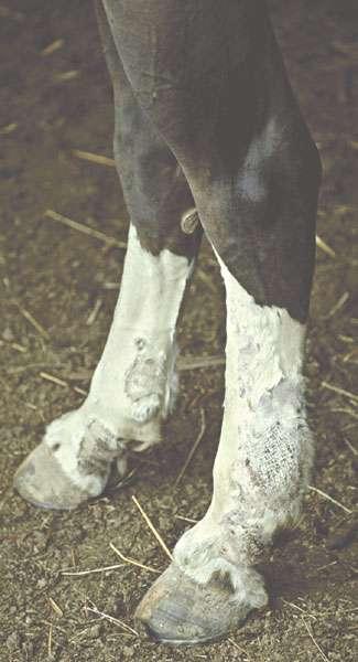
15 Secondary lesions Hyperkeratosis: localized, multifocal or generalized accumulation of adherent keratinaceous material. 4/24/2013 Dr. Khaled Al-Qudah 15

16 Secondary lesions Crust (scab): a dried, solid, adherent consolidation of varying combinations of serum, blood, pus, cutaneous debris, and microorganisms. It may or may not be infected. 4/24/2013 Dr. Khaled Al-Qudah 16

17 Secondary lesions Erosion: loss of epidermis to varying depths, but not penetrating the basement membrane. Does not result in scarring. 4/24/2013 Dr. Khaled Al-Qudah 17

18 Secondary lesions Ulcer: loss of tissue that breaches basement membrane and the dermis, if healing occurs, it results in scar formation. 4/24/2013 Dr. Khaled Al-Qudah 18

19 Secondary lesions Epidermal collarette: circular rim of peeling epidermis surrounding a recent erosion or ulcer. 4/24/2013 Dr. Khaled Al-Qudah 19

20 Secondary lesions Lichenification: thickened skin that is hard, with exaggeration of normal lines and markings. It occurs as a result of chronic inflammation and is due to repeated rubbing or biting at areas affected by pruritus. 4/24/2013 Dr. Khaled Al-Qudah 20

21 Secondary lesions Lichenification Chronic 4/24/2013 Dr. Khaled Al-Qudah 21

22 Secondary lesions Excoriation: Superficial traumatic abrasions and scratches which remove some of the skin substances, commonly caused in animals by rubbing or scratching pruritic skin. 4/24/2013 Dr. Khaled Al-Qudah 22

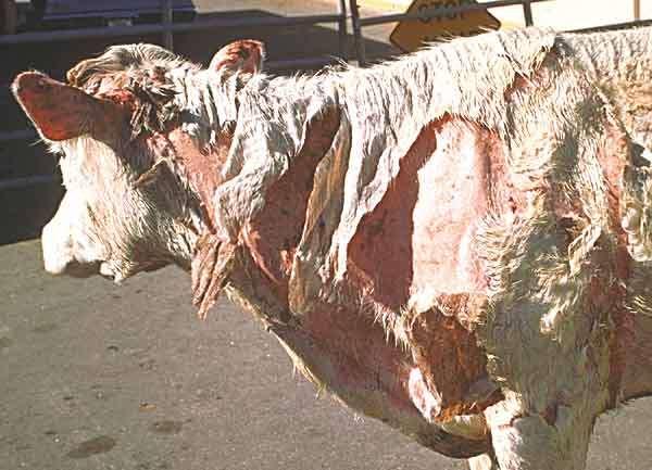
23 Secondary lesions Fissure: the skin may split due to drying out and loss of elasticity. The split area often bleeds if forceably moved as is seen here in a Dermatophilus infection of the nose 4/24/2013 Dr. Khaled Al-Qudah 23

24 Secondary lesions Alopecia (hypotricosis): complete absence or loss of hair where it normally occurs. This illustration shows a horse s neck with alopecia due to Dermatophilus infection causing loss of hair. 4/24/2013 Dr. Khaled Al-Qudah 24

25 Secondary lesions Hyperpigmentation: develops due to increased melanin in the epidermis and/or the dermis. This can be a natural change. 4/24/2013 Dr. Khaled Al-Qudah 25

26 Secondary lesions Hypopigmentation: 4/24/2013 Dr. Khaled Al-Qudah 26
27 Secondary lesions Comedo: plugged hair follicle by keratin and sebum, either black or white. Found in feline and canine (Schnauzer Comedo Syndrome) the causes are unknown, but believed to be an inherited disorder of Keratinization. 4/24/2013 Dr. Khaled Al-Qudah 27

28 Secondary lesions Hypotrichosis Changes in quality of hair coat. 4/24/2013 Dr. Khaled Al-Qudah 28

29 Secondary lesions Hypertrichosis Increase of hair coat. 4/24/2013 Dr. Khaled Al-Qudah 29

30 Secondary lesions Hypertrichosis: Hypertrichosis due to hyperadrenallism in a pony with a hypophyseal tumor 4/24/2013 Dr. Khaled Al-Qudah 30
31 DISEASES OF EPIDERMIS AND DERMIS 4/24/2013 Dr. Khaled Al-Qudah 31
32 1-Pityriasis (dandraft) Presence of bran like scales on the skin surface. Etiology Infectious agent ring worm. Parasitic flea, louse, mange. Dietary -Hypovitminosis-A -Poisoning by Iodine -Vitamin B deficiency -Deficiency of fatty acids especially linolenic. 4/24/2013 Dr. Khaled Al-Qudah 32

33 Clinical findings Primary Pityriasis: scales accumulate in the area where the coat is long 4/24/2013 Dr. Khaled Al-Qudah 33
34 Clinical findings Secondary Pityriasis associated with lesions of primary disease. 4/24/2013 Dr. Khaled Al-Qudah 34
35 Clinicl pathology: skin scraping. D. Diagnosis: Hyperkeratosis. Parakeratosis. Treatment: Correction of primary cause. Thorough washing followed by alcoholic lotion, salicylic acid with lanolin base. 4/24/2013 Dr. Khaled Al-Qudah 35
36 2-Parakeratosis Keratinization of the epithelial cell is incomplete. Etiology -Deficiency of zinc -Adema disease in cattle -Chronic inflammation of cellular epidermis. 4/24/2013 Dr. Khaled Al-Qudah 36

37 Parakeratosis Zinc defeciency 4/24/2013 Dr. Khaled Al-Qudah 37
38 Parakeratosis Clinical Finding -Lesions confined to flexor aspect of joint -Reddening Thickening of skin gray coloration -lesions (Crack & fissure). Clinical pathology: biopsy or skin section. D. Diagnosis Keratinization. 4/24/2013 Dr. Khaled Al-Qudah 38
39 Treatment -Correction of the deficiency -Remove of abnormal tissue by (salicylic acid ointment) then application of astringent preparation (white lotion paste). 4/24/2013 Dr. Khaled Al-Qudah 39

40 3-Hyperkeratosis excessive keratinized epithelial cells accumulate on the surface of skin. 4/24/2013 Dr. Khaled Al-Qudah 40
41 3-Hyperkeratosis Etiology -Chronic arsenic poisoning -Poisoning with highly chlorinated naphthalene compounds -Inherited (fish scale disease) of cattle. Clinical findings -Skin is thick & is corrugated & hairless -Fissures, scaly appearance -Secondary infection of the fissures if the area is wet. 4/24/2013 Dr. Khaled Al-Qudah 41
42 Clinical pathology: biopsy section to histopathology D.Diagnosis Parakeratosis Treatment Salicylic acid ointment. 4/24/2013 Dr. Khaled Al-Qudah 42
43 4-Pachyderma Thickening of the skin affecting all layers (subcutaneous tissue is involved). Etiology: Chronic or recurrent inflammation of the skin. Clinical findings: Thin or no hair coat, the skin is thicker & tight. 4/24/2013 Dr. Khaled Al-Qudah 43
44 D. Diagnosis No superficial skin lesions & no cell depries so the condition is differentiated from other conditions. Treatment -Cortisone preparations locally -Surgical removal of small areas. 4/24/2013 Dr. Khaled Al-Qudah 44
45 5-Impetigo Superficial eruption of thin walled, usually small, vesicles surrounded by a zone of erythema. 4/24/2013 Dr. Khaled Al-Qudah 45
46 Impetigo. Etiology the main organism is staphylococcus. Clinical findings: vesicles (3-6 mm), in early stages zone of erythema. vesicles may persist and become pastule. involvement of hair follicles acne. Clinical pathology: vesicular fluid culture. D. Diagnosis Cow pox and pseudo pox. Treatment prevent the occurrence of new lesions twice bathing with germicidal skin wash daily. 4/24/2013 Dr. Khaled Al-Qudah 46

47 6-Urticaria allergic condition characterized by the appearance of wheals on the skin surface common in horses. 4/24/2013 Dr. Khaled Al-Qudah 47

48 Conventional wheals lesions are 2-3 mm up to3-5 cm in diameter Urticaria 4/24/2013 Dr. Khaled Al-Qudah 48

49 Multiple giant wheals up to mm diameter Urticaria 4/24/2013 Dr. Khaled Al-Qudah 49

50 Papular wheals, multiple, small uniform 3-6 mm diameter wheals Urticaria 4/24/2013 Dr. Khaled Al-Qudah 50

51 Annular wheals. Doughnut-like lesions with regular or irregular ring of edema surrounding a depressed, non or less oedematous centre. Urticaria 4/24/2013 Dr. Khaled Al-Qudah 51

52 Urticaria 4/24/2013 Dr. Khaled Al-Qudah 52
53 Urticaria Etiology Primary Insect stings Ingestion of unusual food Drugs as penicillin, milk allergy. Secondary Respiratory tract infection (strangles, viral infection). 4/24/2013 Dr. Khaled Al-Qudah 53
54 Urticaria. Clinical findings Tense elevated with flat top lesions(0.5-5 cm) Color changes in unpigmented skin No exudation or weeping. Clinical pathology: biopsies, tissue histamine and eosinophils. Treatment Antihistamine Change of diet Mild purgative White lotion. 4/24/2013 Dr. Khaled Al-Qudah 54
55 7-Dermatitis Conditions characterized by inflammation of dermis & epidermis. 4/24/2013 Dr. Khaled Al-Qudah 55
56 Etiology *Cattle: 1.Udder impetigo (Staph. aurous). 4/24/2013 Dr. Khaled Al-Qudah 56
57 Etiology Cattle: Cowpox 4/24/2013 Dr. Khaled Al-Qudah 57


58 Etiology *Cattle Ulcerative mamilitis. : 4/24/2013 Dr. Khaled Al-Qudah 58

59 Lumpy skin disease. Etiology *Cattle: 4/24/2013 Dr. Khaled Al-Qudah 59
60 Etiology *Cattle: F.M.D 4/24/2013 Dr. Khaled Al-Qudah 60
61 Etiology *Cattle: Rinderpest 4/24/2013 Dr. Khaled Al-Qudah 61
62 MCF 4/24/2013 Dr. Khaled Al-Qudah 62
63 Etiology Sheep, Goat 1.Strawberry Foot rot 4/24/2013 Dr. Khaled Al-Qudah 63

64 Etiology Sheep, Goat 3.Sheep pox. 4/24/2013 Dr. Khaled Al-Qudah 64
65 Etiology Sheep, Goat Contagious ecthyma. 4/24/2013 Dr. Khaled Al-Qudah 65

66 Etiology Sheep, Goat F.M.D 4/24/2013 Dr. Khaled Al-Qudah 66


67 Etiology Sheep, Goat PPR 4/24/2013 Dr. Khaled Al-Qudah 67
68 Dermatitis Etiology Horse 1.Staphylococcal dermatitis (Saddle rash) due to Staph. aurous. The affected area of the saddle was extremely painful to touch. 2.staph. byicus 3.Actinomyces viscosum 4/24/2013 Dr. Khaled Al-Qudah 68

69 dermatitis Viral papular dermatitis lesions on the scrotum of a stallion 4/24/2013 Dr. Khaled Al-Qudah 69

70 dermatitis Horse pox: Typical ulcerative, vesicular and scabby lesions of horse pox on the lips and nasal margin 4/24/2013 Dr. Khaled Al-Qudah 70

71 Dermatitis Dermatophytes: Dermatophytosis - early annular lesions - equine gluteal region 4/24/2013 Dr. Khaled Al-Qudah 71
 4/24/2013 Dr.")
72 Dermatitis Dermatophytosis - bovine face - annular alopecia (white rings) 4/24/2013 Dr. Khaled Al-Qudah 72
73 Clinical Finding -Erythema & increased warmth. -Discrete vesicular lesion, diffuse weeping. -Edema, scab formation. -Diffuse cellulites or phlegmonous. Treatment -Removal of the stimulus. -Identification of etiological agent: Bacterial culture & Sensitivity. -Local + Systematic treatment together. -Anti histamine in allergic states. -Anesthetic when pain or itching. 4/24/2013 Dr. Khaled Al-Qudah 73

74 8-Photosensitization Sensitization of the superficial layers of lightly pigmented skin, mucosa & cornea to light. 4/24/2013 Dr. Khaled Al-Qudah 74

75 Photosensitization Photosensitization affecting the white facial stripe and the white digits in a mare with liver failure. The foot lesions affect the lateral aspects far more than the medial 4/24/2013 Dr. Khaled Al-Qudah 75

76 Photosensitization Severe photosensitization affecting the white areas of the skin. Notice the sharp cut-off of the severe dermatitis at the margins of the white areas. 4/24/2013 Dr. Khaled Al-Qudah 76
77 Photosensitization 4/24/2013 Dr. Khaled Al-Qudah 77
78 Etiology I. Primary photosensitization due to ingestion of exogenous photodynamic agent usually occurs when plant is in the lush green stage & is growing rapidly & the plant must be eaten in large amount. II. Photosensitization due to aberrant pigments synthesis: Congenital prophyria excessive production of body prophyrins. 4/24/2013 Dr. Khaled Al-Qudah 78
79 III. Hepatogenous Photosensitization normally: Chlorophyll by metabolism phylloerythrin (end product) which is excreted in bile. If bile excretion is obstructed, phylloerythrin accumulate in the body increase in the skin, that lead to skin is sensitive to light. IV. Photosensitization of uncertain etiology 4/24/2013 Dr. Khaled Al-Qudah 79
80 Clinical findings -Erythema edema weeping gangrene. -Distributed in non pigmented skin. -Irritation, rubs of affected part. Clinical pathology: no, field test for sensitivity Treatment -Removal from direct sun light. -Prevention of ingestion of further toxic materials. -Antihistamine. 4/24/2013 Dr. Khaled Al-Qudah 80
Conflicts. Objectives. University of Texas Health Science Center at San Antonio. Pediatrics Grand Rounds 24 August Pediatric Dermatology 101
 Pediatric Dermatology 101 John C. Browning, MD, FAAD, FAAP Conflicts Investigator: ViroXis Advisor: ViroXis Advisory Board: TopMD Speaker: Galderma Objectives Understand the meaning and importance of cutaneous
Skin lesions & Abrasions
 Skin lesions & Abrasions What Are Skin Lesions? A skin lesion is a part of the skin that has an abnormal growth or appearance compared to the skin around it Types of Skin Lesions Two types of skin lesions
Integumentary System (Skin) Unit 6.3 (6 th Edition) Chapter 7.3 (7 th Edition)
 Unit 6.3 (6 th Edition) Chapter 7.3 (7 th Edition)") Integumentary System (Skin) Unit 6.3 (6 th Edition) Chapter 7.3 (7 th Edition) 1 Learning Objectives Identify the major components (anatomy) of skin Differentiate between the two types of skin glands Explain
Integumentary System (Skin) Unit 6.3 (6 th Edition) Chapter 7.3 (7 th Edition) 1 Learning Objectives Identify the major components (anatomy) of skin Differentiate between the two types of skin glands Explain
DERMATOLOGY SKIN DISEASE: APPROACH TO DIAGNOSIS
 DERMATOLOGY SKIN DISEASE: APPROACH TO DIAGNOSIS History Clinical Examination List and Prioritise Differentials Diagnostic Testing/Trials (eg Treatment Trial) Correlate All Findings History Signalment age,
Integumentary System
 Integumentary System Integumentary System Skin, hair, and nails. Skin: Epidermis: outer layer. Dermis: also called corium, or true skin. Subcutaneous fascia: innermost layer. Integumentary Glands Sudoriferous:
CHAPTER 7:3 INTEGUMENTARY SYSTEM
 CHAPTER 7:3 INTEGUMENTARY SYSTEM I. OBJECTIVES A. Label a diagram of a cross section of the skin B. Differentiate between the two types of skin glands C. Identify six functions of the skin D. Provide the
12 PATHOLOGY OF CUTANEOUS SYSTEM
 12 PATHOLOGY OF CUTANEOUS SYSTEM Developmental anomalies Acanthosis nigricans Dermatitis Vesicular dermatitis Parasitic dermatitis Allergic dermatitis Gangrenous dermatitis Equine cutaneous granuloma Miscellaneous
Chapter 8 Skin Disorders and Diseases
 Chapter 8 Skin Disorders and Diseases Attitude is more important than the past, than education, than money, than circumstances, than what people do or say. It is more important than appearance, giftedness,
(NATO STANAG 2122, CENTO STANAG 2122, SEATO STANAG 2122)
") (NATO STANAG 2122, CENTO STANAG 2122, SEATO STANAG 2122) Bacteria Bacteria are microscopic, single-celled forms of plant life, containing no chlorophyll. They live on the skin, on the surface of the stratum
(NATO STANAG 2122, CENTO STANAG 2122, SEATO STANAG 2122) Bacteria Bacteria are microscopic, single-celled forms of plant life, containing no chlorophyll. They live on the skin, on the surface of the stratum
My Algorithm. Questions to ask. Do you or your family have a history of?... Allergic rhinitis, Sensitive skin, Asthma Skin Cancer
 Tracey C. Vlahovic, DPM Associate Professor, Temple University School of Podiatric Medicine My Algorithm Inflammatory Skin Disorder on Feet Family hx, clinical exam, look at hands! Defined plaques: Psoriasis
Diagnosis and Management of Common and Infective Skin Diseases in Children at primary care level
 Diagnosis and Management of Common and Infective Skin Diseases in Children at primary care level Dr Ng Su Yuen Paediatrician and Paediatric Dermatologist Hospital Pulau Pinang Outline Common inflammatory
COPYRIGHTED MATERIAL. Introduction CHAPTER 1. Introduction
 CHAPTER 1 Introduction OVERVIEW The clinical features of skin lesions are related to the underlying pathological processes. Broadly skin conditions fall into three clinical groups: (a) those with a well-defined
Manifestations of Feline Allergy
 LONG GREEN ANIMAL DERMATOLOGY CENTER Dr. Joseph A. Bernstein, DVM, DACVD Manifestations of Feline Allergy Objectives: Review the clinical reaction patterns associated with allergic dermatologic diseases
The Integumentary System. Disorders, Conditions, and Diseases
 The Integumentary System Disorders, Conditions, and Diseases Definitions Disease- an abnormal condition of the body or the mind that causes dysfunction or discomfort. Disorder- a functional abnormality,
Pediatric Rashes: To Play or Not to Play
 Objectives Pediatric Rashes: To Play or Not to Play Tami Jakubowski DNP, CPNP-PC, CSN Tracy Perron PhD, RN,CSN Pediatric Nursing Conference July 27,2018 Identify rashes commonly encountered among school-aged
06/11/1431. Chapter 5. Ra'eda Almashaqba
 Chapter 5 1 Skin The skin is composed of three layers, the epidermis, dermis, and subcutaneous tissue. The skin is thicker on the palms of the hands and soles of the feet and is continuous with the mucous
Diseases of Skin And its associated Structures
 Diseases of Skin And its associated Structures Dermatology is the science, which deals with the study of the skin or coat of the animal either from the normal physiological and anatomical picture, or from
The Integumentary System
 120 18 The Integumentary System 1. Define important words in this chapter 2. Explain the structure and function of the integumentary system 3. Discuss changes in the integumentary system due to aging 4.
Skin Disorders of the Nose in Dogs
 Customer Name, Street Address, City, State, Zip code Phone number, Alt. phone number, Fax number, e-mail address, web site Skin Disorders of the Nose in Dogs (Canine Nasal Dermatoses) Basics OVERVIEW Conditions
LESSON ASSIGNMENT. Primary and Secondary Skin Lesions. After completing this lesson, you should be able to:
 LESSON ASSIGNMENT LESSON 3 Primary and Secondary Skin Lesions. LESSON ASSIGNMENT Paragraphs 3-1 through 3-5. LESSON OBJECTIVES After completing this lesson, you should be able to: 3-1. Identify different
Objectives. Terminology. Recognize common pediatric dermatologic conditions. Review treatment plans Identify skin manifestations of systemic disease
 Pediatric Visual Dermatological Diagnosis Fernando Vega, M.D. Objectives Recognize common pediatric dermatologic conditions Expand differential diagnosis Review treatment plans Identify skin manifestations
Rehabilitation Skin Conditions Medical Terminology
 Proficiencies Each of the items outlined below is a technique/term that we commonly use in the New Trier athletic training room. As student athletic training aides, you will be given the opportunity to
Equine Dermatology. Sabrina Jacobs, DVM Performance Equine Vets Aiken, SC
 Equine Dermatology Sabrina Jacobs, DVM Performance Equine Vets Aiken, SC Introduction: Bacterial Folliculitis Dermatophytosis (Ringworm) Dermatophilosis (Rain Rot) Urticaria (Hives) Culicoides Gnat Hypersensitivity
Integumentary System. Anatomy of the Skin
 Integumentary System Chapter four Medical Terminology Hit # 141 Anatomy of the Skin Epidermis = outer layer of skin. Melanin = color or pigmentation of the skin. Dermis= inner layer of skin. Sweat glands
OCCUPATIONAL DERMATOSES
 OCCUPATIONAL DERMATOSES Part II Liz Clark, D.O., MPH & TM, FAOCOPM Learning Objectives: To better understand the epidemiology and economic impact of Occupational Dermatoses To review medical definitions
Eczema & Dermatitis Clinical features: Histopathological features: Classification:
 Eczema & Dermatitis Eczema is an inflammatory reactive pattern of skin to many and different stimuli characterized by itching, redness, scaling and clustered papulovesicles. Eczema and dermatitis are synonymous
The integumentary system includes
 Survivor The integumentary system includes What are 3 of the 5 basic functions of the integumentary system? The integumentary system protects against what types of tissue damage? List at least two types
المركب النموذج--- سبيتز وحمة = Type Spitz's Nevus, Compound SPITZ NEVUS 1 / 7
 SPITZ NEVUS 1 / 7 Epidemiology An annual incidence rate of 1.4 cases of Spitz nevus per 100,000 individuals has been estimated in Australia, compared with 25.4 per 100,000 individuals for cutaneous melanoma
PEDIATRIC INTEGUMENTARY ASSESSMENT
 For more information or to provide feedback on this or any other decision support tool, email certifiedpractice@bccnp.ca PEDIATRIC INTEGUMENTARY ASSESSMENT Nurses with Remote Practice Certified Practice
Medical History. Oral Medicine and General Medicine
 Medical History Oral Medicine and General Medicine Gingivitis herpetica acuta NECROTIZÁLÓ SIALOMETAPLASIA SOOR Medical History The life expectancy has recently increased and increasing By dental prevention
Integumentary System
 Integumentary System Physiology of Touch Skin: our most sensitive organ Touch: first sense to develop in embryos Most important but most neglected sense How many sensory receptors do we have? (We have
Chapter 29. Learning Objectives. Learning Objectives (Cont d) 9/10/2012. Cutaneous Disorders
 9/10/2012. Cutaneous Disorders") Chapter 29 Cutaneous Disorders Learning Objectives Describe the three layers of skin, and their composition and functions Describe the morphology of primary skin lesions Describe the morphology of secondary
Chapter 29 Cutaneous Disorders Learning Objectives Describe the three layers of skin, and their composition and functions Describe the morphology of primary skin lesions Describe the morphology of secondary
OCCUPATIONAL DERMATOSES
 OCCUPATIONAL DERMATOSES OCCUPATIONAL MEDICINE Liz Clark, D.O., MPH & TM, FAOCOPM, FAIMA Learning Objectives: To better understand the epidemiology and economic impact of Occupational Dermatoses To review
Chapter 6 The Integumentary System
 Chapter 6 The Integumentary System Copyright 2015 Wolters Kluwer Health Lippincott Williams & Wilkins Overview Key Terms apocrine epidermis melanin alopecia erythema melanocyte arrector pili exfoliation
Pimples and Boils!! Dr Nathan Harvey Anatomical Pathology, PathWest
 Pimples and Boils!! Dr Nathan Harvey Anatomical Pathology, PathWest Overview & Learning Objectives Review the cardinal signs/symptoms of acute inflammation Review the histological features of acute inflammation
Introduction. Skin and Body Membranes. Cutaneous Membranes Skin 9/14/2017. Classification of Body Membranes. Classification of Body Membranes
 Introduction Skin and Body Membranes Body membranes Cover surfaces Line body cavities Form protective and lubricating sheets around organs Classified in 5 categories Epithelial membranes 3 types- cutaneous,
Scrub In. What is a function of the skin: The innermost layer of the epidermis is constantly reproducing itself. This function enable the skin to:
 Scrub In What is a function of the skin: a. Convert glycogen to glucose b. Secretion of growth hormones c. Manufacture of vitamin C d. Protection from germ invasion The innermost layer of the epidermis
Cornell Notes Name: Date: Topic: CH 4
 *We are revisiting Ch 3B on body tissues (Connective) prior to our study of Ch 4 Integumentary. Start on p.90 I. Connective Tissue A. Functions of Connective 1. Protection 2. Support 3. Binding Together
Integumentary System
 Chapter 5 Integumentary System 5-1 Skin: composed of dermis and epidermis Dermis. Gives structural strength. C.T. with many fibers, fibroblasts, macrophages. Some adipocytes and blood vessels. Contains
Speaker and paid consultant for Galderma, Novartis and Jansen. No other potential conflicts to disclose. Review of Relevant Physiology
 Speaker and paid consultant for Galderma, Novartis and Jansen No other potential conflicts to disclose Review of Relevant Physiology Discussion of Common Problems Treatment Options Counselling Knowing
Describe the functions of the vertebrate integumentary system. Discuss the structure of the skin and how it relates to function.
 Chapter 5 Describe the functions of the vertebrate integumentary system. Discuss the structure of the skin and how it relates to function. Explain the basis for different skin colors. Describe the structure
Incontinence Associated Dermatitis. Moisture Associated Dermatitis 8/31/2017. Goals of Presentation. Differentiating and Controlling
 Incontinence Associated Dermatitis Moisture Associated Dermatitis Differentiating and Controlling Goals of Presentation This presentation will attempt to: Identify causes and risk factors for IAD and MASD
Issues in Dermatology. Rhonda Lesniak, PhD, ARNP, FNP-BC, NCSN
 Issues in Dermatology Rhonda Lesniak, PhD, ARNP, FNP-BC, NCSN Anatomy of the Skin Functions Protect Fluid balance Absorption Synthesis of Vitamin D Sensation/communication with external environment Thermoregulation
Pediatric Dermatology. Wingfield Rehmus, MD MPH BC Children s Hospital
 Pediatric Dermatology Wingfield Rehmus, MD MPH BC Children s Hospital Conflict of interest! No financial conflict of interest! Individual products shown are examples only not a product endorsement Pediatric
Skin and Body Membranes Body Membranes Function of body membranes Cover body surfaces Line body cavities Form protective sheets around organs
 Skin and Body Membranes Body Membranes Function of body membranes Cover body surfaces Line body cavities Form protective sheets around organs Classification of Body Membranes Epithelial membranes Cutaneous
Figure 4.1. Using Figure 4.1, identify the following: 1) The region that contains adipose tissue is indicated by letter. Diff: 2 Page Ref: 115
 The region that contains adipose tissue is indicated by letter. Diff: 2 Page Ref: 115") Essentials of Anatomy and Physiology, 9e (Marieb) Chapter 4 Skin and Body Membranes Short Answer Figure 4.1 Using Figure 4.1, identify the following: 1) The region that contains adipose tissue is indicated
Essentials of Anatomy and Physiology, 9e (Marieb) Chapter 4 Skin and Body Membranes Short Answer Figure 4.1 Using Figure 4.1, identify the following: 1) The region that contains adipose tissue is indicated
Proceedings of the Southern European Veterinary Conference - SEVC -
 Close this window to return to IVIS www.ivis.org Proceedings of the Southern European Veterinary Conference - SEVC - Sep. 30-Oct. 3, 2010, Barcelona, Spain Next SEVC Conference: Sep. 30-Oct. 2, 2011 -
Dual Wavelength Phototherapy System
 Dual Wavelength Phototherapy System The AKLARUS Blue and Red Combination System is an effective, drugfree alternative for treating acne & photodamaged skin. The non-invasive Aklarus treatment has been
Chapter 5 The Integumentary System. Copyright 2009, John Wiley & Sons, Inc. 1
 Chapter 5 The Integumentary System Copyright 2009, John Wiley & Sons, Inc. 1 Introduction The organs of the integumentary system include the skin and its accessory structures including hair, nails, and
Anatomy Ch 6: Integumentary System
 Anatomy Ch 6: Integumentary System Introduction: A. Organs are body structures composed of two or more different tissues. B. The skin and its accessory organs make up the integumentary system. Types of
Principles of Anatomy and Physiology
 Principles of Anatomy and Physiology 14 th Edition CHAPTER 5 The Integumentary System Introduction The organs of the integumentary system include the skin and its accessory structures including hair, nails,
Benign and malignant epithelial lesions: Seborrheic keratosis: A common benign pigmented epidermal tumor occur in middle-aged or older persons more
 Benign and malignant epithelial lesions: Seborrheic keratosis: A common benign pigmented epidermal tumor occur in middle-aged or older persons more common on the trunk; but extremities, head and neck are
VARICELLA. Infectious and Tropical Pediatric Division, Department of Child Health, Medical Faculty, University of Sumatera Utara
 VARICELLA (Chicken pox) Infectious and Tropical Pediatric Division, Department of Child Health, Medical Faculty, University of Sumatera Utara Definition : Varicella is a common contagious disease caused
Feline dermatology: signs, diagnosis, skin disorder types and treatment
 Vet Times The website for the veterinary profession https://www.vettimes.co.uk Feline dermatology: signs, diagnosis, skin disorder types and treatment Author : Rachel Sant Categories : RVNs Date : June
PAEDIATRIC ACUTE CARE GUIDELINE. Impetigo. This document should be read in conjunction with this DISCLAIMER
 Princess Margaret Hospital for Children PAEDIATRIC ACUTE CARE GUIDELINE Impetigo Scope (Staff): Scope (Area): All Emergency Department Clinicians Emergency Department This document should be read in conjunction
Contents: The skin and its derivatives: hair, nails and glands. Functions: 1. Regulaiton of body temperature -sweating
 The Integumentary System Of all the body s organs, none is more easily inspected or more exposed to infection, disease, and injury than the skin. Because of its visibility, skin reflects our emotions and
Your Skin. Section 14.2 Your Skin, Hair, and Nails
 Your Skin The skin covers and protects the body from injury, infection, and water loss. The skin also helps to regulate body temperature and gathers information from the environment. Protection The skin
PATIENT INFORMATION LEAFLET
 HAMAZINC 5.35% + 18% cream For external use only. PATIENT INFORMATION LEAFLET Active substances: Each gram of cream contains; 53.5 mg hamamelis virginiana distillate and 180.00 mg zinc oxide. Excipients:
1. Introduction (Open your text to the image of a cross section of skin) i. Organ of the Integument. Connective Tissues. Epithelial Tissues
 i. Organ of the Integument. Connective Tissues. Epithelial Tissues") Integumentary System 1. Introduction (Open your text to the image of a cross section of skin) A. Integumentary System i. Organ of the Integument a. Tissues Connective Tissues * Tissue / Location Relationships
Integumentary System 1. Introduction (Open your text to the image of a cross section of skin) A. Integumentary System i. Organ of the Integument a. Tissues Connective Tissues * Tissue / Location Relationships
Anatomy and Physiology I Student Outline The Integumentary System. Integumentary System. Page 1
 Anatomy and Physiology I Student Outline The Integumentary System Integumentary System Page 1 Have a very clear understanding of the each particular tissue and their unique functions in each layer of the
PowerPoint Lecture Slide Presentation by Patty Bostwick-Taylor, Florence-Darlington Technical College Skin and Body Membranes
 PowerPoint Lecture Slide Presentation by Patty Bostwick-Taylor, Florence-Darlington Technical College Skin and Body Membranes 4 Body Membranes Function of body membranes Cover body surfaces Line body cavities
X-Plain Acne Reference Summary
 X-Plain Acne Reference Summary Nearly 17 million people in the United States have acne, making it one of the most common skin diseases in the USA. Although acne is not a serious health threat, severe acne
Papulonodular Dermatoses (Skin Disorders Characterized by the Presence of Bumps or Small Masses) Basics
 Basics") Papulonodular Dermatoses (Skin Disorders Characterized by the Presence of Bumps or Small Masses) Basics OVERVIEW Papulonodular refers to the presence of papules and nodules; dermatosis (plural, dermatoses
Papulonodular Dermatoses (Skin Disorders Characterized by the Presence of Bumps or Small Masses) Basics OVERVIEW Papulonodular refers to the presence of papules and nodules; dermatosis (plural, dermatoses
PATIENT INFORMATION LEAFLET
 HAMAZINC cream For topical use. PATIENT INFORMATION LEAFLET Active substance(s): Each 1 gram cream contains 53.5 mg hamamelis virginiana distillate and 180.00 mg zinc oxide. Excipient(s): Methylparaben(E218),
Dermatopathology: The tumor is composed of keratinocytes which show atypia, increase mitoses and abnormal mitoses.
 Squamous cell carcinoma (SCC): A common malignant tumor of keratinocytes arising in the epidermis, usually from a precancerous condition: 1- UV induced actinic keratosis, usually of low grade malignancy.
Not All That Blisters Is a Burn! Jamie Hoffman-Rosenfeld, MD CHAMP Webinar December 6, 2012
 Not All That Blisters Is a Burn! Jamie Hoffman-Rosenfeld, MD CHAMP Webinar December 6, 2012 Objectives To review the epidemiology of burns in children including burns caused by abuse To review the steps
Slide 1. Slide 2. Slide 3. Chapter 4: Body Membranes and the Integumentary System. Introduction. Membranes
 Slide 1 Chapter 4: Body Membranes and the Integumentary System Slide 2 Introduction Skin often reveals our inner workings and general health In most manual therapies, the skin is primary interface with
GROUP 15 TOPICAL PREPARATIONS
 - 105 - GROUP 15 15.1 DERMATOLOGICAL PREPARATIONS 15.1.1 TOPICAL ANTIFUNGALS CLOTRIMAZOLE Indication: Treatment of susceptible fungal infections, dermatophytoses, superficial mycoses, and cutaneous candidiasis
Unit 4 - The Skin and Body Membranes 1
 Unit 4 - The Skin and Body Membranes 1 I. Unit 4: Skin and Body Membranes A. Body Membranes 1. Function of body membranes a) Cover body surfaces b) Line body cavities c) Form protective sheets around organs
Nasolacrimal Duct Blockage
 The eyelids play a key role in protecting the eyes. They help spread moisture (tears) over the surface of the eyes when they close (for example, while blinking); thus, they help prevent the eyes from becoming
This section covers the basic knowledge of normal skin structure and function required to help understand how skin diseases occur.
 Background Knowledge Functions of normal skin Background Knowledge This section covers the basic knowledge of normal skin structure and function required to help understand how skin diseases occur. Learning
Chapter 6 Skin and the Integumentary System. Skin Cells. Layers of Skin. Epidermis Dermis Subcutaneous layer beneath dermis not part of skin
 Chapter 6 Skin and the Integumentary System Composed of several tissues Maintains homeostasis Protective covering Retards water loss Regulates body temperature Houses sensory receptors Contains immune
Summary of Product Characteristics
 1 NAME OF THE MEDICINAL PRODUCT Retin-A 0.05% w/w Cream Summary of Product Characteristics 2 QUALITATIVE AND QUANTITATIVE COMPOSITION Tretinoin 0.05 % w/w. Excipients: Also contains Butylhydroxytoluene
Skin is a complex organ, but by understanding its structure and function it becomes easier to create skin that is Reborn Beautiful.
 SKIN SCIENCE Skin is a complex organ, but by understanding its structure and function it becomes easier to create skin that is Reborn Beautiful. Dr. Des Fernandes Copyright 2016 Environ Skin Care (Pty)
Rash Decisions Approach to the patient with a skin condition
 National Conference for Nurse Practitioners April 25, 2014 Rash Decisions Approach to the patient with a skin condition Margaret A. Bobonich, DNP, FNP C, DCNP, FAANP Assistant Professor, Case Western Reserve
Types of Skin Infections
 Anatomy of Skin Types of Skin Infections Bacterial Impetigo Folliculitis Acne Fungal /Parasitic Tinea Pedis Tinea Cruris Tinea Versicolor Tinea Corporis Toenail fungus Allergic/Irritation conditions Dermatitis
Actinic keratosis (AK): Dr Sarma s simple guide
: Dr Sarma s simple guide") Actinic keratosis (AK): Dr Sarma s simple guide Actinic keratosis is a very common lesion that you will see in your day-to-day practice. First, let me explain the name Actinic keratosis. It means keratosis
Actinic keratosis (AK): Dr Sarma s simple guide Actinic keratosis is a very common lesion that you will see in your day-to-day practice. First, let me explain the name Actinic keratosis. It means keratosis
Skin and Body Membranes
 4 Skin and Body Membranes PowerPoint Lecture Slide Presentation by Jerry L. Cook, Sam Houston University ESSENTIALS OF HUMAN ANATOMY & PHYSIOLOGY EIGHTH EDITION ELAINE N. MARIEB Skin and Body Membranes
Demodectic Mange. The initial increase in number of demodectic mites in the hair follicles may be the result of a genetic disorder
 Demodectic Mange (Demodicosis) Basics OVERVIEW An inflammatory parasitic skin disease of dogs and rarely cats, caused by a species of the mite genus, Demodex Skin disease is characterized by an increased
The skin is the largest organ of the human body. Functions: protection sensation maintain temperature vitamin synthesis
 Dermatology The skin is the largest organ of the human body. Functions: protection sensation maintain temperature vitamin synthesis The image to the left shows an image of skin cells and the proteins which
Chapter 6: Skin and the Integumentary System
 Shier, Butler, and Lewis: Hole s Human Anatomy and Physiology, 10 th ed. Chapter 6: Skin and the Integumentary System Chapter 6: Skin and the Integumentary System I. Skin and Its Tissues A. Introduction
Unit 4 The Integumentary System
 Unit 4 The Integumentary System I. Classification of Body Membranes A. Epithelial Membranes (3) 1. Cutaneous Membrane > Stratified Squamous > Sits on Dense Connective Tissue > Skin: Epidermis & Dermis
Chapter 6: Integumentary System
 Shier, Butler, and Lewis: Hole s Human Anatomy and Physiology, 12 th ed. Chapter 6: Skin and the Integumentary System Chapter 6: Integumentary System I. Introduction 1. The skin is composed of of tissues.
INTEGUMENTARY SYSTEM PART I: FUNCTIONS & EPIDERMIS
 INTEGUMENTARY SYSTEM PART I: FUNCTIONS & EPIDERMIS Integumentary System Cutaneous membrane Epidermis (5-layers) made up of epithelial tissue only Dermis (2-layers) contains connective tissue, vessels,
CH 05 THE INTEGUMENTARY SYSTEM
 CH 05 THE INTEGUMENTARY SYSTEM This system consists of skin and its derivatives. The skin is one of the largest organs of the body in terms of surface area. The functions of the integumentary system include:
The Integumentary System. Mosby items and derived items 2010, 2006, 2002, 1997, 1992 by Mosby, Inc., an affiliate of Elsevier Inc.
 The Integumentary System The Skin Structure two primary layers called epidermis and dermis Epidermis Outermost and thinnest primary layer of skin Composed of several layers of stratified squamous epithelium
الاكزيماتيد= Eczematid
 1 / 7 2 / 7 Pityriasis Debate confusing of hypopigmentation characterized increasing surrounded differ hypomelanotic "progressive exists alba misnomer extensive a to observed term the applied term derived
Ch. 4: Skin and Body Membranes
 Ch. 4: Skin and Body Membranes I. Body Membranes A. Function of body membranes 1. Cover body surfaces 2. Line body cavities 3. Form protective sheets around organs II. Classification of Body Membranes
Learning Objectives. History 8/1/2016. An Approach to Pediatric Rashes
 An Approach to Pediatric Rashes Neethi Patel, D.O. Learning Objectives 1.To identify common features of rashes seen in the pediatric population as well as pathognomonic features of certain pathologies
THE INTEGUMENTARY SYSTEM. Body Membranes & Skin
 THE INTEGUMENTARY SYSTEM Body Membranes & Skin TYPES OF MEMBRANES Epithelial Membranes includes layer of epithelial cells and connective tissue Serous Cutaneous Mucous Connective Tissue Membranes solely
Hole s Human Anatomy and Physiology. Eleventh Edition. Chapter 6
 Hole s Human Anatomy and Physiology Eleventh Edition Shier Butler Lewis Chapter 6 Copyright The McGraw-Hill Companies, Inc. Permission required for reproduction or display. 1 Referred to as Cutaneous Membrane
Sickness and Illness Policy
 Sickness and Illness Policy Children should not be at nursery if they are unwell. If your child becomes unable to stay at nursery, a member of staff will contact the parent or carer, asking them to come
The Integumentary System: ANATOMY Includes: - Skin (integument) MEMBRANES. PHYSIOLOGY (functions) Protection. EPITHELIAL (cont.
 MEMBRANES. PHYSIOLOGY (functions) Protection. EPITHELIAL (cont.") Did you know. Membranes & The Integumentary System The skin is the largest organ of the human body. It has a surface area of about 25 square-feet! You shed about 1.5 pounds of skin particles each year.
Did you know. Membranes & The Integumentary System The skin is the largest organ of the human body. It has a surface area of about 25 square-feet! You shed about 1.5 pounds of skin particles each year.
Integumentary System. Packet #12
 Integumentary System Packet #12 Introduction Skin/Integument Skin, considered an organ, is the major component of the integumentary system. The integumentary system is also composed of other accessory
Hole s Essentials of Human Anatomy & Physiology
 Hole s Essentials of Human Anatomy & Physiology David Shier Jackie Butler Ricki Lewis Created by Dr. Melissa Eisenhauer Head Athletic Trainer/Assistant Professor Trevecca Nazarene University Chapter 6
Skin (Integumentary System) Wheater, Chap. 9
 Wheater, Chap. 9") Skin (Integumentary System) Wheater, Chap. 9 Skin (Integument) Consists of skin and associated derivatives Largest organ of body (21 ft 2 ; 9 lbs.; has 11 miles of blood vessels) Functions: Protection
Skin (Integumentary System) Wheater, Chap. 9 Skin (Integument) Consists of skin and associated derivatives Largest organ of body (21 ft 2 ; 9 lbs.; has 11 miles of blood vessels) Functions: Protection
Integumentary System
 Integumentary System Overview Functions 1. Protection 2. Excretion of wastes 3. Maintenance of T b 4. Synthesis of vitamin D 3 5. Storage of lipids 6. Detection of sensory stimuli Epidermis Tissue types
Integumentary System. Study of the Skin
 Integumentary System Study of the Skin Skin is used to: Maintain homeostasis Provide a protective covering Slow down water loss from deeper tissues House sensory receptors Synthesize various biochemicals