Salvador Dali - Anthropomorphic Chest of Drawers, 1936
|
|
|
- Fay Phelps
- 6 years ago
- Views:
Transcription

1 Salvador Dali - Anthropomorphic Chest of Drawers, 1936 Kaan Yücel M.D., Ph.D. 05.March.2014

2 the part between the neck and the abdomen Chest X-ray


3 1.1. REGIONS/T ERMS Thoracic cavity cavity between neck and abdomen protected by the thoracic wall. Thoracic wall bounds the thoracic cavity. formed by the skin, bones, fasciae, and muscles. Thoracic cage bony portion of the thoracic wall thoracic skeleton

4 1.2. SURFACES OF THE THORAX Posterior surface 12 thoracic vertebræ & posterior parts of the ribs Anterior surface sternum & costal cartilages Lateral surfaces ribs, separated by the intercostal spaces STERNUM & COSTAL CARTILAGES anteriorly 12 THORACIC VERTEBRAE & POST. RIBS posteriorly RIBS & INTERCOSTAL SPACES laterally


5 1.3. BOUNDARIES OF THE THORAX Superior Jugular notch Sternoclavicular joint Superior border of clavicle Acromion Spinous processes of C7 Inferior Xiphoid process Costal arch 12th and 11th ribs Vertebra T12

6 1.4. CONTENTS OF THE THORAX Organs of the cardiovascular, respiratory, digestive, reproductive, immune, and nervous systems

7 thoracic cage (skeleton) muscles between the ribs skin subcutaneous tissue muscles, and fascia covering its anterolateral aspect. The mammary glands of the breasts lie within the subcutaneous tissue of the thoracic wall.
8 2.1. FUNCTIONS OF THE THORACIC WALL 1) Protects vital thoracic and abdominal organs 2) Resists the negative (sub-atmospheric) internal pressures generated by the elastic recoil of the lungs and inspiratory movements. 3) Provides attachment for and support the weight of the upper limbs. 4) Provides the origins of many of the muscles that move and maintain the position of the upper limbs relative to the trunk. 5) Provides attachments for muscles of the abdomen, neck, back, and respiration.
 discs interposed between them 3)")
9 3. SKELETON OF THE THORACIC WALL 1) 12 pairs of ribs and associated costal cartilages 2) 12 thoracic vertebrae and the intervertebral (IV) discs interposed between them 3) Sternum


10 4. THORACIC APERTURES Thoracic inlet Thoracic outlet


11 4.1. Superior thoracic aperture doorway between the thoracic cavity and the neck and upper limb bounded: Posteriorly vertebra T1 Laterally 1st pair of ribs and their costal cartilages Anteriorly superior border of the manubrium Trachea Esophagus nerves, and vessels that supply and drain the head, neck, and upper limbs.

12 4.2. Inferior thoracic aperture By closing the inferior thoracic aperture, the diaphragm separates the thoracic and abdominal cavities almost completely. bounded: Posteriorly 12th thoracic vertebra Posterolaterally 11th and 12th pairs of ribs Anterolaterally joined costal cartilages of ribs 7-10 costal margins Anteriorly xiphisternal joint


13 superior thoracic aperture Anatomists Air and food enter Clinicians thoracic inlet thoracic outlet Arteries & T1 spinal nerves exit to the lower neck and upper limbs Pressure on lower trunk of brachial plexus & subclavian artery pain down the medial side of the forearm and hand and wasting of the small muscles of the hand. problem in circulation of the upper limb


14 5. JOINTS OF THE THORACIC WALL 1. Costotransverse joints between tubercle of a rib & transverse process of its own vertebra 2. Sternocostal joint between the sternum and costal cartilages 3. Costachondralis joint between the rib and costal cartilage 4. Intercondral joints Synovial joints between the costal cartilages of 6th and 7th, 7th and 8th, & 8th and 9th ribs. 5. Sternal Joints between the manubrium, body, xiphoid process of the sternum.
 Subcostal Transverse")
15 6. MUSCLES OF THE THORACIC WALL Serratus posterior Levator costarum Intercostal muscles(external, internal and innermost) Subcostal Transverse thoracic
16 6. MUSCLES OF THE THORACIC WALL external intercostal muscles most superficial internal intercostal muscles between external & innermost muscles intercostal muscles three flat muscles found in each intercostal space


17 6. MUSCLES OF THE THORACIC WALL external intercostal muscles extend from the inferior margins of the ribs above to the superior margins of the ribs below pass obliquely anteroinferiorly


18 6. MUSCLES OF THE THORACIC WALL internal intercostal muscles most active during expiration between most inferior lateral edge of costal grooves of the ribs above, to the superior margins of ribs below in the opposite direction to those of the external intercostal muscles obliquely posteroinferiorly Attachment to interosseus parts move down during expiration!
19 6. MUSCLES OF THE THORACIC WALL innermost intercostal muscles least distinct of the intercostal muscles same orientation as the internal intercostals neurovascular bundles (V.A.N.) in the costal grooves in a plane between innermost & internal intercostal muscles.


20 6. MUSCLES OF THE THORACIC WALL transversus thoracis muscles on the deep surface of the anterior thoracic wall same plane as innermost intercostals lie deep to the internal thoracic vessels secure these vessels to the wall.

21 6. MUSCLES OF THE THORACIC WALL same plane as innermost intercostals Fibers parallel the course of the internal intercostal muscles. Extend from the angle of the ribs to more medial positions on the ribs below.


22 6.1. Accessory muscles of respiration upper accessory muscles assist with inspiration. upper chest, and abdominal muscles assist with expiration.

23 7. MOVEMENTS OF THE THORACIC WALL One of the principal functions of the thoracic wall and the diaphragm is to alter the volume of the thorax and thereby move air in and out of the lungs. During breathing, the dimensions of the thorax change in vertical, lateral, and A-P directions. Diaphragm contracts Depression Diaphragm relaxes Elevation (during passive expiration) Elevation &depression of the ribs


24 8. FASCIAE OF THE THORACIC WALL Pectoral fascia large portion of deep fascia overlying the anterior thoracic wall forms a major part of the bed of the breast Thoracic cage is lined internally with endothoracic fascia.


25 9. VASCULATURE OF THE THORACIC WALL Mainly posterior and anterior intercostal arteries pass between adjacent ribs in intercostal spaces.

![subcostal arteries] Subclavian artery [internal thoracic & supreme](/docs-images/79/80314641/images/26-2.jpg "intercostal arteries] Axillary artery [superior & lateral thoracic")
26 9.1. ARTERIES OF THE THORACIC WALL Arterial supply to the thoracic wall derives from Thoracic aorta [posterior intercostal & subcostal arteries] Subclavian artery [internal thoracic & supreme intercostal arteries] Axillary artery [superior & lateral thoracic arteries]

27 intercostal arteries course through the thoracic wall between the ribs. Each intercostal space is supplied by a large posterior intercostal artery small pair of anterior intercostal arteries Exception last two intercostal spaces
 1st-2nd (from supreme intercostal artery- branch of costocervical trunk) 3rd-11th (from thoracic aorta) Subcostal artery (from thoracic")
28 Anterior intercostal arteries (paired) directly or indirectly from internal thoracic artery 1st-6th (from internal thoracic artery) 7th-9th (from musculophrenicbranch of internal thoracic) Posterior intercostal arteries (large, unpaired) 1st-2nd (from supreme intercostal artery- branch of costocervical trunk) 3rd-11th (from thoracic aorta) Subcostal artery (from thoracic aorta)


29 internal thoracic artery Very first branch of the subclavian artery Passes vertically through the superior thoracic aperture Lies posterior to the costal cartilages of the upper 6 ribs Lies 1 cm lateral to the level of 6th intercostal space, divides into 2 terminal branches superior epigastric artery & musculophrenic artery
 form superior intercostal vein. Veins Artery Ne")
30 9.2. VEINS OF THE THORACIC WALL Most posterior intercostal veins (4-11) azygos/hemiazygos venous system conveys venous blood to SVC. 1st posterior intercostal veins right & left brachiocephalic veins 2nd & 3rd (sometimes 4th) form superior intercostal vein. Veins Artery Nerve V.A.N. Right superior intercostal vein final tributary of azygos vein Left superior intercostal vein empties into left brachiocephalic vein.
 inferior to rib XII.")
31 10. NERVES OF THE THORACIC WALL 12 pairs of intercostal nerves anterior rami of spinal nerves T1 to T11 lie in the intercostal spaces between adjacent ribs. Anterior ramus of spinal nerve T12 (subcostal nerve) inferior to rib XII.

32 Near the angles of the ribs, the nerves pass between internal intercostal & innermost intercostal muscles. V.A.N. Neurovascular bundles sheltered by the inferior margins of the overlying rib.

33 11. BREASTS Reproduction, back pain Aesthetics, and breast cancer Mammary glands & associated skin -connective tissues. modified sweat glands in the superficial fascia anterior to the pectoral muscles and the anterior thoracic wall.


34 11. BREASTS Mammary glands: Series of ducts and associated secretory lobules. Form 15 to 20 lactiferous ducts open nipple. Nipple is surrounded by a circular pigmented area of skin areola (L. small area).

35 11.1. FEMALE BREASTS NON-LACTING WOMEN PREDOMINANT COMPONENT: FAT LACTING WOMEN- PREDOMINANT COMPONENT: GLANDULAR TISSUE The breast rests on a bed extends transversely from lateral border of the sternum mid-axillary line vertically from the 2nd through 6th ribs


36 suspensory ligaments (of Cooper) o substantial skin ligaments attaching mammaryg glands to dermis o help support the lobes and lobules of the mammary gland.
 or enlarged")
37 axillary process or tail (of Spence) Confused with a lump (tumor) or enlarged lymph nodes.
 3) Posterior intercostal arteries 2nd-4th (aorta) Venous drainage of the breast Mainly to axillary")
38 Arterial supply of the breast 1) Medial mammary branches (internal thoracic artery) 2) Lateral mammary branches (axillary artery) 3) Posterior intercostal arteries 2nd-4th (aorta) Venous drainage of the breast Mainly to axillary vein
")

39 75% (lateral breast quadrants) Axillary lymph nodes Most of the remaining (medial breast quadrants) parasternal lymph nodes or to the opposite breast Lymph from inferior quadrants may pass deeply to abdominal lymph nodes.
40 Lymphatics in the axilla Drainage from Upper limb An extensive area on the adjacent trunk Regions of the upper back & shoulder, lower neck, chest, upper anterolateral abdominal wall Drainage from ~ 75% of the mammary gland.
41 The axillary nodes are divided into 5 groups - on the basis of location- The groups are arranged in a manner that reflects the pyramidal shape of the axilla. Humeral (lateral) nodes Pectoral (anterior) nodes Subscapular (posterior) nodes Central nodes Apical nodes
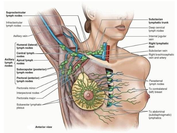
42
43 Efferent vessels from the apical group traverse the cervico-axillary canal.

44 Efferent vessels from the apical group converge to form the subclavian lymphatic trunk, which usually joins the venous system at the junction between right subclavian vein & right internal jugular vein in the neck.

45

46 Axillary process of the mammary gland In some cases, the superolateral region of breast may pass around the margin of pectoral muscle and enters the axilla. This axillary process rarely reaches as high as the apex of the axilla.


47 In metastatic cancer of the apical group, the nodes often adhere to the axillary vein, which may necessitate excision of part of this vessel. Enlargement of the apical nodes may obstruct the cephalic vein superior to the pectoralis minor.

48 The examination of the axillary lymph nodes always forms part of the clinical examination of the breast. With the patient standing or sitting, he or she is asked to place the hand of the side to be examined on the hip and push hard medially. This action of adduction of the shoulder joint causes the pectoralis major muscle to contract maximally so that it becomes hard like a board. The examiner then palpates the axillary nodes. EXAMINATION OF THE LYMPH NODES

49 Anterior & lateral cutaneous branches of the 4th-6th intercostal nerves sensory fibers from the skin of the breast sympathetic fibers to the blood vessels in the breasts smooth muscle in the overlying skin and nipple

50
51 most common cancer among women, other than skin cancer second leading cause of cancer death in women, after lung cancer Chance of a woman having an invasive breast cancer some time during her life about 1 in 8 Understanding the lymphatic drainage of the breasts is of practical importance in predicting the metastasis (dispersal) of cancer cells from a carcinoma of the breast (breast cancer). spreads by means of lymphatic vessels (lymphogenic metastasis), which carry cancer cells from the breast to the lymph nodes, chiefly those in the axilla
52

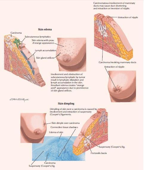
53 Digital mammograms replacing conventional film mammography younger women with dense breast tissue benefit most from this type of mammography. Surgeons use mammography as a guide when removing breast tumors, cysts, and abscesses. jagged density in the mammogram. The skin is thickened over the tumor and the nipple is depressed

54 breast excision simple mastectomy breast is removed down to the retromammary space. radical mastectomy more extensive surgical procedure removal of the breast, pectoral muscles, fat, fascia, and as many lymph nodes as possible in the axilla and pectoral region.

55 accessory nipples
 from which the breasts")
56 rudimentary nipple & areola mistaken for a mole (nevus) anywhere along a line extending from the axilla to the groin embryonic mammary crest (milk line) from which the breasts develop.
57 Polymastia


58 no breast development There may be a nipple and/or areola, but no glandular tissue.

59 59

60 The pectoral region is external to the anterior thoracic wall and anchors the upper limb to the trunk. 60
61 Superficial compartment Contains skin, superficial fascia, breasts Deep compartment Contains muscles & associated structures 61


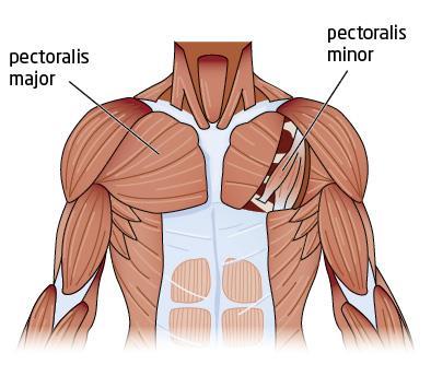
62 Four pectoral muscles move the pectoral girdle Pectoralis major Pectoralis minor Serratus anterior Subclavius Clavicular head: Medial half of clavicle Coracoid Medial process border of of scapula scapula Stabilizes Lateral lip of intertubercular Adducts 3rd-5th scapula and ribs sulcus near of humerus medially their costal rotates cartilages Lateral parts of humerus scapula 1st-8th ribs Protracts & rotates Sternocostal head: Anterior surface of sternum, superior six costal cartilages, aponeurosis of external oblique muscle 62


63 Serratus anterior Pectoralis major Pectoralis minor 63

64 Pectoralis major powerful adduction medial rotation of the arm clavicular head flexing the humerus sternocostal head extending it back 64
65 Pectoralis minor stabilizes the scapula touch an object that is just out of reach. Assists in elevating the ribs Subclavius Anchors and depresses the clavicle, stabilizing it during movements of the upper limb. 65


66 one of the most powerful muscles of the pectoral girdle Strong protractor of scapula Abduction used when punching or reaching anteriorly(boxer's muscle). 66


67 Pectoralis major Medial & Lateral pectoral nerves Pectoralis minor Medial pectoral nerve Subclavius Nerve to subclavius Serratus anterior Long thoracic nerve 67

68 Deep to the pectoral fascia & pectoralis major Descends from the clavicle Clavipectoral triangle cephalic vein can be found. formed by pectoralis major, deltoid & clavicle Deltopectoral groove 68

69 In the subcutaneous tissue overlying the pectoralis major and minor muscles. lateral border of the sternum to the midaxillary line vertically from the 2nd through 6th ribs. 69
thoracic cage inlet and outlet landmarks of the anterior chest wall muscles of the thoracic wall sternum joints ribs intercostal spaces diaphragm
 Thoracic Wall Lecture Objectives Describe the shape and outline of the thoracic cage including inlet and outlet. Describe the anatomical landmarks of the anterior chest wall. List various structures making
Gateway to the upper limb. An area of transition between the neck and the arm.
 Gateway to the upper limb An area of transition between the neck and the arm. Pyramidal space inferior to shoulder @ junction of arm & thorax Distribution center for the neurovascular structures that serve
The Thoracic wall including the diaphragm. Prof Oluwadiya KS
 The Thoracic wall including the diaphragm Prof Oluwadiya KS www.oluwadiya.com Components of the thoracic wall Skin Superficial fascia Chest wall muscles (see upper limb slides) Skeletal framework Intercostal
Note : I put the sheet's info within the slides to easily understand this lecture Done by : Zaid Al-Ghnaneem
 Note : I put the sheet's info within the slides to easily understand this lecture Done by : Zaid Al-Ghnaneem Thoracic Wall Lecture Objectives Describe the shape and outline of the thoracic cage including
THORACIC WALL, ABDOMINAL REGION, MUSCLES OF THE VERTEBRAL COLUMN
 THORACIC WALL, ABDOMINAL REGION, MUSCLES OF THE VERTEBRAL COLUMN 9. 03. 2015 Kaan Yücel M.D., Ph.D. https://fhs122.org Thoracic wall 1. THORAX region between neck 6 abdomen, Chest includes the primary
STERNUM. Lies in the midline of the anterior chest wall It is a flat bone Divides into three parts:
 STERNUM Lies in the midline of the anterior chest wall It is a flat bone Divides into three parts: 1-Manubrium sterni 2-Body of the sternum 3- Xiphoid process The body of the sternum articulates above
THE THORACIC WALL. Boundaries Posteriorly by the thoracic part of the vertebral column. Anteriorly by the sternum and costal cartilages
 THE THORACIC WALL Boundaries Posteriorly by the thoracic part of the vertebral column Anteriorly by the sternum and costal cartilages Laterally by the ribs and intercostal spaces Superiorly by the suprapleural
Upper limb Pectoral region & Axilla
 Upper limb Pectoral region & Axilla 黃敏銓 mchuang@ntu.edu.tw 1 Pectoral region Intercostal nerve Anterior branch of lateral cutaneous branch Lateral cutaneous branch Anterior cutaneous branch Anterior cutaneous
Anatomy of thoracic wall
 Anatomy of thoracic wall Topographic Anatomy of the Thorax 1 Bones of Thoracic wall ribs 1-7"true" ribs -those which attach directly to the sternum true ribs actually attach to the sternum by means of
THE THORAX THORACIC WALL
 THE THORAX THORACIC WALL BREAST THE THORAX THORACIC WALL BREAST THORACIC WALL THE THORAX THE THORAX THE THORAX - BONES & JOINTS The part of the body between the neck and abdomen. The thoracic cage (rib
3 Mohammad Al-Mohtasib Areej Mosleh
 3 Mohammad Al-Mohtasib Areej Mosleh ***Muscles Connecting the Upper Limb to the Vertebral Column 1.Trapezius Muscle ***The first muscle on the back is trapezius muscle, it s called so according
Anatomy of the Thorax
 Anatomy of the Thorax A) THE THORACIC WALL Boundaries Posteriorly by the thoracic part of the vertebral column Anteriorly by the sternum and costal cartilages Laterally by the ribs and intercostal spaces
Diaphragm and intercostal muscles. Dr. Heba Kalbouneh Associate Professor of Anatomy and Histology
 Diaphragm and intercostal muscles Dr. Heba Kalbouneh Associate Professor of Anatomy and Histology Skeletal System Adult Human contains 206 Bones 2 parts: Axial skeleton (axis): Skull, Vertebral column,
THE DESCENDING THORACIC AORTA
 Intercostal Arteries and Veins Each intercostal space contains a large single posterior intercostal artery and two small anterior intercostal arteries. The anterior intercostal arteries of the lower spaces
Conceptual overview 124. Surface anatomy 226. Regional anatomy 139. Clinical cases 235
 Conceptual overview 124 General description 124 Functions 125 Breathing 125 Protection of vital organs 125 Conduit 125 Component parts 125 Thoracic wall 125 Superior thoracic aperture 126 Inferior thoracic
Anatomy notes-thorax.
 Anatomy notes-thorax. Thorax: the part extending from the root of the neck to the abdomen. Parts of the thorax: - Thoracic cage (bones). - Thoracic wall. - Thoracic cavity. ** The thoracic cavity is covered
Chapter 3: Thorax. Thorax
 Chapter 3: Thorax Thorax Thoracic Cage I. Thoracic Cage Osteology A. Thoracic Vertebrae Basic structure: vertebral body, pedicles, laminae, spinous processes and transverse processes Natural kyphotic shape,
Jordan University Faculty Of Medicine. Breast. Dr. Ahmed Salman. Assistant professor of anatomy & embryology
 Jordan University Faculty Of Medicine Breast Dr. Ahmed Salman Assistant professor of anatomy & embryology The breasts are specialized accessory glands of the skin that secretes milk. They are situated
Lecturer: Ms DS Pillay ROOM 2P24 25 February 2013
 Lecturer: Ms DS Pillay ROOM 2P24 25 February 2013 Thoracic Wall Consists of thoracic cage Muscle Fascia Thoracic Cavity 3 Compartments of the Thorax (Great Vessels) (Heart) Superior thoracic aperture
Muscle Action Origin Insertion Nerve Innervation Chapter Page. Deltoid. Trapezius. Latissimus Dorsi
 Muscle Action Origin Insertion Nerve Innervation Chapter Page All Fibers Abduct the shoulder (glenohumeral joint) Deltoid Anterior Fibers Flex the shoulder (G/H joint) Horizontally adduct the shoulder
Region of upper limb attachment to the trunk Proximal segment of limb overlaps parts of the trunk (thorax and back) and lower lateral neck.
 and lower lateral neck.") Region of upper limb attachment to the trunk Proximal segment of limb overlaps parts of the trunk (thorax and back) and lower lateral neck. includes Pectoral Scapular Deltoid regions of the upper limb
Region of upper limb attachment to the trunk Proximal segment of limb overlaps parts of the trunk (thorax and back) and lower lateral neck. includes Pectoral Scapular Deltoid regions of the upper limb
LECTURE -I. Intercostal Spaces & Its Content. BY Dr Farooq Khan Aurakzai. Date:
 LECTURE -I Intercostal Spaces & Its Content BY Dr Farooq Khan Aurakzai Date: 18.04.18 Layers of IC space: Following are the layers of the thoracic region: Skin Subcutaneous CT External IC muscle and membrane
Dr. Weyrich G07: Superior and Posterior Mediastina. Reading: 1. Gray s Anatomy for Students, chapter 3
 Dr. Weyrich G07: Superior and Posterior Mediastina Reading: 1. Gray s Anatomy for Students, chapter 3 Objectives: 1. Subdivisions of mediastinum 2. Structures in Superior mediastinum 3. Structures in Posterior
VENOUS DRAINAGE O US F UPPER UPPER LIM B BY dr.fahad Ullah
 VENOUS DRAINAGE OF UPPER LIMB BY dr.fahad Ullah Venous drainage of the supper limb The venous system of the upper limb drains deoxygenated blood from the arm, forearm and hand It can anatomically be divided
Copyright 2010 Pearson Education, Inc.
 E. VERTEBRAL COLUMN 1. The vertebral column extends from the skull to the pelvis and forms the vertical axis of the skeleton. 2. The vertebral column is composed of vertebrae that are separated by intervertebral
Sports Medicine Part II : ANATOMY OF THE SPINE, ABDOMEN AND SHOULDER COMPLEX
 Sports Medicine 25 1.1 Part II : ANATOMY OF THE SPINE, ABDOMEN AND SHOULDER COMPLEX c.w.p. Wagner High School, Sports Medicine, A. Morgan, T. Morgan & A. Eastlake, 2008 Muscles of the Upper Limbs In this
Brachial plexuses and axillary lymph nodes
 Brachial plexuses and axillary lymph nodes Introduction about nervous system nervous system central nervous system periphral nervous system brain spinal cord 31 pairs of spinal nerves 12 paris of cranial
Mediastinum It is a thick movable partition between the two pleural sacs & lungs. It contains all the structures which lie
 Dr Jamila EL medany OBJECTIVES At the end of the lecture, students should be able to: Define the Mediastinum. Differentiate between the divisions of the mediastinum. List the boundaries and contents of
slide 23 The lobes in the right and left lungs are divided into segments,which called bronchopulmonary segments
 Done By : Rahmeh Alsukkar Date : 26 /10/2017 slide 23 The lobes in the right and left lungs are divided into segments,which called bronchopulmonary segments Each segmental bronchus passes to a structurally
The pectoral region. University of Babylon College of Medicine Dr.HaythemAli Alsayigh M.B.CH.B.-F.I.M.B.S. Surgical Clinical Anatomy
 The pectoral region University of Babylon College of Medicine Dr.HaythemAli Alsayigh M.B.CH.B.-F.I.M.B.S. Surgical Clinical Anatomy Objective Study the Bones and Joints A. Clavicle (collarbone) B. Scapula
Yara saddam & Dana Qatawneh. Razi kittaneh. Maher hadidi
 1 Yara saddam & Dana Qatawneh Razi kittaneh Maher hadidi LECTURE 10 THORAX The thorax extends from the root of the neck to the abdomen. The thorax has a Thoracic wall Thoracic cavity and it is divided
DESCRIPTION: This is the part of the trunk, which is located between the root of the neck and the superior border of the abdominal region.
 1 THE THORACIC REGION DESCRIPTION: This is the part of the trunk, which is located between the root of the neck and the superior border of the abdominal region. SHAPE : T It has the shape of a truncated
Identify the lines used in anatomical surface descriptions of the thorax. median line mid-axillary line mid-clavicular line
 L 14 A B O R A T O R Y Thorax THORACIC WALL Identify the lines used in anatomical surface descriptions of the thorax. median line mid-axillary line mid-clavicular line Identify the surface landmarks of
The Arm and Cubital Fossa
 The Arm and Cubital Fossa Dr. Andrew Gallagher School of Anatomical Sciences University of the Witwatersrand Introduction The ARM (BRACHIUM) is the most proximal segment of the upper limb musculoskeletal
10/14/2018 Dr. Shatarat
 2018 Objectives To discuss mediastina and its boundaries To discuss and explain the contents of the superior mediastinum To describe the great veins of the superior mediastinum To describe the Arch of
Surface anatomy of Cardiovascular system
 Surface anatomy of Cardiovascular system Prof. Abdulameer Al-Nuaimi E-mail: a.al-nuaimi@sheffield.ac.uk E. mail: abdulameerh@yahoo.com The lines cover the front, side, and back of the thorax Midsternal
Large veins of the thorax Brachiocephalic veins
 Large veins of the thorax Brachiocephalic veins Right brachiocephalic vein: formed at the root of the neck by the union of the right subclavian & the right internal jugular veins. Left brachiocephalic
In the Last Three Lectures We Already Discussed the Importance of the Thoracic Cage.
 -This Lecture Will Revise what we took in the last three lectures and will introduce the concept of the chest cavity ( Thoracic Cavity ) In the Last Three Lectures We Already Discussed the Importance of
Anatomy of the Shoulder Girdle. Prof Oluwadiya Kehinde FMCS (Orthop)
") Anatomy of the Shoulder Girdle Prof Oluwadiya Kehinde FMCS (Orthop) www.oluwadiya.com Bony Anatomy Shoulder Complex: Sternum(manubrium) Clavicle Scapula Proximal humerus Manubrium Sterni Upper part of
Anatomy of the Shoulder Girdle Prof Oluwadiya Kehinde FMCS (Orthop) www.oluwadiya.com Bony Anatomy Shoulder Complex: Sternum(manubrium) Clavicle Scapula Proximal humerus Manubrium Sterni Upper part of
Upper Limb Muscles Muscles of Axilla & Arm
 Done By : Saleh Salahat Upper Limb Muscles Muscles of Axilla & Arm 1) Muscles around the axilla A- Muscles connecting the upper to thoracic wall (4) 1- pectoralis major Origin:- from the medial half of
Posterior Triangle of the Neck By Prof. Dr. Muhammad Imran Qureshi
 Posterior Triangle of the Neck By Prof. Dr. Muhammad Imran Qureshi For the purpose of anatomical description the neck is sub divided into two major triangles, the Anterior and the Posterior by muscle bellies
PLEURAE and PLEURAL RECESSES
 PLEURAE and PLEURAL RECESSES By Dr Farooq Aman Ullah Khan PMC 26 th April 2018 Introduction When sectioned transversely, it is apparent that the thoracic cavity is kidney shaped: a transversely ovoid space
OBJECTIVE: To obtain a fundamental knowledge of the root of the neck with respect to structure and function
 The root of the neck Jeff Dupree, Ph.D. e mail: jldupree@vcu.edu OBJECTIVE: To obtain a fundamental knowledge of the root of the neck with respect to structure and function READING ASSIGNMENT: Moore and
Osteology of the Thorax. Prof Oluwadiya K S
 Osteology of the Thorax Prof Oluwadiya K S www.oluwadiya.com The thoracic skeleton consists of the following: 12 pairs of ribs and associated costal cartilages 12 thoracic vertebrae and their intervertebral
Intercostal Muscles LO4
 Intercostal Muscles LO4 4 List the structures, from superficial to deep, in an intercostal space. Describe their relationships to each other, to the associated neurovascular bundle and to the pleural cavity.
Mediastinum and pericardium
 Mediastinum and pericardium Prof. Abdulameer Al-Nuaimi E-mail: a.al-nuaimi@sheffield.ac.uk E. mail: abdulameerh@yahoo.com The mediastinum: is the central compartment of the thoracic cavity surrounded by
THE GOOFY ANATOMIST QUIZZES
 THE GOOFY ANATOMIST QUIZZES 8. BREAST AND LYMPHATICS Q1. Which of the following statements concerning the breast is true? A. The inferomedial region of the breast contains the anterior axillary nodes.
Right lung. -fissures:
 -Right lung is shorter and wider because it is compressed by the right copula of the diaphragm by the live.. 2 fissure, 3 lobes.. hilum : 2 bronchi ( ep-arterial, hyp-arterial ), one artery mediastinal
the part between the neck and the abdomen Chest X-ray
 The Two Fridas 1939 by Frida Kahlo Kaan Yücel M.D., Ph.D. 01. 10. 2013 the part between the neck and the abdomen Chest X-ray 1.1. REGIONS/T ERMS Thoracic cavity cavity between neck and abdomen protected
Bony Thorax. Anatomy and Procedures of the Bony Thorax Edited by M. Rhodes
 Bony Thorax Anatomy and Procedures of the Bony Thorax 10-526-191 Edited by M. Rhodes Anatomy Review Bony Thorax Formed by Sternum 12 pairs of ribs 12 thoracic vertebrae Conical in shape Narrow at top Posterior
Abdomen: Introduction. Prof. Oluwadiya KS
 Abdomen: Introduction Prof. Oluwadiya KS www.oluwadiya.com Abdominopelvic Cavity Abdominal Cavity Pelvic Cavity Extends from the inferior margin of the thorax to the superior margin of the pelvis and the
Syllabus: 6 pages (Page 6 lists corresponding figures for Grant's Atlas 11 th & 12 th Eds.)
") PLEURAL CAVITY AND LUNGS Dr. Milton M. Sholley SELF STUDY RESOURCES Essential Clinical Anatomy 3 rd ed. (ECA): pp. 70 81 Syllabus: 6 pages (Page 6 lists corresponding figures for Grant's Atlas 11 th &
PLEURAL CAVITY AND LUNGS Dr. Milton M. Sholley SELF STUDY RESOURCES Essential Clinical Anatomy 3 rd ed. (ECA): pp. 70 81 Syllabus: 6 pages (Page 6 lists corresponding figures for Grant's Atlas 11 th &
1TRUNK: BODY WALL AND SPINE
 TRUNK: BODY WALL AND SPINE SURFACE ANATOMY SKELETON JOINTS & LIGAMENTS MUSCLES VASCULATURE NERVES SPINAL CORD & VERTEBRAL CANAL ANTERIOR BODY WALL & MAMMARY GLAND LATERAL BODY WALL INGUINAL REGION SUPERFICIAL
Biology 323 Human Anatomy for Biology Majors Lecture 11 Dr. Stuart S. Sumida. Peripheral Circulation
 Biology 323 Human Anatomy for Biology Majors Lecture 11 Dr. Stuart S. Sumida Peripheral Circulation Structures of the Splanchnopleure: receive unpaired vessels of the abdominal aorta. Structures of the
The posterior abdominal wall. Prof. Oluwadiya KS
 The posterior abdominal wall Prof. Oluwadiya KS www.oluwadiya.sitesled.com Posterior Abdominal Wall Lumbar vertebrae and discs. Muscles opsoas, quadratus lumborum, iliacus, transverse, abdominal wall
Welcome to the Structure & Development Dissector. Section I
 Welcome to the Structure & Development Dissector The vast majority of questions will be drawn from structures present in the checklist; however, we reserve the right to use a structure or two that is not
CHAPTER 4. Thorax THORACIC CAVITY
 62 CHAPTER 4 Thorax THORACIC CAVITY CHEST WALL VERSUS THORACIC WALL BREAST (MAMMARY GLAND) SKELETAL COMPONENTS OF THE THORACIC WALL Vertebral Bodies Sternum Ribs MUSCULAR COMPONENTS OF THE ANTERIOR CHEST
213: HUMAN FUNCTIONAL ANATOMY: PRACTICAL CLASS 1: Proximal bones, plexuses and patterns
 213: HUMAN FUNCTIONAL ANATOMY: PRACTICAL CLASS 1: Proximal bones, plexuses and patterns CLAVICLE Examine an isolated clavicle and compare it with a clavicle on an articulated skeleton. Viewed from above,
Axial Skeleton: Vertebrae and Thorax
 Axial Skeleton: Vertebrae and Thorax Function of the vertebral column (spine or backbone): 1) 2) 3) Composition of Vertebral column The vertebral column is formed by 33 individual vertebrae (some of which
Upper limb Arm & Cubital region 黃敏銓
 Upper limb Arm & Cubital region 黃敏銓 1 Arm Lateral intermuscular septum Anterior (flexor) compartment: stronger Medial intermuscular septum Posterior (extensor) compartment 2 Coracobrachialis Origin: coracoid
G24: Shoulder and Axilla
 G24: Shoulder and Axilla Syllabus - Pg. 2 ANAT 6010- Medical Gross Anatomy David A. Morton, Ph.D. Objectives Upper limb Systemically: Bones (joints) Muscles Nerves Vessels (arteries/veins) Fascial compartments
Thoracolumbar Anatomy Eric Shamus Catherine Patla Objectives
 1 2 Thoracolumbar Anatomy Eric Shamus Catherine Patla Objectives List the muscular and ligamentous attachments of the thoracic and lumbar spine Describe how the muscles affect the spine and upper extremity
Scapular and Deltoid Regions
 M1 Gross and Developmental Anatomy Scapular and Deltoid Regions Dr. Peters 1 Outline I. Skeleton of the Shoulder and Attachment of the Upper Extremity to Trunk II. Positions and Movements of the Scapula
In the Name of God, the Most Merciful, the Most Compassionate. Breast
 In the Name of God, the Most Merciful, the Most Compassionate What is the Breast? Breast Definition: a modified sweat gland (sometimes called skin gland) present in both genders, well developed in females
Introduction to anatomy
 Introduction to anatomy Dr. Maher Hadidi Fareed Halteh 3 7/2/2013 Subscapularis: It is located on the anterior side of the scapula. It has a triangular shape. It is like Pectoralis major and Teres major
بسم الله الرحمن الرحيم
 بسم الله الرحمن الرحيم * Last lecture we talked about : thoracic wall sternum ribs (according to their features they are divided into typical and atypical) vertebral column ( which is made of 33 vertebrae
The Neck the lower margin of the mandible above the suprasternal notch and the upper border of the clavicle
 The Neck is the region of the body that lies between the lower margin of the mandible above and the suprasternal notch and the upper border of the clavicle below Nerves of the neck Cervical Plexus Is formed
Lab Activity 11: Group I
 Lab Activity 11: Group I Muscles Martini Chapter 11 Portland Community College BI 231 Origin and Insertion Origin: The place where the fixed end attaches to a bone, cartilage, or connective tissue. Insertion:
Axilla and Brachial Region
 L 4 A B O R A T O R Y Axilla and Brachial Region BRACHIAL PLEXUS 5 Roots/Rami (ventral rami C5 T1) 3 Trunks Superior (C5, C6) Middle (C7) Inferior (C8, T1) 3 Cords Lateral Cord (Anterior Superior and Anterior
The Thoracic Cage. OpenStax College
 OpenStax-CNX module: m46350 1 The Thoracic Cage OpenStax College This work is produced by OpenStax-CNX and licensed under the Creative Commons Attribution License 3.0 By the end of this section, you will
Copyright 2010 Pearson Education, Inc. Copyright 2010 Pearson Education, Inc. Figure Sectioned spinous process. Interspinous.
 PowerPoint Lecture Slides prepared by Janice Meeking, Mount Royal College C H A P T E R 7 The Skeleton: Part B Vertebral Column Transmits weight of trunk to lower limbs Surrounds and protects spinal cord
Ventilation 7/28/2013. Clarification of Terminology. Osteology of Ventilation
 Ventilation Clarification of Terminology Ventilation: the mechanical process by which air is inhaled and exhaled through the lungs. It describes only the movement of air. Respiration: a term used to describe
Pectoral region. Lecture 2
 Pectoral region Lecture 2 Muscle Action Each muscle has: Origin Beginning. Insertion End. Body (belly). Law: When a muscle performs its action, its insertion, moves towards its origin. Spring 2016 Dr.
Dana Alrafaiah. - Moayyad Al-Shafei. -Mohammad H. Al-Mohtaseb. 1 P a g e
 - 6 - Dana Alrafaiah - Moayyad Al-Shafei -Mohammad H. Al-Mohtaseb 1 P a g e Quick recap: Both lungs have an apex, base, mediastinal and costal surfaces, anterior and posterior borders. The right lung,
The Upper Limb III. The Brachial Plexus. Anatomy RHS 241 Lecture 12 Dr. Einas Al-Eisa
 The Upper Limb III The Brachial Plexus Anatomy RHS 241 Lecture 12 Dr. Einas Al-Eisa Brachial plexus Network of nerves supplying the upper limb Compression of the plexus results in motor & sensory changes
Chest cavity, vertebral column and back muscles. Respiratory muscles. Sándor Katz M.D., Ph.D.
 Chest cavity, vertebral column and back muscles. Respiratory muscles. Sándor Katz M.D., Ph.D. Chest cavity - bony structures Chest cavity- bony structures Sternum Ribs True ribs: first seven pairs connect
ABDOMINAL WALL & RECTUS SHEATH
 ABDOMINAL WALL & RECTUS SHEATH Learning Objectives Describe the anatomy, innervation and functions of the muscles of the anterior, lateral and posterior abdominal walls. Discuss their functional relations
The Thoracic Cage ANATOMY 2: THORACIC CAGE AND VERTEBRAL COLUMN
 ANATOMY 2: THORACIC CAGE AND VERTEBRAL COLUMN PSK 4U Mr. S. Kelly North Grenville DHS The Thoracic Cage 7 true ribs 3 false ribs 2 floating ribs Clavicle = collarbone Manubrium Sternum Xiphoid Process
Chapter 5: Other mediastinal structures. The Large Arteries. The Aorta. Ascending aorta
 Chapter 5: Other mediastinal structures The Large Arteries The Aorta The aorta is the main arterial trunk of the systemic circulation and in the healthy state its wall contain a large amount of yellow
26/9/2016. Anatomy. 1 Nour Erekat Wejdan Amer
 26/9/2016 Anatomy st 1 Nour Erekat Wejdan Amer Notes before we start the lecture. Bring any colored Atlas with you to the lab. The main reference is clinical anatomy by regions by Richard snell the 9 th
MUSCLE MECHANICS AND CONTROL
 MUSCLE MECHANICS AND CONTROL STRUCTURE OF A SKELETAL MUSCLE NEUROVASCULAR BUNDLE - CONTAINS THE BLOOD VESSELS AND THE NERVES TO A MUSCLE SKELETAL MUSCLE - LONGITUDINAL SECTION MUSCLE ATTACHMENTS: TENDONS
Anatomy of the thorax
 2018 Anatomy of the thorax Sameh S. Akkila THE THORACIC CAGE The thoracic cage consists of the sternum anteriorly, the twelve thoracic vertebrae and their intervertebral discs posteriorly and the twelve
This figure (of humerus) is from Dr. Maher's newest slides. -Its added here just for consideration-
 is from Dr. Maher's newest slides. -Its added here just for consideration-") This figure (of humerus) is from Dr. Maher's newest slides. -Its added here just for consideration- Slides of Anatomy Please note : These slides are Dr. Maher Hadidi s slides of spring 2016 and were edited
This figure (of humerus) is from Dr. Maher's newest slides. -Its added here just for consideration- Slides of Anatomy Please note : These slides are Dr. Maher Hadidi s slides of spring 2016 and were edited
The Thoracic Cage. Role of the Thoracic Cage 2/13/2019. Anatomy 2: Thoracic Cage and Vertebral Column
 PSK 4U Mr. S. Kelly North Grenville DHS Anatomy 2: Thoracic Cage and Column The Thoracic Cage 7 true ribs 3 false ribs 2 floating ribs Clavicle = collarbone Manubrium Sternum Xiphoid Process 12 thoracic
Overview of the Skeleton: Bone Markings
 Name Overview of the Skeleton: Bone Markings Match the terms in column B with the appropriate description in column A. Column A 1. sharp, slender process* 2. small rounded projection* 3. narrow ridge of
Muscles involved in respiration
 Muscles involved in respiration Respiratory block-anatomy-lecture 1 Editing file Objectives Describe the components of the thoracic cage and their articulations. Describe in brief the respiratory movements.
Main Menu. Trunk and Spinal Column click here. The Power is in Your Hands
 1 The Trunk and Spinal Column click here Main Menu K.9 http://www.handsonlineeducation.com/classes/k9/k9entry.htm[3/27/18, 2:00:55 PM] The Trunk and Spinal Column Vertebral column complex 24 intricate
BRACHIAL PLEXUS. DORSAL SCAPULAR NERVE (C5) supraclavicular branch innervates rhomboids (major and minor) and levator scapulae
 supraclavicular branch innervates rhomboids (major and minor) and levator scapulae") THE BRACHIAL PLEXUS DORSAL SCAPULAR NERVE (C5) supraclavicular branch innervates rhomboids (major and minor) and levator scapulae SCHEMA OF THE BRACHIAL PLEXUS THE BRACHIAL PLEXUS PHRENIC NERVE supraclavicular
THE BRACHIAL PLEXUS DORSAL SCAPULAR NERVE (C5) supraclavicular branch innervates rhomboids (major and minor) and levator scapulae SCHEMA OF THE BRACHIAL PLEXUS THE BRACHIAL PLEXUS PHRENIC NERVE supraclavicular
Candidate s instructions Look at this cross-section taken at the level of C5. Answer the following questions.
 Section 1 Anatomy Chapter 1. Trachea 1 Candidate s instructions Look at this cross-section taken at the level of C5. Answer the following questions. Pretracheal fascia 1 2 5 3 4 Questions 1. Label the
Day 5 Respiratory & Cardiovascular: Respiratory System
 Day 5 Respiratory & Cardiovascular: Respiratory System Be very careful not to damage the heart and lungs while separating the ribs! Analysis Questions-Respiratory & Cardiovascular Log into QUIA using your
MUSCLES. Anconeus Muscle
 LAB 7 UPPER LIMBS MUSCLES Anconeus Muscle anconeus origin: distal end of dorsal surface of humerus insertion: lateral surface of ulna from distal margin of the semilunar notch to proximal end of the olecranon
Pectoral region. Lecture 2
 Pectoral region Lecture 2 Muscle Action Each muscle has: Origin Beginning. Insertion End. Body (belly). Law: When a muscle performs its action, its insertion, moves towards its origin. Spring 2016 Dr.
Breasts (mammae) In female breast:
 In female breast:") اهداف جلسه ا شناي ی با ساختمان پستان عضلات قفسه سينه ا شناي ی با ديافراگم ا شناي ی با عضلات شکم ا شناي ي با Breasts (mammae) In female breast: Modified sweat glands a secondary sexual Source of nutrition
اهداف جلسه ا شناي ی با ساختمان پستان عضلات قفسه سينه ا شناي ی با ديافراگم ا شناي ی با عضلات شکم ا شناي ي با Breasts (mammae) In female breast: Modified sweat glands a secondary sexual Source of nutrition
Main Menu. Shoulder Girdle click here. The Power is in Your Hands. 1:07:11 PM]
![Main Menu. Shoulder Girdle click here. The Power is in Your Hands. 1:07:11 PM]](/thumbs/86/94418253.jpg "Main Menu. Shoulder Girdle click here. The Power is in Your Hands. 1:07:11 PM]") 1 The Shoulder Girdle click here Main Menu K.2 http://www.handsonlineeducation.com/classes//k2entry.htm[3/23/18, 1:07:11 PM] Bones Scapula and Clavicle Move as a unit Clavicle s articulation with sternum
1 The Shoulder Girdle click here Main Menu K.2 http://www.handsonlineeducation.com/classes//k2entry.htm[3/23/18, 1:07:11 PM] Bones Scapula and Clavicle Move as a unit Clavicle s articulation with sternum
Pectoral girdle, SUPERIEUR ARM AND HAND. Danil Hammoudi.MD
 Pectoral girdle, SUPERIEUR ARM AND HAND Danil Hammoudi.MD The pectoral girdle is the set of bones which connect the upper limb to the axial skeleton on each side. It consists of the clavicle scapula in
The abdominal Esophagus, Stomach and the Duodenum. Prof. Oluwadiya KS
 The abdominal Esophagus, Stomach and the Duodenum Prof. Oluwadiya KS www.oluwadiya.com Viscera of the abdomen Abdominal esophagus: Terminal part of the esophagus The stomach Intestines: Small and Large
GI module Lecture: 9 د. عصام طارق. Objectives:
 GI module Lecture: 9 د. عصام طارق Objectives: To list structures forming posterior abdominal wall. To follow aorta & its main branches. To describe IVC & its main tributaries. To list nerves of posterior
Anatomy and Physiology II. Spine
 Anatomy and Physiology II Spine Bones and Other Structures Vertibrae Contains Cervical, Thoracic, Lumbar, Sacral and Coccygeal regions We use Capital letters to refer to these (C, T, L, S, and Co) and
The Human Body. Lesson Goal. Lesson Objectives 9/10/2012. Provide a brief overview of body systems, anatomy, physiology, and topographic anatomy
 The Human Body Lesson Goal Provide a brief overview of body systems, anatomy, physiology, and topographic anatomy Medial Lateral Proximal Distal Superior Inferior Anterior Lesson Objectives Explain the
Prime movers provide the major force for producing a specific movement Antagonists oppose or reverse a particular movement Synergists
 Dr. Gary Mumaugh Prime movers provide the major force for producing a specific movement Antagonists oppose or reverse a particular movement Synergists Add force to a movement Reduce undesirable or unnecessary