Gateway to the upper limb. An area of transition between the neck and the arm.
|
|
|
- Sophia Stokes
- 6 years ago
- Views:
Transcription

1


2 Gateway to the upper limb An area of transition between the neck and the arm.


3 Pyramidal space inferior to junction of arm & thorax Distribution center for the neurovascular structures that serve the upper limb. Protected by the adducted upper limb.


4 BLOOD COMES ARTERIES BLOOD GOES VEINS MUSCLES,VESSELS, SWEAT GLANDS INNERVATED NERVES

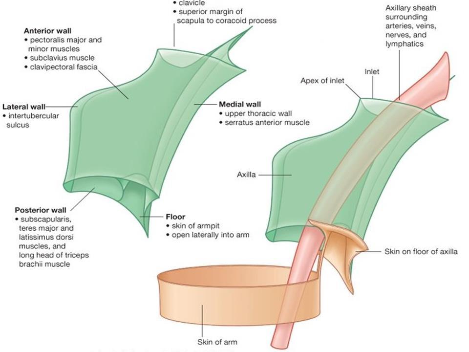
5 Axilla is formed by: Clavicle Scapula Upper thoracic wall Humerus & related muscles


6 Axilla is an irregularly shaped pyramidal space with: Four walls An inlet (apex) A floor (base)
7 Axillary inlet (Apex) Cervico-axillary canal passageway between neck and axilla bounded by: 1st rib Clavicle Superior edge of the scapula The arteries, veins, lymphatics, and nerves traverse this superior opening of the axilla to pass to or from the arm.

8 At the axillary inlet, A P Axillary vein- axillary artery--trunks of brachial plexus

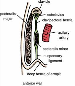
9 Anterior wall Formed by: lateral part of the pectoralis major muscle, underlying pectoralis minor & subclavius muscles, clavipectoral fascia
10

11 Medial wall Formed by the thoracic wall (1st-4th ribs and intercostal muscles) & the overlying serratus anterior. The only major structure that passes directly through the medial wall and into the axilla: intercostobrachial nerve Lateral cutaneous branch of the second intercostal nerve (anterior ramus of T2)

12 Lateral wall A narrow bony wall formed by Intertubercular groove in the humerus

13 Posterior wall Bone framework is formed by the costal surface of the scapula. Formed by scapula & subscapularis on anterior surface teres major & latissimus dorsi, inferiorly

14
")

15 From this distribution center, neurovascular structures pass Superiorly via cervico-axillary canal to (or from) the root of the neck. Anteriorly via clavipectoral triangle to pectoral region. Inferiorly & laterally into limb itself.
.")
16 From this distribution center, neurovascular structures pass Posteriorly via quadrangular space to scapular region Inferiorly & medially along the thoracic wall to the inferiorly placed axioappendicular muscles (serratus anterior and latissimus dorsi).

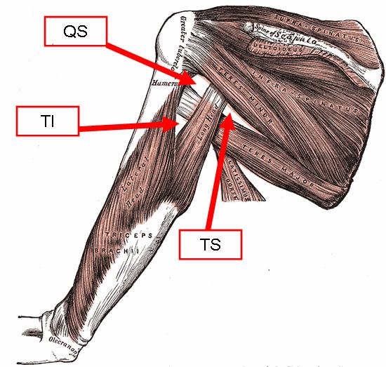
17 Quadrangular space Passageway for nerves & vessels passing between the axilla and the more posterior scapular and deltoid regions.

18 Quadrangular space Viewed from anteriorly, boundaries formed by: Superior: inferior margin of the subscapularis muscle Lateral: surgical neck of the humerus Inferior: superior margin of the teres major muscle Medial: lateral margin of the long head of triceps brachii muscle
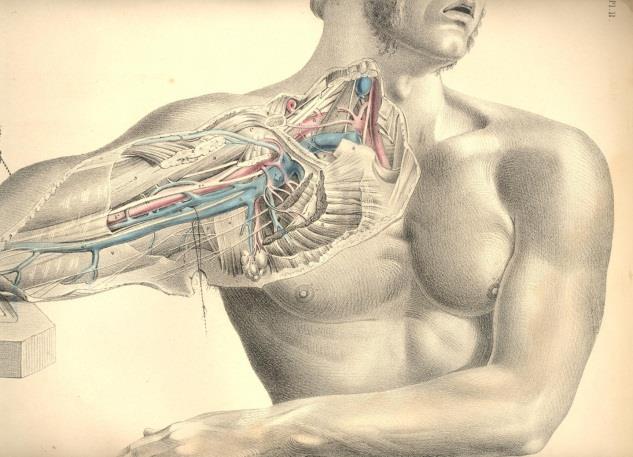
19 Quadrangular space Structures passing through: Axillary nerve Posterior circumflex humeral artery & vein
20 Triangular space Area of communication between axilla and posterior scapular region
21 Triangular space [Medial triangular space] Viewed from anteriorly, formed by: medial margin of the long head of the triceps brachii superior margin of the teres major muscle inferior margin of the subscapularis muscle
22 Triangular space Structures passing through: Circumflex scapular artery & vein
![Triangular interval [Lateral triangular space] formed by: lateral margin of the long](/docs-images/79/80314756/images/23-2.jpg "head of the triceps brachii shaft of the humerus inferior margin of the teres major")
23 Triangular interval [Lateral triangular space] formed by: lateral margin of the long head of the triceps brachii shaft of the humerus inferior margin of the teres major muscle
 & associated")
24 Triangular interval Structures passing through Radial nerve & profunda brachii artery (deep artery of arm) & associated veins
25

26 Formed by the Concave skin Subcutaneous tissue Axillary (deep) fascia Bounded by the anterior and posterior axillary folds, the thoracic wall, and the medial aspect of the arm. The base is supported by the clavipectoral fascia. Floor (Base)
27

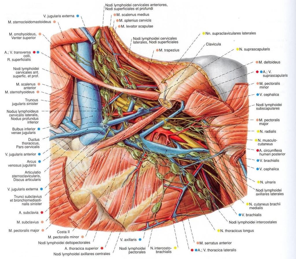
28 Contents of the axilla Axillary artery and its branches Axillary vein and its tributaries Lymph vessels and lymph nodes Brachial plexus These structures are embedded in fat.
29
30
31 Proximally, the neurovascular structures are ensheathed in a sleeve-like extension of the cervical fascia, axillary sheath.


32 The space also contains the proximal parts of two muscles of the arm; biceps brachii & coracobrachialis muscles and axillary process of the breast.


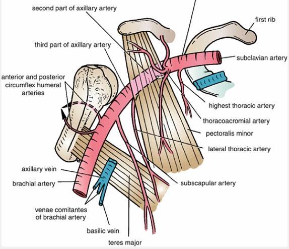
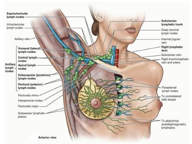
33 Axillary artery Supplies the walls of the axilla & related regions. Before: Subclavian artery After: Brachial artery From lateral border of 1st rib to Inferior border of teres major

34 Throughout its course, the artery is closely related to the cords of the brachial plexus and their branches and is enclosed with them in a connective tissue sheath called the axillary sheath. If this sheath is traced upward into the root of the neck, it is seen to be continuous with the prevertebral fascia.

35 Separated into 3 parts by the pectoralis minor muscle : 1st part proximal to pectoralis minor medial part of pectoralis minor & lateral part of first rib 2nd part posterior to pectoralis minor 3rd part distal to pectoralis minor from lateral part of pectoralis minor to inferior border of teres major
36
 subscapular artery, anterior circumflex humeral artery, posterior circumflex")
37 6 branches of axillary artery 1 branch (1st part) superior thoracic artery 2 branches (2nd part) thoraco-acromial artery & lateral thoracic artery 3 branches (3nd part) subscapular artery, anterior circumflex humeral artery, posterior circumflex humeral artery

38


39 Branches of lateral thoracic artery contribute to the vascular supply of the breast.

40 Subscapular artery, largest branch of the axillary artery, terminates by dividing into: circumflex scapular & thoracodorsal arteries. The anterior circumflex humeral artery anastomoses with the posterior circumflex humeral artery. Along with these two arteries, the circumflex scapular artery and throcadorsal artery participate in the anastomoses around the scapula.

41 Axillary vein Before: Basilic vein After: Subclavian vein From inferior border of teres major to lateral border of 1st rib

42 Tributaries of the axillary vein generally follow the branches of the axillary artery. Other tributaries include brachial veins & cephalic vein.
43 Lymphatics in the axilla Drainage from Upper limb An extensive area on the adjacent trunk Regions of the upper back & shoulder, lower neck, chest, upper anterolateral abdominal wall Drainage from ~ 75% of the mammary gland.
44 The axillary nodes are divided into 5 groups - on the basis of location- The groups are arranged in a manner that reflects the pyramidal shape of the axilla. Humeral (lateral) nodes Pectoral (anterior) nodes Subscapular (posterior) nodes Central nodes Apical nodes
45
46 Efferent vessels from the apical group traverse the cervico-axillary canal.

47 Efferent vessels from the apical group converge to form the subclavian lymphatic trunk, which usually joins the venous system at the junction between right subclavian vein & right internal jugular vein in the neck.

48 Axillary process of the mammary gland In some cases, the superolateral region of breast may pass around the margin of pectoral muscle and enters the axilla. This axillary process rarely reaches as high as the apex of the axilla.


49 In metastatic cancer of the apical group, the nodes often adhere to the axillary vein, which may necessitate excision of part of this vessel. Enlargement of the apical nodes may obstruct the cephalic vein superior to the pectoralis minor.
50 The examination of the axillary lymph nodes always forms part of the clinical examination of the breast. With the patient standing or sitting, he or she is asked to place the hand of the side to be examined on the hip and push hard medially. This action of adduction of the shoulder joint causes the pectoralis major muscle to contract maximally so that it becomes hard like a board. The examiner then palpates the axillary nodes. EXAMINATION OF THE LYMPH NODES
51

 & T 1")

52 a somatic nerve plexus - upper limb formed by intercommunications among ventral rami of lower 4 cervical nerves ( C 5 - C 8) & T 1 responsible for motor innervation to all of muscles of upper limb exception trapezius.
 (intercostobrachial nerve) an area just above the")
 ommunicates with the sympathetic nervous system..")
53 supplies all of the cutaneous innervation of the upper limb exception area of the axilla (armpit) (intercostobrachial nerve) an area just above the point of shoulder (supraclavicular nerves) dorsal scapular area (cutaneous branches of dorsal rami) ommunicates with the sympathetic nervous system..
54 The nerves entering the upper limb provide the following important functions: Sensory innervation to skin & deep structures (e.g. joints) Motor innervation to the muscles Influence over the diameters of the blood vessels by the sympathetic vasomotor nerves Sympathetic secretomotor supply to the sweat glands.
55 At the root of the neck, the nerves form Brachial plexus Nerve fibers derived from different segments of the spinal cord arranged and distributed efficiently in various parts of the upper limb.


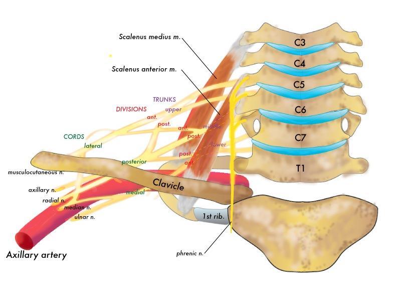
56 begins in the neck and extends into the axilla. Almost all branches of the brachial plexus arise in the axilla (after the plexus has crossed the 1st rib). "Randy Travis Drinks Cold Beer" Roots Trunks Divisions Cords Branches Alternatively: "Read The Damn Cadaver Book!" Alternatively: "Real Texans Drink Coors Beer".

57 Originates in the neck, passes laterally and inferiorly over rib I, and enters the axilla.

58 The parts of the brachial plexus, from medial to lateral, are roots, trunks, divisions, and cords. All major nerves that innervate the upper limb originate from the brachial plexus, mostly from the cords.
59


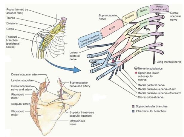
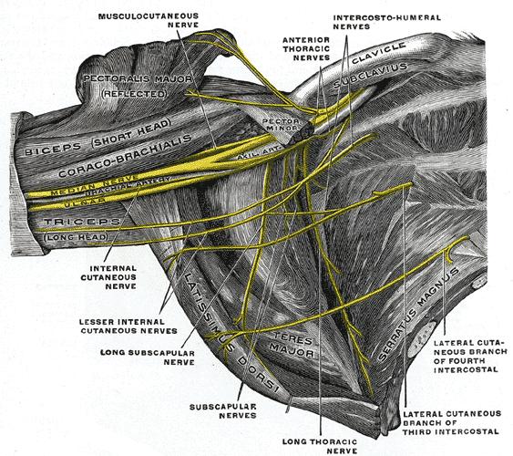
60 Long T h o r a c İ c nerve Dorsal scapular nerve C5 C6 C7 C8 Suprascapular nerve The nerve to subclavius muscle Superior trunk Middle trunk Lateral cord Posterior cord Lateral pectoral nerve Musculocutraneous nerve Lateral root of median nerve Sup. & Inf. Subscapular nerves Thoracodorsal nerve Axillary nerve Radial nerve T1 Inferior trunk Medial cord Medial pectoral nerve Medial cutaneous nerve of arm Medial cutanoues nerve of forearm Ulnar nerve Median nerve Red: Anterior division Blue: Posterior division "Randy Travis Drinks Cold Beer" Roots Trunks Divisions Cords Branches Alternatively: "Read The Damn Cadaver Book!" Alternatively: "Real Texans Drink Coors Beer".

61 Proximal posterior to the subclavian artery in the neck More distal regions surround the axillary artery The roots of the plexus usually pass through the gap between anterior & middle scalene muscles.


62 Musculocutaneous nerve Median nerve Ulnar nerve

63 ROOTS Anterior rami of C5 to C8, and most of T1. The roots & trunks enter the the neck by passing between the anterior scalene and middle scalene muscles. Close to their origin, the roots receive gray rami communicantes from the sympathetic trunk. These carry postganglionic sympathetic fibers onto the roots for distribution to the periphery.
64 .

65 TRUNKS In the inferior part of the neck, the roots of the brachial plexus unite to form three trunks: Superior trunk formed by union of C5 & C6 roots Middle trunk a continuation of C7 root Inferior trunk formed by the union of C8 & T1 roots The inferior trunk lies on rib I posterior to the subclavian artery; the middle and superior trunks are more superior in position.
 compartments of the upper limb Posterior divisions of the trunks supply posterior (extensor) compartments.")
66 DIVISIONS Each trunk divides into: anterior & posterior divisions as the plexus passes through the cervicoaxillary canal posterior to the clavicle. Anterior divisions of the trunks supply anterior (flexor) compartments of the upper limb Posterior divisions of the trunks supply posterior (extensor) compartments. No peripheral nerves originate directly from the divisions of the brachial plexus.
67

68 CORDS The three cords of the brachial plexus originate from the divisions and are related to the second part of the axillary artery.
 Posterior cord Union of all three posterior divisions (C5 to")
69 Lateral cord Union of anterior divisions of upper & middle trunks (C5-C7) Medial cord Continuation of anterior division of inferior trunk (C8-T1) Posterior cord Union of all three posterior divisions (C5 to T1)
 innervates rhomboid")
 innervates serratus anterior muscle")
70 Branches Branches of the roots Dorsal scapular nerve (C5 root of the brachial plexus) innervates rhomboid major and minor muscles Long thoracic nerve (anterior rami of C5 to C7) innervates serratus anterior muscle
 innervates supraspinatus & infraspinatus muscles The nerve to subclavius muscle (C5 & C6) innervates")
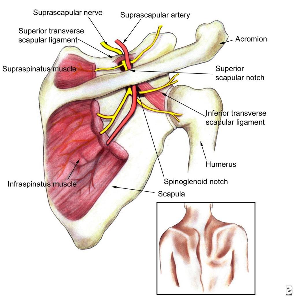
71 Branches of the trunks The only branches from the trunks of the brachial plexus are 2 nerves ;originate from the superior trunk. Suprascapular nerve (C5 & C6) innervates supraspinatus & infraspinatus muscles The nerve to subclavius muscle (C5 & C6) innervates subclavius muscle
72

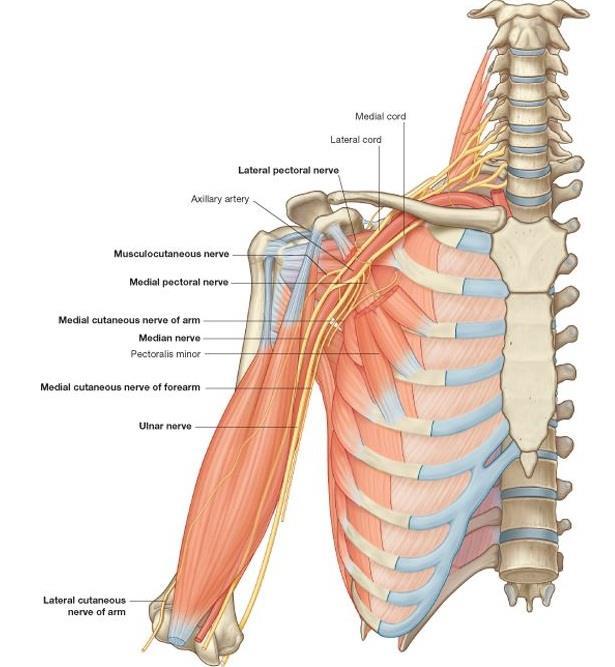
73 Branches of the lateral cord Lateral pectoral nerve pectoralis major muscle
74


75 Branches of the lateral cord Musculocutaneous nerve Coracobrachialis Biceps brachii Brachialis All 3 flexor anterior compartment of the arm Terminates as lateral cutaneous nerve of forearm.

76 Branches of the lateral cord Lateral root of median nerve Largest terminal branch of the lateral cord Passes medially to join a similar branch from the medial cord to form the median nerve. 1. Lateral cord 2. Posterior cord 3. Medial cord 4. Lateral root of median nerve 5. Medial root of median nerve 6. Radial nerve 7. Ulnar nerve 8. Median nerve
77
78
79 Branches of the medial cord 1) Medial pectoral nerve pectoralis major & minor 2) Medial cutaneous nerve of arm (medial brachial cutaneous nerve) medial 1/3 of distal arm 3) Medial cutaneous nerve of forearm (medial antebrachial cutaneous nerve) medial surface of the forearm down to the wrist 4) Ulnar nerve 5) Median nerve

80 Musculocutaneous nerve, lateral root of the median nerve, median nerve, medial root of the median nerve, ulnar nerve form an M over the third part of the axillary artery. This feature, together with penetration of the coracobrachialis muscle by the musculocutaneous nerve, can be used to identify components of the brachial plexus in the axilla
81 Branches of the posterior cord 1) Superior subscapular nerve subscapularis muscle 2) Thoracodorsal nerve latissimus dorsi 3) Inferior subscapular nerve subscapularis & teres major 4) Axillary nerve deltoid and teres minor 5) Radial nerve All these nerves except the radial nerve innervate muscles associated with the posterior wall of the axilla; the radial nerve passes into the arm and forearm.
3 Mohammad Al-Mohtasib Areej Mosleh
 3 Mohammad Al-Mohtasib Areej Mosleh ***Muscles Connecting the Upper Limb to the Vertebral Column 1.Trapezius Muscle ***The first muscle on the back is trapezius muscle, it s called so according
Upper limb Pectoral region & Axilla
 Upper limb Pectoral region & Axilla 黃敏銓 mchuang@ntu.edu.tw 1 Pectoral region Intercostal nerve Anterior branch of lateral cutaneous branch Lateral cutaneous branch Anterior cutaneous branch Anterior cutaneous
Region of upper limb attachment to the trunk Proximal segment of limb overlaps parts of the trunk (thorax and back) and lower lateral neck.
 and lower lateral neck.") Region of upper limb attachment to the trunk Proximal segment of limb overlaps parts of the trunk (thorax and back) and lower lateral neck. includes Pectoral Scapular Deltoid regions of the upper limb
Region of upper limb attachment to the trunk Proximal segment of limb overlaps parts of the trunk (thorax and back) and lower lateral neck. includes Pectoral Scapular Deltoid regions of the upper limb
Brachial plexuses and axillary lymph nodes
 Brachial plexuses and axillary lymph nodes Introduction about nervous system nervous system central nervous system periphral nervous system brain spinal cord 31 pairs of spinal nerves 12 paris of cranial
G24: Shoulder and Axilla
 G24: Shoulder and Axilla Syllabus - Pg. 2 ANAT 6010- Medical Gross Anatomy David A. Morton, Ph.D. Objectives Upper limb Systemically: Bones (joints) Muscles Nerves Vessels (arteries/veins) Fascial compartments
Upper limb Arm & Cubital region 黃敏銓
 Upper limb Arm & Cubital region 黃敏銓 1 Arm Lateral intermuscular septum Anterior (flexor) compartment: stronger Medial intermuscular septum Posterior (extensor) compartment 2 Coracobrachialis Origin: coracoid
Key Relationships in the Upper Limb
 Key Relationships in the Upper Limb This list contains some of the key relationships that will help you identify structures in the lab. They are organized by dissection assignment as defined in the syllabus.
Axilla and Brachial Region
 L 4 A B O R A T O R Y Axilla and Brachial Region BRACHIAL PLEXUS 5 Roots/Rami (ventral rami C5 T1) 3 Trunks Superior (C5, C6) Middle (C7) Inferior (C8, T1) 3 Cords Lateral Cord (Anterior Superior and Anterior
VENOUS DRAINAGE O US F UPPER UPPER LIM B BY dr.fahad Ullah
 VENOUS DRAINAGE OF UPPER LIMB BY dr.fahad Ullah Venous drainage of the supper limb The venous system of the upper limb drains deoxygenated blood from the arm, forearm and hand It can anatomically be divided
The arm: *For images refer back to the slides
 The arm: *For images refer back to the slides Muscles of the arm: deltoid, triceps (which is located at the back of the arm), biceps and brachialis (it lies under the biceps), brachioradialis (it lies
MUSCLES. Anconeus Muscle
 LAB 7 UPPER LIMBS MUSCLES Anconeus Muscle anconeus origin: distal end of dorsal surface of humerus insertion: lateral surface of ulna from distal margin of the semilunar notch to proximal end of the olecranon
BRACHIAL PLEXUS. DORSAL SCAPULAR NERVE (C5) supraclavicular branch innervates rhomboids (major and minor) and levator scapulae
 supraclavicular branch innervates rhomboids (major and minor) and levator scapulae") THE BRACHIAL PLEXUS DORSAL SCAPULAR NERVE (C5) supraclavicular branch innervates rhomboids (major and minor) and levator scapulae SCHEMA OF THE BRACHIAL PLEXUS THE BRACHIAL PLEXUS PHRENIC NERVE supraclavicular
THE BRACHIAL PLEXUS DORSAL SCAPULAR NERVE (C5) supraclavicular branch innervates rhomboids (major and minor) and levator scapulae SCHEMA OF THE BRACHIAL PLEXUS THE BRACHIAL PLEXUS PHRENIC NERVE supraclavicular
*Our main subject is the brachial plexus but it's important to understand the spinal cord first in order to understand the brachial plexus.
 *Our main subject is the brachial plexus but it's important to understand the spinal cord first in order to understand the brachial plexus. *Vertebral column is formed by the union of 33 sequential vertebrae
Al-Balqa Applied University
 Al-Balqa Applied University Faculty Of Medicine *You can use this checklist as a guide to you for the lab. the items on this checklist represent the main features of the models that you have to know for
Upper Limb Muscles Muscles of Axilla & Arm
 Done By : Saleh Salahat Upper Limb Muscles Muscles of Axilla & Arm 1) Muscles around the axilla A- Muscles connecting the upper to thoracic wall (4) 1- pectoralis major Origin:- from the medial half of
Fascial Compartments of the Upper Arm
 Fascial Compartments of the Upper Arm The upper arm is enclosed in a sheath of deep fascia and has two fascial septa: 1- Medial fascial septum (medial intermuscular septum): attached to the medial supracondylar
The Arm and Cubital Fossa
 The Arm and Cubital Fossa Dr. Andrew Gallagher School of Anatomical Sciences University of the Witwatersrand Introduction The ARM (BRACHIUM) is the most proximal segment of the upper limb musculoskeletal
region of the upper limb between the shoulder and the elbow Superiorly communicates with the axilla.
 1 region of the upper limb between the shoulder and the elbow Superiorly communicates with the axilla. Inferiorly, a number of important structures pass between arm & forearm through cubital fossa. 2 medial
The Upper Limb III. The Brachial Plexus. Anatomy RHS 241 Lecture 12 Dr. Einas Al-Eisa
 The Upper Limb III The Brachial Plexus Anatomy RHS 241 Lecture 12 Dr. Einas Al-Eisa Brachial plexus Network of nerves supplying the upper limb Compression of the plexus results in motor & sensory changes
Scapular and Deltoid Regions
 M1 Gross and Developmental Anatomy Scapular and Deltoid Regions Dr. Peters 1 Outline I. Skeleton of the Shoulder and Attachment of the Upper Extremity to Trunk II. Positions and Movements of the Scapula
213: HUMAN FUNCTIONAL ANATOMY: PRACTICAL CLASS 1: Proximal bones, plexuses and patterns
 213: HUMAN FUNCTIONAL ANATOMY: PRACTICAL CLASS 1: Proximal bones, plexuses and patterns CLAVICLE Examine an isolated clavicle and compare it with a clavicle on an articulated skeleton. Viewed from above,
Pectoral region. Lecture 2
 Pectoral region Lecture 2 Muscle Action Each muscle has: Origin Beginning. Insertion End. Body (belly). Law: When a muscle performs its action, its insertion, moves towards its origin. Spring 2016 Dr.
Introduction to anatomy
 Introduction to anatomy Dr. Maher Hadidi Fareed Halteh 3 7/2/2013 Subscapularis: It is located on the anterior side of the scapula. It has a triangular shape. It is like Pectoralis major and Teres major
This figure (of humerus) is from Dr. Maher's newest slides. -Its added here just for consideration-
 is from Dr. Maher's newest slides. -Its added here just for consideration-") This figure (of humerus) is from Dr. Maher's newest slides. -Its added here just for consideration- Slides of Anatomy Please note : These slides are Dr. Maher Hadidi s slides of spring 2016 and were edited
This figure (of humerus) is from Dr. Maher's newest slides. -Its added here just for consideration- Slides of Anatomy Please note : These slides are Dr. Maher Hadidi s slides of spring 2016 and were edited
STRUCTURAL BASIS OF MEDICAL PRACTICE EXAMINATION 5 October 6, 2006
 STRUCTURAL BASIS OF MEDICAL PRACTICE EXAMINATION 5 October 6, 2006 PART l. Answer in the space provided. (8 pts) 1. Identify the structures. (2 pts) B C A. _pisiform B. _ulnar artery A C. _flexor carpi
Netter's Anatomy Flash Cards Section 6 List 4 th Edition
 Netter's Anatomy Flash Cards Section 6 List 4 th Edition https://www.memrise.com/course/1577581/ Section 6 Upper Limb (66 cards) Plate 6-1 Humerus and Scapula: Anterior View 1.1 Acromion 1.2 Greater tubercle
Biology 323 Human Anatomy for Biology Majors Lecture 11 Dr. Stuart S. Sumida. Peripheral Circulation
 Biology 323 Human Anatomy for Biology Majors Lecture 11 Dr. Stuart S. Sumida Peripheral Circulation Structures of the Splanchnopleure: receive unpaired vessels of the abdominal aorta. Structures of the
Muscle Action Origin Insertion Nerve Innervation Chapter Page. Deltoid. Trapezius. Latissimus Dorsi
 Muscle Action Origin Insertion Nerve Innervation Chapter Page All Fibers Abduct the shoulder (glenohumeral joint) Deltoid Anterior Fibers Flex the shoulder (G/H joint) Horizontally adduct the shoulder
The Clavicle Right clavicle Deltoid tubercle: Conoid tubercle, conoid ligamen Impression for the
 The Clavicle Muscle Attachment Sites in the Upper Limb Pectoralis major Right clavicle Smooth superior surface of the shaft, under the platysma muscle tubercle: attachment of the deltoid Acromial facet
Posterior Triangle of the Neck By Prof. Dr. Muhammad Imran Qureshi
 Posterior Triangle of the Neck By Prof. Dr. Muhammad Imran Qureshi For the purpose of anatomical description the neck is sub divided into two major triangles, the Anterior and the Posterior by muscle bellies
Supplied in part by the musculocutaneous nerve. Forms the axis of rotation in movements of pronation and supination
 Anatomy: Upper limb (15 questions) 1. Latissimus Dorsi: Is innervated by the dorsal scapular nerve Lies above feres major muscle Medially rotates the humerus All of the above 2. Supinator muscle is: Deep
Muscles of the Upper Limb
 Muscles of the Upper Limb anterior surface of ribs 3 5 coracoid process Pectoralis minor pectoral nerves protracts / depresses scapula Serratus anterior Subclavius ribs 1-8 long thoracic nerve rib 1 ----------------
Pectoral region. Lecture 2
 Pectoral region Lecture 2 Muscle Action Each muscle has: Origin Beginning. Insertion End. Body (belly). Law: When a muscle performs its action, its insertion, moves towards its origin. Spring 2016 Dr.
Salvador Dali - Anthropomorphic Chest of Drawers, 1936
 Salvador Dali - Anthropomorphic Chest of Drawers, 1936 Kaan Yücel M.D., Ph.D. 05.March.2014 the part between the neck and the abdomen Chest X-ray 1.1. REGIONS/T ERMS Thoracic cavity cavity between neck
BRACHIAL PLEXUS 11/12/2014 كيف تتكون الضفيرة FORMATION ENLARGEMENT (INTUMESCENCE) OF THE SPINAL CORD. Grey matter. Cervical intumescence - C 6 - T 2
 OF THE SPINAL CORD. Grey matter. Cervical intumescence - C 6 - T 2") BRACHIAL PLEXUS Prof. Fawzy Elnady ENLARGEMENT (INTUMESCENCE) OF THE SPINAL CORD Grey matter Cervical intumescence - C 6 - T 2 Lumbar intumescence - L 4 S 2 كيف تتكون الضفيرة FORMATION The ventral rami
BRACHIAL PLEXUS Prof. Fawzy Elnady ENLARGEMENT (INTUMESCENCE) OF THE SPINAL CORD Grey matter Cervical intumescence - C 6 - T 2 Lumbar intumescence - L 4 S 2 كيف تتكون الضفيرة FORMATION The ventral rami
Dr. Mahir Alhadidi Anatomy Lecture #9 Feb,28 th 2012
 Quick Revision: Upper arm is divided into two compartments: 1. Anterior Compartment: Contains three muscles (Biceps brachii, Coracobrachialis, Brachialis). Innervated by Musculocutaneous nerve. 2. Posterior
Anatomy of the Shoulder Girdle. Prof Oluwadiya Kehinde FMCS (Orthop)
") Anatomy of the Shoulder Girdle Prof Oluwadiya Kehinde FMCS (Orthop) www.oluwadiya.com Bony Anatomy Shoulder Complex: Sternum(manubrium) Clavicle Scapula Proximal humerus Manubrium Sterni Upper part of
Anatomy of the Shoulder Girdle Prof Oluwadiya Kehinde FMCS (Orthop) www.oluwadiya.com Bony Anatomy Shoulder Complex: Sternum(manubrium) Clavicle Scapula Proximal humerus Manubrium Sterni Upper part of
Peripheral Nervous Sytem: Upper Body
 Peripheral Nervous Sytem: Upper Body MSTN121 - Neurophysiology Session 10 Department of Myotherapy Cervical Plexus Accessory nerve (CN11 + C1-5) Motor: trapezius and sternocleidomastoid Greater auricular
*the Arm* -the arm extends from the shoulder joint (proximal), to the elbow joint (distal) - it has one bone ; the humerus which is a long bone
, to the elbow joint (distal) - it has one bone ; the humerus which is a long bone") *the Arm* -the arm extends from the shoulder joint (proximal), to the elbow joint (distal) - it has one bone ; the humerus which is a long bone - muscles in the arm : *brachialis muscle *Biceps brachii
*the Arm* -the arm extends from the shoulder joint (proximal), to the elbow joint (distal) - it has one bone ; the humerus which is a long bone - muscles in the arm : *brachialis muscle *Biceps brachii
FUNCTIONAL ANATOMY OF SHOULDER JOINT
 FUNCTIONAL ANATOMY OF SHOULDER JOINT ARTICULATION Articulation is between: The rounded head of the Glenoid cavity humerus and The shallow, pear-shaped glenoid cavity of the scapula. 2 The articular surfaces
26/9/2016. Anatomy. 1 Nour Erekat Wejdan Amer
 26/9/2016 Anatomy st 1 Nour Erekat Wejdan Amer Notes before we start the lecture. Bring any colored Atlas with you to the lab. The main reference is clinical anatomy by regions by Richard snell the 9 th
Human Anatomy Biology 351
 1 Human Anatomy Biology 351 Upper Limb Exam Please place your name on the back of the last page of this exam. You must answer all questions on this exam. Because statistics demonstrate that, on average,
OBJECTIVE: To obtain a fundamental knowledge of the root of the neck with respect to structure and function
 The root of the neck Jeff Dupree, Ph.D. e mail: jldupree@vcu.edu OBJECTIVE: To obtain a fundamental knowledge of the root of the neck with respect to structure and function READING ASSIGNMENT: Moore and
Abduction of arm until your hand rich your head. Flexion of forearm at elbow joint. Extension of arm at elbow joint. Flexion of fingers 10.
 Num. answer 1. Medialy With the manubrium ( sternum ), and laterally with the acromion of the scapula 2. 1. Trapezius 2. Levator scapulae 3. Rhomboids 3. 1. Pectoralis major 2. Pectoralis minor 3. Latissiumus
LIST OF STRUCTURES TO BE IDENTIFIED IN LAB: UPPER EXTREMITY REVIEW 2016
 LIST OF STRUCTURES TO BE IDENTIFIED IN LAB: UPPER EXTREMITY REVIEW 2016 BONES Ribs, sternum, clavicle Humerus: Head, greater tubercle, lesser tubercle, intertubercular sulcus, surgical neck, anatomical
BOGOMOLETS NATIONAL MEDICAL UNIVERSITY. Department of Human Anatomy GUIDELINES. The theme of the lesson The vessels of the upper limb.
 BOGOMOLETS NATIONAL MEDICAL UNIVERSITY Department of Human Anatomy GUIDELINES Academic discipline HUMAN ANATOMY Module 2 The theme of the lesson The vessels of the upper limb. Course Faculties І Medical
The pectoral region. University of Babylon College of Medicine Dr.HaythemAli Alsayigh M.B.CH.B.-F.I.M.B.S. Surgical Clinical Anatomy
 The pectoral region University of Babylon College of Medicine Dr.HaythemAli Alsayigh M.B.CH.B.-F.I.M.B.S. Surgical Clinical Anatomy Objective Study the Bones and Joints A. Clavicle (collarbone) B. Scapula
THE SHOULDER JOINT T H E G L E N O H U M E R A L ( G H ) J O I N T
 J O I N T") THE SHOULDER JOINT T H E G L E N O H U M E R A L ( G H ) J O I N T CLARIFICATION OF TERMS Shoulder girdle = scapula and clavicle Shoulder joint (glenohumeral joint) = scapula and humerus Lippert, p115
THE SHOULDER JOINT T H E G L E N O H U M E R A L ( G H ) J O I N T CLARIFICATION OF TERMS Shoulder girdle = scapula and clavicle Shoulder joint (glenohumeral joint) = scapula and humerus Lippert, p115
The shoulder girdle consists of the glenohumeral, acromioclavicular, sternoclavicular and scapulothoracic joints
 Anatomy of Shoulder Girdle The shoulder girdle consists of the glenohumeral, acromioclavicular, sternoclavicular and scapulothoracic joints Glenohumeral Joint A ball and socket synoval joint with a large
Practical 2 Worksheet
 Practical 2 Worksheet Upper Extremity BONES 1. Which end of the clavicle is on the lateral side (acromial or sternal)? 2. Describe the difference in the appearance of the acromial and sternal ends of the
7/31/2012 THE SHOULDER JOINT CLARIFICATION OF TERMS OSTEOLOGY OF THE GH JOINT(BONES)
") THE SHOULDER JOINT T H E G L E N O H U M E R AL ( G H ) J O I N T CLARIFICATION OF TERMS Shoulder girdle = scapula and clavicle Shoulder joint (glenohumerual joint) = scapula and Lippert, p115 OSTEOLOGY
THE SHOULDER JOINT T H E G L E N O H U M E R AL ( G H ) J O I N T CLARIFICATION OF TERMS Shoulder girdle = scapula and clavicle Shoulder joint (glenohumerual joint) = scapula and Lippert, p115 OSTEOLOGY
Nerve Injury. 1) Upper Lesions of the Brachial Plexus called Erb- Duchene Palsy or syndrome.
 Upper Lesions of the Brachial Plexus called Erb- Duchene Palsy or syndrome.") Nerve Injury - Every nerve goes to muscle or skin so if the nerve is injured this will cause paralysis in the muscle supplied from that nerve (paralysis means loss of function) then other muscles and other
Nerve Injury - Every nerve goes to muscle or skin so if the nerve is injured this will cause paralysis in the muscle supplied from that nerve (paralysis means loss of function) then other muscles and other
MUSCLES OF SHOULDER REGION
 Dr Jamila EL Medany OBJECTIVES At the end of the lecture, students should: List the name of muscles of the shoulder region. Describe the anatomy of muscles of shoulder region regarding: attachments of
Multiple variations involving all the terminal branches of the brachial plexus and the axillary artery a case report
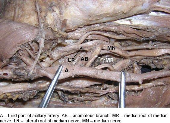
 SHORT REPORT Eur J Anat, 10 (3): 61-66 (2006) Multiple variations involving all the terminal branches of the brachial plexus and the axillary artery a case report K. Ramachandran, I. Kanakasabapathy and
Candidate s instructions Look at this cross-section taken at the level of C5. Answer the following questions.
 Section 1 Anatomy Chapter 1. Trachea 1 Candidate s instructions Look at this cross-section taken at the level of C5. Answer the following questions. Pretracheal fascia 1 2 5 3 4 Questions 1. Label the
G25: Brachium. ANAT Medical Gross Anatomy. David A. Morton, Ph.D.
 G25: Brachium ANAT 6010- Medical Gross Anatomy David A. Morton, Ph.D. Brachial Plexus Randy Travis Drinks Cold Beer What muscle(s) enable her to do the following exercise? What muscle(s) enable her to
The Thoracic wall including the diaphragm. Prof Oluwadiya KS
 The Thoracic wall including the diaphragm Prof Oluwadiya KS www.oluwadiya.com Components of the thoracic wall Skin Superficial fascia Chest wall muscles (see upper limb slides) Skeletal framework Intercostal
STRUCTURAL BASIS OF MEDICAL PRACTICE EXAMINATION 5. September 30, 2011
 STRUCTURAL BASIS OF MEDICAL PRACTICE EXAMINATION 5 September 30, 2011 PART l. Answer in the space provided. (12 pts) 1. Identify the structures. (2 pts) EXAM NUMBER A. Suprascapular nerve B. Axillary nerve
Anatomy Workshop Upper Extremity David Ebaugh, PT, PhD Workshop Leader. Lab Leaders: STATION I BRACHIAL PLEXUS
 Anatomy Workshop Upper Extremity David Ebaugh, PT, PhD Workshop Leader Lab Leaders: STATION I BRACHIAL PLEXUS A. Posterior cervical triangle and axilla B. Formation of plexus 1. Ventral rami C5-T1 2. Trunks
The grouping of nerves connecting the C4 to Th1 junctions of the spinal cord to the left and right arms.
 THE BRACHIAL The grouping of nerves connecting the C4 to Th1 junctions of the spinal cord to the left and right arms. CONTENTS Brachial plexus Brachial plexus anatomy MRI of brachial plexus Dermatome(C8-T1)
Anatomy of the Musculoskeletal System
 Anatomy of the Musculoskeletal System Kyle E. Rarey, Ph.D. Department of Anatomy & Cell Biology and Otolaryngology University of Florida College of Medicine Outline of Presentation Vertebral Column Upper
Slides of Anatomy. Spring Dr. Maher Hadidi, University of Jordan
 Slides of Anatomy Please note : These slides are Dr. Maher Hadidi s slides of spring 2016 and were edited by the Premed Academic Team to fit the slides of spring 2019. Spring 2019 Dr. Maher Hadidi, University
Sports Medicine Part II : ANATOMY OF THE SPINE, ABDOMEN AND SHOULDER COMPLEX
 Sports Medicine 25 1.1 Part II : ANATOMY OF THE SPINE, ABDOMEN AND SHOULDER COMPLEX c.w.p. Wagner High School, Sports Medicine, A. Morgan, T. Morgan & A. Eastlake, 2008 Muscles of the Upper Limbs In this
Anatomy of thoracic wall
 Anatomy of thoracic wall Topographic Anatomy of the Thorax 1 Bones of Thoracic wall ribs 1-7"true" ribs -those which attach directly to the sternum true ribs actually attach to the sternum by means of
Nerves of the upper limb Prof. Abdulameer Al-Nuaimi. E. mail:
 Nerves of the upper limb Prof. Abdulameer Al-Nuaimi E-mail: a.al-nuaimi@sheffield.ac.uk E. mail: abdulameerh@yahoo.com Brachial plexus Median nerve After originating from the brachial plexus in the axilla,
Bilateral Variations in the Branching Pattern of the Axillary Artery in a Single Cadaver
 Case Report Bilateral Variations in the Branching Pattern of the Axillary Artery in a Single Cadaver Dr. Purnendu Rang 1, Dr. Parijat Mukherjee 2, Dr. Aradhana Sanga 3, Dr. Arunima Nag (Ray) 4, Dr. Champak
Synapse Homework. Back page last question not counted. 4 pts total, each question worth 0.18pts. 26/34 students answered correctly!
 Synapse Homework Back page last question not counted 26/34 students answered correctly! 4 pts total, each question worth 0.18pts Business TASS hours extended! MWF 1-2pm, Willamette 204 T and Th 9:30-10:30am,
Spinal nerves and cervical plexus Prof. Abdulameer Al Nuaimi. E mail: a.al E. mail:
 Spinal nerves and cervical plexus Prof. Abdulameer Al Nuaimi E mail: a.al nuaimi@sheffield.ac.uk E. mail: abdulameerh@yahoo.com Branches of ophthalmic artery Muscles of face A spinal nerve Spinal
David G. Simpson, Ph.D.
 David G. Simpson, Ph.D. ARM & CUBITAL FOSSA Revised 7/08 Text References Moores 3 rd ed., p402 408, 436 439, 439 443, 478, 481 LEARNING OBJECTIVES: 1. Describe the humerus, indicating the sites of muscle
Multiple Neurovascular... Pit Baran Chakraborty, Santanu Bhattacharya, Sumita Dutta.
 Multiple Neurovascular... Pit Baran Chakraborty, Santanu Bhattacharya, Sumita Dutta. Fig-3: Showing high formation of Median nerve. Fig-1: Showing atypical formation of cords of Brachial plexus. 1 = Upper
Pectoral girdle, SUPERIEUR ARM AND HAND. Danil Hammoudi.MD
 Pectoral girdle, SUPERIEUR ARM AND HAND Danil Hammoudi.MD The pectoral girdle is the set of bones which connect the upper limb to the axial skeleton on each side. It consists of the clavicle scapula in
REFERENCE DIAGRAMS OF UPPER LIMB MUSCLES: NAMES, LOCATIONS, ATTACHMENTS, FUNCTIONS MUSCLES CONNECTING THE UPPER LIMB TO THE AXIAL SKELETON
 REFERENCE DIAGRAMS OF UPPER LIMB MUSCLES: NAMES, LOCATIONS, ATTACHMENTS, FUNCTIONS MUSCLES CONNECTING THE UPPER LIMB TO THE AXIAL SKELETON A25LAB EXERCISES: UPPER LIMB MUSCLES Page 1 MUSCLES CONNECTING
MUSCLE MECHANICS AND CONTROL
 MUSCLE MECHANICS AND CONTROL STRUCTURE OF A SKELETAL MUSCLE NEUROVASCULAR BUNDLE - CONTAINS THE BLOOD VESSELS AND THE NERVES TO A MUSCLE SKELETAL MUSCLE - LONGITUDINAL SECTION MUSCLE ATTACHMENTS: TENDONS
BILATERAL RARE NEURO VASCULAR VARIATIONS OF UPPER LIMB A CASE REPORT
 BILATERAL RARE NEURO VASCULAR VARIATIONS OF UPPER LIMB A CASE REPORT *N. B. S. Parimala Department of Anatomy, Dr. Pinnamaneni Siddhartha Institute of Medical Sciences & Research Foundation, Chinnaoutpalli,
Muscular Nomenclature and Kinesiology - One
 Chapter 16 Muscular Nomenclature and Kinesiology - One Lessons 1-3 (with lesson 4) 1 Introduction 122 major muscles covered in this chapter Chapter divided into nine lessons Kinesiology study of human
Misc Anatomy. Upper Limb! 2. Lower Limb! 5. Venous Drainage! Head & neck! 8
 Misc Anatomy Upper Limb! 2 Arteries!... 2 Veins!... 2 Spaces!... 4 Lower Limb! 5 Arteries!... 5 Venous Drainage!... 6 Spaces!... 7 Head & neck! 8 Artery!... 8 Ultrasound View for IJ CVL!... 8 Arteries
Muscle Anatomy Review Chart
 Muscle Anatomy Review Chart BACK Superficial (5) Trapezius Transverse cervical a. Latissimus dorsi Thoracodorsal a. Rhomboideus major Dorsal scapular a. Rhomboideus minor Levator scapulae Intermediate
Chapter 14. The Nervous System. The Spinal Cord and Spinal Nerves. Lecture Presentation by Steven Bassett Southeast Community College
 Chapter 14 The Nervous System The Spinal Cord and Spinal Nerves Lecture Presentation by Steven Bassett Southeast Community College Introduction The Central Nervous System (CNS) consists of: The spinal
Spinal nerves. Aygul Shafigullina. Department of Morphology and General Pathology
 Spinal nerves Aygul Shafigullina Department of Morphology and General Pathology Spinal nerve a mixed nerve, formed in the vicinity of an intervertebral foramen, where fuse a dorsal root and a ventral root,
Figure 27: The synovial membrane of the shoulder joint (anterior view)
") The coracoacromial ligament; is an accessory ligament that protects the superior aspect of the joint extending from the coracoid process to the acromion over the tendon of supraspinatus. The synovial membrane
The coracoacromial ligament; is an accessory ligament that protects the superior aspect of the joint extending from the coracoid process to the acromion over the tendon of supraspinatus. The synovial membrane
Gross Anatomy Faculty: Gross Anatomy Faculty: Gross Anatomy Faculty: Dr. Melissa McGinn. Welcome to Gross and Developmental Anatomy
 Welcome to Gross and Developmental Anatomy M1 Anatomy Gross Anatomy Faculty: Dr. Richard Krieg Dr. Milton Sholley Dr. David Simpson 1 2 Gross Anatomy Faculty: Gross Anatomy Faculty: Dr. Steve Gudas Dr.
Muscles in the Shoulder, Chest, Arm, Stomach, and Back
 Muscles in the Shoulder, Chest, Arm, Stomach, and Back Shoulder Muscles Deltoid Supraspinatus Infraspinatus Teres Major Teres Minor Subscapularis Deltoid (Delts) Function: Raises the upper arm Origin:
Joint G*H. Joint S*C. Joint A*C. Labrum. Humerus. Sternum. Scapula. Clavicle. Thorax. Articulation. Scapulo- Thoracic
 A*C Joint Scapulo- Thoracic Articulation Thorax Sternum Clavicle Scapula Humerus S*C Joint G*H Joint Labrum AC Ligaments SC Ligaments SC JOINT AC Coracoacromial GH GH Ligament Complex Coracoclavicular
SUPERIEUR ARM AND HAND
 Pectoral girdle, SUPERIEUR ARM AND HAND Danil Hammoudi.MD The pectoral girdle is the set of bones which connect the upper limb to the axial skeleton on each side. It consists of the clavicle scapula in
Copyright 2010 Pearson Education, Inc.
 E. VERTEBRAL COLUMN 1. The vertebral column extends from the skull to the pelvis and forms the vertical axis of the skeleton. 2. The vertebral column is composed of vertebrae that are separated by intervertebral
BLUE SKY SCHOOL OF PROFESSIONAL MASSAGE AND THERAPEUTIC BODYWORK. Musculoskeletal Anatomy & Kinesiology II REVIEW
 BLUE SKY SCHOOL OF PROFESSIONAL MASSAGE AND THERAPEUTIC BODYWORK Musculoskeletal Anatomy & Kinesiology II REVIEW MSAK101-II Session 4 LEARNING OBJECTIVES: By the end of this session, the student will be
Neck-2. Dr. Heba Kalbouneh Associate Professor of Anatomy and Histology
 Neck-2 ` Dr. Heba Kalbouneh Associate Professor of Anatomy and Histology Triangles of the neck Side of the neck Midline Lower border of mandible Line between angle of mandible and mastoid Superior nuchal
e- Lateral pectoral nerve
 1. All of the following muscles have double innervations except: a- Brachialis b- Flexor digitorum profundus c- Trapezius d- Pectoralis major e- Subscapularis 2. You can t put your hand over your head
In the Last Three Lectures We Already Discussed the Importance of the Thoracic Cage.
 -This Lecture Will Revise what we took in the last three lectures and will introduce the concept of the chest cavity ( Thoracic Cavity ) In the Last Three Lectures We Already Discussed the Importance of
Day 5 Respiratory & Cardiovascular: Respiratory System
 Day 5 Respiratory & Cardiovascular: Respiratory System Be very careful not to damage the heart and lungs while separating the ribs! Analysis Questions-Respiratory & Cardiovascular Log into QUIA using your
The arterial system of upper limb begins with the
 Kathmandu University Medical Journal (2009), Vol. 7, No. 3, Issue 27 Case Note Multiple arterial anomalies in upper limb Baral P 1, Vijayabhaskar P 2, Roy S 1, Kumar S 2, Ghimire S 3, Shrestha U 3 1 Lecturer,
Year 2004 Paper one: Questions supplied by Megan
 QUESTION 47 A 58yo man is noted to have a right foot drop three days following a right total hip replacement. On examination there is weakness of right ankle dorsiflexion and toe extension (grade 4/5).
Breast conservation surgery and sentinal node biopsy: Dr R Botha Moderator: Dr E Osman
 Breast conservation surgery and sentinal node biopsy: Dr R Botha Moderator: Dr E Osman Breast anatomy: Breast conserving surgery: The aim of wide local excision is to remove all invasive and in situ
Forelimb Amputation. Indications. Pre operatively 11/12/2008. Technique Preparing for surgery. Bone neoplasia. Severe trauma/paralysis
 Forelimb Amputation January 10, 2007 Amy Bringardner Becky Pentecost Indications Bone neoplasia Osteosarcoma Fibrosarcoma Chondrosarcoma Severe trauma/paralysis Brachial plexus avulsion Infection Congenital
Anatomage Table Instructors Guide- Upper Limb
 The Upper Limb Anatomage Table Instructors Guide- Upper Limb Table of Contents Upper Limb 1- The Skeletal System...3 1: Clavicle...3 2: Scapula...5 3: Shoulder (Glenohumeral) and Proximal Humerus...7 4:
Scapular & Parascapular flap FLAP TERRITORY ANATOMY. is normally accompanied by two venae comitantes.
 Scapular & Parascapular flap FLAP TERRITORY This is a composite flap that is situated over the scapula with various incisional arrangements. It can be harvested as a skin and subcutaneous tissue flap,
Lina Al Qerem Zaid R Al Najdawi Ahmad Gharaibeh Farah Khreisat Satani Sharkas Ahmad Ar ar
 Lina Al Qerem Zaid R Al Najdawi Ahmad Gharaibeh Farah Khreisat Satani Sharkas Ahmad Ar ar Osama Al Zoubi Mamoon Qatameen Lecture 1 Done by Lina Al Qerem Cytology: the science of the cell. Lysosome-> Lyso:
cardiac plexus is continuous with the coronary and no named branches pain from the heart and lungs
 Nerves of the Thoracic Region Nerve Source Branches Motor Sensory Notes cardiac plexus cardiac brs. of the vagus n. and cervical ; thoracic l nn. the heart and lungs cardiac, cervical cardiac, vagal vagus
divided by the bones ( redius and ulna ) and interosseous membrane into :
 and interosseous membrane into :") fossa Cubital Has: * floor. * roof : - Skin - superficial fasica - deep fascia ( include bicipital aponeurosis ) Structures within the roof : -cephalic and basilic veins -and between them median cubital
fossa Cubital Has: * floor. * roof : - Skin - superficial fasica - deep fascia ( include bicipital aponeurosis ) Structures within the roof : -cephalic and basilic veins -and between them median cubital