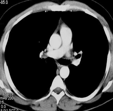
CT Chest. Verification of an opacity seen on the straight chest X ray
|
|
|
- Avis Patrick
- 6 years ago
- Views:
Transcription
1 CT Chest Indications: To assess equivocal plain x-ray findings Staging of lung neoplasm Merastatic workup of extra thoraces malignancies Diagnosis of diffuse lung diseases with HRCT Assessment of bronchietasis Assessment of suspected posttraumatic complications Diagnosis of mediastinal and chest wall lesions Diagnosis of suspected pulmmary embolism Verification of an opacity seen on the straight chest X ray Patient preparation: Fasting 4-6 hours before examination [most of the patients are usually injected with contrast material for adequate delineation of the intrathoracic vascular structures] Water soluble contrast material (urographin,..) is injected IV before start the examination [ 1-2 ML /kgm body weight ] NB: Contrast is not usually needed in these conditions: Evaluation of bronchiectasis Evaluation of diffuse pulmonary parenchymal disease Searching for pul. parenchymal deposits Some cases of trauma Patient s position: Supine Scanogram: Frontal [should include the lower neck and upper abdomen] Sections: 1cm sections from the lung apex to the level of the suprarenal glands in the upper abdomen. Mediastinal window and lung window for all images Bone window for sections showing lesions affecting bones [ribs, spine,.. ]
2 Scanning parameters Standard routine CT chest High resolution CT [HRCT] using special scanning parameters and filters to get more detailed images of the lung paranchyma.. usually needed for assessment of diffuse paranchymal lung diseases and bronchiectasis Helical CT= spiral CT = volumetric CT Allows scanning of the whole thorax in a single breath hold Advantages: Rapid examination technique suitable for children and uncooperative patients Ensures adequate vascular opacification with relatively smaller volume of contrast material injected Avoid respiratory misregestration [ missing small lesions because of respiratory movements ] Mulridetector CT Recently introduced scanners with multiple detectors instead of one detector in old scanners. These machines allow multiple sections per tube rotation. The number of slices increases with the increase in the number of detectors. The available machines now include from detectors. Advantages: o Very rapid scanning time o Image reformation in different planes as Coronal, sagittal, 3D carded reformatted images as well as coloured images. o Non invasive vascular imaging by injecting contrast material intravenously and imaging any of the arterial or venous circulation o Reduction of the contrast material used o Triphasic and function studies o The best modality for diagnosis of pul. embolism
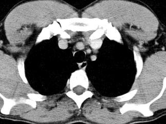
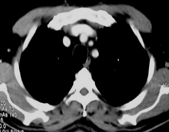
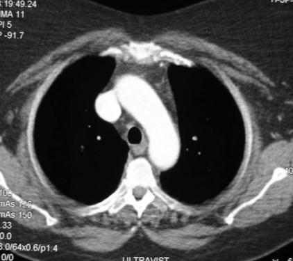
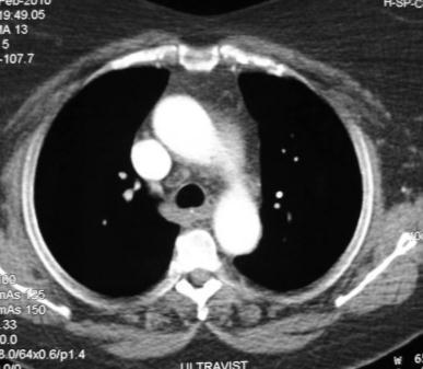
3 Anatomy The aortic arch is our anatomic landmark. At this level we identify the SVC, trachea and esophagus. The space between the sternum and aortic arch represents the anatomic site of the thymus which is normally seen up to the age of 2 years. Sections above the level of the aortic arch show the major aortic branches (left subclavian, left common carotid and innominate arteries) as well as both innominate veins + trachea and esophagus Sections below the aortic arch show the ascending and descending aorta with the pulmonary artery in between. The SVC is seen postrolateral to the ascending aorta. Lower down sections will show different cardiac chambers, descending aorta and esophagus Anatomic sites of intrathoracic lymph nodes: NB Normal lymph nodes are not usually seen in CT scan with minor exceptions Internal mammary lymph nodes along the postro lateral aspect of the sterum on both sides Retrocaval (Rt paratracheal) lymph nodes posterior to the SVC Pre vascular (retrosternal) lymph nodes along the antrolateral aspect of the aortic arch Aortic window lymph nodes between the ascending and descending aorta above the pulmonary artery Carinal lymph nodes around the tracheal bifurcation Subcarinal lymph nodes between the mainstem bronchi Hilar (broncho -pulmonary) lymph nodes at the left and Rt hilar regions Circum cardiac lymph nodes around the pericardium Posterior mediastinal (Zygo esophageal) lymph nodes near the lower esophagus
4

5

6
7 Segmental anatomy The anatomic land mark for labor and segmental lung anatomy is the tracheal bifurcation Sections above the level of tracheal bifurcation (upper sections) Sections at the tracheal bifurcation (middle sections) Sections below the tracheal bifurcation (lower sections) In the upper sections the trachea appears as rounded air filled structure. The 1 st two sections in this group pass through the lung apex [On the Rt side known as the apical segment of the upper lobe while on the left side it is called the apicoposterior segment of the upper lobe] Lower down in this group, one can identify 3 segments on the Rt side [anterior, apical and posterior segments] while on the side the apical and posterior segments form only one segment, so according to the figure, we can identify only 2 segments [ anterior and apico posterior] In middle sections the trachea has divided into two mainstem bronchi. At this level 2 segments are seen on both sides [the anterior segment of the upper lobe and the superior segment of the lower lobe]. No difference between the Rt and left side in this group In the lower sections the mainstem bronchi are not seen, subsegmental bronchi may be identified and the cardiac shadow is seen These sections are divided in an anterior one third which corresponds to the middle lobe on the Rt side and the lingula on the left side The posterior two thirds correspond to the lower lobe segments Differences between Rt and left side: The segments of the middle lobe are medial and lateral The segments of the lingula are superior and inferior Four segments are seen in the Rt lower lobe [anterior, posterior, medial and lateral] No medial segment in the left lower lobe because of the heart left


8 In order to know the lobar anatomy of the lung on CT basis we have to divide the CT images into 3 major groups: Group I: The images where you can see the trachea Group II: The level of the tracheal bifurcation Group III: The images below the tracheal bifurcation R UL UL L In this image the trachea is seen as an air filled rounded midline structure. Wherever you see the trachea, you are cutting in the upper lobe (UL)
 and the posterior half belongs to")

9 UL UL LL In this image you can see the tracheal bifurcation. Whenever the trachea is divided into 2 main stem bronchi, the section is also divided into two halves, the anterior half belongs to the upper lobe (UL) and the posterior half belongs to the lower lobe (LL) ML Ln LL LL In this image you can see the cardiac shadow, no trachea, no main stem bronchi. This section is divided into an anterior 1/3 and posterior 2/3. The anterior 1/3 on the left side belongs to the lingua (Ln) while the anterior 1/3 on the right side belongs to the middle lobe (ML). The posterior 2/3 on both sides belong to the lower lobe (LL)
Undergraduate Teaching
 Prof. James F Meaney Undergraduate Teaching Chest X-Ray Understanding the normal anatomical by reference to cross sectional imaging Radiology? It s FUN! Cryptic puzzle Sudoku (Minecraft?) It s completely
Mediastinum It is a thick movable partition between the two pleural sacs & lungs. It contains all the structures which lie
 Dr Jamila EL medany OBJECTIVES At the end of the lecture, students should be able to: Define the Mediastinum. Differentiate between the divisions of the mediastinum. List the boundaries and contents of
DESCRIPTION: This is the part of the trunk, which is located between the root of the neck and the superior border of the abdominal region.
 1 THE THORACIC REGION DESCRIPTION: This is the part of the trunk, which is located between the root of the neck and the superior border of the abdominal region. SHAPE : T It has the shape of a truncated
Chest X-ray Interpretation
 Chest X-ray Interpretation Introduction Routinely obtained Pulmonary specialist consultation Inherent physical exam limitations Chest x-ray limitations Physical exam and chest x-ray provide compliment
Lecturer: Ms DS Pillay ROOM 2P24 25 February 2013
 Lecturer: Ms DS Pillay ROOM 2P24 25 February 2013 Thoracic Wall Consists of thoracic cage Muscle Fascia Thoracic Cavity 3 Compartments of the Thorax (Great Vessels) (Heart) Superior thoracic aperture
Lab #3. Mohammad Hisham Al-Mohtaseb. Jumana Jihad. Ammar Ramadan. 0 P a g e
 Lab #3 Mohammad Hisham Al-Mohtaseb Jumana Jihad Ammar Ramadan 0 P a g e Last anatomy lab: Lungs and structure on the mediastinal surfs: 1-the right lung: How do we know it s the right lung??? -the 3 lobes
Lung & Pleura. The Topics :
 Lung & Pleura The Topics : The Trachea. The Bronchi. The Brochopulmonary Segments. The Lungs. The Hilum. The Pleura. The Surface Anatomy Of The Lung & Pleura. The Root & Hilum. - first of all, the lung
Chest and cardiovascular
 Module 1 Chest and cardiovascular A. Doss and M. J. Bull 1. Regarding the imaging modalities of the chest: High resolution computed tomography (HRCT) uses a slice thickness of 4 6 mm to identify mass lesions
Dr. Weyrich G07: Superior and Posterior Mediastina. Reading: 1. Gray s Anatomy for Students, chapter 3
 Dr. Weyrich G07: Superior and Posterior Mediastina Reading: 1. Gray s Anatomy for Students, chapter 3 Objectives: 1. Subdivisions of mediastinum 2. Structures in Superior mediastinum 3. Structures in Posterior
Monitor Images for Respiratory System Dissection
 Monitor Images for Respiratory System Dissection **This document includes extra images of the radiology of the bronchopulmonary segments. These imaged are an excellent way to review the three-dimensional
Large veins of the thorax Brachiocephalic veins
 Large veins of the thorax Brachiocephalic veins Right brachiocephalic vein: formed at the root of the neck by the union of the right subclavian & the right internal jugular veins. Left brachiocephalic
Mediastinum and pericardium
 Mediastinum and pericardium Prof. Abdulameer Al-Nuaimi E-mail: a.al-nuaimi@sheffield.ac.uk E. mail: abdulameerh@yahoo.com The mediastinum: is the central compartment of the thoracic cavity surrounded by
Identify the lines used in anatomical surface descriptions of the thorax. median line mid-axillary line mid-clavicular line
 L 14 A B O R A T O R Y Thorax THORACIC WALL Identify the lines used in anatomical surface descriptions of the thorax. median line mid-axillary line mid-clavicular line Identify the surface landmarks of
Do you want to be an excellent Radiologist? - Focus on the thoracic aorta on lateral chest image!!!
 The lateral chest radiograph: Challenging area around the thoracic aorta!!! Do you want to be an excellent Radiologist? - Focus on the thoracic aorta on lateral chest image!!! Dong Yoon Han 1, So Youn
International Association for the Study of Lung Cancer lymph node map Lymph node stations Imaging CT
 Review of the International Association for the Study of Lung Cancer Lymph Node Classification System Localization of Lymph Node Stations on CT Imaging Hamza Jawad, MBBS a, Arlene Sirajuddin, MD b, Jonathan
OBJECTIVE: To obtain a fundamental knowledge of the root of the neck with respect to structure and function
 The root of the neck Jeff Dupree, Ph.D. e mail: jldupree@vcu.edu OBJECTIVE: To obtain a fundamental knowledge of the root of the neck with respect to structure and function READING ASSIGNMENT: Moore and
Sectional Anatomy Quiz - III
 Sectional Anatomy - III Rashid Hashmi * Rural Clinical School, University of New South Wales (UNSW), Wagga Wagga, NSW, Australia A R T I C L E I N F O Article type: Article history: Received: 30 Jun 2018
Right lung. -fissures:
 -Right lung is shorter and wider because it is compressed by the right copula of the diaphragm by the live.. 2 fissure, 3 lobes.. hilum : 2 bronchi ( ep-arterial, hyp-arterial ), one artery mediastinal
11.1 The Aortic Arch General Anatomy of the Ascending Aorta and the Aortic Arch Surgical Anatomy of the Aorta
 456 11 Surgical Anatomy of the Aorta 11.1 The Aortic Arch 11.1.1 General Anatomy of the Ascending Aorta and the Aortic Arch Surgery of the is one of the most challenging areas of cardiac and vascular surgery,
CT Depiction of Regional Nodal Stations for Lung Cancer Staging
 ownloaded from www.ajronline.org by 37.44.204.189 on 11/24/17 from IP address 37.44.204.189. opyright RRS. For personal use only; all rights reserved T epiction of Regional Nodal Stations for Lung ancer
Pulmonary vascular anatomy & anatomical variants
 Review Article Pulmonary vascular anatomy & anatomical variants Asha Kandathil, Murthy Chamarthy Department of Radiology, University of Texas Southwestern Medical Center, Dallas, TX, USA Contributions:
Chest X-ray (CXR) Interpretation Brent Burbridge, MD, FRCPC
 Interpretation Brent Burbridge, MD, FRCPC") Chest X-ray (CXR) Interpretation Brent Burbridge, MD, FRCPC An approach to reviewing a chest x-ray will create a foundation that will facilitate the detection of abnormalities. You should create your own
Chest X-ray (CXR) Interpretation Brent Burbridge, MD, FRCPC An approach to reviewing a chest x-ray will create a foundation that will facilitate the detection of abnormalities. You should create your own
10/14/2018 Dr. Shatarat
 2018 Objectives To discuss mediastina and its boundaries To discuss and explain the contents of the superior mediastinum To describe the great veins of the superior mediastinum To describe the Arch of
Anatomy Sheet #5. In the previous lecture, we finished discussion about the larynx; now we continue with trachea, lungs and pleura.
 Anatomy Sheet #5 In the previous lecture, we finished discussion about the larynx; now we continue with trachea, lungs and pleura. Trachea and lungs The knowledge about the pleura and lungs is very important
THE GOOFY ANATOMIST QUIZZES
 THE GOOFY ANATOMIST QUIZZES 7. LUNGS Q1. Fill in the blanks: the lung has lobes and fissures. A. Right, three, two. B. Right, two, one. C. Left, three, two. D. Left, two, three. Q2. The base of the lung
This lab activity is aligned with Visible Body s A&P app. Learn more at visiblebody.com/professors
 1 This lab activity is aligned with Visible Body s A&P app. Learn more at visiblebody.com/professors 2 PRE-LAB EXERCISES: A. Watch the video 29.1 Heart Overview and make the following observations: 1.
UERMMMC Department of Radiology. Basic Chest Radiology
 UERMMMC Department of Radiology Basic Chest Radiology PHYSICS DENSITIES BONE SOFT TISSUES WATER FAT AIR TELEROENTGENOGRAM Criteria for an Ideal Chest Radiograph 1. Upright 2. Posteroanterior View 3. Full
Thoracic Imaging: A Case of Metastatic Adenocarcinoma of Unknown Primary
 January 28, 2009 Thoracic Imaging: A Case of Metastatic Adenocarcinoma of Unknown Primary Kristina Mirabeau-Beale, Harvard Medical School Year III Gillian Lieberman, MD Agenda Introduce Patient RS Discuss
FUNDAMENTALS OF CXR INTERPRETATION THE BASICS
 FUNDAMENTALS OF CXR INTERPRETATION THE BASICS PART I QUALITY ASSESSMENT 1 PATIENT-DEPENDENT FACTORS 3 REVIEW OF IMPORTANT ANATOMY 7 LUNGS AND PLEURA 11 DIAPHRAGMS 13 BONES AND SOFT TISSUES 14 A BRIEF LOOK
Mediastinum. Respiratory block-anatomy-lecture 6. Editing file
 Mediastinum Respiratory block-anatomy-lecture 6 Editing file Objectives At the end of the lecture, students should be able to: Define the Mediastinum. Differentiate between the divisions of the mediastinum.
Lab CT scan. Murad Kharabsheh Yaman Alali
 Lab CT scan Murad Kharabsheh Yaman Alali Some rules to read The CT Scan : 1. Remember that it s a transverse section across the body and we are looking at the inferior part of the section (not the superior),
AJCC-NCRA Education Needs Assessment Results
 AJCC-NCRA Education Needs Assessment Results Donna M. Gress, RHIT, CTR Survey Tool 1 Survey Development, Delivery, Analysis THANKS to NCRA for the following work Developed survey with input from partners
Signs in Chest Radiology
 Signs in Chest Radiology Jonathan H. Chung, MD Disclosures No pertinent disclosures Jonathan H. Chung, MD Assistant Professor Institute t of fadvanced d Biomedical Imaging National Jewish Health Denver,
Superior and Posterior Mediastinum. Assoc. Prof. Jenny Hayes
 Superior and Posterior Mediastinum Assoc. Prof. Jenny Hayes WARNING This material has been provided to you pursuant to section 49 of the Copyright Act 1968 (the Act) for the purposes of research or study.
Introduction to Chest CT Interpretation. Objectives 8/28/2017
 Introduction to Chest CT Interpretation Deborah Stein ACNP BC, CCRN NP Education Specialist Department of Anesthesia and Critical Care Medicine August 28, 2017 Objectives Basic Principles Thoracic Anatomy
Anatomy of the Lungs. Dr. Gondo Gozali Department of anatomy
 Anatomy of the Lungs Dr. Gondo Gozali Department of anatomy 1 Pulmonary Function Ventilation and Respiration Ventilation is the movement of air in and out of the lungs Respiration is the process of gas
Advances in MDCT of Thoracic Trauma
 Baltic Congress of Radiology, Riga 2010 Advances in MDCT of Thoracic Trauma Robert A. Novelline, MD Professor of Radiology, Harvard Medical School Director of Emergency Radiology, Massachusetts General
Sheet lab 5 Anatomy: CT Scans
 Sheet lab 5 Anatomy: CT Scans In the orientation we see the picture from downward to upward. The first picture is a CT scan at the level of the heart. Left border of the heart is the left ventricle and
Imaging of Thoracic Trauma: Tips and Traps. Arun C. Nachiappan, MD Associate Professor of Clinical Radiology University of Pennsylvania
 Imaging of Thoracic Trauma: Tips and Traps Arun C. Nachiappan, MD Associate Professor of Clinical Radiology University of Pennsylvania None Disclosures Objectives Describe blunt and penetrating traumatic
Assignable revenue codes: Explanation of services:
 computed tomography Chest/Cardiac Assignable revenue codes: Explanation of services: 0350 CT Scan General Classification 0351 CT Scan Head Scan 0352 CT Scan Body Scan 0359 CT Scan Other CT Scans Known
Atlases for Organs at Risk (OARs) in Thoracic Radiation Therapy
 in Thoracic Radiation Therapy") Atlases for Organs at Risk (OARs) in Thoracic Radiation Therapy Feng-Ming (Spring) Kong MD PhD Leslie Quint MD Mitchell Machtay MD Jeffrey Bradley MD 1 Outline of Content Atlas for lung, esophagus, and
Atlases for Organs at Risk (OARs) in Thoracic Radiation Therapy Feng-Ming (Spring) Kong MD PhD Leslie Quint MD Mitchell Machtay MD Jeffrey Bradley MD 1 Outline of Content Atlas for lung, esophagus, and
CT abdomen and pelvis
 CT abdomen and pelvis General indications: Assessment of vague abdominal symptoms (pain, colics,distenstion,...) Varifecation of a lesion discovered by other diagnostic modalities as US, barium,ivp, Staging
After the Chest X-Ray:
 After the Chest X-Ray: What To Do Next Alan S. Brody Professor of Radiology and Pediatrics Chief of Thoracic Imaging Cincinnati Children s Hospital Cincinnati, Ohio USA What Should We Do Next? CT scan?
Sectional Anatomy Quiz II
 Sectional Anatomy II Rashid Hashmi Rural Clinical School, University of New South Wales, Wagga Wagga, New South Wales, Australia A R T I C L E I N F O Article type: Article history: Received: 3 Aug 2017
slide 23 The lobes in the right and left lungs are divided into segments,which called bronchopulmonary segments
 Done By : Rahmeh Alsukkar Date : 26 /10/2017 slide 23 The lobes in the right and left lungs are divided into segments,which called bronchopulmonary segments Each segmental bronchus passes to a structurally
Cardiovascular system:
 Cardiovascular system: Mediastinum: The mediastinum: lies between the right and left pleura and lungs. It extends from the sternum in front to the vertebral column behind, and from the root of the neck
Anatomy. Contents Brain (Questions)
") Anatomy 12 Contents 12.1 Brain (Questions).................................................... 683 12.2 Head and Neck (Questions)............................................. 685 12.3 Thorax (Questions)...................................................
Anatomy 12 Contents 12.1 Brain (Questions).................................................... 683 12.2 Head and Neck (Questions)............................................. 685 12.3 Thorax (Questions)...................................................
Theme 30. Structure, topography and function of the lungs and pleura. Mediastinum and its contents. X -ray films digestive and respiratory systems.
 Theme 30. Structure, topography and function of the lungs and pleura. Mediastinum and its contents. X -ray films digestive and respiratory systems. STRUCTURE, TOPOGRAPHY AND FUNCTІON OF LUNGS AND PLEURA.
Chapter 5: Other mediastinal structures. The Large Arteries. The Aorta. Ascending aorta
 Chapter 5: Other mediastinal structures The Large Arteries The Aorta The aorta is the main arterial trunk of the systemic circulation and in the healthy state its wall contain a large amount of yellow
THYROID & PARATHYROID. By Prof. Saeed Abuel Makarem & Dr. Sanaa Al-Sharawy
 THYROID & PARATHYROID By Prof. Saeed Abuel Makarem & Dr. Sanaa Al-Sharawy 1 OBJECTIVES By the end of the lecture, the student should be able to: Describe the shape, position, relations and structure of
Radiological Anatomy of Thorax. Dr. Jamila Elmedany & Prof. Saeed Abuel Makarem
 Radiological Anatomy of Thorax Dr. Jamila Elmedany & Prof. Saeed Abuel Makarem Indications for Chest x - A chest x-ray may be used to diagnose and plan treatment for various conditions, including: Diseases/Fractures
B-I-2 CARDIAC AND VASCULAR RADIOLOGY
 (YEARS 1 3) CURRICULUM FOR RADIOLOGY 13 B-I-2 CARDIAC AND VASCULAR RADIOLOGY KNOWLEDGE To describe the normal anatomy of the heart and vessels including the lymphatic system as demonstrated by radiographs,
The External Anatomy of the Lungs. Prof Oluwadiya KS
 The External Anatomy of the Lungs Prof Oluwadiya KS www.oluwadiya.com Introduction The lungs are the vital organs of respiration Their main function is to oxygenate the blood by bringing inspired air into
PRACTICAL GUIDE TO FETAL ECHOCARDIOGRAPHY IC Huggon and LD Allan
 PRACTICAL GUIDE TO FETAL ECHOCARDIOGRAPHY IC Huggon and LD Allan Fetal Cardiology Unit, Harris Birthright Research Centre for Fetal Medicine, King's College Hospital, London, UK IMPORTANCE OF PRENATAL
Cardiac Radiography. Jared D. Christensen, M.D.
 Cardiac Radiography Jared D. Christensen, M.D. Cardiac radiography Jared D. Christensen, M.D. Overview Basic Concepts Technique Normal anatomy Cases Technique 3 Standard Views Posterior-Anterior (PA) Anterior-Posterior
Tests Your Pulmonologist Might Order. Center For Cardiac Fitness Pulmonary Rehab Program The Miriam Hospital
 Tests Your Pulmonologist Might Order Center For Cardiac Fitness Pulmonary Rehab Program The Miriam Hospital BASIC ANATOMY OF THE LUNGS Lobes of Lung 3 lobes on the Right lung 2 lobes on the Left Blood
Anterior Mediastinal Masses: The 4 T s
 May 2001 Anterior Mediastinal Masses: The 4 T s Rachel Van Sambeek, Harvard Medical School, Year III 1 Mediastinal Compartments 3 arbitrary divisions that do not correlate with anatomic planes: Anterior
Chapter 2 Cardiac Interpretation of Pediatric Chest X-Ray
 Chapter 2 Cardiac Interpretation of Pediatric Chest X-Ray Ra-id Abdulla and Douglas M. Luxenberg Key Facts The cardiac silhouette occupies 50 55% of the chest width on an anterior posterior chest X-ray
Geography of Pulmo Park: Landmarks. Cards 1A
 Geography of Pulmo Park: Landmarks Cards 1A Directions: Cut out the cards, fold and laminate and then hand out to students (one each). If you have more cards than students, some students will have more
Surface anatomy of Cardiovascular system
 Surface anatomy of Cardiovascular system Prof. Abdulameer Al-Nuaimi E-mail: a.al-nuaimi@sheffield.ac.uk E. mail: abdulameerh@yahoo.com The lines cover the front, side, and back of the thorax Midsternal
THE DESCENDING THORACIC AORTA
 Intercostal Arteries and Veins Each intercostal space contains a large single posterior intercostal artery and two small anterior intercostal arteries. The anterior intercostal arteries of the lower spaces
BOGOMOLETS NATIONAL MEDICAL UNIVERSITY DEPARTMENT OF HUMAN ANATOMY. Guidelines. Module 2 Topic of the lesson Aorta. Thoracic aorta.
 BOGOMOLETS NATIONAL MEDICAL UNIVERSITY DEPARTMENT OF HUMAN ANATOMY Guidelines Academic discipline HUMAN ANATOMY Module 2 Topic of the lesson Aorta. Thoracic aorta. Course 1 The number of hours 3 1. The
Dana Alrafaiah. - Moayyad Al-Shafei. -Mohammad H. Al-Mohtaseb. 1 P a g e
 - 6 - Dana Alrafaiah - Moayyad Al-Shafei -Mohammad H. Al-Mohtaseb 1 P a g e Quick recap: Both lungs have an apex, base, mediastinal and costal surfaces, anterior and posterior borders. The right lung,
Collaborative Stage. Site-Specific Instructions - LUNG
 Slide 1 Collaborative Stage Site-Specific Instructions - LUNG In this presentation, we are going to review the AJCC Cancer Staging criteria for the lung primary site. Slide 2 Reading Assignments As each
Anatomy Lecture 8. In the previous lecture we talked about the lungs, and their surface anatomy:
 Anatomy Lecture 8 In the previous lecture we talked about the lungs, and their surface anatomy: 1-Apex:it lies 1 inch above the medial third of clavicle. 2-Anterior border: it starts from apex to the midpoint
Thorax Lecture 2 Thoracic cavity.
 Thorax Lecture 2 Thoracic cavity. Spring 2016 Dr. Maher Hadidi, University of Jordan 1 Enclosed by the thoracic wall. Extends between (thoracic inlet) & (thoracic outlet). Thoracic inlet At root of the
1\ :_:'f~~---;-f_il_~_q V\_ re_--:- ( L t-j ~~ ) s ( Jc (/6 t~--l -i~,jl..f- t:j Ll f1 ilb}.c
 s ( Jc (/6 t~--l -i~,jl..f- t:j Ll f1 ilb}.c") t-=-=e.~~~t.:::_"~ :.fl...:...-c..:... 1\ :_:'f~~---;-f_il_~_q V\_ re_--:- ( L t-j ~~ ) s ( Jc (/6 t~--l -i~,jl..f- t:j Ll f1 ilb}.c c6 ~ { oo d! Lt 1e ce i v-u /-r-(j'r\-'1. 4-.z.)/ {) Pt c/o -~ ~,:u
t-=-=e.~~~t.:::_"~ :.fl...:...-c..:... 1\ :_:'f~~---;-f_il_~_q V\_ re_--:- ( L t-j ~~ ) s ( Jc (/6 t~--l -i~,jl..f- t:j Ll f1 ilb}.c c6 ~ { oo d! Lt 1e ce i v-u /-r-(j'r\-'1. 4-.z.)/ {) Pt c/o -~ ~,:u
Chest imaging (Case-based teaching) 胸腔病例教學 謝叔強 財團法人恩主公醫院主治醫師萬芳醫院放射科兼任主治醫師.
 胸腔病例教學 謝叔強 財團法人恩主公醫院主治醫師萬芳醫院放射科兼任主治醫師.") Chest imaging (Case-based teaching) 胸腔病例教學 謝叔強 財團法人恩主公醫院主治醫師萬芳醫院放射科兼任主治醫師 e510019@gmail.com Check List(1) 1. Check patient data, position, technical quality and normal anatomy. 2. Review systematically
Chest imaging (Case-based teaching) 胸腔病例教學 謝叔強 財團法人恩主公醫院主治醫師萬芳醫院放射科兼任主治醫師 e510019@gmail.com Check List(1) 1. Check patient data, position, technical quality and normal anatomy. 2. Review systematically
Anatomy notes-thorax.
 Anatomy notes-thorax. Thorax: the part extending from the root of the neck to the abdomen. Parts of the thorax: - Thoracic cage (bones). - Thoracic wall. - Thoracic cavity. ** The thoracic cavity is covered
CARDIOVASCULAR DANIL HAMMOUDI.MD
 CARDIOVASCULAR DANIL HAMMOUDI.MD 18 Systemic Circulation Figure 19.19 Pulmonary Circulation Figure 19.18b 1. Thyroid gland 2. Trachea 3. Brachiocephalic 4. Common carotid 5. Internal jugular 6. Superior
Manage TB Dr. A. Chitrakumar Madras Medical College and RGGGH Institute of Thoracic Medicine, Chennai
 Manage TB Dr. A. Chitrakumar Madras Medical College and RGGGH Institute of Thoracic Medicine, Chennai Lecture 16 Radiology in diagnosis of Tuberculosis Session 01 So, welcome to the session Radiology in
ABSTRACT. Journal of Radiation Research, Vol. 58, No. 1, 2017, pp doi: /jrr/rrw076 Advance Access Publication: 8 September 2016
 Journal of Radiation Research, Vol. 58, No. 1, 2017, pp. 86 105 doi: 10.1093/jrr/rrw076 Advance Access Publication: 8 September 2016 The Japan Lung Cancer Society Japanese Society for Radiation Oncology
BMS101 INTRODUCTION TO THE HUMAN BODY STUDY NOTES EXTRACTS FOR SAMPLE
 BMS101 INTRODUCTION TO THE HUMAN BODY STUDY NOTES EXTRACTS FOR SAMPLE What is anatomy? Anatomy is the study of structure. Greek for to cut up or cut open What is physiology? Physiology studies the function
10/17/2016. Nuts and Bolts of Thoracic Radiology. Objectives. Techniques
 Nuts and Bolts of Thoracic Radiology October 20, 2016 Carleen Risaliti Objectives Understand the basics of chest radiograph Develop a system for interpreting chest radiographs Correctly identify thoracic
The Human Body. Lesson Goal. Lesson Objectives 9/10/2012. Provide a brief overview of body systems, anatomy, physiology, and topographic anatomy
 The Human Body Lesson Goal Provide a brief overview of body systems, anatomy, physiology, and topographic anatomy Medial Lateral Proximal Distal Superior Inferior Anterior Lesson Objectives Explain the
Syllabus: 6 pages (Page 6 lists corresponding figures for Grant's Atlas 11 th & 12 th Eds.)
") PLEURAL CAVITY AND LUNGS Dr. Milton M. Sholley SELF STUDY RESOURCES Essential Clinical Anatomy 3 rd ed. (ECA): pp. 70 81 Syllabus: 6 pages (Page 6 lists corresponding figures for Grant's Atlas 11 th &
PLEURAL CAVITY AND LUNGS Dr. Milton M. Sholley SELF STUDY RESOURCES Essential Clinical Anatomy 3 rd ed. (ECA): pp. 70 81 Syllabus: 6 pages (Page 6 lists corresponding figures for Grant's Atlas 11 th &
CHEST CT PROTOCOL FOR MULTIPLE DETECTOR ROW SCANNERS
 1. Standard ed 2. Standard ed & Abdomen 3. Standard ed, Abdomen, & Pelvis 4. Aortic Dissection arch dome thru adrenals apex arch + 2 arch dome thru abdomen apex arch + 2 arch dome to crests apex arch +
This is not a required assignment but it is recommended.
 SU 12 Name: This is not a required assignment but it is recommended. BIO 116 - Anatomy & Physiology II Practice Assignment 2 - The Respiratory and Cardiovascular Systems 1. The exchange of oxygen and carbon
Chapter 2 The Human Cardiovascular System
 Chapter 2 The Human Cardiovascular System 2.1 Introduction Before delving into the computational methods of CHD, this chapter provides a preliminary understanding of the circulatory system from a physiological
Seventh Edition of the Cancer Staging Manual and Stage Grouping of Lung Cancer. Quick Reference Chart and Diagrams
 CHEST Special Features Seventh Edition of the Cancer Staging Manual and Stage Grouping of Lung Cancer Quick Reference Chart and Diagrams Omar Lababede, MD ; Moulay Meziane, MD ; and Thomas Rice, MD, FCCP
Mediastinal and Hilar Lymphadenopathy: Cross-Referenced Anatomy on Axial and Coronal Images Displayed by Using Multi-detector row CT 1
 Mediastinal and Hilar Lymphadenopathy: Cross-Referenced natomy on xial and Coronal Images Displayed by Using Multi-detector row CT 1 Ju-Hyun Lee, M.D., Kyung Soo Lee, M.D., Tae Sung Kim, M.D., Chin Yi,
Surgical anatomy of thyroid and parathyroid glands
 Head & Neck Surgery Course Surgical anatomy of thyroid and parathyroid glands Dr Pierfrancesco PELLICCIA Pr Benjamin LALLEMANT Service ORL et CMF CHU de Nîmes CH de Arles Thyroid glands Dr Pierfrancesco
Case 47 Clinical Presentation
 93 Case 47 C Clinical Presentation 45-year-old man presents with chest pain and new onset of a murmur. Echocardiography shows severe aortic insufficiency. 94 RadCases Cardiac Imaging Imaging Findings C
Disclosure. Clinical Chest Radiography Interpretation Part I
 Clinical Chest Radiography Interpretation Part I Anthony M. Angelow, PhD(c), MSN, ACNPC, AGACNP-BC, CEN Associate Lecturer, Fitzgerald Health Education Associates Clinical practice Division of Trauma Surgery
Lecture 2: Clinical anatomy of thoracic cage and cavity II
 Lecture 2: Clinical anatomy of thoracic cage and cavity II Dr. Rehan Asad At the end of this session, the student should be able to: Identify and discuss clinical anatomy of mediastinum such as its deflection,
Attenuation value in HU From -500 To HU From -10 To HU From 60 To 90 HU. From 200 HU and above
 Brain Imaging Common CT attenuation values Structure Air Fat Water Brain tissue Recent hematoma Calcifications Bone Brain edema and infarction Normal liver parenchyma Attenuation value in HU From -500
Parenchyma-sparing lung resections are a potential therapeutic
 Lung Segmentectomy for Patients with Peripheral T1 Lesions Bryan A. Whitson, MD, Rafael S. Andrade, MD, and Michael A. Maddaus, MD Parenchyma-sparing lung resections are a potential therapeutic option
New Horizons in the Imaging of the Lung
 New Horizons in the Imaging of the Lung Postprocessing. How to do it and when do we need it? Peter M.A. van Ooijen, MSc, PhD Principal Investigator, Radiology, UMCG Discipline Leader Medical Imaging Informatics
Thyroid and Parathyroid Glands
 Thyroid and Parathyroid Glands Please view our Editing File before studying this lecture to check for any changes. Color Code Important Doctors Notes Notes/ explanation Objectives: By the end of the lecture,
Neck Ultrasound. Faculty Info: Amy Kule, MD
 Neck Ultrasound Date: Friday, October 19, 2018 Time: 11:00 AM Location: SMALL GROUP LABORATORY SSOM L71 Watch: Ø Neck Ultrasound Scanning Protocol (4:00): https://www.youtube.com/watch?v=zozd2x2ll4q Faculty
Mohammad Hisham Al-Mohtaseb. Lina Mansour. Enas Ajarma
 6 Mohammad Hisham Al-Mohtaseb Lina Mansour Enas Ajarma Some recommended videos are attached to this sheet ( if u are studying online click on them, if not u can reach them by typing their names on the
Sheet. April/14 th /2013. Introduction to Anatomy. Dr. Maher Hadidi. Muna Abu Hijleh. 1 P a g e
 Sheet Introduction to Anatomy Dr. Maher Hadidi Muna Abu Hijleh 1 P a g e 29 April/14 th /2013 Superior & Posterior Mediastinum ***Superior mediastinum * is bounded from: -Anterior by manubrium sterni -posterior
Chapter 2. Relevant Thoracic Anatomy. Jed A. Gorden. 1. Central Airway Anatomy. 2. Upper Airway
 Chapter 2 Relevant Thoracic Anatomy Jed A. Gorden 1. Central Airway Anatomy This section outlines the basic anatomy of the tracheobronchial tree, including lengths and diameters (Fig. 2.1). This serves
JlntSocPlastination, Vol4:16-22,
 JlntSocPlastination, Vol4:16-22, 1990 16 SECTIONAL ANATOMY: STANDARDIZED METHODOLOGY Alexander Lane, Coordinator of Anatomy and Physiology, Triton College, Visiting Associate Professor, University of Illinois
Interpreting thoracic x-ray of the supine immobile patient: Syllabus
 Interpreting thoracic x-ray of the supine immobile patient: Syllabus Johannes Godt Dep. of Radiology and Nuclear Medicine Oslo University Hospital Ullevål NORDTER 2017, Helsinki Content - Why bedside chest
Mediastinum. Lecture 5. Please check our Editing File. ھذا العمل لا یغني عن المصدر الا ساسي للمذاكرة
 Mediastinum Lecture 5 Please check our Editing File. ھذا العمل لا یغني عن المصدر الا ساسي للمذاكرة Objectives At the end of the lecture, students should be able to: Define the Mediastinum. Differentiate
Lies in front and sides of the neck. Consists of two lobe connected anterior to the trachea by an isthmus.
 THYROID GLAND 1 Lies in front and sides of the neck. Consists of two lobe connected anterior to the trachea by an isthmus. A small pyramidal lobe projects upwards from the left lobe in 40% of cases. The
Focused Assessment Sonography of Trauma (FAST) Scanning Protocol
 Scanning Protocol") Focused Assessment Sonography of Trauma (FAST) Scanning Protocol Romolo Gaspari CHAPTER 3 GOAL OF THE FAST EXAM Demonstrate free fluid in abdomen, pleural space, or pericardial space. EMERGENCY ULTRASOUND
Focused Assessment Sonography of Trauma (FAST) Scanning Protocol Romolo Gaspari CHAPTER 3 GOAL OF THE FAST EXAM Demonstrate free fluid in abdomen, pleural space, or pericardial space. EMERGENCY ULTRASOUND
Research Article Three-Dimensional Reconstruction of Thoracic Structures: Based on Chinese Visible Human
 Hindawi Publishing Corporation Computational and Mathematical Methods in Medicine Volume 0, Article ID 0, pages http://dx.doi.org/./0/0 Research Article Three-Dimensional Reconstruction of Thoracic Structures:
Clinical Applications
 C H A P T E R 16 Clinical Applications In selecting pulse sequences and measurement parameters for a specific application, MRI allows the user tremendous flexibility to produce variations in contrast between