Advances in MDCT of Thoracic Trauma
|
|
|
- Barrie Lamb
- 5 years ago
- Views:
Transcription


1 Baltic Congress of Radiology, Riga 2010 Advances in MDCT of Thoracic Trauma Robert A. Novelline, MD Professor of Radiology, Harvard Medical School Director of Emergency Radiology, Massachusetts General Hospital
2 Instructional Objectives: 1. Describe MDCT Protocol for Chest Trauma 2. Review MDCT findings with important injuries of: 1. Pleural spaces 2. Lungs 3. Thoracic Aorta 4. Heart 5. Extra-Aortic Thoracic Arteries 6. Airways 7. Thoracic skeleton Disclosure Statement: I have no relevant financial relationships to disclose


3 Chest Trauma: MDCT Protocol Contrast Material: cc of 370mg concentration IV contrast at 2.5 to 3.0cc/sec Scan at 30 sec delay Scanning Techniques: 4 Slice Detector configuration of 4X1.25mm, view at 2.5/5mm slices 16 Slice Detector configuration of 16x1.25mm, view at 2.5/5mm slices 64 Slice Acquired at 0.625mm, view at 2.5/5mm slices Routine coronal and sagittal reformations Optional volumetric, MIP or CTA reformations 3D workstation available 24/7 May be performed as total body trauma scan
4 1. Injuries of the Pleural Space Hemothorax Occult pneumothorax Tension pneumothorax Pneumothorax size


5 Hemothorax? Active Bleed? Always Measure HU of Pleural Fluid Effusion 7 HU Hemothorax 44 HU with Blood Clots Hemothorax 46 HU With Active Bleed


6 Occult Pneumothorax Definition: Seen at CT but not on chest film >50% pneumothoracies occult In supine patient, air accumulates anteriorly Signs of pneumothorax in supine patient: Increased lucency lower chest Better definition of mediastinum Deep lateral sulcus sign


7 Bilateral Occult Pneumothoraces Increased lucency lower chest Better definition of mediastinum Deep lateral sulcus sign


8 Radiological Signs of Tension Pneumothorax Large pneumothorax with hyperexpansion of ipsilateral chest Splaying ipsilateral ribs Mediastinal shift to opposite side Flattening/inversion ipsilateral hemidiaphragm; deep sulcus sign Before Chest Tube After Chest Tube


9 Do not Overlook Tension Pneumothorax at CT Check scout view and coronal reformats


10 Measuring Pneumothorax Size How Big are They? Right PNX is 496cc; Left PNX is 53.5cc
11 2. Injuries of the lung Lung Contusion Lung Laceration Lung Herniation
12 Pulmonary Contusion Most common lung injury with blunt trauma Hemorrhage and edema in air spaces and interstitium without parenchymal disruption Coup and contra-coup locations Outlines injury track with penetrating trauma


13 Pulmonary Contusion May be solitary or multiple opacities


14 Differentiate Pulmonary Contusion & Aspiration Contusion Contusion Non-segmental, patchy, ill-defined parenchymal opacities Aspiration usually segmental! Thin margin of sparing at pleural surfaces 85% seen in 6 hours; 100% in hours If appearing after 24 hours, not contusion Starts clearing 2-4 days; resolves 10 days Aspiration


15 Pulmonary Contusion with Overlying Right Rib Fracture (Coup Injury)
16 Pulmonary Lacerations Tears of pulmonary parenchyma Due to elastic recoil of lung, tears appear ovoid at CT Caused by blunt forces, or penetrating injuries (stabs, gunshots, rib fractures) Lacerations may be: Air-filled (traumatic pneumatocele) Blood-filled (traumatic hematocele) Both air/blood (traumatic hematopneumocele) Usually accompanied by surrounding contusion Contusion can hide lacerations on radiographs


17 Pulmonary Lacerations
18 Types of Lung Lacerations Blunt Trauma: 1. Compression rupture Laceration within lung parenchyma from chest wall compression causing lung rupture 2. Compression shear Laceration paravertebral location due to shear injury from sudden shifting of lower lobe across the spine 3. Rib penetration tear Small peripheral laceration where a fractured rib has punctured lung 4. Adhesion tear Laceration at lung periphery due to pre-existing adhesion limiting shifting of lung with trauma
19 Types of Lung Lacerations: Penetrating Trauma: 1. Stab Wound Tract of knife or other penetrating object through lung 2. Gunshot Wound Tract of bullet through lung


20 Pulmonary Laceration 1. Compression Rupture (Small)


21 Pulmonary Laceration 1. Compression Rupture (Large)
 Initial Scan")

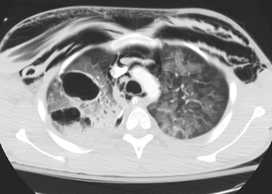
22 Pulmonary Laceration 2. Compression Shear (Small) Initial Scan Vs 7 Days Later Note Paravertebral Location At 7 Days Laceration Looks Like a Pulmonary Nodule


23 Pulmonary Laceration 2. Compression Shear (Large)


24 Pulmonary Laceration 3. Rib Penetration Tear


25 Pulmonary Laceration Stab Wound to Left Posterior Chest


26 Gunshot Wound to Right Chest Contusion outlines bullet track


27 Lung Herniation Protrusion of pulmonary tissue beyond confines of thoracic cage Traumatic form occurs with rib and costochondral fractures Herniated lung may undergo incarceration and strangulation Beware positive pressure ventilation with herniation
28 3. Injuries of the Thoracic Aortic 8000 cases per year in USA Accounts for 15-20% of all MVC fatalities 90% die before reaching hospital Most involve junction of posterior aortic arch and descending aorta Proposed mechanism is rapid deceleration *99.8% accuracy with 64-slice scanner *Steenburg SD, Ravenel JG, Acute traumatic aortic injury: Experience with 64-MDCT, AJR 2008;191:

29 Thoracic Aorta Trauma Radiographic Findings Mediastinal widening Loss of normal aortic arch Enlarged, irregular aortic arch Left apical pleural cap Displacement NG tube and tracheal air shadow to right Loss of descending aortic line


30 Thoracic Aortic Trauma: Coned-down chest film, CT scout view Left apical pleural cap Displacement of trachea and nasogastric tube to the right


31 Thoracic Aortic Trauma CT Findings Indirect Sign Mediastinal hemorrhage Para-aortic hematoma associated with aortic trauma Direct Signs Intimal tear, flap, disruption False aneurysm formation Aortic contour abnormality Thrombus protruding into the lumen Pseudocoarctation Extravasation of IV contrast

32 Initial Portable Chest, Aortic Trauma Another Patient: Less Impressive Mediastinal Widening


33 CT: Intimal Disruption & False Aneurysm


34 Volume Rendered Aortic Arch; Patient Treated with Aortic Stent Provide Volumetric Reformations 24/7


35 Aortic Injury Multiple Trauma


36 Volumetric Images Aortic Trauma
 CT may show evidence of")

37 4. Injuries of the Heart Occurs with anterior chest trauma (Often an unrestrained driver who struck the steering wheel) CT may show evidence of cardiac contusion, cardiac laceration, pericardial laceration CT findings include hemopericardium, pneumopericardium, extravasation contrast material, pericardial defect

38 Right Ventricular Laceration with Active Bleed Courtesy of J. Klein


39 Pericardial Laceration with Pneumopericardium
40 5. Extra-Aortic Vascular Injuries Brachiocephalic arteries and branches Internal mammary arteries Intercostal arteries Any of the above may show intimal disruption, false aneurysm, traumatic occlusion or active bleeding Post-process the positive cases


41 Subclavian Artery Traumatic Occlusion


42 Subclavian Artery Injury Without Occlusion Better Detail on MIP Volumetric Axial Scan MIP


43 Internal Mammary Artery Bleed Post-Embolization

44 6. Airway Injuries: Tracheal Laceration: Tracheal lacerations usually longitudinal and posterior at junction of cartilagenous and membranous trachea See secondary signs of subcutaneous and mediastinal emphysema


45 Cervical Tracheal Laceration 2 ½ year trauma victim intubated in the field


46 Tracheal Laceration: CT


47 Adult with Tracheal Laceration Suffered in MVC From T. Eglin
48 6. Airway Injuries (Continued) Bronchial laceration or fracture Often single transverse laceration within 2cm carina Right main stem bronchus more common than left Often associated with a large pneumothorax, pneumomediastinum, and/or subcutaneous air Pneumothorax fails to resolve with chest tube Bronchial stenosis and occlusion may occur later


49 Right Bronchus Laceration

50 Right Bronchus Laceration


51 One Year Later


52 15 year old with Blunt Chest Trauma; 3 Days Later, Left Lung Collapse from Traumatic Left Bronchus Obstruction


53 Volumetric Airway-Lung Reformation
54 7. Injuries of the Thoracic Skeleton Displaced Rib Fractures Flail Chest Posterior Sternoclavicular dislocation Sternal Fracture Thoracic Spine Fracture


55 Displaced Rib Fractures with Chest Deformity


56 Matrix Rib Fixation Plates Rib Fracture Repair

57 Flail Chest 3 or more sequential ribs fractured in 2 or more places Fractured segment moves paradoxically with respiration Normal respiratory motion inhibited; ventilation impaired 3-6% mortality

58 Flail Chest: 73 year old man with chest pain and difficulty breathing after MVC


59 Right Flail Chest: CT Bone Window


60 Bilateral Flail Chest


61 Posterior Sternoclavicular Dislocation


62 Sternal Fracture


63 Do Not Miss Signs of T-Spine Fracture on Portable Chest Radiograph Look for paraspinal hematoma on all trauma chest radiographs


64 T7 Fracture with Paraspinal Hematoma
65 Summary Recommendations MDCT of Thoracic Trauma 1. Perform thoracic trauma MDCT scans with IV contrast material at optimum arterial opacification 2. Routine coronal and sagittal reformations 3. Volumetric/CTA reformations when indicated


66 Thank You! Robert A. Novelline, MD Professor of Radiology, Harvard Medical School Director of Emergency Radiology, Massachusetts General Hospital
Imaging of Thoracic Trauma: Tips and Traps. Arun C. Nachiappan, MD Associate Professor of Clinical Radiology University of Pennsylvania
 Imaging of Thoracic Trauma: Tips and Traps Arun C. Nachiappan, MD Associate Professor of Clinical Radiology University of Pennsylvania None Disclosures Objectives Describe blunt and penetrating traumatic
Children are not small adults Children are Not Small Adults Anatomic considerations Pliable bony & cartilagenous structures - Significant thoracic inj
 PEDIATRIC CHEST TRAUMA Children are not small adults Role of imaging Spectrum of injury Children are not small adults Children are Not Small Adults Anatomic considerations Pliable bony & cartilagenous
CT of Acute Thoracic Aortic Syndromes Stuart S. Sagel, M.D.
 CT of Acute Thoracic Aortic Syndromes Stuart S. Sagel, M.D. Thoracic Aortic Aneurysms Atherosclerotic Dissection Penetrating ulcer Mycotic Inflammatory (vasculitis) Traumatic Aortic Imaging Options Catheter
Acute Aortic Syndromes
 Acute Aortic Syndromes Carole J. Dennie, MD Acute Thoracic Aortic Syndromes Background Non-Traumatic Acute Thoracic Aortic Syndromes Carole Dennie MD FRCPC Associate Professor of Radiology and Cardiology
MISSED FINDINGS IN EMERGENCY RADIOLOGY: CASE BASE SESSION 5 th Nordic Trauma Radiology Course Oslo, Norway
 MISSED FINDINGS IN EMERGENCY RADIOLOGY: CASE BASE SESSION 5 th Nordic Trauma Radiology Course Oslo, Norway K.SHANMUGANATHAN M.D. EASILY MISSED FINDINGS IN EMERGENCY RADIOLOGY OBJECTIVES Commonly missed
General Imaging. Imaging modalities. Incremental CT. Multislice CT Multislice CT [ MDCT ]
![General Imaging. Imaging modalities. Incremental CT. Multislice CT Multislice CT [ MDCT ]](/thumbs/76/74079340.jpg "General Imaging. Imaging modalities. Incremental CT. Multislice CT Multislice CT [ MDCT ]") General Imaging Imaging modalities Conventional X-rays Ultrasonography [ US ] Computed tomography [ CT ] Radionuclide imaging Magnetic resonance imaging [ MRI ] Angiography conventional, CT,MRI Interventional
General Imaging Imaging modalities Conventional X-rays Ultrasonography [ US ] Computed tomography [ CT ] Radionuclide imaging Magnetic resonance imaging [ MRI ] Angiography conventional, CT,MRI Interventional
B-I-2 CARDIAC AND VASCULAR RADIOLOGY
 (YEARS 1 3) CURRICULUM FOR RADIOLOGY 13 B-I-2 CARDIAC AND VASCULAR RADIOLOGY KNOWLEDGE To describe the normal anatomy of the heart and vessels including the lymphatic system as demonstrated by radiographs,
Advances in Emergency Imaging
 Hampton Symposium,, October 16 th, 2010 Advances in Emergency Imaging Robert A. Novelline, MD Professor of Radiology, Harvard Medical School Director of Emergency Radiology, Massachusetts General Hospital
Interpreting thoracic x-ray of the supine immobile patient: Syllabus
 Interpreting thoracic x-ray of the supine immobile patient: Syllabus Johannes Godt Dep. of Radiology and Nuclear Medicine Oslo University Hospital Ullevål NORDTER 2017, Helsinki Content - Why bedside chest
CT Chest. Verification of an opacity seen on the straight chest X ray
 CT Chest Indications: To assess equivocal plain x-ray findings Staging of lung neoplasm Merastatic workup of extra thoraces malignancies Diagnosis of diffuse lung diseases with HRCT Assessment of bronchietasis
In ESH we usually see blunt chest trauma but penetrating injuries also treated here (usually as single injuries, like stab wound)
") Chest Trauma Dr Csaba Dioszeghy MD PhD FRCEM FFICM FERC East Surrey Hospital Emergency Department Scope Thoracic injuries are common and can be life threatening In ESH we usually see blunt chest trauma
Chest Trauma Dr Csaba Dioszeghy MD PhD FRCEM FFICM FERC East Surrey Hospital Emergency Department Scope Thoracic injuries are common and can be life threatening In ESH we usually see blunt chest trauma
CHEST TRAUMA. Dr Naeem Zia FCPS,FACS,FRCS
 CHEST TRAUMA Dr Naeem Zia FCPS,FACS,FRCS Learning objectives Anatomy of chest wall and thoracic viscera Physiology of respiration and nerve pathways for pain Enumerate different thoracic conditions requiring
Shedding Light on Neonatal X-rays. Objectives. Indications for X-Rays 5/14/2018
 Shedding Light on Neonatal X-rays Barbara C. Mordue, MSN, NNP-BC Neonatal Nurse Practitioner LLUH Children s Hospital, NICU Objectives Utilize a systematic approach to neonatal x-ray interpretation Identify
Chapter 29 - Chest Injuries
 1 2 3 4 5 6 7 8 9 National EMS Education Standard Competencies (1 of 5) Trauma Applies fundamental knowledge to provide basic emergency care and transportation based on assessment findings for an acutely
The Management of Chest Trauma. Tom Scaletta, MD FAAEM Immediate Past President, AAEM
 The Management of Chest Trauma Tom Scaletta, MD FAAEM Immediate Past President, AAEM Trichotomizing Rib Fractures Upper 1-3 vascular injuries Middle 4-9 Lower 10-12 12 liver/spleen injuries Management
Chest X rays and Case Studies. No disclosures. Outline 5/31/2018. Carlo Manalo, M.D. Department of Radiology Loma Linda University Children s Hospital
 Chest X rays and Case Studies Carlo Manalo, M.D. Department of Radiology Loma Linda University Children s Hospital No disclosures. Outline Importance of history Densities delineated on radiography An approach
10/17/2016. Nuts and Bolts of Thoracic Radiology. Objectives. Techniques
 Nuts and Bolts of Thoracic Radiology October 20, 2016 Carleen Risaliti Objectives Understand the basics of chest radiograph Develop a system for interpreting chest radiographs Correctly identify thoracic
The Trauma Pan Scan A SYSTEMATIC APPROACH TO NOT KILLING THE PATIENT
 The Trauma Pan Scan A SYSTEMATIC APPROACH TO NOT KILLING THE PATIENT On-call duty Trauma patient Questions by ER doctors Questions by consultants What to do when you don t have time to think?! Questions
Imaging of chest trauma
 Imaging of chest trauma Poster No.: C-1117 Congress: ECR 2014 Type: Educational Exhibit Authors: G. Ironi, A. Esposito, R. Nicoletti, F. De Cobelli, R. Faccincani, 1 2 2 1 1 1 1 1 1 2 C. Martinenghi, M.
4/16/2017. Learning Objectives. Interpretation of the Chest Radiograph. Components. Production of the Radiograph. Density & Appearance
 Interpretation of the Arthur Jones, EdD, RRT Learning Objectives Identify technical defects in chest radiographs Identify common radiographic abnormalities This Presentation is Approved for 1 CRCE Credit
Learning Radiology: Recognizing the Basics. Text with Student Consult Online Access Code
 Learning Radiology: Recognizing the Basics. Text with Student Consult Online Access Code Herring, W ISBN-13: 9780323074445 Table of Contents 1. Recognizing Anything The "colorful" world of radiology A
Lecture 2: Clinical anatomy of thoracic cage and cavity II
 Lecture 2: Clinical anatomy of thoracic cage and cavity II Dr. Rehan Asad At the end of this session, the student should be able to: Identify and discuss clinical anatomy of mediastinum such as its deflection,
Signs in Chest Radiology
 Signs in Chest Radiology Jonathan H. Chung, MD Disclosures No pertinent disclosures Jonathan H. Chung, MD Assistant Professor Institute t of fadvanced d Biomedical Imaging National Jewish Health Denver,
Chest X-ray (CXR) Interpretation Brent Burbridge, MD, FRCPC
 Interpretation Brent Burbridge, MD, FRCPC") Chest X-ray (CXR) Interpretation Brent Burbridge, MD, FRCPC An approach to reviewing a chest x-ray will create a foundation that will facilitate the detection of abnormalities. You should create your own
Chest X-ray (CXR) Interpretation Brent Burbridge, MD, FRCPC An approach to reviewing a chest x-ray will create a foundation that will facilitate the detection of abnormalities. You should create your own
Typical and atypical imaging of thoracic and abdominal aortic rupture
 Typical and atypical imaging of thoracic and abdominal aortic rupture Poster No.: C-0453 Congress: ECR 2014 Type: Educational Exhibit Authors: J. Isogai, T. Ichihara, T. Inoue, T. Kanamori ; Asahi/JP,
CHEST INJURIES. Jacek Piątkowski M.D., Ph. D.
 CHEST INJURIES Jacek Piątkowski M.D., Ph. D. CHEST INJURIES 3-4% of all injuries 8% of patients hospitalized due to injuries 65% of patients who died at the accident place CLASSIFICATION OF THE CHEST INJURIES
A pictorial review of thoracic imaging of intensive care patients
 A pictorial review of thoracic imaging of intensive care patients Poster No.: C-1003 Congress: ECR 2010 Type: Educational Exhibit Topic: Chest Authors: E. Y. P. Lee, H. C. Mathias; Cardiff/UK Keywords:
UERMMMC Department of Radiology. Basic Chest Radiology
 UERMMMC Department of Radiology Basic Chest Radiology PHYSICS DENSITIES BONE SOFT TISSUES WATER FAT AIR TELEROENTGENOGRAM Criteria for an Ideal Chest Radiograph 1. Upright 2. Posteroanterior View 3. Full
INJURIES CHEST, ABDOMEN, LIMBS. FN Brno November 2011
 INJURIES CHEST, ABDOMEN, LIMBS FN Brno November 2011 Injury Chest Abdomen Limbs Injury to the rib cage Fractured one or more ribs Sharp pain at the site of fracture Pain on taking a deep breath Shallow
Sectional Anatomy Quiz - III
 Sectional Anatomy - III Rashid Hashmi * Rural Clinical School, University of New South Wales (UNSW), Wagga Wagga, NSW, Australia A R T I C L E I N F O Article type: Article history: Received: 30 Jun 2018
Approach to CXR. Terminology. 1.Identification. Greg Blecher SCH Respir Fellow. Correct patient Correct date and time Correct examination
 Approach to CXR Greg Blecher SCH Respir Fellow From Rob Posteraro http://home.earthlink.net/~rhpos/cxr_interpret.txt.html ; http://home.earthlink.net/~rhpos/cxr_main.txt.html) Approach to viewing Chest
Trauma Activation 7/18/17
 Blunt Rupture of the Thoracic Duct after Severe Thoracic Trauma Samuel Brown, MD Trauma Activation 7/18/17 53 year old male, rear end MVC, exited vehicle and was struck by a semi truck. Denies LOC, complaints
The ABC s of Chest Trauma
 The ABC s of Chest Trauma J Bradley Pickhardt MD, FACS Providence St Patrick Hospital What s the Problem? 2/3 of trauma patients have chest trauma Responsible for 25% of all trauma deaths Most injuries
Chest x-ray in Trauma Pearls and pitfalls. Mats O. Beckman. Stockholm
 Chest x-ray in Trauma Pearls and pitfalls Mats O. Beckman Radiology Karolinska University Hospital Stockholm 3 chestmb08 4 chestmb08 5 chestmb08 6 chestmb08 7 chestmb08 Traumaroom 8 chestmb08 When to do
CT Imaging of Blunt and Penetrating Vascular Trauma DENNIS FOLEY MEDICAL COLLEGE WISCONSIN
 CT Imaging of Blunt and Penetrating Vascular Trauma DENNIS FOLEY MEDICAL COLLEGE WISCONSIN THORACO ABDOMINAL TRAUMA 0 10 20 30 40 50 60 5 cc/sec 30 secs 1.25 mm/ 55 mm Z1.375 2.5 mm/ 55 mm Z 1.375 Grade
Interactive Lecture. Lecture 7 - Interactive. Radiology of cardiorespiratory disease. Editing File. Done By. Color Coding Important Notes Extra
 Lecture 7 - Interactive 436 Teams Interactive Lecture Radiology of cardiorespiratory disease Done By Team Leaders: Khalid Alshehri Hanin Bashaikh Team Members: Ghaida Alsaeed Maha Alissa Nawwaf AlHarbi
Intrathoracic extrapleural lesions: MDCT
 Intrathoracic extrapleural lesions: MDCT Poster No.: C-0994 Congress: ECR 2010 Type: Educational Exhibit Topic: Chest Authors: S. S. Kim, Y. T. Kim, S. S. Jou, J. K. Han; Cheonan/KR Keywords: Thoracic
Case Report Fractured Ribs and the CT Funky Fat Sign of Diaphragmatic Rupture
 Case Reports in Radiology Volume 2016, Article ID 6723632, 4 pages http://dx.doi.org/10.1155/2016/6723632 Case Report Fractured Ribs and the CT Funky Fat Sign of Diaphragmatic Rupture Iclal Ocak 1 and
Thoracic trauma. 1 The majority of deaths occur after the. FEBRUARY 2001 The Surgical Technologist
 Thoracic trauma Richard Wills, MD, MBA, ACSM, Michael Norton, DC, and Kathryn DeLaney Approximately 25% of nonmilitary trauma-related deaths are due to thoracic trauma. 1 The majority of deaths occur after
Chest XRay interpretation INTERPRETATIONS Identifications: Name & Date Technical evaluation Basic Interpretations
 Chest XRay interpretation INTERPRETATIONS Identifications: Name & Date Technical evaluation Basic Interpretations TECHNICAL EVALUATION 1. Projection: AP/PA view To differentiate between AP & PA films,
Chest X-ray Interpretation
 Chest X-ray Interpretation Introduction Routinely obtained Pulmonary specialist consultation Inherent physical exam limitations Chest x-ray limitations Physical exam and chest x-ray provide compliment
Blunt Thoracic Aortic Injury
 September 2004 Blunt Thoracic Aortic Injury Richelle Williams, Harvard Medical School, Year III Blunt Aortic Injury ~8000 deaths/year in the U.S. Most common cause of sudden death following: - high-speed
Medical NREMT-PTE. NREMT Paramedic Trauma Exam.
 Medical NREMT-PTE NREMT Paramedic Trauma Exam https://killexams.com/pass4sure/exam-detail/nremt-pte Question: 41 Which of the following most accurately describes the finding of jugular venous distension
Case Report 1. CTA head. (c) Tele3D Advantage, LLC
 Tele3D Advantage, LLC") Case Report 1 CTA head 1 History 82 YEAR OLD woman with signs and symptoms of increased intra cranial pressure in setting of SAH. CT Brain was performed followed by CT Angiography of head. 2 CT brain Extensive
Case Report 1 CTA head 1 History 82 YEAR OLD woman with signs and symptoms of increased intra cranial pressure in setting of SAH. CT Brain was performed followed by CT Angiography of head. 2 CT brain Extensive
Tracheal Trauma: Management and Treatment. Kosmas Iliadis, MD, PhD, FECTS
 Tracheal Trauma: Management and Treatment Kosmas Iliadis, MD, PhD, FECTS Thoracic Surgeon Director of Thoracic Surgery Department Hygeia Hospital, Athens INTRODUCTION Heterogeneous group of injuries mechanism
TEVAR FOR! THORACIC AORTIC TRAUMA"
 10th HKL Vascular Surgery Conference and Workshop" TEVAR FOR! THORACIC AORTIC TRAUMA" Dr Hanif Hussein" Vascular and General Surgeon" Department of Surgery" Hospital Kuala Lumpur" Source: MIROS! Thoracic
Post-Op Aorta: Differentiating Normal Post-Op vs. Complications. Linda C. Chu, MD Assistant Professor of Radiology Johns Hopkins University
 Post-Op Aorta: Differentiating Normal Post-Op vs. Complications Linda C. Chu, MD Assistant Professor of Radiology Johns Hopkins University No disclosures Disclosures Goals and Objectives To review CT technique
Pneumothorax. Defined as air in the pleural space which can occur through a number of mechanisms
 Pneumothorax Defined as air in the pleural space which can occur through a number of mechanisms Traumatic pneumothorax Penetrating chest trauma Common secondary to bullet or knife penetration Chest tube
Werner Glinz. Chest Trauma. Diagnosis and Management. With 133 Figures
 Werner Glinz Chest Trauma Diagnosis and Management With 133 Figures Springer-Verlag Berlin Heidelberg New York 1981 Priv.-Doz. Dr. WERNER GLINZ Universitatsspital ZUrich Chirurgische Klinik B RamistraBe
New Horizons in the Imaging of the Lung
 New Horizons in the Imaging of the Lung Postprocessing. How to do it and when do we need it? Peter M.A. van Ooijen, MSc, PhD Principal Investigator, Radiology, UMCG Discipline Leader Medical Imaging Informatics
Alexander A Schult, M.D., FCCP. October 21, 2017 Revised 1/10/18
 Alexander A Schult, M.D., FCCP October 21, 2017 Revised 1/10/18 Identifying normal anatomy Identifying various pathologic states Identifying placement of hardware Identifying limitations of portable CXR
Pediatric Isolated Trachea Rupture Treated with a Conservative Approach İ Akdulum 1, M Öztürk 2, N Dağ 1, A Sığırcı 1 ABSTRACT
 Pediatric Isolated Trachea Rupture Treated with a Conservative Approach İ Akdulum 1, M Öztürk 2, N Dağ 1, A Sığırcı 1 ABSTRACT Tracheobronchial rupture as a result of blunt thoracic trauma is extremely
H. Mitchell Shulman MDCM FRCPC CSPQ Assistant Professor, Dept. of Surgery, McGill Medical School Attending Physician, Royal Victoria Hospital,
 H. Mitchell Shulman MDCM FRCPC CSPQ Assistant Professor, Dept. of Surgery, McGill Medical School Attending Physician, Royal Victoria Hospital, Montreal General Hospital, McGill University Health Center
Imminent Cardiac Collapse: The Catastrophe You Cannot Afford To Miss
 Imminent Cardiac Collapse: The Catastrophe You Cannot Afford To Miss Presenting Authors Ameya J Baxi, MD (baxi@uthscsa.edu) Carlos Restrepo, MD Disclaimer: We do not have any conflict of interest or financial
Introduction to Chest CT Interpretation. Objectives 8/28/2017
 Introduction to Chest CT Interpretation Deborah Stein ACNP BC, CCRN NP Education Specialist Department of Anesthesia and Critical Care Medicine August 28, 2017 Objectives Basic Principles Thoracic Anatomy
CHEST INJURY PULMONARY CONTUSION
 CHEST INJURY PULMONARY CONTUSION Introduction Pulmonary contusion refers to blunt traumatic lung parenchymal injury which results in oedema and haemorrhaging into alveolar spaces. It may also result in
Definitive Care Phase: Chest Injuries
 Página 1 de 29 Copyright 2001 Lippincott Williams & Wilkins Greenfield, Lazar J., Mulholland, Michael W., Oldham, Keith T., Zelenock, Gerald B., Lillemoe, Keith D. Surgery: Scientific Principles & Practice,
Role of imaging in evaluation of genitourinary i trauma Spectrum of GU injuries Relevance of imaging findings in determining management Focus on MDCT
 Genitourinary Tract Injuries 6 th Nordic Course Scott D. Steenburg, MD Assistant Professor University of Maryland Department of Radiology Division of Trauma and Emergency Radiology R Adams Cowley Shock
Undergraduate Teaching
 Prof. James F Meaney Undergraduate Teaching Chest X-Ray Understanding the normal anatomical by reference to cross sectional imaging Radiology? It s FUN! Cryptic puzzle Sudoku (Minecraft?) It s completely
Thoracostomy: An Update on Imaging Features and Current Surgical Practice
 Thoracostomy: An Update on Imaging Features and Current Surgical Practice Robert D. Ambrosini, MD, PhD, Christopher Gange, MD, Katherine Kaproth-Joslin, MD, PhD, Susan Hobbs, MD, PhD Department of Imaging
Chapter 29 - Chest_and_Abdominal_Trauma
 Introduction to Emergency Medical Care 1 OBJECTIVES 29.1 Define key terms introduced in this chapter. Slides 11, 15, 18, 27 29.2 Describe mechanisms of injury commonly associated with chest injuries. Slides
ACUTE AORTIC SYNDROMES
 ACUTE AORTIC SYNDROMES AGNETA FLINCK MD, PhD Dept. of Thoracic Radiology Sahlgrenska University Hospital ACUTE AORTIC SYNDROMES Aortic dissection Intramural hematoma (IMH) 5-20% Penetrating atherosclerotic
Neonatal Chest X-Ray Interpretation
 CHAPTER 7 Neonatal Chest X-Ray Interpretation Prof. Praveen Kumar Neonatal unit, Department of Pediatrics, PGIMER, Chandigarh Learning Objectives At the end of this session, you should be able to: 1. Schematically
9/10/2012. Chapter 49. Learning Objectives. Learning Objectives (Cont d) Thoracic Trauma
 Thoracic Trauma") Chapter 49 Thoracic Trauma 1 Learning Objectives Explain relevance of thoracic injuries as part of the overall mortality rate from major trauma List thoracic injuries that may result in early death if
Chapter 49 Thoracic Trauma 1 Learning Objectives Explain relevance of thoracic injuries as part of the overall mortality rate from major trauma List thoracic injuries that may result in early death if
Disclosure. Clinical Chest Radiography Interpretation Part I
 Clinical Chest Radiography Interpretation Part I Anthony M. Angelow, PhD(c), MSN, ACNPC, AGACNP-BC, CEN Associate Lecturer, Fitzgerald Health Education Associates Clinical practice Division of Trauma Surgery
THE GOOFY ANATOMIST QUIZZES
 THE GOOFY ANATOMIST QUIZZES 7. LUNGS Q1. Fill in the blanks: the lung has lobes and fissures. A. Right, three, two. B. Right, two, one. C. Left, three, two. D. Left, two, three. Q2. The base of the lung
Assignable revenue codes: Explanation of services:
 computed tomography Chest/Cardiac Assignable revenue codes: Explanation of services: 0350 CT Scan General Classification 0351 CT Scan Head Scan 0352 CT Scan Body Scan 0359 CT Scan Other CT Scans Known
Aortic Coarctation: Evaluation with Computed Tomography Angiography in Pediatric Patients
 Med. J. Cairo Univ., Vol. 83, No. 2, June: 63-70, 2015 www.medicaljournalofcairouniversity.net Aortic Coarctation: Evaluation with Computed Tomography Angiography in Pediatric Patients MOHAMED ZAKI, M.D.
Four-year Surgical Results for Traumatic Aortic Injury in China Medical University Hospital, Mid-Taiwan
 Four-year Surgical Results for Traumatic Aortic Injury in China Medical University Hospital, Mid-Taiwan Yi-Chun Lin 林怡均 (5 th grade medical student), MingLi Li 李明禮, Chih-Hsiang Hsu, Ching-Feng Wu, Hui-Han
Chapter 3: Thorax. Thorax
 Chapter 3: Thorax Thorax Thoracic Cage I. Thoracic Cage Osteology A. Thoracic Vertebrae Basic structure: vertebral body, pedicles, laminae, spinous processes and transverse processes Natural kyphotic shape,
Concepts in Small Animal Thoracic Radiology Thoracic Radiology
 Concepts in Small Animal Thoracic Radiology + Radiology of the Pleural Space VMB 960 2/21/2011 Optimizing Image Quality Inherent subject contrast Thorax has high inherent subject contrast c/f abdomen Primarily
Case 47 Clinical Presentation
 93 Case 47 C Clinical Presentation 45-year-old man presents with chest pain and new onset of a murmur. Echocardiography shows severe aortic insufficiency. 94 RadCases Cardiac Imaging Imaging Findings C
Traumatic Diaphragmatic Rupture
 Beth Israel Deaconess Medical Center Harvard Medical School December 2009 Traumatic Diaphragmatic Rupture Kapil Verma, Harvard Medical School Year III Gillian Lieberman, MD Agenda Background Menu of Tests
Right lung. -fissures:
 -Right lung is shorter and wider because it is compressed by the right copula of the diaphragm by the live.. 2 fissure, 3 lobes.. hilum : 2 bronchi ( ep-arterial, hyp-arterial ), one artery mediastinal
Update on Acute Aortic Syndrome
 SUNDAY Update on Acute Aortic Syndrome Diana Litmanovich, MD Learning objectives To be familiar with the definition, natural history, and imaging findings of acute aortic syndrome, including: I. Aortic
cardiac imaging planes planning basic cardiac & aortic views for MR
 cardiac imaging planes planning basic cardiac & aortic views for MR Dianna M. E. Bardo, M. D. Assistant Professor of Radiology & Cardiovascular Medicine Director of Cardiac Imaging cardiac imaging planes
Radiological Anatomy of Thorax. Dr. Jamila Elmedany & Prof. Saeed Abuel Makarem
 Radiological Anatomy of Thorax Dr. Jamila Elmedany & Prof. Saeed Abuel Makarem Indications for Chest x - A chest x-ray may be used to diagnose and plan treatment for various conditions, including: Diseases/Fractures
Head CT Scan Interpretation: A Five-Step Approach to Seeing Inside the Head Lawrence B. Stack, MD
 Head CT Scan Interpretation: A Five-Step Approach to Seeing Inside the Head Lawrence B. Stack, MD Five Step Approach 1. Adequate study 2. Bone windows 3. Ventricles 4. Quadrigeminal cistern 5. Parenchyma
PATIENT DATA EVALUATION AND RECOMMENDATION: IMAGING STUDIES
 PATIENT DATA EVALUATION AND RECOMMENDATION: IMAGING STUDIES Robert Harwood, MSA, RRT-NPS Objectives At the end of this presentation the student should be able to: Describe the indications of a chest radiograph.
Chest Trauma.
 Chest Trauma www.fisiokinesiterapia.biz Objectives Anatomy of Thorax Main Causes of Chest Injuries S/S of Chest Injuries Different Types of Chest Injuries Treatments of Chest Injuries Anatomy of the chest
Web Chapter 3. Image Gallery: Lesion detection on low dose chest CT
 Web Chapter 3 Image Gallery: Lesion detection on low dose chest CT Sarabjeet Singh, MD Mannudeep K. Kalra, MD *Eugene J. Mark, MD *James Stone, MD James H. Thrall, MD Department of Radiology and *Department
Radiology Afterhours: ATAI and Interesting Cases
 Radiology Afterhours: ATAI and Interesting Cases Jeff Dunkle, MD February 21, 2011 Disclosures: I have nothing to disclose. Assistant Professor of Clinical Radiology, Department of Radiology and Imaging
Chapter 13. Injuries to the Thorax and Abdomen
 Chapter 13 Injuries to the Thorax and Abdomen Anatomy Review Thoracic cage has 12 pairs of ribs. The first 7 pairs connect directly to sternum. Pairs 8 through 10 connect via common costal cartilage. Pairs
Imaging of Cervical Spine Trauma Tudor H Hughes, M.D.
 Imaging of Cervical Spine Trauma Tudor H Hughes, M.D. General Considerations Most spinal fractures are due to a single episode of major trauma. Fatigue fractures of the spine are unusual except in the
Aurora Health Care EMS Continuing Education Spring 2011 Packet THORACIC TRAUMA THE PREHOSPITAL APPROACH TO CHEST INJURY MANAGEMENT
 Aurora Health Care EMS Continuing Education Spring 2011 Packet THORACIC TRAUMA Chest injuries are significant contributors to death from major trauma and can be difficult to assess adequately in the pre-hospital
CT angiography techniques. Boot camp
 CT angiography techniques Boot camp Overview Basic concepts Contrast administration arterial opacification Time scan acquisition during the arterial phase Protocol examples Helical non-gated CTA Pulmonary
Radiological conference. Left upper lobe collapse. Citation Hong Kong Practitioner, 1998, v. 20 n. 9, p
 Title Radiological conference. Left upper lobe collapse Author(s) Wong, LLS; Peh, WCG Citation Hong Kong Practitioner, 1998, v. 20 n. 9, p. 513-517 Issued Date 1998 URL http://hdl.handle.net/10722/44672
Computed tomography of the chest: I. Basic principles
 BJA Education, 15 (6): 299 304 (2015) doi: 10.1093/bjaceaccp/mku063 Advance Access Publication Date: 2 February 2015 Matrix reference 1A03, 2A12 Computed tomography of the chest: I. Basic principles P
Lines and tubes. 1 Nasogastric tubes Endotracheal tubes Central lines Permanent pacemakers Chest drains...
 Lines and tubes 1 Nasogastric tubes... 15 2 Endotracheal tubes.... 19 3 Central lines... 21 4 Permanent pacemakers.... 25 5 Chest drains... 30 This page intentionally left blank 1 Nasogastric tubes Background
Esophageal Perforation
 Esophageal Perforation Dr. Carmine Simone Thoracic Surgeon, Division of General Surgery Head, Division of Critical Care May 15, 2006 Overview Case presentation Radiology Pre-operative management Operative
CHEST CT PROTOCOL FOR MULTIPLE DETECTOR ROW SCANNERS
 1. Standard ed 2. Standard ed & Abdomen 3. Standard ed, Abdomen, & Pelvis 4. Aortic Dissection arch dome thru adrenals apex arch + 2 arch dome thru abdomen apex arch + 2 arch dome to crests apex arch +
1. In a rear-impact motor vehicle crash, which area of the spine is most susceptible to injury? A. Cervical B. Thoracic C. Lumbar D.
 1. In a rear-impact motor vehicle crash, which area of the spine is most susceptible to injury? A. Cervical B. Thoracic C. Lumbar D. Sacral-coccygeal 2. A 36-year-old male sustains blunt force thoracic
Emergency Approach to the Subclavian and Innominate Vessels
 Emergency Approach to the Subclavian and Innominate Vessels Joseph J. Amato, M.D., Robert M. Vanecko, M.D., See Tao Yao, M.D., and Milton Weinberg, Jr., M.D. T he operative approach to an acutely injured
Internal Injury Documentation Guidelines
 Internal Injury Documentation Guidelines General Open Wound of Thorax Injury to Heart Identify episode of care Initial Subsequent Sequela Laterality Sequela of injury Place of occurrence of injury Activity
Thoracic Imaging: A Case of Metastatic Adenocarcinoma of Unknown Primary
 January 28, 2009 Thoracic Imaging: A Case of Metastatic Adenocarcinoma of Unknown Primary Kristina Mirabeau-Beale, Harvard Medical School Year III Gillian Lieberman, MD Agenda Introduce Patient RS Discuss
Diagnostic Imaging
 www.fisiokinesiterapia.biz Diagnostic Imaging Diagnostic Imaging is no longer limited to radiography. Major technological advancements have lead to the use of new and improved imaging technologies. The
RCH Trauma Guideline. Management of Traumatic Pneumothorax & Haemothorax. Trauma Service, Division of Surgery
 RCH Trauma Guideline Management of Traumatic Pneumothorax & Haemothorax Trauma Service, Division of Surgery Aim To describe safe and competent management of traumatic pneumothorax and haemothorax at RCH.
SUPPLEMENTAL MATERIAL
 SUPPLEMENTL MTERIL Marie erna, Martin Kocher, Rohit Philip Thomas. cute aorta, overview of acute T findings and endovascular treatment options (doi: 10.5507/bp.2016.060) Fig. 1. : Non-enhanced T, hemopericardium
You Are the Emergency Medical Responder
 Lesson 32: Injuries to the Chest, Abdomen and Genitalia You Are the Emergency Medical Responder Your police unit responds to a call in a part of town plagued by violence. When you arrive, you find the
Case Acute ascending thoracic aortic rupture due to penetrating atherosclerotic ulcer
 Case 12305 Acute ascending thoracic aortic rupture due to penetrating atherosclerotic ulcer Lopes Dias J, Costa NV, Leal C, Alves P, Bilhim T Section: Chest Imaging Published: 2014, Dec. 19 Patient: 68